Original article:
TIAGABINE TREATMENT IN KAINIC ACID INDUCED CEREBELLAR LESION OF DYSTONIA RAT MODEL Tsui-chin Wang
1, Sukonthar Ngampramuan
2*, Naiphinich Kotchabhakdi*
Research Center for Neuroscience, Institute of Molecular Biosciences, Mahidol University, Salaya campus, Nakhon Pathom 73170, Thailand
* Corresponding authors:
Sukonthar Ngampramuan: sukonthar.nga@mahidol.ac.th, Tel: +66-81-954-2959 Naiphinich Kotchabhakdi: naiphinich@gmail.com, Tel: +66-81-483-6066
http://dx.doi.org/10.17179/excli2016-482
This is an Open Access article distributed under the terms of the Creative Commons Attribution License (http://creativecommons.org/licenses/by/4.0/).
ABSTRACT
Dystonia is a neurological disorder characterized by excessive involuntary muscle contrac- tions that lead to twisting movements. The exaggerated movements have been studied and have implicated basal ganglia as the point of origin. In more recent studies, the cerebellum has also been identified as the possible target of dystonia, in the search for alternative treatments.
Tiagabine is a selective GABA transporter inhibitor, which blocks the reuptake and recycling of GABA. The study of GABAergic drugs as an alternative treatment for cerebellar induced dystonia has not been reported. In our study, tiagabine was i.p. injected into kainic acid in- duced, cerebellar dystonic adult rats, and the effects were compared with non-tiagabine in- jected and sham-operated groups. Beam walking apparatus, telemetric electromyography (EMG) recording, and histological verification were performed to confirm dystonic symptoms in the rats on post-surgery treatment. Involuntary dystonic spasm was observed with repetitive rigidity, and twisting movements in the rats were also confirmed by a high score on the dys- tonic scoring and a high amplitude on the EMG data. The rats with tiagabine treatment were scored based on motor amelioration assessed via beam walking. The result of this study sug- gests and confirms that low dose of kainic acid microinjection is sufficient to induce dystonia from the cerebellar vermis. In addition, from the results of the EMG recording and the behav- ioral assessment through beam walking, tiagabine is demonstrated as being effective in reduc- ing dystonic spasm and may be a possible alternative therapeutic drug in the treatment of dys- tonia.
Keywords: dystonia, cerebellum, kainic acid, tiagabine, telemetry recording
Abbreviations in the experiment: preS: pre-surgery, postS: post-surgery, sham: sham-
operated group, KA: kainic acid/non-treated group, KA+TGB: kainic acid tiagabine treated
group, TGB: tiagabine group, BW: beam walking, EMG: electromyography, DV: dorsal-
ventral coordinates, i.p.: intraperitoneal injection.
INTRODUCTION
Dystonia is a neurological disorder char- acterized by involuntary and prolonged mus- cle contractions that leads to twisting move- ments or abnormal posturing (Neychev et al., 2008). The dystonic symptoms are stereo- typed and repetitive as a result of the agonist and antagonist muscle co-contraction that in- duce affected body parts to over twist (Tana- be et al., 2009). When the abnormal move- ment continues for a period of time, unaf- fected body parts are forced into stiff abnor- mal posture, relatively (LeDoux and Brady, 2003; LeDoux, 2011). Dystonia can also be task-specific, where patients are normal until they begin to perform certain tasks, such as the writing or playing a musical instrument resulting in writer’s cramp or musician’s cramp (Quartarone et al., 2006). Dystonia is regionally specific such as oromandibular dystonia that interferes with speech, blephar- ospasm that leads to the involuntary closing of the eyelids (Breakefield et al., 2008), and cervical dystonia that causes uncontrolled neck movement (van de Warrenburg et al., 2007).
It was believed that dystonia is an inher- ited disorder and is age-onset dependent, considered as primary dystonia, since no neu- rological lesions have been found in associa- tion with the disorder (Bhidayasiri, 2006).
Some cases of dystonia may accompany oth- er neurological disorders such as brain injury or Parkinson’s disease; they are categorized as secondary dystonia (Breakefield et al., 2008). Although dystonia has recently been successfully treated by deep brain stimulation (DBS) in specific regions of basal ganglia (Krauss, 2002; Ostrem and Starr, 2008), the origin and neural mechanisms of dystonia in- volving alternative brain circuitry have not been explained.
Many GABA transporter subtypes have been cloned as the target for treatment of convulsions and seizures (Madsen et al., 2007; Roth and Draguhn, 2012). The GABA molecules that encounter uptake by trans- porters (GAT1, GAT3) at the neuronal ter- minals can be recycled in the process of GABA transmission (Madsen et al., 2010).
GAT1 has been found in the cerebellar lay- ers, with high concentration in the Purkinje cell layer and molecular layer (Itouji et al., 1996). Tiagabine (TGB) is a GAT1 inhibitor, which enhances GABA effects (Madsen et al., 2011) and controls CNS excitability (Schousboe et al., 2004). As a lipophilic de- rivative of nipecotic acid, TGB crosses the blood-brain barrier to modulate intra- synaptic GABA reuptake (Azar and Abou- Khalil, 2010), but does not increase GABA concentration in the cerebrospinal fluid (CSF) (Angehagen et al., 2003). Tiagabine has been investigated as an anti-seizure treatment in the GABAergic system. In hu- man clinical studies, tiagabine was well tol- erated and did not affect GABA plasma con- centration (Azar and Abou-Khalil, 2010;
Crawford et al., 2001). Although some pa- tients experienced side effects including diz- ziness, there were significant reductions in partial, complex partial and generalized ton- ic-clonic seizures (Crawford et al., 2001), as well as transient acute dystonic movements (Wolanczyk and Grabowska-Grzyb, 2001).
TGB also reduces convulsions and ataxia (Madsen et al., 2011), and alleviates muscle spasticity induced by transient spinal ische- mia (Kakinohana et al., 2012).
Some studies have investigated involve- ment of cerebellar lesion in dystonic rigidity;
however, research on possible pharmacologi- cal treatment for dystonia remains limited.
The aim of the present study was to investi- gate tiagabine as a possible treatment for kainic acid induced dystonia in the cerebel- lum.
MATERIALS AND METHODS Animals
Adult male Sprague Dawley rats (11 weeks old, weighing 250-270 g) were ob- tained from the National Laboratory Animal Center, Thailand. The rats were kept individ- ually in the stainless steel, solid bottom, opened top cages with a 12/12 light-dark cy- cle, and with rat chow and water ad libitum.
Animals were weighted and fed every day at
the same time. All experimental procedures
were approved by the Animal Care and Use
Committee of the Mahidol University, Thai- land (MB-ACUC 2012/02). The experi- mental procedure of this study is summarized in Figure 1.
Concentration and coordinate assessment Kainic acid and tiagabine concentration assessment was performed before the onset of the experiment in order to select kainic ac- id concentration; microinjection coordinates in the cerebellum, the animal tolerance to the drug and the treatment outcome. In the con- centration comparison testing, kainic acid concentration and dorsal-ventral (DV) coor- dinates were observed as part of the rat’s re- sponse after they awaked from anesthesia. In addition, the tolerance to drugs the next few days after the kainic acid microinjection was recorded (Figure 2A). Tiagabine concentra- tion was also investigated in the concentra- tion comparison testing (Figure 2B). The aim of the second concentration comparison test- ing was to consider different concentrations of tiagabine i.p. injection and to observe the drug efficiency 90 minutes after the first in- jection. Dystonia behavior phenotype scoring (Table 1) and beam walking scoring (Table
2) were used. Abnormal posture of dystonic rats were also observed as in Figure 2C.
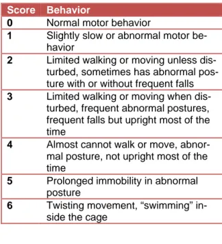
Table 1: Dystonia behavioral phenotype
The behavior table describes the posture, walk- ing ability and steadiness of the rats after kainic acid surgery. Dystonia of score 0 is normal while 6 represents a serious dystonic condition with seizures and involuntary rigidity. Normal rats scored 0 to 1 and dystonic rats scored 4-6.
Score Behavior
0
Normal motor behavior
1
Slightly slow or abnormal motor be- havior
2
Limited walking or moving unless dis- turbed, sometimes has abnormal pos- ture with or without frequent falls
3Limited walking or moving when dis-
turbed, frequent abnormal postures, frequent falls but upright most of the time
4
Almost cannot walk or move, abnor- mal posture, not upright most of the time
5
Prolonged immobility in abnormal posture
6
Twisting movement, “swimming” in- side the cage
Figure 1: Experimental outline
The experimental outline is divided into 2 parts, pre-surgery and post-surgery, where the day of the
stereotaxic surgery is regarded as day 0. About one week before the stereotaxic surgery (-8 and -7
day), the rats were trained for beam walking for the 1
sttime (BW1). The next day, the rats received a
telemetry implant (-6 day) and were allowed to rest for a few days before their 2
ndtraining session for
beam walking and the pre-surgery EMG recording (-1 day, BW2, preS EMG). On the day of the sur-
gery, they were divided into 4 groups: the sham group with saline injection, the KA group with kainic
acid injection, the KA+TGB group with kainic acid injection and tiagabine i.p. injection, and the TGB
group with tiagabine i.p. injection only. All rats were recorded for post-surgery EMG (postS EMG) im-
mediately after they recovered from surgery. Another recording was conducted at 90 minutes, 7
th,
22
nd, 30
th, and 48
thhours after surgery for the sham and the KA group, while the KA+TGB and the
TGB groups received another tiagabine i.p. injection at 90 minutes after surgery and recorded for EMG
one hour later (1.30 h), and continued at the 7
th, 22
nd, 30
th, and 48
thhours post-surgery. At the 49
thhour post-surgery, all rats were tested in the 3
rdbeam walking session and then sacrificed 3 days after
the surgery. BW1 and BW2 are considered as pre-surgery BW testing. BW3 is considered as post-
surgery testing. BW, beam walking; preS, pre-surgery; EMG, electromyography; sham, sham-
operated; KA, kainic acid; KA+ TGB, kainic acid with tiagabine treatment; TGB, tiagabine only; i.p., in-
traperitoneal; h, hour; postS, post-surgery
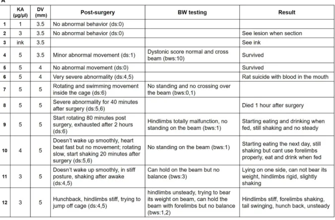
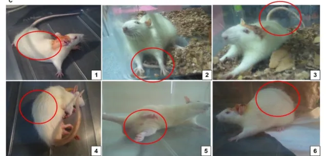
Figure 2: Concentration-coordinates assessment
(A) Kainic acid concentrations were matched with distinct dorsal-ventral coordinates of the rat brain to
determine the rat behavior and response after the stereotaxic surgery. In the first group of testing (trial 1 to 4), kainic acid at 3 and 5 µg/µl was injected to a depth of 3.5 mm, which revealed normal move- ments, giving a dystonia score of 0 and a beam walking score of 10. With further injection to the dorsal location of the brain, where DV was at 4 or 5 mm (trial 5 to 9), the rats showed rotating and over- excited epileptic-like seizures, with a dystonia score of 6 and a beam walking score of 0 and 1. There- fore, the kainic acid concentration was reduced to 4 µg/µl and then re-tested with 3 µg/µl (trial 10 to 12) the rats showed stiffness, with the dystonia scores of 4 and 5, and the capacity to hold on the beam (beam walking scores of 3).
(B) With the confirmation of kainic acid concentration at 3 µg/µl and DV 5 mm, tiagabine concentration
was set at with 2 mg/kg. The figure shows that the rats are steadier in the upright position, with less ri-
gidity and a capacity to walk on the beam after one dose of tiagabine. The dystonia score was 2 to 3
and the beam walking score was 4 to 5.
(C) The rats were observed with trunk twisting (1, 5) and hunchback (1, 4, 6), head and tail unsteadily
swinging (3, 4), limb stiffness (2, 3, 6) and with dystonic seizure after awakening from the stereotaxic surgery. KA, kainic acid; DV, dorsal-ventral coordinates; BW, beam walking; TGB, tiagabine; i.p., in- traperitoneal; ds, dystonia scores from Table 1; bws, beam walking scores from Table 2.
Table 2: Beam walkingobservation
T
his table describes the motor behavior and as- signs the scores to individual rats while they were moving along the beam walk apparatus. The scores depended on the rats’ behaviors, which allowed a score range of 0 to 10.
Score Behavior
0
No behavior, complete paralysis, no weight bearing
1
Poor weight bearing, cannot step up, fall
2
Minor weight bearing, can walk only 1-2 steps, fall
3
Mild weight bearing, can walk more than 2 steps, fall
4
Good weight bearing, can walk few steps, many falls
5
Good walking, but taken time long- er than 60 seconds
6
Good walking, taken time within 60 seconds, 4-5 foot slips
7Good walking, taken time within
60 seconds, 3-4 foot slips
8Good walking, taken time within
60 seconds, 1-2 foot slips
9Good walking, taken time within
30 seconds, no foot slips
10Good walking, taken time within
15 seconds, no foot slips
Telemetric transmitter implantation
Telemetric transmitter (TA11CTA-F40, Data Sciences International, St. Paul, MN, USA) was prepared before the surgery. Insu- lation tubing of the transmitter electrode was removed from the distal part of the lead and a loop was formed (Cesarovic et al., 2011).
The transmitter was then sterilized with Sep- tichlor. Rats were anesthetized with Isoflu- rane, 3.5 % in 100 % oxygen). Following midline abdominal incision, the transmitters were implanted intraperitoneally. The elec- trodes were tunneled subcutaneously and fixed to the gastrocnemius and tibialis anteri- or muscles using silk sutures (Cesarovic et al., 2011). Carproten (Laboratonos, Pfizer, rail) 2.5 mg/kg and Enrofloxacin (General Drugs House Co., LTD, BKK, Thailand) were injected to reduce pain and prevent in- flammation every 12 hours, for 3 days.
Stereotaxic surgery and tiagabine injection
After the telemetry implantation, the rats
were randomly divided into 4 groups. There
were the sham-operated group (sham), the
kainic acid group (KA), the kainic acid with
tiagabine treatment group (KA+TGB), and
the tiagabine group (TGB). The sham, KA,
and KA+TGB groups received stereotaxic surgery. On the day of the surgery, all the rats were weighed and abstained from food and water from the night before. Isoflurane (3.5 % in 100 % oxygen) was used in this surgery as anesthesia. After the rats were ful- ly anesthetized, a midline incision was made in between the rat’s’ ears. The skull was drilled at the midline above the cerebellum and 1 µl of kainic acid (Sigma-Aldrich, 3 µg/µl) was microinjected (AP from interau- ral line: -2.4 mm, DV: -5 mm), for both the KA and the KA+TGB groups (Paxinos and Watson, 1982). Tiagabine (Sigma-Aldrich, 2 mg/kg) was injected i.p. 5 minutes after the kainic acid microinjection. The sham group was microinjected with normal saline in placed of kainic acid. The tiagabine group re- ceived tiagabine injection at the same time, with the same concentration instead of re- ceiving stereotaxic surgery. The concentra- tion of kainic acid (3 µg/µl) and tiagabine (2 mg/kg), as well as dorsal ventral coordi- nates (DV: -5 mm) had been confirmed with preliminary experimentation (Figure 2). Once the rats were awakened from the anesthesia, as mentioned previously in the section of te- lemetry implantation, 2.5 mg/kg each of Carproten and Enrofloxacin were injected in- traperitoneally and subcutaneously to reduce pain and prevent inflammation every 12 hours, for 3 days.
Dystonic movement observation and EMG recording
The rats were moved from the surgery room to record their EMG amplitude and movements immediately after they recovered from anesthesia. Involuntary twisting and ri- gidity were scored using the dystonic pheno- type table (modified from Pizoli et al., 2002;
Jinnah et al., 2005; and Alvarez-Fischer et al., 2012, Table 1).
A research assistant observed and meas- ured the scores according to the assigned Ta- ble 1 to avoid possible bias. A score of 1 rep- resents a behavior pattern that reflects normal motor behavior, with normal posture. A score of 2-4 indicates dystonia with increasing se- verity. If limited walking and movement
were shown, the rat was disturbed by touch.
If there was no walking or moving at all and the rat couldn’t stay in an upright position, it was lifted by tail and placed down again to encourage movement, which often preceded the dystonia (Pizoli et al., 2002). The rat was observed lying still with continuous pro- longed rigidity, giving a score of 5. If one showed twisting movement, it would be giv- en score of 6. According to previous studies, dystonic rats scored about 4, 5, and 6. Nor- mal rats scored 0 or 1. Therefore, the present study would also consider scores 4, 5, and 6 for dystonic rats, and scores 0 and 1 for sham-operated rats.
At the same time, for dystonic testing and scoring, the rats were recorded for their 1
stpost-surgery EMG amplitude. Each EMG re- cording was 30 minutes and the rats were taken for recordings at 90 minutes, 7 hours, 22 hours, 30 hours and 48 hours after surgery (Figure 1). Each EMG recording was video- taped for further comparison and analysis.
Beam walking assessment
The aim of the beam walking apparatus
was to measure the foot slip and the latency
to transverse the beam for motor coordina-
tion and balance (Carter et al., 2001). The
size of the beam used in this parameter was
50 mm x 150 cm. The starting point was
marked at the 0 cm line and the endpoint at
the 110 cm. Every 10 cm was labelled with a
permanent ink to record the distance. A net
was attached under the beam to prevent inju-
ries from falling and escaping. Two 40-watt
lamps were set above and at the starting point
as an adverse stimulus and a goal box was set
by the end point to encourage forward
movement (Carter et al., 2001). A video re-
cording camera was fixed in front of the
starting point (Stanley et al., 2005). Each rat
was trained with 3 consecutive trials in 3
days (Luong et al., 2011), including 2 days
before the telemetry implant (BW1, Figure 1)
and 1 day after the implant (BW2, Figure 1),
as pre-surgery session. For the post-surgery
session, the rats were given 60 seconds on
the bridge per trial and the one that fell
would be returned to the spot of the fall
(Stanley et al., 2005). For the ones that failed to pass the bridge within 60 seconds, the dis- tance travelled was recorded. The time and distance of travel were recorded for each tri- al, before and after the surgery. The rat be- havior on the beam was observed and scored for comparison between groups using the beam walking testing (Table 2, modified from Anand et al., 2011). Weight bearing and walking ability were used to determine the scores. Animals with no weight bearing were given 0 to 1 points, ones with minor or mild weight bearing were given 2 to 3, and ones with good weight bearing were given 4-5 points. The ones able to walk within 60 sec- onds with minor foot slips were given 6 to 10 points (Anand et al., 2011). The immobilized ones on the beam were given less points and the ones that passed the beam were given higher points. The scores given are different from the dystonic scores. The scores were as- signed by a research assistant to avoid possi- ble bias.
Animal sacrifice and brain collection
After the perfusion procedures, brains were collected and post fixed with 4 % for- maldehyde for one week and then changed to 30 % sucrose solution. The brain was sec- tioned in paraffin sections with microtome.
The sections were stained with hematoxylin and eosin staining (H&E staining), and taken for whole brain imaging for needle tracing. A stereomicroscope was used to take a whole brain image, with an objective lens of 1.25X.
EMG data acquisition and analysis
The analogue EMG was digitized at 1 KHz and acquired using PowerLab A/D con- verter and Chart 5.4 software (AD Instru- ments, Australia) (Ngampramuan et al., 2008). The results measured the peak ampli- tude of muscle contraction after kainic acid microinjection and/or tiagabine i.p. injection for 30 minutes. EMG variability analysis in the amplitude domain was performed using Chart 5.4 software.
Data collection and analysis
One-way ANOVA with Turkey’s multi- ple comparison tests was used to calculate the statistical differences for dystonic behav- ioral phenotype score, EMG amplitude com- parison, and beam walking observational scoring test. Sham-operated group and exper- imental groups were compared to reach sta- tistical difference when p value is less than 0.05. Analysis was performed using Prism (GraphPad) software.
RESULTS
Determination of drug concentration and coordinates for the microinjection
Using concentration-coordinates table (Figure 2A), the animal status was scored af- ter awakening from surgery. It indicated the immediate tolerance of a rat towards the drug. When tested with various concentra- tions of kainate (1, 3, and 5 µg/µl) to DV 3.5 mm, only the concentration of 5 µg/µl re- sulted in the expression of minor abnormal movements, with a dystonia score of 1 (ds:1), obtained from Table 1. However, when run- ning on the beam, there was no loss of bal- ance or the capability of weight bearing, and the rats that could walk across the beam scored a 10 for the beam walking observation (bws:10, obtained from Table 2). When mi- croinjection was made at the depth of 4 to 5 mm, the rats could not tolerate the drug tox- icity, expressing their intolerance by over- twisting and running in circles inside the cage (ds: 6), which resulted in bleeding from their mouth. The rats became over exhausted and paralyzed the day after the surgery (ds:
6), some even died before the end point of the experiment. As DV was confirmed to cause the lesion, we needed to keep the ani- mals alive by reducing the kainic acid con- centration. After a few trials of 3 µg/µl vs.
the 5 mm testing, we agreed on the degree of
stiffness and rigidity (ds: 4, 5) as well as the
ability to hold on the beam (bws: 3), which
was considered as the optimal concentration
and coordinates (Figure 2A). In this study,
2 mg/kg tiagabine concentration was i.p. in-
jected post-kainic acid microinjection (Figure
2B). This concentration was also examined
with kainic acid concentration and DV coor- dinate obtained from Figure 2A. The animals showed more steadiness and calmness 80 to 90 minutes post-surgery (ds: 2, 3) and were able to walk on the beam (bws: 4, 5).
The abnormal behavior was expressed af- ter the rats awakened from the surgery. Limb stiffness and rigidity, and twisting trunk could be observed (Figure 2C, 1 and 5).
Some rats expressed a hunchback posture, ly- ing on one side with limbs freely swinging (Figure 2C, 4). Some could stand on their feet, but if the tail was lifting up, their limbs would attach against their trunk, indicating no support from the lower part of the body and hind limbs (Figure 2C, 6). The day after the surgery, although the rats started eating and drinking, they still experienced limb stiffness with their tails swinging in various directions from time to time (Figure 2C, 2 and 3).
Test for disability scoring for dystonic severity
After the animals awakened from anes- thesia, they had post-surgery EMG record-
ings. The abnormality and rigidity of the dys- tonic seizure was videotaped for a later dys- tonic scoring by a third person. The severity of the dystonic seizure within the period of 120 minutes post-surgery was given scores according to Table 1.
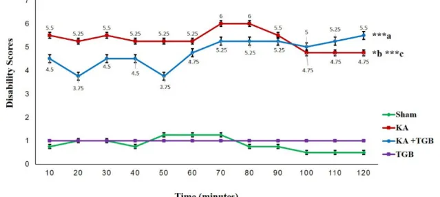
According to the graph, the KA group maintained a score between 5 and 6; in con- trast, there was a score variation in the KA+TGB group, which finally stabilized at 60 minutes post-surgery (Figure 3). There was a little shift in scores from 100 minutes until 120 minutes post-surgery, where the KA group (scores 4.75, 4.75, 4.75, in aver- age, p<0.05) scored slightly less compared with the KA+TGB group (scores 5, 5.25, 5.5, in average), which correlates with data on the EMG observational scoring table at the 90 minutes (Figure 4). These phenomena sug- gest that tiagabine treatment meets the peak efficiency, and the acute injection of kainic acid functioned as the stabilizing effect of the dystonic seizures. Please refer to supple- mental data (S1-Table 1) for complete scores of individual rats.
Figure 3: Dystonic behavioral phenotype score
This Figure shows the behavioral observation within 2 hours duration post-surgery for 4 groups: sham, KA, KA+TGB, and TGB. The scores were obtained from Table 1. The KA group shows a gradually higher score compared to sham (***c) and KA+TGB (*b). KA+TGB also had higher scores compared with sham (***a). The TGB group did not go through the stereotaxic surgery, so they remained steady.
Significantly different value: ***a/c, p<0.001; *b, p<0.05; Sham, sham-operated; KA, kainic acid; KA+
TGB, kainic acid with tiagabine treatment; TGB, tiagabine only group.
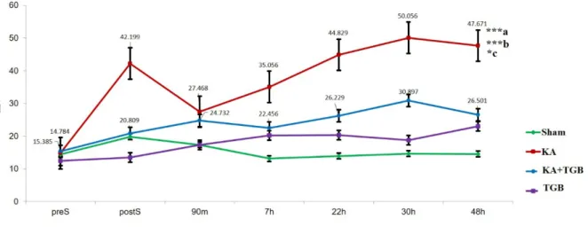
Figure 4: EMG amplitude comparison
This Figure compares the average EMG amplitude between the 4 groups; sham, KA, KA+TGB and TGB. The kainic acid group had significant gradual bursting in the EMG with more severity in dystonic rigidity and muscle spasm compared with sham (***a), with KA+TGB (*c,); with TGB group (***b). The numbers above, or beside the plotted points, show the mean ± SEM (standard error of mean) for only the treatment group (KA+TGB) and the non-treatment group (KA). Significantly different value: ***a/b, p<0.001, *c, p<0.05. preS, pre-surgery; postS, post-surgery; m, minute; h, hour; Sham, sham- operated; KA, kainic acid; KA+ TGB, kainic acid with tiagabine treatment. TGB, tiagabine only.
Intra-muscular EMG recording
After the rats received the EMG teleme- try implantation, there were seven EMG re- cordings (preS, postS, 90 minutes, 7 hours, 22 hours, 30 hours and 48 hours) (Figure 1).
Each EMG recording was 30 minutes, except for the pre-surgery, 15 minutes.
For post-surgery recordings, the rats were recorded only after awakening from anesthe- sia. The KA group showed most exaggerated muscle contractions compared to the sham, KA+TGB and TGB groups (Figure 4). There was a high bursting frequency post-surgery (42.199±11.122), although muscle was re- laxed with less amplitude at 90 minutes post- surgery (27.468±11.091), the amplitude was stabilized at the 7
thhour post-surgery (35.056±8.924) and gradually increased until the 30
thhour post-surgery (50.056±5.862).
Please refer to supplemental data (S2-Table 1) for the complete standard deviation value.
Kainic acid-injected rats expressed high- est amplitude in muscle contraction, which was significantly different when compared to sham-operated animals (p< 0.001). The KA group also expressed significantly higher
amplitude compared with the KA+TGB group (p< 0.05). In addition, the KA group also showed significant difference compared to the TGB group (p< 0.001). In comparison with the KA group, the KA+TGB group showed an increase in amplitude gradually from postS (20.809±4.058) to 90 minutes (24.732±5.341), and also stabilized at 7 hours (22.456±3.885). From the graph, the KA and the KA+TGB groups composed a correlated slope from 7 hours to 48 hours (Figure 4).
Please refer to supplemental data (S2-Table 1) for the complete standard deviation value.
Beam walking analysis
All rats were trained for beam walking
for 2 sessions before the kainic acid mi-
croinjection (BW1 and BW2, Figure 1). Af-
ter full recovery from the kainic acid mi-
croinjection, the rats were required to walk
on the beam again (BW3, Figure 1). The
purpose of the third attempt was to record the
efficacy of the tiagabine treatment in that
case of kainic acid excitotoxicity in the cere-
bellar cortex, as well as to make a compari-
son with the non-treated and pre-surgery
groups. In this behavioral apparatus, we quantified not only the locomotion, but also the observation scores on how the animals responded to the beam walking after the sur- gery, with/without tiagabine treatment (Table 2). The scoring was given by a third person to avoid possible bias.
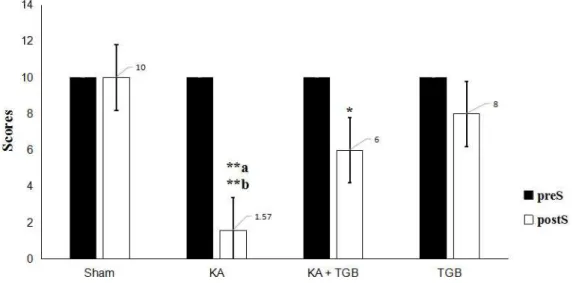
As illustrated in Figure 5, the post- surgery response reached a statistical differ- ence compared to the pre-surgery condition.
The kainic acid group had difficulty in weight bearing and was very sensitive when touching the beam (scored 1.57 in average).
Post-surgery KA expressed a significantly different reaction towards the beam when compared with the post-surgery sham group (scored 10 in average, p<0.01), KA+TGB (scored 6 in average, p<0.5), and TGB (scored 8 in average, p<0.01) groups.
KA+TGB rats had limited weight bearing, but could hold onto the beam with their limbs and crawl across the beam with some help (scored 6 in average), although it took longer time (Figure 6). TGB had no significantly different motion when touching the beam (scored 8 in average). They had good weight
bearing and good crossing time, except for a few foot slips. Please refer to supplemental data (S3-Table 1) for the complete scores of individual rats.
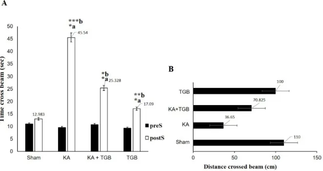
Each rat was compared before and after the surgery to observe their behavioral changes, especially with the tiagabine treat- ment. In Figure 6A, KA group had taken a significantly longer duration in holding on the beam (45 seconds in average), when compared with the sham (12 seconds in aver- age, p<0.0005) and the TGB groups (17 sec- onds in average, p<0.005), which also spent more than 50 % of the time crossing the beam when compared with KA+TGB. De- spite spending longer time on the beam walk- ing task, rats in the KA group failed to pass the 110 cm beam completely (average 36 cm, Figure 6B). For the treatment group, alt- hough the rats did not completely traverse the beam either, they managed to spend signifi- cantly less time on the beam (25 seconds in average, p<0.05) and completed the task de- spite foot slips and the fails, with the distance travelled of 100 cm.
Figure 5: Beam walking behavioral observation
The scores were obtained from Table 2; when the rats were running or holding onto the beam, with the
objective of quantifying the rats’ motor behavior and balance. The KA group, compared to the sham
group (**a) and the TGB group (**b), expressed most severe dystonic seizures with difficulty staying
on the beam or starting walking. The average score was 1.57 for the KA+TGB group compared with to
KA group (*p<0.5). The average score of 6 was generally given to the rats for walking through the
beam within 60 seconds with fewer falls and significantly different value: **a/b p< 0.01, *p<0.5. preS,
pre-surgery; postS, post-surgery; Sham, sham-operated; KA, kainic acid; KA+TGB, kainic acid with ti-
agabine treatment. TGB, tiagabine only.
Figure 6: Time and distance crossed the bridge
These two Figures describe time and distance used to cross the beam in the 4 groups: sham, KA, KA+TGB, and TGB groups. (A) In comparing the pre- and post-surgery data for each group, all the rats used significantly longer time after surgery (KA: *a; KA+TGB: *a; TGB: *a). After the surgery, KA also spent significantly longer time (45 seconds in average) than sham (***b), KA+TGB (*b), and TGB (**b).
(B) Distance in crossing the beam. Although the KA group spent the longest time on the beam,this group only travelled 36 cm on average. KA+TGB, the treatment group, travelled 70 cm on aver- age. Significantly different value: *a/b, p<0.05, **b, p<0.005, ***b, p<0.0005. preS, pre-surgery; postS, post-surgery; Sham, sham-operated; KA, kainic acid; KA+ TGB, kainic acid with tiagabine treatment;
TGB, tiagabine only.
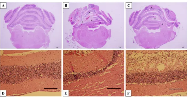
Histological imaging
All brains were collected, sectioned and stained with H&E to observe the morphology of cerebellar tissue and cells, especially the lesion parts. For the KA and KA+TGB groups, there were obvious needle traces, fragmentation, cell loss and fibrous tissue development (Figure 7). When compared with the distinct cerebellar layers in the sham group, cell loss and fragmentation indicated layers of atrophy and enlargement of the in- tra-layer space rather than complete layers (Figure 7B, C). When observed at higher magnification (10X), the KA group showed thinner layers, indicating cell loss, which is in contrast with the KA+TGB sections, where some regions of the cerebellar layer showed fibrosis and fragmentation (Figure 7E, F). Despite different degrees of tissue at- rophy, there was significant degeneration in the cellular morphology of the KA+TGB group and the non-treated KA group.
Limitation of the study
Further molecular analysis or quantitative cell number estimation could be performed if the improvement of motor function resulted from tiagabine treatment and/or cellular plas- ticity and adaptation. Although preliminary study was performed to determine the suita- ble kainic acid concentration, electroenceph- alography (EEG) may also have been re- quired to determine whether the dystonic sei- zure we induced by kainic acid injection is indeed dystonia rather than some other cere- bellar syndromes, such as epilepsy and atax- ia. Pizoli’s study with kainic acid-induced cerebellar dystonia suggests that this kind of stiffness and rigidity can be considered as dystonic symptoms after EEG measurement, which was compared with the effects of other glutamate drugs in their study (Pizoli et al.
2002).
Figure 7: Histological images