Susanne Kirchhof
Diels Alder Hydrogels as Intraocular Drug Delivery Systems for Antibodies
Diels Alder Hydrogels as Intraocular Drug Delivery Systems for Antibodies
DISSERTATION ZUR ERLANGUNG DES DOKTORGRADES DER NATURWISSENSCHAFTEN (DR. RER. NAT.)
DER FAKULTÄT FÜR CHEMIE UND PHARMAZIE DER UNIVERSITÄT REGENSBURG
vorgelegt von Susanne Kirchhof
aus Bobingen
Juli 2015
Diese Arbeit entstand in der Zeit von August 2010 bis Juli 2015 am Lehrstuhl für Pharmazeutische Technologie an der Universität Regensburg.
Die Arbeit wurde von Herrn Prof. Dr. Achim Göpferich angeleitet.
Promotionsgesuch eingereicht am:
Datum der mündlich Prüfung:
Prüfungsausschuss:
31. Juli 2015 09. Oktober 2015
Prof. Dr. Jens Schlossmann (Vorsitzender) Prof. Dr. Achim Göpferich (Erstgutachter) PD Dr. Rainer Müller (Zweitgutachter) Prof. Dr. Sigurd Elz (Drittprüfer)
Diels Alder Hydrogels as Intraocular Drug Delivery Systems for Antibodies
Die Wissenschaft fängt eigentlich erst da an, interessant zu werden, wo sie aufhört.
Justus von Liebig
Table of Contents
1 Introduction and Goal of the Thesis ... 1
Introduction ... 2
Goal of the thesis ... 4
2 Hydrogels in Ophthalmic Applications ... 9
Introduction ... 11
Methods of hydrogel preparation and properties of hydrogels ... 13
Design criteria for hydrogels and challenges associated with their preparation ... 15
Ophthalmic applications of hydrogels ... 16
2.4.1 In situ forming hydrogels for ophthalmic drug delivery... 16
2.4.2 Drug-eluting contact lenses ... 18
2.4.3 Tissue adhesives for ocular wound repair ... 21
2.4.4 Intraocular lenses... 22
2.4.5 Hydrogel-based vitreous substitutes ... 23
2.4.6 Intravitreal drug delivery systems ... 25
2.4.7 Cell-based approaches ... 27
Limitations of hydrogel-based drug delivery systems ... 29
Concluding remarks and future perspectives ... 31
3 Investigation of the Diels-Alder Reaction as a Cross-Linking Mechanism for Degradable Poly(ethylene glycol) Based Hydrogels ... 33
Introduction ... 35 1.1
1.2
2.1 2.2 2.3 2.4
2.5 2.6
3.1
Experimental Section ... 36
3.2.1 General procedure... 36
3.2.2 Materials ... 36
3.2.3 Synthesis of branched PEG-amines ... 37
3.2.4 Synthesis of furyl-substituted four-armed PEG ... 37
3.2.5 Synthesis of furyl-substituted eight-armed PEG, molecular weight 10 kDa 38 3.2.6 Synthesis of furyl-substituted eight-armed PEG, molecular weight 20 kDa 38 3.2.7 Synthesis of 3,6-epoxy-1,2,3,6-tetrahydrophthalimide (ETPI) ... 38
3.2.8 Synthesis of ETPI-substituted four-armed PEG ... 39
3.2.9 Synthesis of 4armPEG10k-maleimide ... 39
3.2.10 Synthesis of 8armPEG10k-maleimide ... 40
3.2.11 Synthesis of 8armPEG20k-maleimide ... 40
3.2.12 Preparation of Diels-Alder hydrogels ... 40
3.2.13 Rheological characterization of Diels-Alder hydrogels ... 41
3.2.14 Calculation of hydrogel network mesh size ... 41
3.2.15 Swelling and degradation of Diels-Alder hydrogels ... 42
Results and discussion ... 43
3.3.1 Synthesis of furyl- and maleimide-substituted PEG-derivatives ... 43
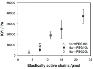
3.3.2 Rheological characterization of Diels-Alder hydrogels ... 43
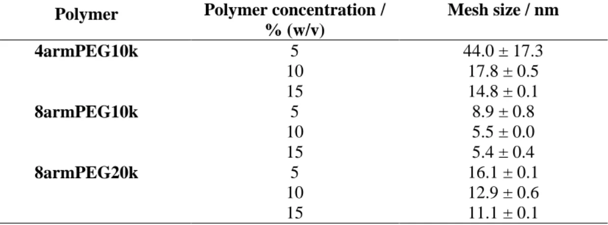
3.3.3 Calculation of hydrogel network mesh size ... 46
3.3.4 Swelling and degradation of Diels-Alder hydrogels ... 48
Conclusion ... 51
4 New Insights into the Cross-Linking and Degradation Mechanism of Diels- Alder Hydrogels ... 53
Introduction ... 55
Experimental section ... 57
4.2.1 Materials ... 57
4.2.2 Hydrolytic stability of 8armPEG10k-maleimide ... 57
4.2.3 Rheological characterization of DA hydrogels ... 58
4.2.4 Swelling and degradation of DA hydrogels ... 58
4.2.5 Molecular modeling studies ... 59
4.2.6 Statistical analysis ... 59
Results and discussion ... 60
4.3.1 Hydrolytic stability of 8armPEG10k-maleimide ... 60
4.3.2 Rheological characterization of DA hydrogels ... 61
4.3.3 Swelling and degradation of DA hydrogels ... 64 3.2
3.3
3.4
4.1 4.2
4.3
Table of Contents
4.3.4 Molecular modeling studies ... 67
Conclusion ... 70
5 Diels-Alder Hydrogels for Controlled Antibody Release: Correlation between Mesh Size and Release Rate ... 71
Introduction ... 73
Materials and methods ... 74
5.2.1 Materials ... 74
5.2.2 Swelling studies... 75
5.2.3 Rheological characterization ... 76
5.2.4 Low field NMR measurements ... 76
5.2.5 In vitro release of fluorescein isothiocyanate-labeled dextran ... 78
5.2.6 In vitro release of bevacizumab ... 78
Results and discussion ... 79
5.3.1 Determination of the network mesh size ... 79
5.3.2 In vitro release of fluorescein isothiocyanate labeled dextran ... 86
5.3.3 In vitro release of bevacizumab ... 90
Conclusion ... 91
6 Diels-Alder Hydrogels with Enhanced Stability: First Steps Towards Controlled Release of Bevacizumab ... 93
Introduction ... 95
Materials and methods ... 97
6.2.1 General procedure... 97
6.2.2 Materials ... 97
6.2.3 Synthesis of macromonomers ... 97
6.2.4 Hydrolytic stability of maleimides ... 98
6.2.5 Kinetics of the Diels-Alder reaction ... 98
6.2.6 Characterization of Diels-Alder hydrogels ... 99
6.2.7 In vitro release of bevacizumab ... 99
6.2.8 Statistical analysis ... 100
Results and discussion ... 100
6.3.1 Synthesis of macromonomers ... 100
6.3.2 Hydrolytic stability of maleimides ... 101
6.3.3 Gelation kinetics of Diels-Alder hydrogels ... 102
6.3.4 Rheological characterization of Diels-Alder hydrogels ... 105
6.3.5 Determination of the average mesh size ... 106 4.4
5.1 5.2
5.3
5.4
6.1 6.2
6.3
6.3.6 Swelling and degradation of Diels-Alder hydrogels ... 107
6.3.7 In vitro release of bevacizumab ... 109
Conclusion ... 111
Supplements ... 112
6.5.1 Synthesis of 8armPEG20k-NH2 (1), 8armPEG20k-Fur (2a) and 8armPEG20k-Mal (2b) ... 112
6.5.2 Synthesis of compound 3 ... 112
6.5.3 Synthesis of compound 4 ... 113
6.5.4 Synthesis of compound 8armPEG20k-Ahx-Fur (5a) ... 114
6.5.5 Synthesis of compound 8armPEG20k-Ahx-Mal (5b) ... 114
6.5.6 Synthesis of compound 6 ... 115
6.5.7 Synthesis of compound 7 ... 115
6.5.8 Synthesis of compound 8 ... 116
6.5.9 Synthesis of compound 9 ... 116
6.5.10 Synthesis of compound 8armPEG20k-Lys-Ahx-Fur2 (10a) ... 117
6.5.11 Synthesis of compound 8armPEG20k-Lys-Ahx-Mal2 (10b) ... 117
7 Summary and Conclusion ... 119
Summary ... 120
Conclusion and outlook ... 122
References ... 125
List of Figures... 153
List of Schemes 155 List of Tables ... 157
Appendix ... 158 6.4
6.5
7.1 7.2
Chapter 1
Introduction and Goal of the Thesis
Introduction
In recent years, the therapeutic use of antibodies has become a powerful new treatment option for various serious diseases including asthma, rheumatoid arthritis, Crohn's disease, different types of cancer and wet age related macular degeneration (AMD) [1–5]. Since the market launch of the first antibody muromonab-CD3 in 1986, the number of therapeutically successful antibodies has increased continuously. Nowadays, antibodies, antibody fragments, conjugates, and fusion proteins represent the most important and valuable product class within the biopharmaceutical market. These antibody products can be applied either systemically or locally. Although the systemic application of antibodies is associated with several disadvantages, including high dosing and serve side effects, most antibodies are currently administered by intravenous injections. The mentioned drawbacks can be overcome by local application in order to achieve higher drug concentrations at the application site. This results in lower required drug dosages and improved therapeutic effects. Moreover, the systemic antibody exposure is reduced causing less systemic side effects. Currently, a variety of potential, local antibody application sites are investigated or have already been established. For instance, antibodies are directly administered at the tumor site for anti-cancer application, are intraarticularly injected for the treatment of arthritic joint diseases or are being used to minimize or even cure injuries of the central nervous system after intrathecal delivery [6–8]. The most used method for local antibody delivery is the intravitreal administration of anti-vascular endothelial growth factor (VEGF) antibodies for the therapy of diseases at the posterior segment of the eye, such as AMD and proliferative diabetic retinopathy (PDR) [5,9]. In this case the blood-retina-barrier represents an insurmountable obstacle for the systemic application of macromolecules, such as antibodies. Thus, intraocular injections are essential for a successful antibody therapy (Figure 1.1).
However, there are several limitations minimizing the success of those strategies: short drug residence times at the application site and hence frequent antibody administration as well as side effects of injections, such as pain and inflammation, reduce the compliance [10]. Consequently, suitable drug delivery systems for sustained antibody delivery are required to overcome these limitations and enable more effective therapies [11]. During the last decades different types of drug delivery systems based on
1.1
1.1 Introduction microparticles, nanoparticles, liposomes and hydrogels were investigated [12,13]. These drug delivery systems lower drug dosage and enhance drug efficacy while reducing systemic side effects.
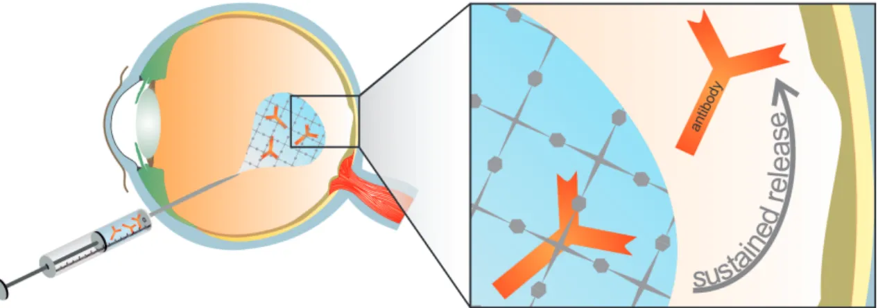
Figure 1.1. Sustained release of entrapped antibodies at the posterior segment of the eye after intraocular injection of an in situ gelling hydrogel.
The use of hydrogels for antibody delivery offers several advantages, such as reduced antibody degradation [13,14]. Generally, hydrogels are regarded to be biocompatible because of their high water content and soft nature similar to the extracellular matrix.
Their porous structure is extremely suitable to accommodate high loads of water-soluble proteins either by post-fabrication partitioning or in situ encapsulation [14,13]. The simple preparation procedures in aqueous medium at room temperature contribute to preserving protein stability. Moreover, the entrapped antibodies have limited mobility in hydrogel networks with high cross-linking density resulting in sustained release controlled by the swelling and degradation behavior of the gels. Finally, in situ gelling hydrogels can be easily injected and form drug delivery systems at the application site.
Important for the success of the system is a precise control over the cross-linking density and hence the mechanical properties, the swelling capacity, the degradation behavior of the hydrogel and the release profile of entrapped antibodies [15]. However, it is more or less impossible to obtain a defined network structure through widespread radical photopolymerization [16]. Moreover, the use of photoinitiators generates free radicals during cross-linking, which may induce toxicity and affect the bioactivity and availability of proteins [14,17,18]. Additionally, a lower cross-linking conversion due to un-reacted functional groups and network non-idealities may negatively influence antibody release.
Therefore, alternative cross-linking reactions have to be developed for the successful utilization of antibody release systems based on hydrogels. An appropriate reaction
should occur in water at body temperature without using any catalyst or initiator. A cross- linking reaction that meets the criteria is the Diels-Alder (DA) click reaction. This [4 + 2]
cyclo-addition is a highly effective reaction between electron-rich diens and dienophiles.
Despite being widely used in organic chemistry for the synthesis of steroid hormones and six-membered rings, its application for hydrogel preparation is less common [19,20].
Nevertheless, few examples have been described in literature using natural polymers in combination with relatively short cross-linkers, which results in rather undefined networks [21,22]. Although natural polymers are regarded to be non-toxic and principally biodegradable, they are less suitable for hydrogel preparation due to their non- reproducible chemical and physical properties. Therefore, synthetic polymers are favored for hydrogel preparation. Especially hydrogels based on poly(ethylene glycol) (PEG) are widespread because of their favorable polymer properties, such as high solubility, excellent biocompatibility and broad acceptance by regulatory agencies [14,23].
Goal of the thesis
This thesis is focused on the development and characterization of DA hydrogels as injectable drug delivery system for the intraocular application of antibodies.
Currently, hydrogels are already used for a number of different applications in ophthalmology. For instance, they are used as in situ gelling eye drops, soft contact lenses (SCL), intraocular lenses (IOL) and adhesives for ocular wound repair or are investigated as vitreous substitutes and intravitreal drug delivery systems (Chapter 2). Although significant progress has been made in the field of SCL, in situ gelling eye drops and adhesives for ocular wound repair, many challenges remain to improve the safety and clinical performance of intraocularly applied hydrogels. More research is particularly needed to improve hydrogels as intravitreal drug delivery systems for the treatment of severe, vision-threatening diseases, such as AMD or PDR.
There are a number of general requirements that intraocularly applied hydrogels must fulfill. Ideally, these hydrogels are injectable and offer sustained drug release over several weeks. So-called in situ gelling hydrogels, which are injected prior to gelation and form a semi-solid drug depot at the application site, offer such characteristics. After gelation the mechanical properties of the resulting hydrogel should be comparable to those of the environment in order to cause no foreign body sensation. Moreover, the hydrogel should be designed for simple and effective drug loading procedures, especially for proteins and
1.2
1.2 Goal of the thesis antibodies. Therefore, suitable cross-linking processes are necessary to permit fast gelation without inactivating the entrapped proteins. Besides appropriate loading procedures, a complete, yet sustained release of still active drugs is of particular importance. One of the main challenges of hydrogels is the control of drug release over several weeks due to their high water content. After complete drug release, the hydrogel should degrade without releasing any toxic degradation product. Although, a multitude of polymers and cross-linking possibilities have already been tested, there is no product commercially available until today. Promising materials are chemically cross-linked PEG-based hydrogels. Due to their excellent biocompatibility they are used for several biomedical applications. As in situ gelling systems they can be easily injected by minimally invasive techniques and hold great promise as drug delivery systems.
Nevertheless, most established cross-linking mechanisms are associated with significant disadvantages, such as the formation of potentially harmful radicals or unwanted side reactions with incorporated protein drugs.
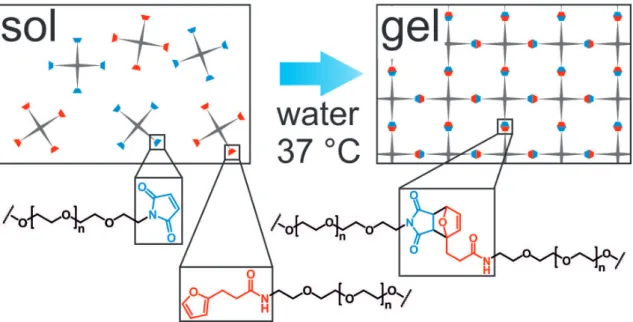
To overcome these limitations, new cross-linking reactions for PEG-based hydrogels have to be developed. Since the DA reaction proceeds in water without any initiator or metal catalyst, it is considered as an effective and non-toxic cross-linking possibility (Chapter 3). For the hydrogel preparation, two complementary macromonomers were synthesized by functionalizing star-shaped PEG with furyl and maleimide groups (Figure 1.2).
Figure 1.2. Principle of DA reaction for in situ hydrogel formation. Furyl- and maleimide functionalized star shaped PEG were dissolved in water and combined, e.g. by using a two-chamber syringe. Directly after mixing both components the formation of highly elastic hydrogels is initiated.
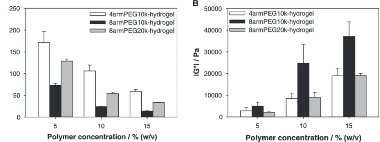
The influence of the macromonomer concentration, molecular weight and branching factor on gel characteristics, such as rheology and swelling behavior, were investigated.
Surprisingly, the covalently cross-linked hydrogels dissolved within days to weeks. To investigate this unexpected degradation behavior of PEG-based DA hydrogels in detail, further experiments were performed (Chapter 4). UV spectroscopy was used to analyze the hydrolytic stability of maleimide functionalized star-shaped PEG as a function of temperature and pH. Finally, molecular modeling studies of DA and retro-Diels-Alder (rDA) moieties were performed to investigate the influence of these reactions for the degradation process.
Besides the degradability, the permeability of entrapped drugs, such as proteins or antibodies, is crucial for success or failure of the system. The mesh size or correlation length ξ is an important parameter that characterizes the permeability. It is defined as “the average distance between consecutive cross-links” and indicates the maximum size of solutes that can pass through the gel network. In Chapter 5 swelling studies, rheology and low field NMR spectroscopy were used to calculate ξ of the prepared DA hydrogels.
The knowledge of ξ, in combination with the size of the entrapped molecule, allows the estimation of the drug release rate and the evaluation of the general suitability of the resulting hydrogels for controlled release of entrapped, therapeutic antibodies. As a proof of concept, fluorescein-labeled dextrans of different molecular weight were entrapped into the hydrogel and release studies were performed. Finally, the data of those in vitro studies were compared to theoretical predictions.
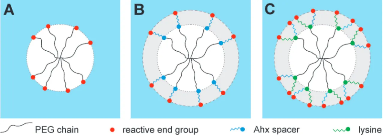
Figure 1.3 Modification of eight-armed PEG (A) by introducing Ahx spacer between PEG and reactive end-groups to increase hydrolytic stability (B). Modification with Lys and Ahx yields in doubled number of reactive end-groups and increased hydrolytic stability (C).
1.2 Goal of the thesis However, slow gelation and comparatively fast degradation may limit the application of DA hydrogels as injectable drug delivery systems. To overcome these limitations the macromonomers were modified with lysine (Lys) and/or 6-aminohexanoic acid residues (Ahx) in order to increase the hydrolytic stability of maleimide and to double the number of reactive groups per macromonomer (Chapter 6) (Figure 1.3).
To prove the feasibility of the optimized DA hydrogels as drug delivery systems, the gels were loaded with bevacizumab, a VEGF-neutralizing antibody used in the treatment of AMD. Afterwards, the release profiles of the entrapped antibody were determined by fluorescence spectroscopy.
Chapter 2
Hydrogels in Ophthalmic Applications
This chapter was published as: S. Kirchhof, A. M. Goepferich and F. P. Brandl, Eur. J.
Pharm. Biopharm. (2015), doi: org/10.1016/j.ejpb.2015.05.016
Abstract
More and more people worldwide are affected by severe eye diseases eventually leading to visual impairment or blindness. In most cases, the treatment involves the application of ophthalmic dosage forms such as eye drops, suspensions or ointments. Unfortunately, some of the therapeutic approaches have major shortcomings, especially in the treatment of the posterior segment of the eye, where many vision-threatening diseases originate.
Therefore, research focuses on the development of new materials (e.g., for vitreous substitution) and more advanced drug delivery systems. Hydrogels are an extremely versatile class of materials with many potential applications in ophthalmology. They found widespread application as SCL, foldable IOL, in situ gelling formulations for ophthalmic drug delivery and ocular adhesives for wound repair; their use as vitreous substitutes and intravitreal drug delivery systems is currently under investigation. In this article, we review the different applications of hydrogels in ophthalmology with special emphasis placed on the used polymers and their suitability as ocular drug delivery systems.
2.1 Introduction
Introduction
More and more people worldwide are affected by severe eye diseases, such as glaucoma, PDR and AMD [24–27]. Without adequate treatment, these diseases will eventually lead to severe visual impairment or blindness. Today, two major strategies for the treatment and management of ocular disorders can be identified: (1) the attempt to specifically deliver drug molecules to diseased ophthalmic tissues and (2) surgical procedures to repair or replace damaged tissues, such as the lens or the vitreous body [28–30].
Unfortunately, most currently available therapeutic approaches have major shortcomings, especially in the treatment of the posterior segment of the eye, where most vision- threatening diseases originate. The main challenges are as follows: (1) poor adherence of patients to the currently available therapeutic regimens, (2) maintaining effective drug levels over extended time periods and extending the required dosing intervals, (3) the delivery of drug molecules to the posterior segment of the eye, (4) the administration of therapeutic proteins and nucleic acids, and (5) the repair or substitution of dysfunctional cells or tissues. For example, over 50% of 500 surveyed glaucoma patients were found to be either non-adherent to their therapeutic regimens or demonstrated improper administration techniques [31]. While patients can be educated on the importance of adherence and instructed on proper eye drop administration, the inherent shortcomings of eye drops, such as low ocular bioavailability of the applied drug, are less easy to overcome. For example, it has been shown that less than 5% of the administered dose reaches the target tissue due to drainage and limited corneal permeability [32–34].
Moreover, only the anterior segment of the eye can be treated by eye drop installation, whereas different application routes (e.g., intravitreal injections) and/or more complex drug delivery systems (e.g., intraocular implants) may be required to reach the posterior segment [35–38]. For example, the development of anti-VEGF drugs, such as monoclonal antibodies or antibody fragments, revolutionized the therapy of neovascular AMD and other vision-threatening diseases [5,9,39]. However, the required intravitreal injections are associated with significant discomfort and complications including endophthalmitis, retinal tears or detachment, and cataract [10]. It is obvious that sustained delivery systems for anti-VEGF drugs would make the current therapeutic regimens more patient-friendly and cost-effective.
2.1
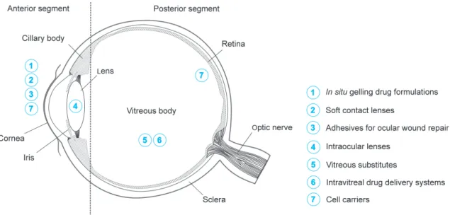
Figure 2.1. Application sites of hydrogels in ophthalmology.
The given examples illustrate the need for new developments in the field of ophthalmic drug delivery, and indicate the great market potential of those drug delivery systems.
Compared to the different materials and dosage forms that have been developed for ophthalmic applications, such as colloidal drug delivery systems [40–42] or polymeric implants [43], hydrogels may offer several advantages. For example, the mild preparation conditions and high water content of hydrogels are beneficial in preserving the activity of biopharmaceuticals such as peptides, proteins or nucleic acids [13,14,44]. Moreover, many hydrogels, such as temperature-responsive or in situ chemically cross-linked systems, can be administered by minimally invasive methods [13,14,44,45]. Hydrogels have already been approved for several ophthalmic applications, with many more currently being under investigation. For instance, they are successfully marketed as corrective SCL [46], foldable IOL [47,48], or in situ gelling vehicles for ophthalmic drug delivery [49,50]. Most research efforts currently aim at improving existing formulations for antibiotics, anti-inflammatory drugs or β-blockers, or at developing sustained release formulations for therapeutic proteins and nucleic acids. Furthermore, hydrogels are investigated as adhesives for ocular wound repair [51] or potential vitreous substitutes [29,30]. A graphical overview on possible ophthalmic applications of hydrogels is given in Figure 2.1.
In most of these applications, hydrogels are used because of their favorable physicochemical properties (e.g., transparency, high water content, and mechanical flexibility) or in combination with active pharmaceutical ingredients. In this review article, we present a general overview on ophthalmic applications of hydrogels with
2.2 Methods of hydrogel preparation and properties of hydrogels special emphasis on drug delivery systems for the posterior segment of the eye. We particularly focus on the different gel-forming polymers, the physicochemical properties of the resulting hydrogels, and the challenges associated with the preparation of hydrogel- based drug delivery systems. Recent developments in the field of ophthalmic drug delivery are presented and critically reviewed regarding the potential therapeutic benefits.
Methods of hydrogel preparation and properties of hydrogels
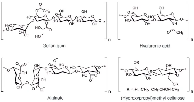
Before discussing the diverse applications of hydrogels in ophthalmology, a short review on the different methods of hydrogel preparation will be presented along with an overview on the characteristic properties of hydrogels. According to the definition by the International Union of Pure and Applied Chemistry, a hydrogel is a polymer network that is expanded throughout its whole volume by water [52]. A wide range of natural, semisynthetic and synthetic polymers can be used as starting materials for hydrogels.
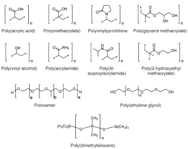
Typical examples for polymers of natural origin include alginate, collagen, and hyaluronic acid (HA) (Figure 2.2). PEG, poly(vinyl alcohol) (PVA), polymers based on acrylate monomers, and siloxanes are important examples for synthetic gel-forming materials (Figure 2.3) [13,44,53–55].
Figure 2.2. Chemical structures of selected polymers from natural sources commonly used for the preparation of hydrogels.
2.2
Figure 2.3. Chemical structures of synthetic polymers commonly used for the preparation of hydrogels.
Polymers of natural origin usually have the advantage of being non-toxic and biodegradable. Furthermore, natural polymers often interact with proteins and cells through non-specific or specific binding, which can be desired in hydrogel-based sustained release systems or cell carriers. However, hydrogels made from natural polymers may suffer from weak mechanical strength, high batch-to-batch variability, and immunogenicity. Synthetic polymers, on the other hand, allow for the preparation of hydrogels with well-defined network architecture, tunable mechanical properties, and prolonged stability. However, many synthetic polymers, such as PEG or PVA, do not inherently interact with proteins or cells; furthermore, biocompatibility and biodegradability must be carefully evaluated.
Hydrogels can be further classified into physical and chemical gels, depending on how the polymer networks are formed [13,44,53–55]. In physical or reversible hydrogels, the networks are formed by physical aggregation of polymer chains, caused by molecular entanglements, hydrogen bonds, hydrophobic interactions, ionic bonding, or complexation. All of these interactions are reversible, and the stability of the formed hydrogels is susceptible to changes in the ionic strength, pH, temperature, or the application of mechanical stress. Chemical or permanent hydrogels, on the other hand,
2.3 Design criteria for hydrogels and challenges associated with their preparation contain covalently cross-linked polymer networks with tunable physicochemical properties such as equilibrium water content, permeability, mechanical strength, and degradability. Covalently cross-linked networks can be formed by radical polymerization of water-soluble monomers (e.g., 2-hydroxyethyl methacrylate or acrylamide) and suitable cross-linking agents (e.g., PEG diacrylate or bisacrylamide). Alternatively, pre- formed polymers (e.g., HA or star-shaped PEG) can be functionalized with reactive groups suitable for polymerization and cross-linked via Schiff base formation, Michael- type addition reaction, azide-alkyne cycloaddition, or DA reaction [13,14,44,45,56]. For a more comprehensive overview on the different hydrogel-forming polymers and cross- linking mechanisms, the reader is referred to several excellent review articles [13,14,44,45,53–56].
Design criteria for hydrogels and challenges associated with their preparation
Cross-linked hydrogels are extremely versatile materials with many favorable properties such as transparency, high water content, good biocompatibility and mechanical flexibility. Consequently, hydrogels found widespread application in ophthalmology as SCL, foldable IOL, in situ gelling vehicles for ophthalmic drug delivery, and adhesives for ocular wound repair; their use as vitreous substitutes and intravitreal drug delivery systems is currently under investigation. All ophthalmic formulations must meet strict requirements regarding sterility and biocompatibility; however, the biocompatibility standards for topically or intraocularly applied hydrogels may be different. Besides these fundamental requirements, hydrogels used in ophthalmology must meet further criteria, which often depend on their specific applications. In the following section, we will discuss general design criteria for hydrogel-based drug delivery systems such as in situ forming hydrogels for topical drug delivery, drug-eluting SCL and intravitreal drug delivery systems. Design criteria for specific applications, such as ocular wound repair, vitreous substitution or cell transplantation, are separately discussed later in this article.
When developing hydrogels for ophthalmic drug delivery, two major design criteria can be identified: availability and stability of the active ingredients [13,14,44]. Availability relates to the capability of delivering therapeutics at the right dosage; it depends on the method of drug loading, the type and molecular weight of the polymer, the polymer concentration, the cross-linking density, the hydrogel degradation rate, and the affinity of
2.3
the payload for the carrier. The stability of the active ingredients is again influenced by the method of drug loading, the type of the polymer, the cross-linking mechanism, the type and concentration of the initiator, and the use of stabilizing agents. Drugs can be loaded into hydrogels by incubating cross-linked gels in concentrated drug solutions (post-fabrication partitioning) or by in situ encapsulation, a method in which drug loading and cross-linking are carried out simultaneously [14]. The former method has the advantage of preserving drug stability, but offers less control over the amount of drug loading. On the other hand, in situ encapsulation permits the preparation of hydrogels loaded with large quantities of drugs in a fast and reproducible way. However, the polymerization reaction may induce undesired reactions between the encapsulated molecules and the polymer network, which negatively affects drug availability and stability [13,14,17,44]. Aside from the above-mentioned methods, drugs can be incorporated within gel matrices through tethering via pH- or enzyme-sensitive linkers, or through the addition of drug-loaded micro- or nanoparticles [13,14,44,57]. The drug release rate is mostly defined by the molecular weight of the active ingredient, the polymer concentration, the cross-linking density of the hydrogel, and the affinity of the active ingredient for the polymer network [13,14,44,58,59]. For example, the release of synthetic drugs, peptides, and small proteins is often diffusion-controlled. On the other hand, the release of large proteins, such as whole antibodies, may be swelling-controlled or chemically controlled if the gel matrix degrades. The release kinetics can be modulated to some degree by varying the concentration of the gel-forming polymers, by carefully adjusting the cross-linking density, by varying the molecular weight of the gel-forming polymers, or by altering the degradation rate. However, it should be kept in mind that this process may also change other parameters of the drug delivery system such as mechanical properties, biocompatibility, and drug stability.
Ophthalmic applications of hydrogels
2.4.1 In situ forming hydrogels for ophthalmic drug delivery
Liquid ophthalmic formulations usually show low bioavailability because of constant lachrymation and fast nasolacrimal drainage [34,60]. As a result, short dosing intervals and high drug concentrations are needed to reach effective therapeutic levels. This may result in non-adherence of patients [31]; moreover, absorption of the drug drained through
2.4
2.4 Ophthalmic applications of hydrogels the nasolacrimal canal may cause systemic side effects. To extend the precorneal residence time of the applied therapeutics, various vehicles, such as suspensions, ointments and inserts, have been developed [38,40,42,61]. Even though some improvements over conventional liquid dosage forms have been reached, these drug delivery systems did not become widely accepted because of blurred vision and foreign body sensation. With regard to patient acceptability, liquid dosage forms that remain in contact with the cornea and sustain drug release over extended periods of time would be ideal. Such a behavior can be achieved by delivery systems that show a sol-gel phase transition due to changes in the pH, temperature, or ionic strength [38,49,50]. These in situ forming hydrogels are instilled in liquid form and shift to the gel phase once in the cul-de-sac of the eye. Cellulose acetate phthalate, which was first introduced in the 1980s, and poly(acrylic acid) (PAA, Carbomer, or Carbopol®) are among the polymers that reversibly gel in response to variations in the pH [62–65]. PAA exhibits a distinct sol-gel phase transition as the pH of the solution is raised above its pKa of about 5.5. Viscosity enhancing polymers, such as (hydroxypropyl)methyl cellulose (HPMC), can be added to reduce the irritant effect of highly concentrated PAA hydrogels [66,67]. Alternatively, temperature-responsive polymers, such as poloxamers (Pluronics®), poly(N-isopropyl- acrylamide) (PNIPAAm), or polysaccharides, can be used to develop in situ gelling formulations. Poloxamers are triblock copolymers composed of a hydrophobic poly(propylene oxide) central block flanked by two hydrophilic poly(ethylene oxide) blocks; they exhibit a reversible sol-gel phase transition, with the phase transition temperature depending on the concentration, molecular weight and poly(ethylene oxide) : poly(propylene oxide) weight ratio. Poloxamer-based formulations were evaluated in multiple studies; the developed systems provided sustained release of timolol and pilocarpine over several hours [68–70]. The combination of pH and temperature- responsive polymers is another promising approach to formulate in situ gelling delivery systems. For example, Lin and Sung [71] showed that physical mixtures of 0.3%
Carbopol® and 14% Pluronic® F-127 gave in situ forming hydrogels. A similar formulation was shown to provide sustained release of puerarin over 8 h [72]. In addition to physical mixtures of poloxamers and PAA, graft copolymers of Pluronic® F-127 and PAA were proposed as in situ gelling vehicles for ophthalmic drug delivery [73].
Aqueous solutions of PNIPAAm also show a temperature-dependent sol-gel phase transition and may have therapeutic benefits in the treatment of glaucoma [74]. Cao et al. [75] developed a novel PNIPAAm-chitosan copolymer for potential use in ophthalmic
drug delivery. In comparison with conventional timolol eye drops, the in situ gelling formulation showed improved pharmacokinetic parameters such as maximum plasma concentration (cmax), time to reach cmax, and area under the curve; it had a stronger capacity to reduce the intraocular pressure over a period of 12 h. Besides PAA, poloxamer and PNIPAAm-based formulations, temperature-responsive materials of natural origin have been investigated as potential alternative. For example, Miyazaki et al. [76]
developed a temperature-sensitive system containing 1.5% xyloglucan for the sustained release of pilocarpine, which showed a miotic response similar to hydrogels containing 25% Pluronic® F-127. Despite this success, there are currently no pH or temperature- sensitive hydrogels approved for human use. Among all stimuli-responsive systems, hydrogels sensitive to the ionic strength are most widespread and have proven to be most successful. Aqueous solutions of certain polymers, such as gellan gum (Gelrite®) or alginate, form hydrogels after instillation into the conjunctival sac through the presence of cations in the tear fluid. Gellan gum is a linear, anionic heteropolysaccharide consisting of glucose, glucuronic acid and rhamnose [77]. A solution of 0.6% gellan gum was demonstrated to enhance the precorneal residence time and, hence, the bioavailability of timolol [78]. These formulations proved to be extremely successful and resulted in the approval of two products (Timoptol® XE and Timoptic® XE). Moreover, delivery systems based on gellan gum were developed for methylprednisolone [79], and the two antibiotics ciprofloxacin [80] and pefloxacin [81]. Similar to gellan gum, alginate forms hydrogels at physiological levels of cations. Alginate was used in combination with HPMC as viscosity-enhancing agent to develop sustained release systems for antibiotics [82,83]. The fluoroquinolone antibiotic gatifloxacin was released from alginate/HPMC hydrogels over a period of 8 h with excellent ocular tolerance [83].
Mixtures of gellan gum and alginate could further improve the bioavailability of matrine [84]. Furthermore, combinations of alginate and Pluronic® have been described for the ophthalmic delivery of pilocarpine [85].
2.4.2 Drug-eluting contact lenses
Corrective SCL are without doubt the most common and most successful commercial application of hydrogels. The idea of using poly(2-hydroxyethyl methacrylate) (pHEMA) hydrogels for corrective SCL was first reported in 1960 by Otto Wichterle [86]. Since that time, new developments have occurred and further improvements of existing materials have been made [46]. Selected hydrogel materials used for the manufacture of
2.4 Ophthalmic applications of hydrogels corrective SCL are listed in Table 2.1. Most research efforts currently aim at improving the oxygen permeability and wearing comfort of SCL [87,88]. Another field of activity is the development of new polymers with antifouling properties to reduce the adsorption of proteins and cells on the lens surface [89]. This may help to improve the biocompatibility of SCL, especially during long-term wear. The second major innovation has been the development of poly(dimethyl siloxane) (PDMS) based lens materials (Table 2.2) [46].
These silicone hydrogels combine the extremely high oxygen permeability of PDMS and the wearing comfort of conventional pHEMA hydrogels. Besides their application as corrective lenses, SCL may serve as drug delivery system for the anterior segment of the eye [90–93], or as protective devices or “bandage lenses” for the treatment of corneal injuries [94]. Although there are currently no approved products on the market, the use of SCL as drug delivery system is an interesting yet challenging approach. The main challenge is to reach adequate levels of drug loading while ensuring controlled drug release at the same time. To achieve this goal, different possibilities of drug loading have been investigated. The applied techniques range from simply soaking the lenses in concentrated drug solutions to more sophisticated approaches such as molecular imprinting, piggybacking of drug-polymer films on SCL, loading with liposomes, surface modification with nanoparticles, and the use of ionic ligands [90–93]. Although most studies used commercial SCL, the development of new lens materials has been described as well. For example, Ribeiro et al. [95] reported the synthesis of bio-inspired pHEMA- based hydrogels for potential application as drug-eluting SCL. By copolymerizing HEMA, 4-vinylimidazole, and zinc methacrylate, hydrogels with high affinity for carbonic anhydrase inhibitors were obtained. In comparison with plain pHEMA hydrogels, the bio-inspired hydrogels showed a higher drug loading efficiency and 50%
lower release rates for acetazolamide and ethoxzolamide. However, further aspects such as optical transparency, influence of the thickness on drug release, and long-term stability of the drug-polymer interactions, need to be studied to evaluate the applicability of bio- inspired hydrogels as drug-eluting SCL. The given example illustrates the challenges researchers face in the development of drug-eluting SCL. Even though well-established polymers are used, biocompatibility, transparency, and oxygen permeability of drug- loaded SCL have to be proven again. However, finding effective ways of drug loading and maintaining therapeutic drug concentrations over extended time periods are by far the greatest challenge. Exhaustive in vitro and in vivo studies will be required to prove the safety and efficacy of the developed drug delivery system.
Table 2.1. Conventional hydrogel materials used for the preparation of corrective SCL (Sources: USAN dictionary, data from the manufactures).
a USAN: United States adopted name
b Dk represents the oxygen permeability (barrers = 10-11 cm2 O2 s-1 mmHg)
AMA: allyl methacrylate; EGDMA: ethylene glycol dimethacrylate; HEMA: 2-hydroxyethyl methacrylate;
MAA: methacrylic acid; MAEBTP: 4-(2-methacryloyloxyethyl)-2-(2H-benzotriazol-2-yl)phenol; MPC: 2- methacryloxyethyl phosphorylcholine; NVP: N-vinylpyrrolidone; PEPGDMA: poly(ethylene propylene glycol) dimethacrylate; PVA: poly(vinyl alcohol); TBHMA: 4-tert-butyl-2-hydroxycyclohexyl methacrylate; SMA: sodium methacrylate; TRIM: 1,1,1-tris(hydroxymethyl)propane trimethacrylate
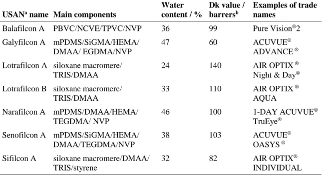
Table 2.2. Silicone hydrogels used for the preparation of corrective SCL (Sources: USAN dictionary, data from the manufactures).
a USAN: United States adopted name
b Dk represents the oxygen permeability (barrers = 10-11 cm2 O2 s-1 mmHg)
EGDMA: ethylene glycol dimethacrylate; DMAA: N,N’-dimethyl acrylamide; HEMA: 2-hydroxyethyl methacrylate; mPDMS: monofunctional polydimethylsiloxane; NCVE: N-carboxyvinyl ester; NVP: N- vinylpyrrolidone; PBVC: poly(dimethylsiloxyl)di-(silylbutanol)bis-(vinyl carbamate); SiGMA: methylbis- (trimethylsiloxy)silylpropylglyceryl methacrylate; TEGDMA: tetraethylene glycol dimethacrylate; TPVC:
tris(trimethylsiloxy)silylpropylvinyl carbamate; TRIS: methacryloxypropyl tris(trimethylsiloxy silane) USANa name Main components
Water content / %
Dk value / barrersb
Examples of trade names
Etafilcon A HEMA/SMA/TRIM 58 28 ACUVUE®2®
Hilafilcon B HEMA/EGDMA/AMA/NVP 59 22 SofLens®59 Nelfilcon A PVA, partially acetalized with
N-(formylmethyl)acrylamide
69 26 DAILIES®
AquaComfort Plus® Nesofilcon A HEMA/NVP/EGDMA/AMA/
PEPGDMA/TBHMA/MAEBTP
78 42 Biotrue® ONEday
Ocufilcon D HEMA/MAA/EGDMA 55 20 Biomedics® 1 Day
extra
Omafilcon A HEMA/MPC/EGDMA 62 33 Proclear®
Polymacon B HEMA/EGDMA 38 9 SofLens®38
USANa name Main components
Water content / %
Dk value / barrersb
Examples of trade names
Balafilcon A PBVC/NCVE/TPVC/NVP 36 99 Pure Vision®2 Galyfilcon A mPDMS/SiGMA/HEMA/
DMAA/ EGDMA/NVP
47 60 ACUVUE®
ADVANCE ® Lotrafilcon A siloxane macromere/
TRIS/DMAA
24 140 AIR OPTIX ®
Night & Day® Lotrafilcon B siloxane macromere/
TRIS/DMAA
33 110 AIR OPTIX ®
AQUA Narafilcon A mPDMS/DMAA/HEMA/
TEGDMA/ NVP
46 100 1-DAY ACUVUE®
TruEye® Senofilcon A mPDMS/SiGMA/HEMA/
DMAA/TEGDMA/NVP
38 103 ACUVUE®
OASYS ® Sifilcon A siloxane macromere/DMAA/
TRIS/styrene
32 82 AIR OPTIX®
INDIVIDUAL
2.4 Ophthalmic applications of hydrogels
2.4.3 Tissue adhesives for ocular wound repair
Another possible application for hydrogels is in the treatment of surgical or traumatic ocular wounds that are routinely sutured with thin nylon threads. Unfortunately, this standard treatment is associated with several disadvantages including infections, inflammation, corneal neovascularization, and impaired wound healing. To overcome the drawbacks of ocular sutures, tissue adhesives have been developed [96]. Tissue adhesives are materials, which are able to glue tissue surfaces together, either by mechanical adhesion or by forming chemical bonds. Soft and flexible hydrogels are among the most promising materials that are currently suggested as ophthalmic adhesives [97]. Many different polymers, either of natural or of synthetic origin, have been investigated for their suitability as ophthalmic sealants [51]. For example, hydrogels were prepared from polysaccharides, such as HA or chondroitin sulfate, gelatin, and different derivatives of PEG. The applied cross-linking reactions were, among other reactions, photoactivated processes and nucleophilic substitutions. For an optimal performance, ophthalmic adhesives must meet a number of requirements. Based on the available literature, Oelker and Grinstaff [51] defined essential design requirements for ideal ophthalmic adhesives, including leak pressure (> 80 mmHg), cross-linking time (< 30 s), mechanical properties (5–200 kPa), swelling (< 200%), diffusion coefficient (2 · 10-7 cm2 s-1), refractive index (1.32–1.40), adhesion strength (> 0.1 kPa), viscosity (5–100 cP), and degradation time (1 week–6 months). Very recently, the U.S. Food and Drug Administration approved ReSure® Sealant; the in situ forming hydrogel is based on PEG and trilysine amine, has a water content of approximately 90% after polymerization, and has been approved for sealing corneal incisions after cataract surgery. Although most tissue adhesives were developed for sealing corneal incisions, a few studies investigated their intraocular use in retinal detachment surgery [98–100]. Moreover, the possibility of incorporating drug molecules into hydrogel-based tissue adhesives has been studied. In particular, doxycycline-loaded hydrogels were investigated for the treatment of corneal injuries resulting from exposure to chemical warfare agents, such as sulfur mustard and nitrogen mustard [101,102]. In comparison with equally dosed doxycycline solutions, in situ forming doxycycline-loaded PEG hydrogels could improve epithelial healing in a vesicant-exposed corneal organ culture model [101]. In a follow-up study, rabbit corneas were exposed to sulfur mustard in vivo; the instillation of doxycycline eye drops resulted in faster edema reduction, but the hydrogels significantly reduced neovascularization [102]. In addition, hydrogels loaded with epidermal growth factor
(EGF) were developed for the treatment of corneal epithelial wounds. As a first proof of concept, Sheardown et al. [103] examined the effects of EGF on corneal epithelial wound healing in rabbits. Compared to control gels without EGF, the treatment with EGF-loaded PAA hydrogels resulted in significant wound healing enhancement. More recently, sustained release of EGF from chemically cross-linked cationized gelatin hydrogels has been reported [104]. The applied hydrogels gradually degraded over 1 week with concomitant release of EGF; the controlled release of EGF accelerated corneal epithelial wound healing in comparison with topically applied EGF solutions. Compared to physically cross-linked hydrogels, such as hydrogels based on gellan gum or alginate, these chemically cross-linked hydrogels are advantageous because they offer a much more prolonged drug release. Furthermore, high levels of drug loading can be reached, which is an advantage over drug-eluting SCL.
2.4.4 Intraocular lenses
In addition to these topical applications, hydrogels may also serve as IOL, vitreous substitutes, or intravitreal drug delivery systems. These intraocular applications are particularly relevant since most vision-threatening diseases, such as cataract, PDR or AMD, originate from the lens or the posterior segment of the eye. In cataract surgery, which is the most frequently performed eye surgery worldwide, the impaired lens is removed and subsequently substituted with an artificial IOL [28]. The first IOL were manufactured from rigid poly(methyl methacrylate). Today, more flexible materials, including hydrophobic acrylate polymers, hydrophilic acrylate polymers (i.e., hydrogels), siloxanes, and collamer (i.e., a copolymer of collagen and HEMA with a water content of 34%), are generally preferred [47,105]. These highly elastic materials are used to manufacture foldable IOL, which can be implanted through smaller incisions. The most frequent complication of cataract surgery is posterior capsule opacification (PCO), which has been associated with the abnormal proliferation of residual lens epithelial cells in the capsular bag [48]. As a consequence, several strategies to prevent PCO have been developed. For example, coating the lens surface with PEG or “bioactive” polymers exhibiting sulfonate and carboxylate groups could successfully prevent cell adhesion and proliferation [106,107]. Surface modification with porphyrins, which generate cytotoxic singlet oxygen, may also improve the biocompatibility of IOL [108]. Another approach to improve the biocompatibility of IOL and reduce the risk for PCO is the incorporation and controlled release of drug molecules. The available studies on drug-loaded IOL have
2.4 Ophthalmic applications of hydrogels been reviewed in two recent articles; the different approaches were compared with regard to the administered drug molecules, the used drug loading method, and potential therapeutic benefits [109,110]. In general, there are three possibilities for drug loading:
soaking the lens in concentrated drug solutions, coating of the lens surface (e.g., by spraying or chemical modification), and coating of the haptics that hold the lens in place.
Apart from the optical portion, IOL are not required to be fully transparent. For example, the haptics can be coated with non-transparent polymers, drug suspensions, or molecules that stain certain regions of the lens material [109]. Such drug-loaded IOL may have potential benefits in the prevention of endophthalmitis, a frequent complication of cataract surgery; they may prevent postoperative inflammation and reduce the risk for PCO. Nevertheless, there are no products approved for human use at the moment.
2.4.5 Hydrogel-based vitreous substitutes
Besides cataract, severe vitreoretinal diseases, such as diabetic retinopathy or retinal detachment, affect more and more people in industrialized nations. After surgical intervention and removal of the dysfunctional vitreous, the injection of vitreous substitutes may be required, either for short-term or for long-term treatment. Because of the complex function of the vitreous and delicate nature of the intraocular environment, potential vitreous substitutes have to meet particular requirements [111]. An ideal vitreous substitute should (1) be clear and transparent, (2) be biologically and chemically inert, (3) possess a refractive index and density comparable to those of the natural vitreous, (4) possess sufficient mechanical rigidity, (5) allow the transfer of metabolites, (6) be non-absorbable and non-biodegradable, (7) be hydrophilic, and (8) be injectable through small-gauge needles. At the moment, various materials are used in clinical practice, including gases (e.g., air, sulfur hexafluoride or perfluoropropane), perfluorocarbon liquids, fluorosilicone, and silicone oils [29,111–114]. However, none of these substances are ideal for long-term or permanent vitreous substitution due to their fast intraocular absorption, toxic reactions or other adverse effects, such as the formation of post-operative cataract or glaucoma. Therefore, alternative vitreous substitutes, based on natural, semisynthetic and synthetic polymers, have been investigated; the tested polymers included HPMC [115], ADCON-L [116], HA/gellan gum [117], pHEMA [118], poly(glycerol methacrylate) [119] Pluronic® [120], siloxanes [121], PVA [122] and poly(vinyl pyrolidone) [123]. In the early days, uncross-linked polymer solutions were investigated; however, these substances had major drawbacks and adverse
effects, such as short retention time, opacification and inflammation, and were, therefore, not useful as long-term vitreous substitutes [29,111,113]. Today’s experimental vitreous substitutes are mostly cross-linked hydrogels; these materials show an enhanced retention time in the eye and are capable to act as a tamponade agent. A comprehensive overview on these polymeric vitreous substitutes is given elsewhere and, therefore, not subject of this article [29]. Recently, new developments and improvements of existing hydrogel- based vitreous substitutes have been reported. For example, Leone et al. [124] reported the synthesis of PVA hydrogels through cross-linking with non-toxic trisodium trimetaphosphate (STMP). Using a STMP : PVA molar ratio of 1 : 8 resulted in rheological properties similar to those of the natural vitreous body. More importantly, these properties were preserved after injection. The developed PVA gel met the requirements for vitreous substitutes regarding transparency, swelling capacity, permeability, and cytotoxicity. However, in vivo studies to prove the long-term compatibility of this promising material remain to be done. Swindle-Reilly et al. [125]
copolymerized acrylamide with bisacryloylcystamine. The disulfide-containing cross- linker was then reduced to give an injectable polymer solution; once inside the eye, the disulfide cross-links reformed by air oxidation to produce a stable hydrogel that matched the mechanical properties of the natural vitreous humor. One week after injection into the vitreous cavity of rabbits, slit lamp examination, dilated fundus examination, electroretinography, and histopathological examination showed no signs of inflammation. Although this pilot study has indicated the biocompatibility of the gel, further in vivo studies on the long-term compatibility and stability are required. A different cross-linking approach was pursued by Tao et al. [126]. A mixture of two reactive PEG derivatives was injected into vitrectomized eyes of rabbits; cross-links were introduced in situ by Michael-type addition reaction. The formed hydrogel was stable during the study period of 9 months; the mechanical and optical properties were very similar to the natural vitreous body. No adverse reactions were detected by measurement of the intraocular pressure (IOP), fundus fluorescein angiography, electroretinography, histopathological examination, and B-scan ultrasound. Altogether, this comprehensive study demonstrated that chemically cross-linked PEG hydrogels might be suitable as long-term vitreous substitute. Besides synthetic polymers, the use of natural polymers for vitreous replacement has been investigated in two recent studies. With HA being an essential part of the human vitreous body [127], the use of this polymer for vitreous substitution seems obvious, even though the injection of uncross-linked HA solutions has
2.4 Ophthalmic applications of hydrogels proven to be unsuccessful [111]. Schramm et al. [128] compared two different HA hydrogels regarding their suitability as vitreous substitute; the cross-links were introduced either with adipic dihydrazide (ADH) and 1-ethyl-3-(3-dimethylaminopropyl)carbodi- imide (EDC), or by photopolymerization of glycidyl methacrylate groups. In cell culture experiments, ADH/EDC cross-linked hydrogels induced mild cytotoxicity, whereas photopolymerized HA gels showed no toxicity. These photopolymerized gels showed excellent in vivo biocompatibility during the study period of 6 weeks, without significant degradation or loss of transparency. In contrast to the results of Schramm et al., another study found no toxic effects of similar ADH cross-linked HA hydrogels, both in vitro and in vivo [129]. In addition to these conventional approaches of using polymeric hydrogels for vitreous replacement, Feng et al. [130] recently proposed using a foldable vitreous- shaped capsule to address the unknown long-term compatibility of these materials. The thin capsule was implanted into the vitreous cavity and subsequently filled with a PVA hydrogel; a prolonged in vivo stability was observed during the investigation period of 180 days. Moreover, the authors mentioned the possibility of incorporating drug molecules into the system. Although significant improvements have been achieved over the past years, there are up until now no artificial vitreous substitutes that reach the properties of the natural vitreous body. Even in situ cross-linkable hydrogels, which are generally favored due to the absence of shear thinning, do not meet all of the requirements to act as long-term vitreous substitute [29,111,113].
2.4.6 Intravitreal drug delivery systems
As mentioned earlier, PDR and AMD are among the leading causes of visual impairment or blindness in industrialized nations [131]. Characteristic for both diseases is an abnormal neovascularization due to increased levels of VEGF [132]. Besides photodynamic therapy and photocoagulation, current therapeutic approaches aim at neutralizing VEGF by intravitreal injections of anti-VEGF antibodies or antibody fragments every 4-6 weeks. These frequent intravitreal injections are associated with high healthcare costs, significant discomfort, and rare complications including endophthalmitis, retinal detachment, and uveitis [10]. Furthermore, it has been shown that VEGF plays an important role in supporting the adult subretinal vasculature, which is important for nurturing the photoreceptors and maintaining central vision [132–134].
Therefore, caution may be required when patients receive long-term treatment with anti- VEGF agents. One promising approach to solve these problems would be the local release
of anti-VEGF agents from controlled release systems over extended time periods. Besides implants, microspheres and nanoparticulate systems, hydrogels may serve as drug delivery systems in the posterior segment of the eye [35–37]. For obvious reasons, in situ forming hydrogels that can be injected by minimally invasive techniques are particularly favorable. In contrast to vitreous substitutes, hydrogel-based drug delivery systems usually occupy only a small part of the vitreous cavity, they are not required to be transparent, and degradability is often desired. Achieving high levels of drug loading and preserving the stability of the active ingredients are significant challenges in the development of intravitreal drug delivery systems. Furthermore, the cross-linking density, swelling capacity, and degradation rate of hydrogels need to be fine-tuned to ensure controlled drug release during the period of application. Special care has to be taken that the applied hydrogel does not increase the IOP in order to avoid severe complications, such as glaucoma. For these reasons, the use of temperature-responsive PNIPAAm hydrogels, which were additionally cross-linked with PEG diacrylate, has been suggested [135,136]. At room temperature, the highly swollen gels can be easily injected through small gauge needles. In the body, above the volume phase transition temperature, the hydrogels collapse resulting in an initial burst release of encapsulated drug followed by sustained release over several weeks. The non-degradability of cross-linked PNIPAAm hydrogels and the use of potentially harmful radical initiators are disadvantages of this material. Therefore, poly(ethylene glycol)-poly-(serinol hexamethylene urethane) (ESHU) hydrogels have been developed [137]. In contrast to PNIPAAm gels, the synthesis of ESHU does not require radical initiators; furthermore, ESHU is biodegradable and does not require surgical removal after drug release. ESHU hydrogels could be easily injected through 31-gauge needles. The gels were well tolerated in vivo; they did not affect the IOP and there was no evidence of inflammation. ESHU hydrogels sustained the release of incorporated bevacizumab for over 9 weeks; the drug concentration was on average 4.7 times higher than in eyes receiving bolus injections of bevacizumab. Brandl et al. [138] suggested using four-armed PEG-succinimidyl propionate that can be cross-linked in situ with PEG-amines to yield transparent hydrogels with high water content. The performed in vitro tests, including rheological characterization of mechanical properties, cytotoxicity studies and release experiments, suggested the suitability of PEG hydrogels as ophthalmic drug delivery system. In a similar approach, four-armed PEG was cross-linked via Michael-type addition reactions between sulfhydryl and maleimide groups [139]. Incorporated bevacizumab was released
2.4 Ophthalmic applications of hydrogels over 14 days; the hydrogels might, therefore, be suitable to treat intraocular neovascularization. And very recently, Yu et al. [59,140] reported the preparation of in situ chemically cross-linked hydrogels from vinyl sulfone functionalized HA and thiolated dextran. To reduce the covalent binding of proteins, both the concentration and the degree of modification of the thiolated dextran were increased relative to the vinyl sulfone functionalized HA. The incorporated bevacizumab was released over 3 months in vitro; the release rate was found to depend on the polymer concentration but not the degree of modification. First in vivo studies demonstrated the biocompatibility of the hydrogel; the hydrogel was able to prolong the retention of bevacizumab at therapeutically relevant concentrations for at least 6 months. Hydrogels have also been proposed for the treatment of retinal detachment. Neffe et al. [141] have synthesized a cross-linked HA hydrogel that can be applied as a patch to cover retinal breaks. The gel patches could be injected into the vitreous cavity, showed excellent intraocular biocompatibility, and good retinal adherence; the developed patches were slowly degrading and provided controlled release of incorporated triamcinolone.
2.4.7 Cell-based approaches
While intravitreal injections of anti-VEGF drugs are able to delay the progression of PDR and AMD, patients who have already lost vision due to severe ocular diseases or trauma may profit from cell-based therapeutic approaches. For example, it has been shown that the implantation of stem cells may have benefits in the treatment of corneal injuries and certain retinal diseases [142–145]. The cornea consists of three major layers: an outer stratified squamous epithelium, a thick stromal layer, and an inner monolayer of specialized endothelial cells. These endothelial cells play an important role in maintaining corneal clarity by regulating fluid and solute transport between the aqueous humor and the corneal stroma [146]. Tissue-engineered corneal epithelial cell sheets have already been used to treat patients with total limbal stem cell deficiencies [147]. Furthermore, the implantation of endothelial cell sheets has proven to be successful in animal models [148]. Human corneal endothelial cells were cultured on thermoresponsive PNIPAAm surfaces for 3 weeks at 37 °C and harvested as transplantable cell sheets by lowering the temperature to 20 °C. The subsequent transplantation to endothelium- denuded rabbit corneas could restore corneal function and clarity [148,149]. Ozcelik et al. [150] developed ultrathin chitosan-PEG hydrogel films for corneal tissue engineering. The films were approximately 50 µm thick, had desirable mechanical and