Cornichon Protein
I n a u g u r a l - D i s s e r t a t i o n zur Erlangung des Doktorgrades
der Mathematisch-Naturwissenschaftlichen Fakultät der Universität zu Köln
vorgelegt von Waldemar Wojciech
aus Laurahütte Köln, 2014
Berichterstatter/in: Prof. Dr. Henrike Scholz
Tag der letzten mündlichen Prüfung: 02.07.2014
Contents I
List of abbreviations IV
1 Introduction 1
1.1 The model organismDrosophila melanogaster . . . . 1
1.2 The early secretory pathway . . . . 2
1.3 COPII vesicle formation and cargo selection . . . . 4
1.4 COPII dependent cargo concentration . . . . 6
1.5 Transmembrane cargo receptors . . . . 6
1.6 The Cornichon protein family: Conserved cargo receptors . . . . 8
1.7 Human Cornichon homolog 4: Protein interactions and specificity for secretory cargo . . . . 10
1.8 Cornichon function: Evidence for diverse roles in neurotransmission . . 11
1.9 Objective . . . . 13
2 Materials and methods 14 2.1 Materials . . . . 14
2.1.1 General laboratory equipment . . . . 14
2.1.2 Chemicals . . . . 14
2.1.3 Reaction kits . . . . 14
2.1.4 Restriction enzymes and buffers . . . . 15
2.1.5 Solutions and media . . . . 15
2.1.6 Fly stocks . . . . 17
2.1.7 Oligonucleotides and primers . . . . 18
2.1.8 Vectors and plasmids . . . . 20
2.1.9 Antibodies and fluorescent dyes . . . . 21
2.1.10 Microscopy . . . . 21
2.1.11 Computer software . . . . 22
2.2 Methods . . . . 22
2.2.1 Fly stock keeping and breeding . . . . 22
2.2.2 Fly stock keeping and breeding for behavioural experiments . . 23
2.2.3 Evaluation of mendelian crosses . . . . 23
2.2.4 Negative gravitaxis assay . . . . 23
2.2.5 Adult survivorship assay . . . . 24
2.2.6 Developmental survivorship assay . . . . 25
2.2.7 Alcohol sensitivity assay . . . . 25
2.2.8 Generation of transgenic flies . . . . 26
2.2.9 Generation ofcnirknock-out flies . . . . 27
2.2.10 Generation of a∆cnir/cniAR55 double mutant . . . . 28
2.2.11 Generation of clones . . . . 28
2.2.12 Extraction of genomic DNA . . . . 29
2.2.13 RNA extraction and cDNA synthesis . . . . 29
2.2.14 Quantification of DNA . . . . 30
2.2.15 Polymerase chain reaction (PCR) . . . . 30
2.2.16 Gel electrophoresis . . . . 31
2.2.17 Sequencing of DNA . . . . 31
2.2.18 Cloning and ligation . . . . 32
2.2.19 Transformation of bacteria . . . . 33
2.2.20 Isolation of plasmid DNA . . . . 33
2.2.21 Cloning ofcnirdonor construct . . . . 33
2.2.22 Preparation of egg shells . . . . 34
2.2.23 Dissection, fixation and antibody staining in ovaries . . . . 34
2.2.24 Dissection, fixation and antibody staining in imaginal discs . . . 34
2.2.25 Phylogenetic analysis of Cornichon proteins and protein mem- brane topology . . . . 35
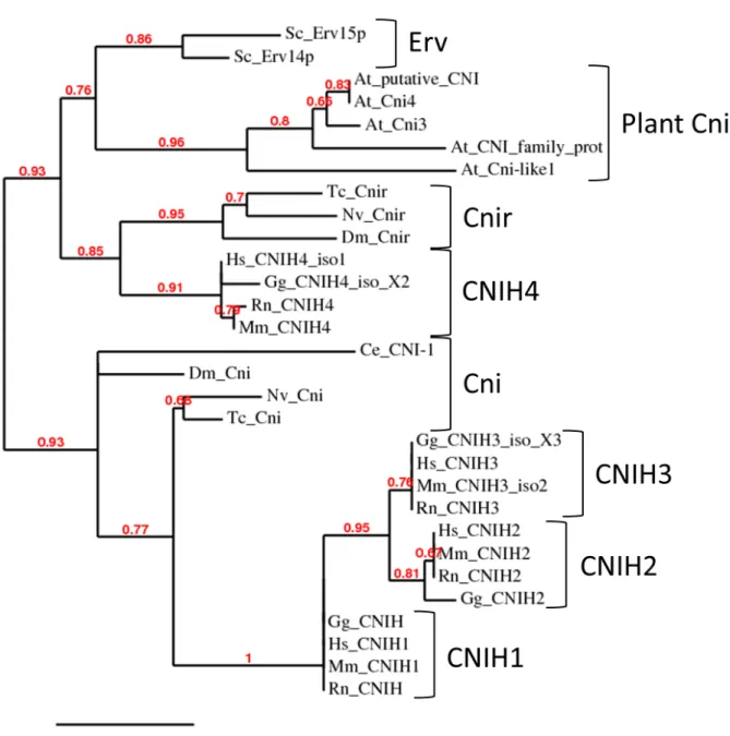
3 Results 36 3.1 Phylogenetic analysis of Cni proteins and the Cnir membrane topology 36 3.2 Generation of acnir knock-out line . . . . 36
3.3 Survivorship of ∆cnirthroughout development . . . . 42
3.4 Survivorship of adult ∆cnirflies . . . . 45
3.5 Locomotion defects of ∆cnir flies . . . . 45
3.6 Ethanol sensitivity of∆cnirflies . . . . 48
3.7 Muscular rescue of locomotion defects . . . . 50
3.8 Neuronal rescue of locomotion defects . . . . 53
3.9 Cnir protein localization and neuronal rescue with GFP:Cnir . . . . 56
3.10 Analysis of a cnir/cnidouble mutant . . . . 61
4 Discussion 67 4.1 Generation of a mutant forcnir: A putative ortholog of humanCNIH4 . 67 4.2 ∆cnir is not lethal but has an impact on mortality throughout develop-
ment and adult life . . . . 68 4.3 Locomotor behaviour depends on Cnir function in neurons but not in
muscles . . . . 69 4.4 Loss of Cnir leads to decreased ethanol sensitivity . . . . 71 4.5 GFP tagged Cnir potentially localizes to the ER but is not functional . . 72 4.6 Drosophila Cni and Cnir do not show strong functional redundancy in
the soma . . . . 73 4.7 Perspectives . . . . 75
References 78
Supplement 89
List of figures 91
List of tables 92
Zusammenfassung 93
Abstract 94
Danksagungen 95
Erklärung 96
Lebenslauf 97
AC adenylyl cyclase
act actin
AMPA α-amino-3-hydroxy-5-methyl-4-isoxazolepropionic acid
AMPAR α-amino-3-hydroxy-5-methyl-4-isoxazolepropionic acid receptor
AR adrenergic receptor
At Arabidopsis thaliana
ATP adenosine triphosphate
b black
bp base pair
BSA bovine serum albumin
°C degree Celsius
cAMP 3’-5’-cyclic adenosine monophosphate
cDNA complementary
cds coding sequence
CA countercurrent apparatus
Cf partition coefficient CFP cyan fluorescent protein
cn cinnabar
Cni Cornichon
CNIH Cornichon homolog
Cnir Cornichon related
COPI/II coat protein complex I/II
CyO Curly of Oster
DAPI 4’,6-diamidino-2-phenylindole DD2R Dopamine D2-like receptor
DNA deoxyribonucleic acid
Dnc Dunce
dNTP deoxynucleotide triphosphate
Dm Drosophila melanogaster
EGFR epidermal growth factor receptor
ER endoplasmic reticulum
ERAD ER associated degradation
eye eyeless
ERES ER exit sites
fig. figure
FLP flipase
FRT flipase recombination target
g gram
GAP GTPase activating protein GEF guanine exchange factor GFP green fluorescent protein
Gg Gallus gallus
Gla glazed
GluR glutamate receptor
GPCR G protein coupled receptor
Grk Gurken
h hour
Hs Homo sapiens
hs heat shock promoter
IF irregular facit
kb kilo base pairs
kDa kilodalton
l liter
M mole
MCP bacteriophage MS2 coat protein
MET mean elution time
mg milligram
mhc myosin heavy chain
min minute
ml milliliter
Mm Mus musculus
mRNA messenger ribonucleic acid
NGS normal goat serum
NMJ neuromuscular junction
no. number
nos nanos
Nv Nasonia vitripennis
OD optical density
PBS phosphate buffered saline
PBT phosphate buffered saline with Triton X-100 PCR polymerase chain reaction
pr purple
Rn Rattus norvegicus
rpm round per minute
ry rosy
s second
Sc Saccharomyces cerevisiae
Sco Scutoid
SM second multiple
SOC super optimal broth with catabolite repression
Sp Sternopleural
SSC saline-sodium citrate
tab. table
Taq Thermus aquaticus
TARP transmembrane AMPAR regulatory protein
Tc Tribolium castaneum
TGFα transforming growth factorα
TMD transmembrane domain
TM2 third multiple2
TM6B third multiple6B
Tris tris(hydroxymethyl)aminomethane
tub tubulin
U unit
UAS upstream activating sequence
UTR untranslated region
UV ultra violet
V volt
w white
wt wild type
YFP yellow fluorescent protein
X-Gal bromo-chloro-indolyl-galactopyranoside β-Gal β-Galactosidase
µg microgram
µl microliter
µM micromolar
1.1 The model organism Drosophila melanogaster
The fruit flyDrosophila melanogaster is one of the best studied model organisms in bi- ology. Besides the classical advantages like the short generation time of only ten days and the high number of progeny, theDrosophilagenome is also completely sequenced since the year 2000 [Adams et al., 2000]. Furthermore, it is estimated that approxi- mately 75% of human disease genes have an obvious ortholog in flies, which makes Drosophila a valuable model for human disease [Chienet al.,2002].
The advantages ofDrosophilaas a model are augmented by the availability of diverse techniques for genetic manipulation that allow the precise study of genes and their roles in cellular processes. For example, transgenesis via the P element-mediated [Rubin and Spradling, 1983] and φC31 integrase transformation [Bischof et al., 2007] systems offer the opportunity for expression studies of tagged proteins to monitor their cellular and subcellular localization. Furthermore, the UAS/Gal4system [Brand and Perrimon,1993] enables misexpression of genes to investigate their temporal and spatial requirement. In addition, the FLP/FRT system [Chou et al., 1993; Xu and Rubin, 1993; Chou and Perrimon, 1996] for generation of mitotic clones can be used to analyze labeled mutant cells in direct comparison to wild type cells in the same tissue. Finally, genetic tools to create gene knock-outs via homologous recombination allow the generation of mutants for any gene of interest [Gong and Golic,2003; Huang et al.,2008].
Overall, Drosophila holds many tissues that are accessible for extensive manipu- lation and are models for many cellular and developmental events. Oogenesis, for example, requires almost all cellular processes for the development of a stem cell into a mature egg, such as cell cycle control, fate specification, cell polarization and epithe- lial morphogenesis [Bastock and St Johnston, 2008]. In addition, Drosophila imaginal discs are a common epithelial model for investigation of pattern formation and cell proliferation [McClure and Schubiger, 2005]. Furthermore, the larval neuromuscu- lar junction (NMJ) poses a comparatively simple system to investigate developmental and functional plasticity at synapses that possess glutamate receptors homologous to those in the mammalian brain [Menonet al., 2013].
Moreover, Drosophila is a complex organism with a rich behavioral repertoire that has been established as a model for larval and adult locomotion [Garganoet al.,2005; Inagaki et al., 2010; Sinadinos et al., 2012], alcohol research [Devineni and Heberlein, 2013], as well as aging [Partridgeet al.,2011].
In this thesis, all of these advantages of Drosophila as a model are applied to in- vestigate the function of Cornichon-related (Cnir). DrosophilaCnir belongs to a highly conserved protein family of cargo receptors, but its function has not been investigated.
In the early secretory pathway of all eukaryotic cells, Cornichon proteins facilitate ef- ficient endoplasmic reticulum (ER) export of numerous secretory proteins. Therefore, the mechanisms of early protein secretion are introduced below.
1.2 The early secretory pathway
In eukaryotic cells many proteins enter the secretory pathway in order to be accu- rately delivered with the correct temporal and spatial localization, such as to the plasma membrane or extracellular space. Therefore, this process is crucial for cell function and development of all eukaryotic organisms [Dancourt and Barlowe, 2010; D’Arcangeloet al.,2013].
Secretory proteins have sorting elements that are recognized by the intracellular transport machinery at multiple stages of the transport process to guide the protein cargo to its proper location. The organization of the secretory pathway, which con- sists of membrane bound compartments, strongly depends on coat protein complexes.
Those complexes recognize sorting signals at the surface of single compartments and selectively sort proteins into transport vesicles. The best studied coat complexes are clathrin and coat protein complexes I and II (COPI and COPII). Each of those com- plexes is a multi subunit structure, and direct binding of a subunit to a cargo protein is required for uptake into a forming carrier vesicle. However, in some cases the ef- ficient incorporation of a cargo into a transport vesicle requires adaptor proteins or transmembrane receptors [Dancourt and Barlowe, 2010].
Translation and folding of nascent secretory proteins take place at the ER. An effi- cient quality control system ensures that unfolded proteins are retained or not recog- nized for uptake into COPII vesicles and subsequent transport to pre Golgi or Golgi compartments until proper folding occurs [Vembar and Brodsky, 2008; Dancourt and Barlowe, 2010]. The transport between ER and Golgi is highly dynamic [Sciaky et al.,
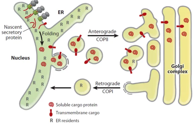
1997; Wardet al., 2001; Losevet al., 2006]. The anterograde transport of secretory pro- teins in COPII vesicles is equilibrated by a retrograde transport in COPI vesicles in order to recycle vesicle components and ER resident escaped proteins (fig. 1.1). How- ever, secretory cargo advances steadily forward, while resident proteins of the early secretory pathway are dynamically transported back into the proper compartments [Dancourt and Barlowe, 2010].
To understand cargo selection for anterograde transport from the ER, it is important to elucidate the composition of COPII vesicles and mechanisms of their formation.
Hence, these processes are considered next.
Figure 1.1 | Bidirectional transport between ER and Golgi
Scheme of the bidirectional transport between ER and Golgi. Nascent secretory proteins are trans- lated and folded in the ER. Completely folded transmembrane and soluble cargo proteins are sub- sequently incorporated into COPII vesicles for anterograde transport to the pre-Golgi and Golgi compartments. The anterograde transport is balanced by a retrograde transport in COPI vesicles in order to recycle vesicle components and retrieve escaped ER resident proteins (R). As a con- sequence of those processes, secretory cargo moves steadily anterograde, while resident proteins localize dynamically to early secretory compartments (figure from Dancourt and Barlowe [2010]).
1.3 COPII vesicle formation and cargo selection
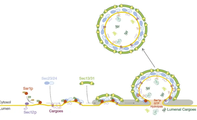
The machinery responsible for budding of COPII vesicles is localized to regions known as ER exit sites (ERES) [Orci et al., 1991; Bannykh et al., 1996; Rossaneseet al., 1999; Dancourt and Barlowe, 2010]. The different steps in COPII vesicle assembly are depicted in fig. 1.2.
The first event in COPII vesicle formation is the activation of the small GTPase Sar1p by its guanine exchange factor (GEF) Sec12p. As a consequence of Sar1p activation through the exchange of GDP to GTP, its hydrophobic N-terminal amphiatic α-helix is exposed and inserted into the ER membrane. That process leads to the bending of the membrane and recruitment of the Sec23-Sec24 complex. This complex serves as a cargo adaptor and furthermore as a specific GTPase activating protein (GAP) com- plex for Sar1p. Lastly the outer layer, consisting of Sec13-Sec31 heterotertrameres, forms around the Sar1-Sec23-Sec24 pre budding complex. This leads to formation of a cage like structure that bends the lipid bilayer of the ER and finally buds vesi- cles [Lee et al., 2004, 2005; Budnik and Stephens, 2009; Dancourt and Barlowe, 2010; D’Arcangelo et al., 2013]. In vitro studies have shown that cage like structures [Stagg et al., 2006], as well as COPII vesicles [Matsuoka et al., 1998], can be formed with merely the corepurified proteins (Sar1p, Sec23-Sec24, Sec13-Sec31) and synthetic lipo- somes [Dancourt and Barlowe, 2010; D’Arcangelo et al., 2013].
In addition to the primary feature of forming vesicles, the COPII recognizes and se- lects cargo proteins for uptake into vesicles, while separating them from ER resident proteins [Salamaet al.,1993; Barloweet al.,1994]. Typically, cargoes can be subdivided into integral membrane proteins and soluble lumenal proteins. Transmembrane pro- teins can have one or multiple membrane spanning segments and a type I topology with the N-terminus facing the inside, or a type II topology with the N-terminus facing the outside of the ER lumen [Dancourt and Barlowe, 2010]. Transmembrane cargoes have sorting signals presented on their cytoplasmic surfaces that direct their uptake into COPII [Bonifacino and Glick, 2004]. Biochemical approaches show that transmembrane cargo proteins bind to the Sec23-Sec24 complex. Furthermore, this interaction is sorting signal dependent [Aridor et al., 1998; Kuehn et al., 1998]. The presence of a non hydrolyzable GTP is able to stabilize the formation of the cargo complex consisting of Sec23-Sec24, Sar1p-GTP and cargo. In contrast, the controlled hydrolysis of GTP by Sar1p enables the complex to dissociate. Thus, the cargo can be released from its sorting subunits and COPII components can be recycled at ERES
Figure 1.2 | COPII vesicle formation and cargo selection
Scheme of the COPII vesicle formation and cargo selection process. The GEF Sec12p activates Sar1p. Subsequently, Sec23-Sec24 binds to the activated membrane-bound Sar1p-GTP and form pre budding complexes. In those complexes Sec24p is responsible for binding to sorting signals present on cargo proteins. As indicated, the binding of cargo proteins to Sec24p can be direct or mediated by transmembrane sorting receptors. Ultimately, the Sec13-Sec31 complex is recruited to the pre budding cargo complexes, forming the outer layer. This leads to curvature of the ER membrane and finally budding of the vesicle (figure modified from D’Arcangeloet al.[2013]).
[Dancourt and Barlowe, 2010]. The binding of Sec24p to well defined sorting signals is possible due to various cargo recognition sites within this protein [Milleret al.,2003; Mossessova et al., 2003]. In addition, the diversity of potentially recognized sorting signals is increased by the presence of several Sec24p isoforms [Miller et al., 2002; Wendeleret al.,2007].
Many soluble cargo proteins do not span the ER membrane and thus cannot be recognized by COPII subunits. Furthermore, not all secretory proteins possess notice- able COPII sorting signals. Hence, transmembrane cargo receptors may be necessary to facilitate efficient export of many types of secretory cargoes from the ER by linking them to the COPII budding complex [Dancourt and Barlowe, 2010].
The cargo recognition is often associated with cargo concentration. Therefore, the mechanisms of cargo concentration will be addressed in the following section.
1.4 COPII dependent cargo concentration
Early studies already suggested that viral transmembrane proteins can be concen- trated during transport from the ER to the Golgi [Quinn et al., 1984] and that some viral glycoproteins can be concentrated up to tenfold during vesicle budding from the ER [Balchet al.,1994]. Those results were suported byin vitrodata [Salamaet al.,1993; Rexach et al., 1994; Aridor et al., 1998; Kuehn et al., 1998] and genetic experiments [Kappeleret al.,1997; Nishimura and Balch, 1997] indicating a COPII dependent con- centrative ER export of integral membrane cargo [Dancourt and Barlowe, 2010]. As previously mentioned, the recognition of signals in transmembrane cargo strongly relies on Sec24p [Milleret al., 2003; Mossessovaet al., 2003].
Although transmembrane cargoes are concentrated during transport, soluble secre- tory proteins show both, concentrative [Mizuno and Singer,1993; Malkus et al., 2002] or bulk flow mechanisms [Martínez-Menárguez et al., 1999]. Which mechanism is used depends mainly on the cargo investigated [Barlowe, 2003; Dancourt and Bar- lowe, 2010]. Therefore, bulk flow and concentrative ER export mechanisms cannot be seen as mutually exclusive [Dancourt and Barlowe, 2010]. Importantly, there is evidence that secretory cargo requires cargo receptors for concentration into COPII during budding from the ER [Barlowe et al., 1994; Kuehn et al., 1998; Dancourt and Barlowe,2010].
As previously discussed, cargo receptors are crucial for recognition and concentra- tion of cargo. How cargo receptor binding to its cargoes is regulated, the impact of cargo receptors on ER quality control and the consequences of mutation of a specific cargo receptor are described below.
1.5 Transmembrane cargo receptors
Many abundant membrane proteins that localize to early secretory compartments and transport intermediates act in cargo sorting and transport between the ER and Golgi.
Cells lacking specific cargo receptors show particular sorting defects characterized by an inefficient export of a subset of secretory proteins from the ER, while other se- cretory proteins traffic at normal rates. Cargo sorting receptors are believed to cycle between ER and Golgi compartments in COPII and COPI vesicles due to cytoplas- mically exposed coat recognition signals. Thus, anterograde transport of a specific cargo through binding in the ER is followed by dissociation in the pre Golgi and
Golgi compartments. The dissociation is induced by a lower pH and potentially by Ca2+ gradients. This in turn leads to conformational changes in cargo receptors to decrease their affinity for cargo. In addition, every characterized cargo receptor forms oligomeric complexes, allowing major conformational shifts in slightly different pH conditions, which is a widely used mechanism for regulation of binding affinity [Dan- court and Barlowe, 2010].
Cargo receptors can be subdivided into canonical and non canonical. The first link lumenal cargo to the COPII coat while the latter facilitate transport of integral membrane proteins, which could exhibit their own ER export motifs [D’Arcangelo et al.,2013].
While cargo receptors appear not to be involved in cargo folding, the binding of cargo to a receptor is directly connected to the ER quality control. For instance, yeast strains lacking certain cargo receptors show activation of the unfolded protein response pathway [Belden and Barlowe, 2001a; Bue et al., 2006; Jonikas et al., 2009; Dancourt and Barlowe, 2010]. Furthermore, a terminally misfolded ER associated degradation (ERAD) substrate in yeast has a reduced turnover rate when its cargo receptor is lacking. It could be that in this case susceptibility of the misfolded cargo for ERAD depends on prior binding to its receptor for ER exit and subsequent re- trieval from post ER compartments [Kincaid and Cooper, 2007]. Yet, an affinity of a cargo receptor for its misfolded cargo can also be reduced, which might help to guide the misfolded cargo away from the ER folding chaperones, making it more prone to ERAD [Dancourt and Barlowe, 2009, 2010]. Furthermore, some data demonstrate that several cargo receptors recognize preferentially completely folded and assem- bled cargo [Otte and Barlowe, 2004; Appenzeller-Herzog et al., 2005; Dancourt and Barlowe,2009,2010]. Thus many different mechanisms possibly couple cargo binding to its receptor to the ER quality control.
Although cargo receptors share most of the common features described in the pre- vious sections, each of them has specific activities. The following section will address the function of the Cornichon protein family of cargo receptors in more detail.
1.6 The Cornichon protein family: Conserved cargo receptors
Drosophila Cornichon (Cni) is the founding member of a conserved protein family of cargo receptors [Rothet al.,1995]. At least two Cni paralogs can be found in almost all eukaryotes analyzed so far, ranging from plants to vertebrates. In Drosophila, the cni mutation is characterized by a ventralized embryo due to a failure in ER export of the TGFα ligand Gurken (Grk). For proper signaling, Grk must be processed and trans- ported to the oocyte surface [Rothet al.,1995; Herpers and Rabouille,2004; Bökelet al., 2006; Dancourt and Barlowe, 2010]. It has been shown molecularly that the first 30 membrane proximal residues of Grk are necessary for interaction with the N-terminal half of Cni. TheDrosophilagenome encodes a second Cni paralog known asCni-related (Cnir). Although very little is known aboutcnir, there are hints that the twoDrosophila cni genes have partially overlapping functions. For example, the lack of onecnircopy in acni amorphic mutant background leads to synthetic lethality. Furthermore, it has been shown that expression of cnir under the control of a cni promoter rescues the synthetic lethality, as well as some somatic phenotypes ofcnimutant flies [Bökelet al., 2006]. Mechanistically, the recognition of cargo by Cni proteins, as well as its role as a cargo receptor, seem to be conserved, since studies of the mammalian Cornichon homolog 1 (CNIH1) shows that the protein colocalizes with makers of the early se- cretory pathway and that it affects secretion of mammalian TGFα [Castroet al., 2007; Dancourt and Barlowe, 2010].
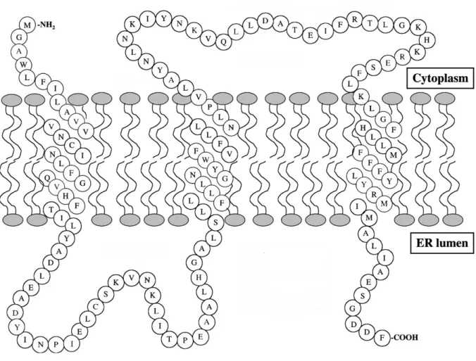
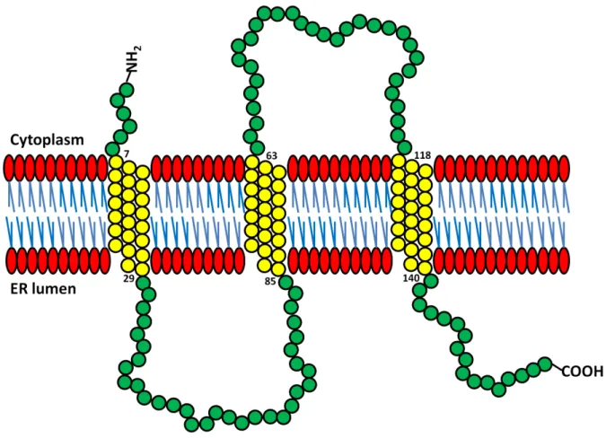
One of the molecularly best studied Cni proteins is the yeast Erv14p, a small hy- drophobic protein, which spans the ER membrane three times and is a non canonical cargo receptor (fig. 1.3). Hence, it has one cytoplasmic loop and one that faces the ER lumen. Erv14p is a component of COPII vesicles that mediate cargo export of the transmembrane secretory protein, Axl2p, to the cell surface. The delivery of Axl2p is important for budding site selection [Powers and Barlowe, 1998, 2002]. It has been demonstrated that Erv14p physically interacts with Axl2p, as well as the COPII pre budding Sec23-Sec24-Sar1-GTP complex. Binding of Erv14p to subunits of the COPII coat is believed to depend on conserved residues in its cytoplasmic second loop do- main [Powers and Barlowe,2002].
Furthermore, Erv14p is involved in the transport of Sma2p which is important for prospore membrane formation during yeast sporulation. Prospore membrane defects
Figure 1.3 | Model of Erv14p
Model of the Erv14p protein structure with its N-terminus being cytoplasmatically localized, while its C-terminus resides in the ER lumen. The cytoplasmic second loop is believed to be crucial for Erv14p binding to the COPII coat (figure modified from Powers and Barlowe [2002]).
inerv14mutants can be partially rescued by overexpression of its paralog Erv15p and can be enhanced in the double mutant. This indicates functional redundancy of both yeast Cni paralogs [Nakanishi et al., 2007]. Moreover, it has been demonstrated that a large group of transmembrane bitopic and polytopic proteins requires Erv14p as a cargo receptor [Castillon et al., 2009; Herziget al., 2012]. Strikingly, those proteins do not share a known functional or structural similarity, nor a sequence motif. However, all of them reside in late secretory pathway membranes that are populated with pro- teins of longer transmembrane domains (TMDs) compared to TMDs of ER resident proteins [Sharpe et al., 2010; Herzig et al., 2012]. Indeed, it was shown that cargo specificity of Erv14p depends on a TMD length of at least 22 amino acids to accel- erate cargo export from the ER. Thus, recognition of cargo occurs through physical
properties, rather than sequence motifs [Herzig et al., 2012]. It could be that Erv14p functions as a chaperone to protect cargo TMDs from degradation, which is possibly triggered by hydrophobic mismatches between the thin lipid bilayer of the ER and the long TMDs of those cargoes [D’Arcangelo et al., 2013].
Although TMD length of cargo proteins is also crucial for their Golgi exit and localization to the plasma membrane, Erv14p does not play a role in this later sorting process. It has been speculated that in this case Golgi exit of cargo requires another cargo receptor, or depends on a different vesicle composition [Herzig et al., 2012].
1.7 Human Cornichon homolog 4: Protein interactions and specificity for secretory cargo
Novel investigations identify the human CNIH4as an interaction partner of members from the three major families of G protein coupled receptors (GPCRs). GPCRs repre- sent the largest superfamily of cell surface receptors and although they do not possess a high sequence homology they share a seven TMD topology, with each TMD being 20-30amino acids long. GPCRs are categorized into six families (A-F), with A-C rep- resenting the main families [Caerset al.,2012; Sauvageauet al.,2014]. GPCRs regulate an immense number of physiological and cellular processes like proliferation, devel- opment, sensory perception, metabolism, nerve transmission, neuromodulation and locomotion [Bendenaet al.,2012]. Thus, they are activated by a broad range of ligands.
For example, family A GPCRs are activated by odorants, biogenic amines, neuropep- tides, peptidergic hormones, lipids, nucleotides, proteases, and even photons. Family B GPCRs bind to hormones and peptides, while family C (metabotropic glutamate) binds to amino acids, ions, and tastants [Allen and Roth, 2011]. Yet, all known neu- ropeptide GPCRs belong to family A (rhosopsin-like) or family B (secretin-like) [Caers et al.,2012]
After synthesis, folding and assembly, GPCRs are packed into COPII vesicles at ERES [Dupré et al., 2006; Dong et al., 2008; Sauvageau et al., 2014] and transported through the pre Golgi and Golgi compartments to the plasma membrane. During the trafficking process, many GPCRs undergo consecutive post translational modifica- tions like N- and O-glycosylation, which can be used as readouts for their maturation state [Donget al., 2007; Sauvageauet al., 2014]. Strikingly, the ER exit has been shown
to be the bottleneck in maturation and cell surface transport of GPCRs [Petaja-Repo et al.,2000; Sauvageauet al.,2014].
Human CNIH4 interacts selectively with members from the three major families of GPCRs (A-C) and the COPII components Sec23 and Sec24. Furthermore, it does not bind single TMD proteins like EGFR, the T-cell receptors CD4 and CD8, or the 12TMD adenylyl cyclase, which implies CNIH4specificity in cargo selection. CNIH4 localizes to the early secretory pathway and overexpression, as well as knock down of CNIH4, causes retention of GPCRs in the ER. However, low levels of CNIH4 are cru- cial for maturation and cell surface expression of the G protein coupled β2-adrenergic receptor (family A). In contrast to the knock-down of CNIH4, the overexpression leads to proteasome mediated degradation of receptors. This indicates an active function of CNIH4 in degradation of ER retained cargo. CNIH4 does not colocal- ize with GPCRs at the plasma membrane and selectively binds to the immature ER form of β2-adrenergic receptor, indicating no permanent interaction. Taken together, the data suggest an important function of CNIH4in regulation of GPCR export levels [Sauvageau et al., 2014].
Interestingly, many Drosophila GPCRs, neuropeptides and GPCR signaling path- ways elements are important models for their vertebrate homologs due to high func- tional conservation. Even minor changes in GPCRs, or their regulatory proteins can result in behavioral plasticity because of changes in GPCR controlled pathways [Ben- dena et al., 2012]. However, there is no evidence for involvement of Drosophila Cni proteins in the control of GPCR trafficking and the potentially resulting behavioral alterations.
Although Cni proteins represent a conserved family of cargo receptors, there is ev- idence that they also possess a role beyond trafficking of transmembrane cargo. New studies show that some Cni paralogs are involved in regulation of neurotransmission through Glutamate receptors. Therefore, the Cni function in neurons is highlighted next.
1.8 Cornichon function: Evidence for diverse roles in neurotransmission
Several recent studies identify Cni proteins as a functional subunit of ionotropic gluta- mate receptors (GluRs) of the AMPA (α-amino-3-hydroxy-5-methyl-4-isoxazolepropio-
nic acid) subtype [Schwenk et al., 2009; Kato et al., 2010; Shi et al., 2010; Coombs et al., 2012]. The tetrameric AMPA receptors (AMPARs) consist of the pore-lining α- subunits GluA1-4 and auxiliary β-subunits that regulate their gating properties and trafficking. Thereby, the β-subunits mediate swift excitatory synaptic transmission in the mammalian brain [Harmel et al., 2012]. The transmembrane AMPAR regulatory proteins (TARPs) have six isoforms and are β-subunits of most AMPARs [Gill et al., 2011]. Nevertheless, it has been shown that the majority of AMPARs in the rat brain are co-assembled with Cornichon homolog 2and 3(CNIH2 and CNIH3), rather than TARPs. In heterologous cells, CNIH proteins increase surface expression of AMPARs and furthermore alter channel gating by slowing deactivation and desensitization ki- netics [Schwenk et al., 2009; Kato et al., 2010; Shi et al., 2010; Coombset al., 2012]. The picture is complicated further by a diverse localization of CNIH2 in different neuron types depending on the expressed TARP isoform. For instance, CNIH2 can be found in the surface of hippocampal neurons, while it is absent at the surface of Purkinje neurons of stargazer mice expressing a different TARP isoform [Gill et al., 2011]. In addition, CNIH2 differentially modulates AMPAR kinetics depending on the TARP isoform composition in the receptor complex [Gill et al., 2012].
The first in vivo analysis of Cni proteins was performed in CNIH2/CNIH3 condi- tional knock-out mice. Glutamate gated currents are strongly reduced in CNIH2/
CNIH3 mutant hippocampal neurons due to the selective binding of CNIH2 and CNIH3to GluA1[Herring et al., 2013]. Thus only GluA1 containing AMPARs, which are predominant in hippocampal neurons and deactivate slowly, can be localized to the plasma membrane [Lu et al., 2009; Herring et al., 2013]. It is reasoned that in- teraction of CNIH2 and CNIH3 with other GluAα-subunits is prevented depending on the TARP isoform expressed in hippocampal neurons. Therefore, transport and gating of different AMPARs seems to be regulated by the interaction of itsα-subunits, CNIHs and TARPs [Herring et al., 2013]. In keeping with the importance of CNIH2 in regulation of AMPARs, its deletion has been reported to be involved in mild intel- lectual disorders in human disease [Floor et al., 2012]. Furthermore, elevation of the CNIH1-3, but notCNIH4, mRNA levels have been reported in the prefrontal cortex of schizophrenia patients [Drummondet al.,2012].
Other studies indicate that CNIH2 still possesses its conserved function as a cargo receptor continuously cycling between ER and Golgi in a COPII dependent manner. In the ER, CNIH2is believed to alter the glycosylation pattern of GluA2, thus regulating
AMPAR maturation and thereby possibly influencing AMPAR function at synapses [Harmelet al.,2012; Brockie et al., 2013].
A study of the sole Cni homolog (CNI-1) inCaenorhabditis elegansshows that it colo- calizes with the AMPAR subunit GLR-1and the Sec24COPII component, indicating a role in regulation of GLR-1 trafficking. Furthermore, CNI-1colocalizes with synaptic GLR-1. In contrast to the reports on CNIH2 and CNIH3 function in hippocampal neurons of knock-out mice [Herring et al., 2013], nematode mutants forcni-1 possess elevated synaptic transmission through AMPARs. Consistently, worms lacking CNI-1 function display a higher number of GluRs at synapses [Brockieet al., 2013]. In addi- tion, reconstitution experiments with the vertebrate CNIH1 and CNIH2show similar results. Therefore, although Cni proteins seem to have an evolutionarily conserved function in the regulation of AMPARs there might be additional regulatory effects on AMPAR transport in vertebrate neurons [Brockie et al., 2013].
Although the primary neurotransmitter in excitatory synapses in the fly brain is acetylcholine [Yasuyama and Salvaterra, 1999], the Drosophila larval neuro muscular junction (NMJ) synapses use ionotropic GluRs homologous to AMPARs in the mam- malian brain. Moreover, many synaptic components are conserved betweenDrosophila and vertebrates [Chen et al., 1986; Davis et al., 1989; Lahey et al., 1994; Tabuchi and Südhof, 2002; Banovic et al., 2010; Sun et al., 2011; Menon et al., 2013]. However, no connection between Cni proteins and Gulatmate receptors has been described in the fly.
1.9 Objective
The aim of this thesis was to investigate the loss of function phenotype of theDrosophila cnirgene and its impact on the viability and behavior of the fly. Furthermore, the goal was to analyze potential functional overlaps with its paralogcni.
2.1 Materials
2.1.1 General laboratory equipment
All plastic laboratory equipment used was ordered from the companies Eppendorf (Wesseling-Berzdorf), Sarstedt (Nümbrecht), Simport Plastics Ltd. (Beloeil, QC Cana- da), Regina Industries Ltd. (Newcastle, England), Sorenson BioScience (West Salt Lake City, USA) and Ratiolab (Dreieich).
2.1.2 Chemicals
All chemicals used during this thesis were ordered from the companies Roth (Karls- ruhe), Sigma Aldrich (Steinheim), Invitrogen (Karlsruhe), VWR International GmbH (Darmstadt) and Polysciences Europe GmbH (Eppelheim).
2.1.3 Reaction kits
Table 2.1 shows all used reaction kits and the company they were manufactured by.
The kits were all used according to the supplied manuals.
Table 2.1 | Reaction Kits
Reaction Kit Company
GenElute Plasmid Midiprep Kit Sigma Aldrich, Steinheim HiSpeed Plasmid Midi Kit Qiagen, Hilden
Zymoclean Gel DNA Recovery Kit Zymo Research, Orange, USA ZR Plasmid Miniprep Classic Zymo Research, Orange, USA TOPO TA Cloning Kit Dual Promoter Invitrogen, Karlsruhe
pENTR Directional TOPO Cloning Kit Invitrogen, Karlsruhe SuperScript II Reverse Transcriptase Invitrogen, Karlsruhe
2.1.4 Restriction enzymes and buffers
Table 2.2 shows all restriction enzymes that were used, as well as the company they ordered from and the buffers they were used in. Each enzyme was used according to suggestions of the manufacturer.
Table 2.2 | Restriction enzymes
Enzyme Company Buffer
NotI Thermo Scientific, Schwerte Buffer O NdeI Thermo Scientific, Schwerte Buffer O BglII Thermo Scientific, Schwerte Buffer BamHI AscI NEB, Ipswich, England Buffer BamHI
2.1.5 Solutions and media
All solutions and media used for this thesis are listed in table 2.3 in alphabetical or- der and were made with distilled H2O (Milli-Q Water Purification System, Millipore, Eschborn) or labeled individually if not.
Table 2.3 | Solutions and media
Solution/Medium Components
Ampicillin: 100mg/mlstock solution in50% ethanol Apple juice agar: 40g agar
1l H2O
333.4ml commercial apple juice 6.4g commercial sugar
2.66g liquid nipagin
BSA (10%): BSA in PBS
Fly food1: 85g agar-agar
766g maize groats (Küper, Oberhausen)
180g dry yeast (Biospringer, Maisons Alford, France) 100g soy flour (Edelsoja, Hamburg)
816g malt extract (Leyh-Pharma GmbH, Trusetal)
408g beet treacle (Grafschafter Krautfabrik, Mecken-
150ml nipagin solution 45ml propionic acid Fly food2 (20l): 160g agar-agar
1200g polenta 300g dry yeast 1600ml beet treacle 57ml proprionic acid 160ml nipagin
filled with H2O to20l Homogenization buffer: 160mM sucrose
80mM EDTA pH8 100mM Tris pH8 0.5% SDS
0.1mg/mlProteinase K
Injection buffer: 0.1mM phosphate buffer pH7.4 5mM KCl
LB-medium: 0.5% NaCl
1% peptone140 0.5% Bacto yeast
adjusted to pH7 with2M NaOH LB-agar: LB-medium with15g/lagar NGS100%: normal goat serum in H2O
PBS 10x: 80g NaCl
2g KCl
14.4g of Na2HPO4 2.4g of KH2PO4
dissolved in800ml H2O, adjusted pH7.4, filled up to1l with H2O and autoclaved
PBT: PBS with Triton-X100
Proteinase K: 50mg/mldiluted in PBT SOC medium (1l): 20g bacto-tryptone
5g bacto-yeast extract 0.5g NaCl
10ml250mM KCl pH7
5ml2M MgCl2
autoclaved and20ml of sterile 1M glucose added TAE buffer50x (1l): 242g Tris
57.1ml acetic acid
100ml0.5M EDTA pH8 TE buffer 10x: 100mM Tris pH8
10mM EDTA
X-Gal: 100mg/mlstock solution in DMF (dimethylformamide)
2.1.6 Fly stocks
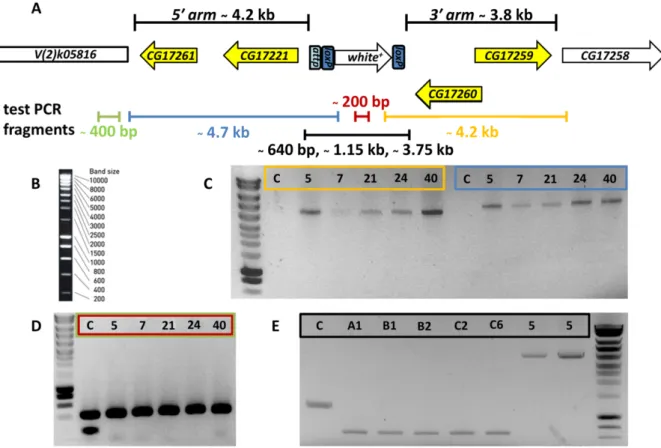
Table 2.4 shows all fly stock used during this thesis, as well as their source. The fly stocks from the collection of Prof. Dr. Siegfried Roth (Institute for Developmental Biol- ogy, University of Cologne) are labeled by SCR (stock collection Roth). Stocks received from other groups from the university of cologne are labeled as: SCS (Stock collection Scholz), SCU (Stock collection Uhlirova), SCL (Stock collection Leptin). ∆cnir [w+] stocks without specific labeling derive from line no. 5.
Table 2.4 | Fly stocks
Fly stock Source
w1118 SCR
w1118 SCS
w− ; IF/CyO ; MKRS/TM6B SCR
w− ; Gla/CyO ; MKRS/TM2 SCR
w1118 ; Sp/CyO ; TM2/TM6B SCS
w1118 ;; appl::Gal4 SCS
w− ; IF/CyO ; appl::Gal4/TM6B this thesis
w− ;; act::Gal4 SCL
w− ;; mhc::Gal4 SCL
y− w− hs::FLP ; Sp/SM6; TM6 SCR
hs::Cre ; Sco/CyO SCL
y− w− ; Ubi::GFP Ubi::GFP FRT40A/CyO BL#5198
eye::FLP ; FRT40A tub::Gal80/CyO ; act::Gal4UAS::GFP/TM6B SCU