Cathepsin D Serum Mass Concentrations in Patients with Hepatocellular Carcinoma and/or Liver Cirrhosis
Gaetano Leto1, Francesca Maria Tumminello\ Giuseppe Pizzolanli1, Giuseppe Montalto2, Maurizio Soresi2, Irene Ruggeri2 and Nicola Gebbia1
1 Servizio di Chemioterapia
2 Cattedra di Medicina Interna
Facolta di Medicina e Chirurgia, Universit di Palermo; Policlinico "P. Giaccone", Palermo, Italy
Summary: Cathepsin D serum mass concentrations were determined by enzyme immunoassay in patients with hepatocellular carcinoma (n = 51) and/or liver cirrhosis (n = 92) or benign steatosis (n = 16) and correlated with some biochemical and clinical properties of these diseases. Increased cathepsin D serum mass concentrations (P < 0.001) were observed in all these groups of patients as compared to normal subjects (n = 98). However, patients with steatosis had serum mass concentrations of this enzyme significantly lower (mean 2—3 fold) than those measured in cancer patients (P < 0.05) or cirrhotic patients (P < 0.001). Interestingly, significantly higher cathepsin D serum mass concentrations (mean + 62%) (P < 0.006) were determined in the cirrhosis group as compared to cancer patients. No correlation between cathepsin D and a number of clinical and biochemical proper- ties examined, namely, α-foetoprotein, number of neoplastic lesions and tumour size in cancer patients or, Child- Pugh grade of severity of cirrhosis and other enzymes of liver function tests in the cirrhotic group was found. The present data and those from other studies which indicate that cathepsin D may be involved in carcinogenesis suggest that this enzyme may be potentially useful as an additional biochemical marker to identify cirrhotic patients who may develop precancerous hepatic nodules.
Introduction.
Cathepsin D1) is a lysosomal acidic endopeptidase widely distributed in animal and human tissues (1). This enzyme has been suggested to be mainly involved, in physiological conditions, in intracellular protein catabol- ism (1). However, growing experimental evidence indi- cates that cathepsin D may also promote tumour pro- gression, invasion and metastasis (2, 3). In fact several investigations have shown that this proteinase appears to facilitate, at least in vitro, oncogenic transformation (4). Moreover, it has been shown that a number of hu- man carcinoma cell lines actively secrete a precursor form of cathepsin D which, following autoactivation at an acid pH, may degrade extracellular matrix or may activate latent precursor forms of other proteolytic en- zymes involved in the metastatic cascade (2, 3). In addi- tion, it has been recently shown that this precursor form may be endowed with mitogenic activity, thus facilitat- ing tumour cell proliferation (5, 6). Although evident proof that cathepsin D may also facilitate these pro-
l) Enzymes
Cathepsin D (EC 3.4.23.5) Alkaline phosphatase (EC 3.1.3.1) Alanine aminotransferase (EC 2.6.1.2) Aspartate aminotransferase (EC 2.6.1.1) γ-Glutamyl-transferase (EC 2.3.2.2)
cesses in vivo has still not been provided, a consistent bulk of clinical studies have shown that this enzyme is highly expressed, in terms of mass and/or catalytic activ- ity concentrations, in tumour cells and tissues of a number of human neoplastic diseases as compared to their normal counterpart and that these levels may corre- late, in some cases, with the malignant progression of these tumours (7—13). In this context several studies have also shown increased cathepsin D catalytic activity concentrations in human liver tumours as compared to normal liver tissues (14) as well elevated plasma mass concentrations of this enzyme in patients with malignant and non-malignant liver diseases as compared to healthy subjects (15). Interestingly, recent experimental in vitro and in vivo studies (16, 17) have evidenced in rat hepa- toma cells altered intracellular processing and increased secretion of a precursor form of cathepsin D which ap- pears to be endowed with mitogenic activity and a sig- nificant increase of the catalytic activity concentration of this enzyme in the ascites and in plasma of rats trans- planted with Yoshida AH-130 hepatoma. An important consequence related to these phenomena may be, as sug- gested (14, 15, 18), that cathepsin D produced in excess or unusually distributed at extracellular levels following these pathological processes, may be actively secreted into the bloodstream. Therefore, increased cathepsin D
serum content may reflect the presence of neoplastic nodules in liver tissue or may also be related to the ma- lignant transformation of cirrhosis, a non-neoplastic liver disease which may evolve in hepatocellular carci- noma (19). In this latter case the pattern of serum levels of this enzyme may be useful as an additional biochemi- cal marker to identify cirrhotic patients at risk to develop hepatocellular carcinoma. This finding may have clinical relevance as, to date, no serum marker including a-foe- toprotein has been found specifically reliable for the early detection of precancerous lesions (20, 21). On the basis of these considerations we have assessed cathep- sin D serum mass concentrations in groups of patients with hepatocellular carcinoma and/or liver cirrhosis and evaluated its potential clinical interest in non-malignant and malignant liver diseases.
Materials and Methods
Patients
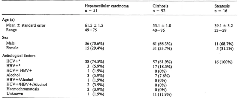
This study was undertaken on a total of 159 patients with malignant and non-malignant liver diseases whose clinical features are re- ported in table 1. Ninety-eight registered healthy blood donors of both sex were used as control. Diagnosis of hepatocellular carci- noma was established by ultrasonography and by histological find- ings following ultrasound-assisted fine-needle biopsy, evidence from positive diagnostic imaging and clinical and biochemical findings as well. Liver cirrhosis was diagnosed by histological findings and on the basis of unequivocal clinical and biochemical data. The Child-Pitgh classification (22) was used to evaluate the grade of severity of cirrhosis. Liver steatosis was diagnosed by histological findings. All subjects with steatosis had a positive se- rum test for hepatitis C virus. Immunoenzymatic and biochemical assays were performed on sera, previously stored at -80 °C, ob- tained from venous blood samples of patients and healthy blood donors.
Determination of cathepsin D serum mass concentrations
Cathepsin D concentrations were determined by a commercially available antibody-based immunoenzymatic kit (Ciba Corning
Diagnostics, Alarneda, USA) in sera of both healthy subjects and patients and diluted 1 : 50 with the appropriate sample diluent in- cluded in the kit. The assay detected both precursors (procathepsin D, Mr 52 000, MT 48 000) and mature (Mr 34 000) forms of cathep- sin D. The standard curve was linear from 0 up to 2.0 nmol/1. The minimum detectable concentration, according to the manufacturer, is 0.012 nmol/1. Mean cathepsin D serum mass concentrations in normal subjects, + 2-fold the standard deviation value or the high- est serum value of the enzyme determined in healthy subjects, were taken as cut-off limits.
Determination of serum α-foetoprotpin and other biochemical tests
α-Foetoprotein, the most sensitive and reliable serum marker for the diagnosis of hepatocellular carcinoma (23), was determined in healthy subjects and in patients by a commercially available immu- noluminometric based assay kit (Byk Sangtec, Milan, Italy). Nor- mal range values, according to the manufacturer, were 0-8 kIU/1.
Additionally, serum levels of 200 and 400 kIU/1 were used as cut- off values as these concentrations have been reported to be highly diagnostic for this tumour (20, 23, 24). Furthermore, the following serum biochemical constituents were also assayed and their corre- lation with cathepsin D was assessed: alkaline phosphatase1), albu- min, alanine aminotransferase1), aspartate aminotfansferase1), bili- rubin, γ-glutamyl-transferase1), prothrornbin. These assays were done with commercially available standard kits.
Statistical analysis
Data analysis was computed by the Mann-Whitney and the Kruskall-Wallis non-parametric tests. Linear regression analysis was used to compute the relationship between cathepsin D and a- foetoprotein serum concentrations in cancer patients and patients with cirrhosis. Due to the uneven distribution of the data, the Spearman rank correlation test was used to assess the correlation between cathepsin D serum levels and Child-Pugh grade of sever- ity of cirrhosis as well as between cathepsin D and the other bio- chemical properties examined. P values < 0.05 were taken as sig- nificant.
Results
Cathepsin D serum mass concentrations
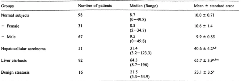
Table 2 reports the mean ± standard error, the median and the range of total serum cathepsin D content in all
Tab. 1 Patients features.
Hepatocellular carcinoma
n = 51 Cirrhosis
n = 92 Steatosis
n = 16 Age (a)
Mean ± standard error Range
Sex MaleFemale
61.5 ± 1.5 49-75 36 (70.6%) 15(29.4%)
55.1 ± 1.0 40-76 61 (66.3%) 31 (33.7%)
39.1 ± 3.2 23-59 11 (68.7%)
5(31.2%) Aetiological factors
HCV+a HBV+b
HCV+ HBV+
Alcohol HBV-h/Alcohol HCV+/HBV+/Alcohol Haemochromatosis Unknown
38 (74.5%) 3 (5.9%) 1 (1.9%) 3 (5.9%) 1 (1.9%) 2 (3.9%) 2 (3.9%) 1 (1.9%)
57(61.9%) 17(18.5%)
0 (0%) 7 (7.6%) 0 (0%) 0 (0%) 0 (0%) 11(11.9%)
16(100%)
Patients with positive serum test for hepatitis C virus. b Patients with positive serum test for hepatitis B virus.
Tab. 2 Cathepsin D serum mass concentrations (nmol/1) in normal subjects and patients with hepatocellular carcinoma and/or liver cirrhosis or benign steatosis.
Groups Normal subjects
— Female
— Male
Hepatocellular carcinoma Liver cirrhosis
Benign steatosis
Number of patients 98
31 67 51 92 16
Median (Range) 8.7(0-49.8) 8.5 (2-34.7) 9.5(0-49.8) 31.4 (3.2-123.3) 64.3 (8.7-196) 21.5(5.3-54.9)
Mean ± standard error 10.0 ± 0.71
10.6 ± 1.4 9.9 ± 0.85 40.6 ± 4.2a-b 65.7 ± 3.9a-b-c
23.1 ±3.5a Data analysis was computed by the Mann-Whiiney and Kniskall-
Wallis non-parametric test.
a P < 0.001 as compared to normal subjects;
b P < 0.04 as compared to benign steatosis;
c P < 0.006 as compared to hepatocellular carcinoma.
the examined groups. No significant difference in the serum mass concentrations of this enzyme was observed between female and male normal subjects (tab. 2). Sig- nificantly higher cathepsin D serum mass concentrations (mean 4—6 fold) were determined either in cancer or cirrhotic patients as compared to normal subjects (P < 0.001). Interestingly, cathepsin D serum mass con- centrations in cirrhotic patients were significantly higher (mean + 62%) than those measured in cancer patients (P = 0.006) (tab. 2). Statistically significant increased cathepsin D serum mass concentrations were also ob- served in subjects with steatosis as compared to normal subjects (P < O.O 1). However, these concentrations were, in turn, significantly lower (mean 2—3 fold) than those determined in the serum of cancer patients (P < 0.04) or patients from cirrhosis (P < 0.001) (tab.
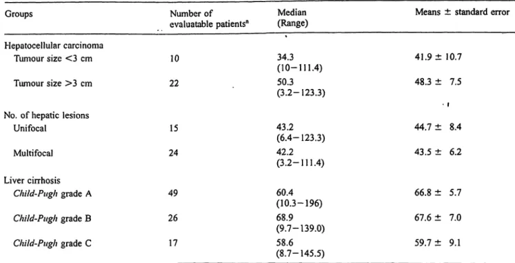
2). No significant correlation between cathepsin D se- rum mass concentrations and tumour size (r — 0.23;
P = 0.23) or number of neoplastic lesions (r = 0.19;
P = 0.23) was evidenced in those patients evaluatable for these properties (tab. 3). Furthermore, no correlation between cathepsin D serum mass concentrations and grade of severity of cirrhosis was further evidenced (r = -0.78; P = 0.48) (tab. 3).
α-Foetoprotein serum concentrations
α-Foetoprotein serum concentrations were markedly ele- vated (mean 842 fold) in cancer patients as compared to normal subjects or patients with cirrhosis (P < 0.001) (tab. 4). These concentrations were also significantly increased (mean 6 fold) in patients with cirrhosis as compared to normal subjects (P < 0.01). However, this phenomenon occurred at a very lessened extent as com- pared to cancer patients (tab. 4). The serum concentra- tions of this protein were significantly different in pa- tients with tumour size larger or smaller than 3 cm
(P < 0.05) and also significantly different in patients with single or multiple neoplastic lesions (P < 0.02).
Furthermore, this tumour marker was significantly correlated with tumour size (r = 0.49, P = 0.004) and the number of malignant lesions (r = 0.47, P = 0.004) (tab. 4). Finally, a significant correlation (r = 0.112, P = 0.004), was evidenced between α-foetoprotein se- rum levels and grade of severity of cirrhosis (tab. 4).
Correlation between cathepsin D and biochemical properties of hepatocellular carcinoma and cirrhosis
No relationship between cathepsin D and a-foetoprotein levels was observed either in hepatocellular carcinoma (r = -0.027, P = 0.87) or cirrhotic patients (r = 0.19, P = 0.065) (data not shown). No further correlation with the other examined biochemical properties of cholestasis or cytolysis was evidenced (albumin: r = 0.13; alanine aminotransferase: r = -0.3; aspartate aminotransferase:
r = —0.2; alkaline phosphatase: r = 0.27; bilirubin:
r = 0.03; γ-glutamyl-transferase: r = 0.28: prothrombin:
r = 0.103). P values were not significant in any of the cases (data not shown).
Rate of increased cathepsin D and
α-foetoprotein in patients with hepatocellular carcinoma or cirrhosis
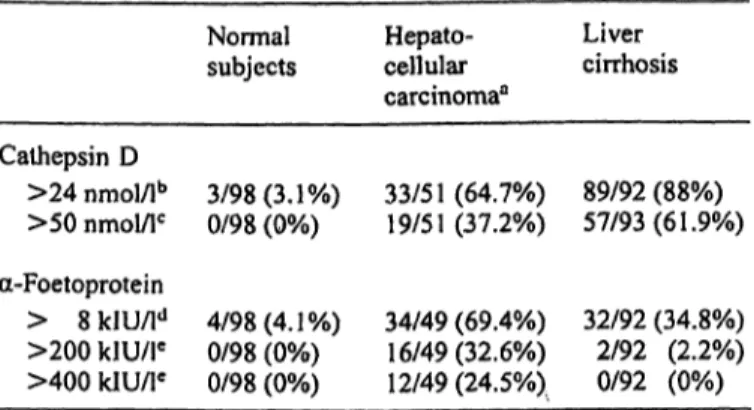
Cathepsin D serum mass concentrations higher than the cut-off level of 24 nmol/1 (mean serum content in nor- mal subjects + 2 standard deviations) were measured in 64.7% of cancer patients and in 88.0% of patients with cirrhosis (tab. 5). At a cut-off level of 50 nmol/1 (highest serum mass concentration measured in healthy subjects) this phenomenon was observed in 37.2% of cancer pa- tients and in 61.9% of patients with cirrhosis (tab. 5).
Tab, 3 Cathepsin D scrum mass and/or liver cirrhosis: distribution Groups
Hepatocellular carcinoma Tumour size <3 cm Tumour size >3 cm
No. of hepatic lesions Unifocal
Multifocal Liver cirrhosis
Child-Pugh grade A Child-Pugh grade B Child-Pugh grade C
concentrations (nmol/l) in patients according to clinical findings.
Number of
evaluatable patients"
10 22
15 24
49 26 17
with hepatocellular carcinoma
Median Means ± standard error (Range)
34.3 41.9 ±10.7 (10-111.4)
50.3 48.3 ± 7.5 (3.2-123.3)
• I
43.2 44.7 ± 8.4 (6.4-123.3)
42.2 43.5 ± 6.2 (3.2-111.4)
60.4 66.8 ± 5.7 (10.3-196)
68.9 67.6 ± 7.0 (9.7-139.0)
58.6 59.7 ± 9.1 (8.7-145.5)
a Due to some missing samples, number of patients may be vari-
able. Data analysis was computed by Mann-Whitney, Kruskall-Wallis and
Spearman rank correlation non-parametric tests. No significant dif- ference or correlation between or among groups was evidenced.
Tab. 4 α-Foetoprotein serum concentrations (kIU/1) in normal subjects and patients with hepatocellular carcinoma and/or liver cirrhosis.
Groups Normal subjects
Hepatocellular carcinoma Tumour size <3 cm Tumour size >3 cm No. of hepatic lesions
Unifocal Multifocal Liver cirrhosis Child-Pugh grade A Child-Pugh grade B Child-Pugh grade C
Number of
evaluatable patients51 98
43 10 24
15 22 92 49 26 17
Median (Range) (0.8-10.1)1.9
33.3 (0.6-78000) 9.3(0.6-459) 161.0 (1.5-78000)
14.8 (0.6-459.9) 316.4 (2.4-78000) 5.7
(0.6-225.9) 5.1(0.8-141.0) 5.9
(0.6-225.9) 5.9(2.0-220.7)
Means ± standard error
2.8 ± 0.32
2359 ± 1811.3"
76.4 ± 45.6 4116±3228.7b-c
55.1 ± 30.6 4357 ± 3519.7^
16.7 ± 3.6f
14.6 ± 3.4 17.1 ± 8.9 23.5 ± 11.98
a Variability in the number of patients is due to some missing sam- ples.
Data analysis was computed by Mann-Wliitney, Kruskall-Wallis and Spearman rank correlation non-parametric tests.
a P < 0.001 as compared to normal subjects and liver cirrhosis;
b P < 0.05 as compared to tumour size <3 cm;
c r = 0.49, Ρ < 0.004;
d Ρ < 0.05 as compared to unifocal;
e r = 0.47, Ρ < 0.004;
f Ρ < 0.01 as compared to normal subjects;
8 r = 0.112, Ρ = 0.004.
Tab. 5 Rate of increased cathepsin D and α-foetoprotein serum concentrations in patients with hepatocellular carcinoma and/or liver cirrhosis.
Cathepsin D
>24 nmol/lb
>50 nmol/lc a-Foetoprotein
> 8 kIU/ld
>200 klU/le
>400 kIU/le
Normal subjects
3/98(3.1%) 0/98 (0%)
4/98(4.1%) 0/98 (0%) 0/98 (0%)
Hepato- cellular carcinoma3
33/51 (64.7%) 19/51(37.2%)
34/49 (69.4%) 16/49(32.6%) 12/49(24.5%)
Liver cirrhosis
89/92 (88%) 57/93(61.9%)
32/92 (34.8%) 2/92 (2.2%) 0/92 (0%)
a Due to some missing samples, number of evaluable subjects in this group is variable.
b Mean serum levels ± 2 SD in normal subjects.
c Highest cathepsin D serum values determined in normal subjects.
d Upper normal serum level according to the manufacturer's in- structions.
c Cut-off levels which have been reported to be diagnostic for hep- atocellular carcinoma (20, 23, 24).
Only 4/16 (25%) or 1/16 (6.2%) of subjects with steatosis had a cathepsin D serum mass concentration higher than 24 or 50 runol/1 respectively (data not shown). Further, α-foetoprotein serum levels higher than 8 kIU/1 (normal upper limit) were observed in 69.4% of patients with hepatocellular carcinoma and in 34.8% of patients with cirrhosis (tab. 5). When 200 or 400 kIU/1 were selected as cut off values, 32.6%
and 24.5% of these patients showed increased levels of this protein while, at a 200 kIU/1 cut-off limit, this phenomenon occurred only in 2.2% of patients with cirrhosis (tab. 5).
Discussion
Total cathepsin D serum mass concentrations (i. e. pro- enzyme + mature form) were observed to be signifi- cantly increased in patients with hepatocellular carci- noma and/or liver cirrhosis as compared to normal sub- jects. Interestingly, in cirrhotic patients these concentra- tions were significantly higher (mean + 62%) than those measured in cancer patients. Increased levels of this pro- teinase were also observed in subjects with benign steatosis, however these values were markedly (2—3 fold) lower than those measured in cancer patients and patients with cirrhosis. These data further confirm, in part, and extend some previous observations ofBrouillet et al. (15) and Zuhlsdorftf. al. (25) who found evidence of elevated mass concentrations of cathepsin D in the plasma or sera of patients with various liver diseases including hepatocellular carcinoma and cirrhosis. The increased serum content of cathepsin D observed in both cirrhotic and cancer patients may not only be the conse- quence of cytolytic processes which occur during the
evolution of these pathological events but also related to an active secretion of this enzyme by intact hepatocytes, which contain most of the enzyme present in the human liver tissue (26). This hypothesis, supported by previous observations of Ryvnyak et al. (18) who showed that, in rats with tetrahydrocarbon chloride-induced cirrhosis, cathepsin D was largely secreted by intact heptocytes and other connective tissue cells into the intercellular space during cirrhosis, is further suggested by the pre- sent data which showed the lack of a significant correla- tion between cathepsin D serum mass concentrations and some biochemical markers of cytolysis. As an al- tered secretion of this enzyme has been reported to be associated, at least in vitro, with some stages of onco- genic transformation in rat fibroblasts and with differen- tiation stages of some carcinoma cell lines (4, 27), it would be conceivable to hypothesize that the increased serum levels of this proteinase in cirrhotic patients may also be associated with carcinogenetic processes which may occur during the evolution of this disease. More- over, a number of experimental in vitro and in vivo in- vestigations have shown that cathepsin D may induce tumour cell proliferation by acting as an autocrine mito- gen (2, 5, 6) and that intraperitoneal injections of puri- fied preparations of cathepsin D may stimulate DNA synthesis and mitosis in intact mouse liver (28, 29). In addition, other studies have suggested that cathepsin D may be implicated in liver regeneration, remodelling and resorption of fibrous tissues in cirrhosis (18, 29, 30) as well as in the activation of latent precursor forms of other proteolytic enzymes, such as cathepsin B and L, involved in the degradation of collagen in fibrotic liver and in tumour invasion and metastasis (31, 32). There- fore, these findings lead further to the hyopthesis that cathepsin D might be able to promote the malignant transformation of cirrhotic tissue. These observations may fit well with the higher cathepsin D serum mass concentrations observed in cirrhotic patients as com- pared to cancer patients. In conclusion, although these results indicate that cathepsin D does not seem to be more reliable than α-foetoprotein as a tool for the diag- nosis of hepatocellular carcinoma, the increased serum levels of this enzyme, in addition to other proposed po- tential prognostic factors (33, 34), may be of interest as an additional biochemical marker to identify patients with cirrhosis who may develop precancerous nodules.
Prospective clinical investigations to better assess this hypothesis are currently in progress.
Acknowledgements
This work was supported by grants from the Associazione Italiana per la Ricerca sul Cancro (A1RC, Milan, Italy) and Consiglio Nazionale delle Ricerche (CNR), Progetto Finalizzato ACRO, SP4 (Contract No. 95.00369.PF39).
References
1. Barrett AJ. Cathepsin D and other carboxyi proteinases. In:
Barrett AJ, editor. Proteinases in mammalian cells and tissues.
Amsterdam: Elsevier/North Holland Biomed Press, 1977:209-29.
2. Rochefort H. Biological and clinical significance of cathepsin D in breast cancer. Acta Oncol 1992; 31:125-30.
3. Leto G, Gebbia N, Rausa L, Tumminello FM. Cathepsin D in the malignant progression of neoplastic disease [review].
Anticancer Res 1992; 12:235-40.
4. Solovyeva NI, Balayevskaya TO, Dilakyan EA, Zakaldamaz- ina-Zama TA, Podznev VF, Topol LZ, et al. Proteolytic en- zymes at various stages of oncogenic transformation of rat fi- broblasts. I. Aspartyl and cysteine proteinases. Int J Cancer
1995; 60:495-500.
5. Fusek M, Vetvicka V. Mitogenic function of procathepsin D:
the role of propeptide. Biochem J 1994; 303:775-80.
6. Vetvicka V, Vektivickova J, Fusek M. Effects of human proca- thepsin D on proliferation of human cell lines. Cancer Lett
1994; 79 (2): 131-5.
7. Fontanini G, Bigini D, Vignati S, Ribechini A, Angeletti CA, Pingitore R. Immunostaining of cathepsin D in non-small cell lung cancer: correlation with morphological and biological parameters. Int J Oncol 1994; 4:169-73.
8. Leto G, Tumminello FM, Russo A, Pizzolanti G, Bazan V, Gebbia N. Cathepsin D activity levels in colorectal cancer:
correlation with cathepsin B and L and other biological and clinical parameters. Int J Oncol 1994; 5:509-15.
9. Marsigliante S, Biscozzo L, Resta L, Leo G, Mottaghi A, Maiorano E, et al. Immunohistochemical and immunoradiome- tric evaluations of total cathepsin D in human larynx. Eur J Cancer 1994; 30B:51-5.
10. Nazeer T, Malfetano JR Rosano GT, Ross JS. Correlation of tumor cytosol cathepsin D with differentiation and invasive- ness of endometrial adenocarcinoma. Am J Clin Pathol 1992;
97:764-9.
11. Podhajcer ÖL, Bover L, Bravo AI, Ledda F, Kariyama C, Calb I, et al. Expression of cathepsin D in primary and metastatic human melanoma and dysplastic nevi. J Invest Dermatol
1995; 104:340-4.
12. Scambia G, Panici BP, Ferrandina G, Salerno G, D'Agostino G, Distefano M, et al. Clinical significance of cathepsin D in primary ovarian cancer. Eur J Cancer 1994; 30A:935—40.
13. Zeillinger R, Swoboda H, Machacek D, Nekham D, Sliutz G, Knogler W, et al. Expression of cathepsin D in head and neck cancer. Eur J Cancer 1992; 28A (8/9):1413-5.
14. Maguchi S, Taniguchi N, Makita A. Elevated activity and increased mannose-6-phosphate in the carbohydrate moiety of cathepsin D from human hepatoma. Cancer Res 1988;
48:362-7.
15. Brouillet JP, Hanslick B, Maudelonde T, Pivat MP, Grenier J, Blanc F, et al. Increased plasma cathepsin D concentrations in hepatic carcinoma and cirrhosis but not in breast cancer. Clin Biochem 1991; 24:491-6.
16. Isidore C, Demoz M, De Stefanis D, Mainferme F, Wattiaux R, Baccino FM. Altered intracellular processing and enhanced secretion of procathepsin D in a highly deviated rat hepatoma.
Int J Cancer 1995; 60:61-4.
17. Isidoro C, Demoz M, Baccino FM, Bonelli G. High levels of proteolytic enzymes in the ascitic fluid and plasma of rats bearing the Yoshida AH-130 hepatoma. Invasion Metastasis
1995; 15:116-24.
18. Ryvnyak W, Gudumak VS, Onya ES. Intracellular and extra- cellular cathepsin D activity in the liver during cirrhosis and involution. Bull Exp Biol Med Engl Trans 1990; 109 (2):250-3.
19. Johnson PJ, Williams K. Cirrhosis and aetiology of hepatocel- lular carcinoma. J Hepatol 1987; 4:140-7.
20. Colombo M, De Franchis D, Del Ninno E, Sangiovanni A, De Fazio C, Tommasini M, et al. Hepatocellular carcinoma in Ital- ian patients with cirrhosis. New Engl J Med 1991; 325 (10):675-80.
21. Theisen ND, Fiel IM, Hytiroglou P,'Schwarz M, Miller C, Thung SN. Macroregenerative nodules in cirrhosis are not as- sociated with elevated serum or stainable tissue alpfaa-fetopro- tein. Liver 1995; 15:30-4.
22. Pugh RNH, Murray Lyon IM, Dawson JL, Pietroni MC, Wil- liams R. Transection of the oesophagus for bleeding oesopha- geal varices. Br J Surg 1973; 60:646-9.
23. Taketa K. Alpha-fetoprotein: reevaluation in hepatology. Hep- atology 1990; 12:1420-32.
24. Okuda K. Early recognition of hepatocellular carcinoma. Hep- atology 1986; 6:729-38.
25. Zuhldorf M, Imort M, Hasilik A, von Figura K. Molecular forms of beta-hexosaminidase and cathepsin D in serum and urine of healthy subjects and patients with elevated activity of lysosomal enzymes. Biochem J 1983; 213:733-40.
26. Reid W, Valler MJ, Kay J. Immunolocalisation of cathepsin D in normal and neoplastic human tissues. J Clin Pathol 1986;
39:1323-30.
27. Huet G, Zerimech F, Dieu MC, Hemon B, Grard G, Balduyck M, et al. The state of differentiation of HT-29 colon carcinoma cells alters the secretion of cathepsin D and of plasminogen activator. Int J Cancer 1994; 57:875-82.
28. Mori oka M, Terayama H. Cathepsin D stimulates DNA-syn- thesis and mitosis in mouse liver in vivo. Exp Cell Res 1982;
151:273-6.
29. Terayama H, Morioka M, Koij T. Mitogenic effects of certain cathepsins and calciferin on intact liver in vivo. Int J Biochem 1985; 17:945-55.
30. Terayama H, Shimuzu N, Fukuzumi R. Calciferin and cathep- sin D-like acid protease in serum in acute and chronic liver injuries in rats and humans. Clin Chem 1989; 35/11:2202-6.
31. Murawaki Y, Yamada S, Koda M, Hirayama C. Collagenase and collagenolytic cathepsin in normal and fibrotic rat liver. J Biochem 1990; 108:241-4.
32. Duffy MJ. The role of proteolytic enzymes in cancer invasion and metastasis. Clin Exp Met 1992; 10:145-55.
33. Hann H-WL, Kim CJ, London WT, Blumberg BS. Increased serum ferritin in chronic liver disease: a risk factor for primary hepatocellular carcinoma. Int J Cancer 1989; 43:376-9.
34. Tsai JF, Jeng JE, Ho MS, Chang W Y, Lin ZY, Tsai JH. Clinical evaluation of serum alpha-fetoprotein and circulating immune complexes as tumor markers of hepatocellular carcinoma. Br J Cancer 1995; 72 (2):442-6.
Received January* IB/May 7, 1996
Corresponding author: Dr. G. Leto, Section of Chemotherapy, Institute of Pharmacology, Policlinico "P. Giaccone", Via del Vespro, 1-90127 Palermo, Italy


