Schmid, Mall an4 Bockhorn: Enzyme activities in human kidney allografts with impaired function 961 J. Clin. Chem. Clin. Biochem.
Vol. 24, 1986, pp. 961-970
© 1986 Walter de Gruyter & Co.
Berlin · New York
Catalytic Activities
of Alkaline Phosphatase and N-Acetyl-ß-Z)-Glucosaminidase in Human Cortical Nephron Segments:
Heterogeneous Changes in Acute Renal Failure and Acute Rejection Following Kidney Allotransplantation
By Heide Schmid, Adelheid Mall
Pathologisches Institut der Universität Tübingen and H. Bockhorn
Chirurgische Klinik und Poliklinik der Universität Tübingen
(Received December 19, 1985/June 23, 1986) *
Dedicated to Professor Dr. med. Adalbert Bohle, Tübingen, on the occasion ofhis 65th birthday
Summary: The catalytic activities of alkaline phosphatase and N-acetyl-ß-Z>-glucosaminidase, constituents of luminal bnish-border membranes and lysosomes of kidney tubular cells, were measured in human kidney allografts in the mäintenance and recovery phases of acute renal failure and in acute rejection crisis. The enzyme activities were fluorometrically determined in single microdissected cortical nephron Segments of biopsies from 4 kidney allografts taken intraoperatively and postoperatively at different periods, which exhibited either goöd function or dysfunction. For comparison, the unaffected part of a human kidney nephrectomized due to hypernephroma äs well äs a biopsy of a morphologically normal human kidney were examined.
Both enzymes displayed highest activities in the proximal part of the human nephron. In some intraoperative and ppstoperätive biopsies with acute renal failure, alkaline phosphatase activity was reduced in proximal tubules, predominantly in the straight portion. This reduction could not be correlated with function. In acute rejection, very low alkaline phosphatase activities were uniformly found in proximal convoluted and straight tubules. Fürthermore, intraoperative biopsies and biopsies of the functioning allograft have only approxi- mately 50% of normal N-acetyl-ß-D-glucosaminidase activity in proximal convoluted tubules, but generally normal values in the straight portion. However, in acute renal failure, this enzyme activity was several-fold enhanced along the whole nephron, when compared with intraoperative values. In acute rejection, N-acetyl- ß-jp-glucpsaminidase activity was slightly reduced in proximal convoluted tubules, when compared with biopsies showing gööd function.
It is suggested that the decrease of proximal tubular enzyme activities is the consequence of increased enzymuria and inadequate enzyme regeneration. On the other hand, the overshoot of N-acetyl-ß-/)-glucos- amißidase activity in the maintenance phase of acute renal failure appears to indicate increased degradative capacity, associated with cellular regeneration along the whole nephron.
J. Clin. Chem. Clin. Biochem. / Vol. 24,1986 / No. 12
962 Schmid, Mall and Bockhorn: Enzyme activities in human kidney ailografts with impaired function
Kataly tische Aktivitäten von alkalischer Phosphatase undN-Acetyl-ß-D-glucosaminidase in menschlichen cortica- len Nephronsegmenten: Heterogene Veränderungen bei akutem Nierenversagen und akuter Abstoßung nach Nierentransplantation
Zusammenfassung: Die katalytischen Aktivitäten von alkalischer Phosphatase und N-Acetyl-ß-Z>-glucosami- nidase, Bestandteilen von luminalen Bürstensaum-Membranen und Lysosomen der Nierentubulus-Zellen, wurden in menschlichen Nierentransplantaten in Spät- und Erholungsphase akuten Nierenversagens und bei akuter Abstoßungskrise gemessen. Die Enzymaktivitäten wurden fluorometrisch in einzelne^mikrodissezier- ten corticalen Nephronsegmenten von Biopsien aus 4 Nierentransplantaten bestimmt, die intraoperativ und postoperativ zu verschiedenen Zeiten bei guter oder schlechter Funktion gewonnen wurden. Zum Vergleich wurden der normale Teil einer menschlichen Niere mit Hypernephroin und die Biopsie einer morphologisch normalen menschlichen Niere untersucht.
Beide Enzyme haben ihre höchsten Aktivitäten im proximalen Teil des menschlichen Nephrons. In einigen intraoperativen und postoperativen Biopsien bei akutem Nierenversagen war die Aktivität der alkalischen Phosphatase in den proximalen Tubuli reduziert, und zwar bevorzugt in den geraden Abschnitten. Diese Reduktion verlief nicht parallel dem klinischen Funktionszustand. Am stärksten und einheitlich in den gewundenen und geraden proximalen Tubuli war diese Enzymaktivität reduziert in der akuten Abstoßungsre- aktion. In den intraoperativen Biopsien und in den Biopsien des funktionsfähigen Transplantats war die katalytische Aktivität der N-Acetyl-ß-Z)-glucosaminidase in den proximalen Konvoluten etwa auf die Hälfte der normalen reduziert, während sie in den geraden proximalen Tubuli im allgemeinen auf Normalniveau blieb. Dagegen wurde im akuten Nierenversagen eine gegenüber den intraoperativen Werten mehrfach erhöhte Aktivität in alleij Nephronsegmenten gefunden. Bei der akuten Abstoßungsreaktion war diese Enzymaktivität in den proximalen Konvoluten leicht reduziert gegenüber den Biopsien im funktionsfähigen Zustand.
Die Abnahme der Enzymaktivitäten in den proximalen Tubuli wird als das Ergebnis vermehrter Enzymurie und nicht angepaßter Enzym-Neusynthese angesehen. Andererseits wird die Erhöhung der Aktivität der N-Acetyl-ß-/)-glucosaminidase in der Spätphase akuten Nierenversagens als Zeichen von vermehrter Abbau- kapazität bei der Zellregeneration im ganzen Nephron gedeutet.
Introduction , , . . , . , . . ,
kidneys, normal distnbution pattern and pathological Alkaline phosphatase (orthophosphoric-monoester changes of these organelles along the nephron have phosphohydrolase, alkaline Optimum, EC 3.1.3.1) not yet been quantified. Morphological analysis öf and N-acetyl-ß-D-glucosaminidase (2-acetamido-2- human kidney biopsies taken in acute renal failure deoxy-ß-Z)-glucoside acetamidodeoxyglucohydro- revealed a reduced luminal surface area in proximal läse, EC 3.2.1.30) are kidney tubular enzymes which tubules (11,12) and an increased number of vacuolar are known to be excreted into urine. Enzymuria in- structures in proximal and distal tubules (11). In creases, when renal function is impaired (l, 2, 3), addition, Gregoire & Gepts (13) observed reduced indicating renal damage. However, the renal pro- activity of microvillar alkaline phosphatase and un- cesses involved in enzymuria are not yet understood. changed activity of lysosomal acid phosphatase in proximal convoluted tubules of human renal homo- in the kidney, alkaline phosphatase is essentially transplants, which had been removed due to chronic found within the proximal part of the nephron (4), insufficiency.
constituting an integral part of luminal brush-border _,
membranes (2). The function of this enzyme is not The present Study 1S based on reoent observations on
yet known (5, 6). N-acetyl-ß-Z)-glucosaininidase is marker 6 3™68 öf cellular compartments along the distributed along the whole nephron with the highest human nePhron- These eariier ßndings showed that activity in proximal tubules (7). It is localized within the enzymes of basolateral interdigitations and mito- the lysosomes of tubular cells (8) and contributes to chondrial inner membranes, Na+-K+-ATPase and the intracellular degradation of carbohydrate-con- succinate dehydrogenase were selectively reduced in taining macromolecules (9). distal tubules of biopsies taken from human kidney allografts in the maintenance phase o£äcute renal Brush-border membranes and lysosomes are cellular failure (14). These enzyme activities were found to be compartments which undergo alterations in exper- unchanged in proximal tubules, but in this part of imentally mduced acute renal failure (10). In human the nephron, marked activity changes were found
Schmid, Mall and.Bockhorn: Enzyme activities in human kidney allografts with impaired function 963 with respect to lysosomal N-acetyl-ß-D-glucos-
aminidase (15). This fmding suggests that in these biopsies, proximal tubules had also undergone altera- tions, but of a different kind. In order to substantiate this, the measurement of N-acetyl-ß-Z)-glucosamini- dase activity was performed in all available cortical nephron segments, in addition to the determination of microvillar alkaline phosphatase activity. The same biopsies äs in the earlier study were examined, in addition to a biopsy taken in acute rejection crisis (15). The study of intranephronal distribution of both enzyme activities in correlation with renal function should give further insight into the nature and loca- tion of cellular events and of enzymuria associated with impaired renal function in hunians.
Materials and Methods Chemicals
Chemicals were of analytical grade from E. Merck AG, D- 6100 Darmstadt (buffer substances, liquid paraffin), from Serva Feinbiochemica GmbH & Co., D-6900 Heidelberg (4-methyl- umbelliferone, 4-methylumbelliferyl-N-acetyl-ß-Z)-glucosamin- ide, bovine serum albumin), and from Sigrna Chemical Co., St.
Louis, U. S. A., Mo 63178 (4-methylumbelliferyl phosphate).
Tissue preparation
Biopsies were taken from 4 human kidney allografts (A, B, C, D) which had been perfused with Euro-Collins solution and preserved on ice för up to 26—32 hours prior to transplanta- tion. Immediately after transplantation, allografts A, B, and C were anurie and did not show äny Symptoms of cellular or humoral rejection at the time of biopsy (A2, B?, C2), while allograft D displayed diuresis developing into normuria (D2, D3) and then into acute rejection crisis (D4) manifested by decreased creatinine clearance (15) and interstitial round-cell Infiltration. Diuretics were not given. Immunosuppressive treat- ment was conventionäl. In addition, biopsies of these allografts had been obtained intraoperatively about 30 min after anast- omosis (Aj, BJ, Q, Dj). In comparispn with these transplant biopsies, the unaffected part of a human kidney nephrectomized due to hypernephrpma grade I (E) and a morphologically normal biopsy taken due to microscopic haematuria (F) were examined.
Tissue samples were prepared for microdissection (16) by imme- diate shock-freezing in liquid nitrogen and freeze-cütting into 16 thick serial sections (Dittes-Duspiva, D-6900 Heidel- berg). The first of three consecutive sections was stained with periodic acid/Schiffs base reagent, the secohd lyophilized, and the third was stained for succinate dehydfpgenase activity, äs described previously (17). Dry weighi of microdissected cortical nephron segments was in the ränge of l — 40 ng.
Determination pf enzyme catalytic activities
Enzyme assays for single nephron segments (4,7) were modifled with respect to incubation conditions and the use of the oil- well technique (16). Sodram carbonate buffer, 0.1 mol/1, pH 10.0, containing 0.2 g/l bovine serum albumin, or sodium citrate buffer, 0.1. mol/1, pH 5.0, containing 0.2 g/l bovine serum albumin (630 nl), was placed into the wells of a teflon rack, loaded with a tissue specimen, and then covered with 2 droplets
of liquid paraffin. After cooling to 4 °C, 630 nl of enzyme reagent were added. After incubation for 32 min at 37 °C (2 min for temperature equilibration), the rack was cooled again to 4 °C. Incubation solution (950 nl) was pipetted into 200 of 0.5 mol/1 glycine buffer, 0.5 mol/1 NaCl, pH 10.4. Reagent blanks and 4-methylumbelliferone Standards (2-20 / ) were treated in the same manner. Tissue bianks were obtained by omitting incubation for 32 min at 37 °C. Fluorescence meas- urements were performed at 365/450 nm with a Fanand Mark I fluorometer.
Enzyme reagents were composcd of: 2 mmol/l 4-methylumbel- liferyl phosphate and 8 mmol/l MgSO4 in 0.1 mol/1 sodium carbonate buffer, pH 10.0, 0.2 g/l bovine serum albumin, for alkaline phosphatase, the Substrate concentration being around Saturation; 1.6 mmol/l 4-raethylumbelliferyl-N-acetyl-ß-/)-glu- cosaminide in 0.1 mol/1 sodium citrate buffer, pH 5.0, 0.2 g/l bovine serum albumin, for N-acetyl-ß-Z)-glucosaminidase, the Substrate concentration being in the ränge of half-saturation.
Specific enzyme activities were expressed in · min"1 4- methylumbelliferone formcd per g tissue dry weight. Since tissue blanks of both enzyme assays were at the detection limits for glomeruli and distal nephron segments and were below 6%
for proximal tubular values, they were neglected. Differences between the means of enzyme catalytic activities were tested for statistical significance with Studenfs t test for unrelated samples.
Results ,
Control biopsies
In addition to the earlier study on enzymes in the human nephron (14), a morphologically normal kid- ney biopsy (F) was taken äs normal tissue. As shown in table l, alkaline phosphatase activity in proximal tubules is about 10 times that of distal nephron seg- ments in accordance with the high brush-border membrane content in proximal regions (18). No dif- ference was found between the convoluted and straight part of cortical proximal tubules. In contrast, N-acetyl-ß-Z>-glucosaminidase activity appears to be distributed inhomogeneously along proximal tubules with higher activities in the straight portion (tab. 2).
Interestingly, in this segment, N-acetyl-ß-£)-glucos- aminidase activities were found to differ strongly among biopsies E and F. In glomeruli and distal nephron segments, N-acetyl-ß-Z)-glucosaminidase ac- tivity is relatively high compared with alkaline phos- phatase activity.
Biopsies taken during transplantation operations (Al5
BI, Ci, Dj) displayed different activities of both en- zymes in proximal tubules (tabs. l —4). In biopsy DI, alkaline phosphatase activities were reduced to about 50% irrespective of post-transplantation kidney func- tion (tab. 1), whereas the proximal convoluted tubules of biopsies Aj, Bj, and Ct äs well äs the proximal straight tubules of biopsies AI and B! contained activ- ities similar to those in normal tissues E and F (tab.
3). In functioning allograft D, alkaline phosphatase activities in proximal convoluted and straight tubules J. Clin. Chem. Clin. Biochem. / Vol. 24,1986 / No. 12
964 Schmid, Mali and Bockhorn: Enzyme activities in human kidney allografts with impaired function Tab l Specific catalytic activities of alkaline phosphatase in single cortical nephron segments of human kidneys (4-methylumbel- ' liferone, μηιοί · min-1 · g-1 dry weight). Values are given s the mean ± Standard devialion with the number of determinalions in parenthescs. The indices represent statistically significant differences: (a) = p< 0.05, (b) = p < 0.02, (c) = p< 0.001, (d) = p< 0.0001. Biopsy Dj was compared with biopsies E and F(*), biopsies D2 and D3 were compared with biopsyD,.
Abbreviations:
G = glomerulus,
PCT = proximal convoluted tubule, cPST = cortical proximal straight tubule,
cTAL = cortical thick ascending limb of Henle's loop, DCT = distal convoluted tubule,
cCD = cortical collecting duct.
Segment Kidney E (normal part of a kidney with
Kidney F (microscopic haematuria,
Kidney allograft D (functioning) kidney with haematuria,
hypernephroma) morphologically normal)
Biopsy D!
(intraoperative)
Biopsy D2
(normuria, 7 days after transplantation)
Biopsy D3
(normuria, 20 days after transplantation) PCTG
cPSTcTAL DCTcCD
0.5 ± 0.2(10) 25.9 + 13.9(49) 23.4+ 8.5(33) 3.2+ 1.4 (6) 3.7 ± 1.7 (7) 4.1 + 1.4 (5)
1.2+ 0.8 (6) 28.2 + 10.1 (33) 25.0 + 8.4 (20) 3.2+ 1.2(10) 2.6 ± 1.9 (7)
1.2 + 0.7 (5) 14.9 ± 7.4 (31) (d, d*) 12.2 ±6.1 (25) (d,d*)
2.4 ±0.9 (6) 1.9 ±0.6 (4) (a)
2.2 ± 0.8 (7) (a) 35.8 ±13.5 (20) (d) 26.2 ±12.8 (21) (d)
1.6+ 0.7 (6) 2.6 ± 1.6 (8) 1.5+ 0.8 (4)
1.2+ 0.4 (8) 25.4 + 13.9 (31) (c) 26.3 + 15.0 (16) (c)
3.6+ 1.8 (6) 3.6+ 1.3 (8)(b) 3.0+ 1.3 (4) Tab. 2. Specific catalytic activities of N-acetyl- -Z)-giucosaminidase in single cortical nephron segments of human kidneys (4^·
methylumbelliferone, μπιοί · min"1 · g"1 dry weight). Values are given s the mean ± Standard deviation with the number of detenninations in parentheses. The indices represent statistically significant differences: (a) = p < 0.02, (b) = p < 0.01, (c) = p < 0.002, (d) = p < 0.001, (e) = p < 0.0001. Biopsy F was compared with biopsy E, biopsy D! with biopsies E and F(*), and biopsies D2 and D3 were compared with biopsy Dj.
The same abbreviations for nephron segments s in table l were used.
Segment
GPCT cPSTcTAL DCTcCD
Kidney E (normal part of a kidney with hypernephroma)
5.4 ± 0.9(17) 33.8 + 26.3 (81) 77.0 + 41.0(31) 15.3 + 7.3(15) 13.7+ 5.5(17) 10.7 ± 5.4 (5)
Kidney F (microscopic haematuria, morphologically normal)
4.2 ± 0.8 (4) (a) 27.3 ±19.6 (18) 41 .6 ± 16.2 (17) (d) 15.6 + 4.5 (10)
8.4 ± 3.7 (8)(b)
Kidney allograft D (functioning) Biopsy Dj
(intraoperative) 6.3 ± 0.7 (10) (b, d*) 17.5 ± 12.4 (30) (e) 89.2 ± 48.6 (26) (e*)
8.5 + 6.1 (12) (a, b*) 10.1 ± 4.5 (13)
Biopsy D2 (normuria, 7 days after transplantation)
7.0 ± 0.8 (8) 18.6 ± 5.3(26) 57.4 ±32.6 (29) (b) 12.3+ 4.5 (9) 11.9 ± 2.0(11) 18.2
Biopsy D3
(normuria, 20 days after transplantation)
4.9 ± 0.9 (10) (c) 22.3 ± 9.9 (42) 39.0 + 18.1 (22) (e) . 7.7+ 4.6(15) '13.0 ± 4.7(14) 13.0+ 4.6 (4)
had reached the levels of biopsies E and F at the 7th postoperative day (D2) (tab. 1). In contrast to this, N-acetyl- -D-glucosaminidase activity was found to be uniformly reduced to about 50% in proximal convoluted tubules of intraoperative biopsies Al5 BI, G!, and D^ when compared with biopsies E and F (tabs. 2 and 4). This level appears to be maintained in proximal convoluted tubules of functioning allo- graft D, 7 and 20 days after transplantation (D2, D3) (tab. 2). On the other band, in proximal straight tubules, the enzyme activities of intraoperative bi- opsies A! and GI were in the same r nge s those in kidney F (tab. 4). The activity in allograft D intraoperatively resembled that of biopsy E, but de- veloped into that of biopsy F after 7 and 20 days of post-transplantation function (D2, D3) (tab. 2). In proximal straight tubules of biopsy B! only half this activity was found (tab. 4).
Biopsies in acute post-transplant renal fail- ure
In the maintenance and recovery phases of acute post-transplant renal failure, gradual activity changes of alkaline phosphatase were detected after 9 days (A2) and 14 days (B2) of anuria (tab. 3). Biopsy A2
displayed the activity of normal renal tissues E and F in proximal convoluted tubules, but slightly reduced activity (61%, p < 0.01) in proximal straight tubules, when compared with intraoperative values. In biopsy B2, alkaline phosphatase activity was decreased in both proximal convoluted (70%, p < 0.002) and straight tubules (55%, p < 0.001). This activity showed a marked decrease in allograft C recovering from anuria (C2), with 25% activity (p < 0.0001) in proximal convoluted tubules and 60% activity (p <
0.002) in proximal straight tubules, corresponding to 35% ofthe mean vatoe of biopsies E, F, Ar, Bls
Schmid. Mall and Bockhorn: Enzyme activities in human kidney allograrts with impaircd function 965
S § v
f
60!·;
SL*53 8 H
w * M
«2-8,2
n
2 «e T3Λ «·.
'i·
< & §· 2«
«ΈII
& 4£ &
P A* ί^ ***" Cfc*^> r^ 5?» ^r.
^^ ζ^
V) CN CO ~ τ?
*i >O CN ~
4H 4^ 44 44 4-1 ON t*^ ·^ OO CN Ο
<Λ OO CN CN *·*
4H 4H 44 44 44
vO O CN VO OO
«^ co* Ξ-* o* oo* CN*
O 00 ~ ~ 0
44 44 44 4H 44 44 rt co 00 vo vO O
«n co CN ~ CN <«-«
s^+ X^«. X"^ X·^ x1··S
^ CN VO Γ^ ^
^ O v> co ~- O —i <N ~ CN
4H 4-1 44 4H 4-1
vq p <n co *- co co co ^ oo
X-"\ /^*S— oo r- X— »%
CO ^ ^ CO VO t^ CO VO
co oo *-ί O
4^ 44 44 -H 44
vo τΤ co ON O »o
o vo r-^ -^ ^ CN
O VO VO
^ "^
^ V* *·* -wi *-J
4-1 4H 4H -H -H
VO <*·* CO CN ON
*** oo cN co
CO CN
o fc
JtjJI
l?llǤ
•s ' "^ "l ·»
:l"c.s§ II ,§ II t?
e ·—in ifi o
s
y co»a .S
* § i i
c8-§«5i
"d·"
^
ί
Ii
3 εe ··=
oc
o S i. >·
tn ^-Sc
>. o
δ -a
•^& s 5·
S "°
93 .
ί I. sP- v> Π
—
» g.>* O
S. 2
§
Ή,ο\
S -3 8.*!
o o 2Bio (int
o vo r^
ΓΜ *-* *-*
ir> *-* \O CO Γ*»
C^4 CO
4-1 +1 44 4-1 4-1
^ r* vo *-i co f*·
2 5 S S2 S =
*O »O CN OO CN Γ*;
O O ON »/S ^ co 4-1 4-1 4-1 4-1 4-1 4-1 oo «n ON oo p -^
co c^i O co
— oo CN -^
-* os —CN r*
-Hl 4-1 4-1 4-1 r^ ^t
^ ONoo ON »o »o vo i> ON oo CO CN CN CN
VO ON GO VO O <N
^-x rr, <v| *- Π s— '
^-S >«· V-X X»X
r^ vo m t- oo o\
o oo vo co o 44 +1 4-1 4-1 4-1 4-1
SS SS O co vo C*^ ON co
CN vi O «O v oo
•r-i CN
4H 44 4-1 4-1 4-1 4-1
VO t*; CN VO -»-i "^
vo" od ν-ί oi r^
^-i CO m CN CN ^-i
o es -^-j co co
•^i VO CO Tf ^-"
T— 1
+1 4-1 4-1 44 44
^5 CO Tt" CO t*·» CO
r^ *o ON *4 r^ r-^-
J. Clin. Chem. Clin. Biochem. / Vol. 24,1986 / No. 12
966 Schmid, Mall and Bockhorn: Enzyme activities in human kidncy allografts with impaired function D2, and D3. N-acetyl-ß-ö-glucosaminidase activity
showed a very different behaviour in postoperative biopsies A2, B2, and C2 (tab. 4). In all cortical nephron segments, activities were increased, when compared with the respective intraoperative values: in glomeruli to 148 — 179%, in thick ascending limbs of Henle's loop to 141 —297%, and in distal convohited tubules to 163-355%. The highest activities in proximal tubules were measured in biopsy B2: 521% (p <
0.0001) in the convoluted and 582% (p < 0.0001) in the straight part. These high activities exceed that of normal renal tissue two- to three-fold. In proximal convoluted tubules of biopsies A2 and C2, N-acetyl- ß-Z)-glucosaniinidase activities had returned to the level of normal renal tissue (E, F), corresponding to 240% (p < 0.0001) and 288% (p < 0.0001), respectively, of intraoperative values. Whereas; the activity of proximal straight tubules in biopsy A2 remained approximately constant, in biopsy. C2 it amounted to 188% (p < 0.001) of the intraoperative value.
Biopsy in acute rejection
In the acute rejection episode of allograft D (D^, 25 days after transplantation, alkaline phosphatase activity was drastically decreased in proximal convo- luted and straight tubules to 24% (p < 0.0001) and 21% (p < 0.0001) of the values in biopsy D3 (tab.
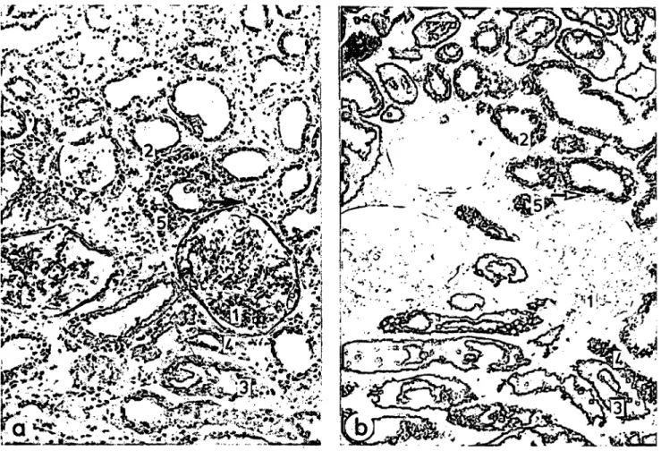
5). In addition, glomeruli and distal tubules displayed reduced enzyme activities. The changes were found to be very heterogeneous according to the histological appearance of the tissue. Figure l demonstrates this heterogeneity with respect to round-cell Infiltration and succinate dehydrogenase activity, äs also de- scribed by Gregoire & Gepts (13). Since microdissec- tion was done in sections adjacent to the stained
sections, isolated specimens could be correlated to normal or reduced succinate dehydrogenase activity (19). This classification permits discrimination be^
tween proximal tubules with alkaline phosphatase activities nearly äs low äs those in distal tubules, corresponding to tissue blanks of normal proximal tubules, and proximal tubules with significantly higher activities (tab. 5). In contrasf/ N-acetyl-ß-J3- glucosaminidase activity showed mainly an additional decrease to 59% (p < 0.0001) in proximal convoluted tubules, when compared with biopsy D3, but was slightly increased (136%, p < 0.02) in proximal straight tubules. Distally, there were only moderate activity changes. The changes of N-acetyl-ß^jD- glucosaminidase activity were independent of round- cell Infiltration density and succinate dehydrogenase agtivity, except in distal convoluted tubules (tab. 6).
Discussion
The present study reports on biochemical chafac- terization of cellular compartments in single nephron segments of human kidney allografts with impaired function. Alterations in brush-border membranes and lysosomes were quantified by means of the marker enzymes alkaline phosphatase and N-acetyl-ß-D-glu- cosaminidase, respectively. Since these enzymes are known to be excreted into urine (l, 3, 20), their renal activity may be balanced by enzymuriä and enzyme regeneration.
Catalytic activities of alkaline phosphatase along the human nephron were considered to be normal in biopsies E and F äs well äs in biopsies D2 and D3 with post-transplantation function. In these biopsies, N-acetyl-ß-D-glucosaminidase activities in glomeruli and distal nephron segments are obviously normal.
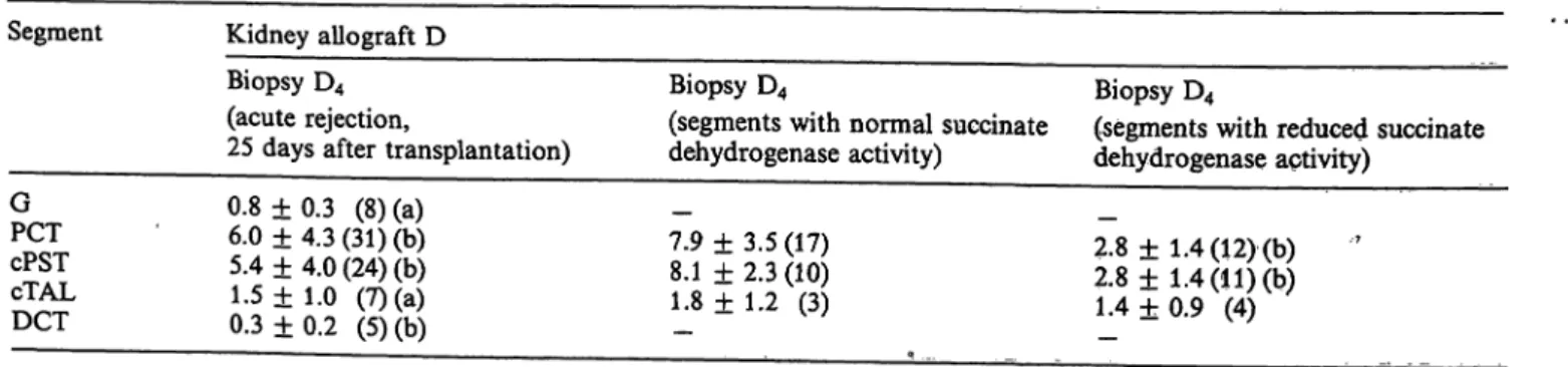
Tab. 5. Specific catalytic activities of alkaline phosphatase in single cortical nephron segments of a human kidney allograft with acute rejection (4-methylumbelliferone, · min"1 · g-1 dry weight). The values of the first column were classified into two groups according to the appearance of tubular staining for succinate dehydrogenase activity (fig. 1). The data represent the mean ± Standard deviation with the number of determinations in parentheses and with the following indices for statistically significant differences: (a) = p < 0.05, (b) = p < 0.0001. The values of biopsy D4 (first column) were compared with those of biopsy D3 (tab. 1). In addition, when classified, the values of the second colunan were compared with those of the third column.
The same abbreviations for nephron segments äs in table l were used.
Segment Kidney allograft D Biopsy D4
(acute rejection,
25 days after transplantation)
Biopsy D4
(segments with normal succinate dehydrogenase activity)
Biopsy D4
(segments with reduced succinate dehydrogenase aptivity)
GPCT cPSTcTAL DCT
0.8±0.3 (8) (a) 6.0 ±4.3 (31) (b) 5.4 + 4.0 (24) (b) 1.5 ± 1.0 (7) (a) 0.3±0.2 (5)(b)
7.9 ± 3.5 (17) 8.1 ± 2.3 (10) 1.8 ± 1.2 (3)
2.8 ±1.4 (12) (b) 7 2.8 ±1.4 (11) (b) 1.4 + 0.9 (4)
Schmid, Mall and Bockhorn: Enzyme activities in human kidney allografts with impaired function 967
Fig. 1. Cryostat sections (16 μπι thick) of kidney allograft D with acute rejection (biopsy D4). Magnification χ 113.
a) First of three consecutive sections stained with periodic acid/Schiffs base reagent with inhomogeneously distributed round-cell Infiltration (-») and with increased interstitial spaces.
b) Third section of this series stained for succinate dehydrogenase activity. This activity appears to be reduced in areas with increased round-cell Infiltration (-*) irrespective of the nephron region (19).
1 = glomerulus,
2 = proximal convoluted tubule, 3 = cortical proximal straight tubule,
4 = cortical thick ascending limb of Henle's loop, 5 = distal convoluted tubule.
Tab. 6. Specific catalytic activities of N-acetyl- -D-glucosaminidase in single cortical nephron segments of a human kidney allograft with acute rejection (4-methylumbelliferone, μιηοΐ · min"1 · g"1 dry weight). The values of the first column were cl ssified into two groups to give the second and the third column, s described in table 5. The data represent the mean ± Standard deviation with the number of determinations in parentheses and with the following indices for stati$tically significant differences: (a) = p < 0.02, (b) = p < 0.002, (c) = p < 0.0001. As in table 5, the values of biopsy D4 were compared with those of biopsy D3 (tab. 2), and those of the second and the third column were compared with each other.
The same abbreviations for nephron segments s in table l were used.
Segment Kidney allograft D Biopsy t>4
(acute rejection,
25 days after transplantation)
Biopsy D4
(segments with normal succinate dehydrogenase activity)
Biopsy D4
(segments with reduced succinate dehydrogenase activity)
GPCT cPSTcTAL DCTcCD
4.6 + 0.8 (9) 13.2 ± 5.1 (24) (c) 53.2 ± 17.3 (20) (a) 4.2+ 2.5 (20) (a) 16.6 ± 10.4 (32)
5.6 ± 3.4 (3)
__
11.6 ± 3.7 (5) 56.9 + 16.3(12) 3.2 ± 1.6(10) 9.7 ± 7.2(12)
—~
13.6 ± 5.4(19)— 47.6 -h 19.8 (7) 5.3 ± 3.0 (9) 21.0 4- 10.2 (19) (b)
J. Clin, Chem. Clin, Biochem. / Vol. 24,1986 / No. 12
968 Schmid, Mall and Bockhorn: Enzyme acüvities in human kidney allografts with impaired function
However, it is more difficult to define normal values in proximal tubules. In proximal convoluted tubules of biopsies E and F, they are thought to be normal.
In proximal straight tubules of biopsy F, the activity was in the ränge of that in the same segments of transplant biopsies D2 and D3, and it was therefore assumed to be normal, confirmed by the respective values obtained from intraoperative transplant bi- opsies A! and Q. Apparently, immunosuppressants had not influenced both enzyme activities. Both en- zymes represent cellular parameters which appear to be highly variable in proximal tubules, äs seen in the functioning allograft with consecutive biopsies Dj, D2, and D3. Such variations may be due to the anam- nesis of the kidneys. In addition, the age of the patients has to be taken into account, äs in case E. In old rats, for example, a decrease of alkaline phosphatase activity (21) and focal brush-border loss äs well äs an increase in the relative volume of auto- phagic vacuoles (22) have been described.
Characteristically, both enzymes display their highest activities in proximal tubules, in parallel with the cellular content of brush-border membranes and lyso- somes, äs also demonstrated in rat kidney (18, 23).
The values of alkaline phosphatase activity in glom- eruli, distal tubules, and collecting ducts are approxi- mately äs low äs tissue blanks of proximal tubules.
In contrast to rat (24) and mouse (4) nephrons, which show the highest alkaline phosphatase activity in proximal convoluted tubules, the activity in human cortical proximal straight tubules is the same äs that in the convoluted part. In addition, the distribution pattern of N-acetyl-ß-Z>-glucosaminidase in proximal tubules of the human nephron, with highest activity in proximal straight tubules, is the inverse of that in rat and rabbit nephrons (7, 25).
The distribution patterns of both enzymes appear to be altered in biopsies of human kidney allografts taken intraoperatively. Thus, loss of microvillar en- zymes äs well äs intracellular and extracellular release of lysosomal enzymes, both described in kidney cryo- preservation (26, 27), do not occur uniformly. How- ever, the outcome of the transplantation does not seem to depend on intact brush-border membranes, äs suggested from alkaline phosphatase activities in proximal tubules of intraoperative biopsies At, Bt, and Dj. Favoured by good metabolic conditions, brush-border membranes can be rapidly restored (28, 29), äs indicated by normal alkaline phosphatase ac- tivities in biopsies D2 and D3, 7 and 20 days after transplantation. N-Acetyl-ß-D-glucosaminidase re- lease may lead to reduction of enzyme activity to nearly 50% of the normal level in proximal convo-
luted tubules of the intraoperative biopsies, irrespec- tive of post-transplantation function. This level is maintained in functioning allograft D (D2, D3), corre- sponding to the well-known increased enzymuria of transplanted kidneys (3, 30). The low activities of N- acetyl-ß-/>-glucosaminidase in proximal convoluted tubules of biopsies AI, Bl5 and Q are only slightly above those of distal tubules. On the basis of the isoenzyme profile in rabbit nephron, compared with that of huinan urine (30), Bourbouze et al. (25) also considered proxiinal convoluted tubules äs the soürce of urinary N-acetyl-ß-Z^glucosammidase. Interest- ingly, in intraoperative biopsy of allograft B (Bi) with severest functional and cellular alterations (14), the proximal straight tubules also showed a decreased N- acetyl-ß-J9-glücosaminidase activity. This may indi- cate that tubular cells are more severely affected by preceding warm and cold ischaemia. This lysosomal·
enzyme release of cryopreserved and transplanted kidneys may be due to ischaemia^ and reperfusion- induced fragility of tubular lysosomes (31).
In the maintenance and recovery phases of acute post-transplant renal failure, renal alkaline phosphat- ase and N-acetyl-ß-D-glucosammidase activities di- verge considerably. The finding of relatively high alkaline phosphatase activities in proximal tubules of anuric allografts (A2, B2) corresponds to the observa- tion of Olsen et al. (12) that the brush-border profile showed only weak cprrelation with renal function in human kidney biopsies. The preferential activity reduction in proximal straight tubules of both biop- sies is consistent with the morphological observations of Venkatachalam et al. (32), who showed that proxi- mal straight tubules display greater vulnerability than proximal convoluted tubules in the early phase of experimentally induced mild renal ischaemia of the rat. The present finding suggests that in the advanced stage of human acute renal failure, the regeneratiön rate of brush-border membranes may be higher in the convoluted part than in the straight part of proximal tubules. Regeneration processes do occur in biopsies taken in anuria (A2, B2) and in the recovery phase from anuria (C2), äs indicated by marked postopera- tive activity increases of N-acetyl-ß-D-glucosaminid- ase along the whole nephron, again with the greätest effect in biopsy B2. This enhanced catabolic capacity may parallel the highly active autophagy that is impli- cated in cellular recovery (31). In biopsies A2 and B2, due to anuria, the renal N-acetyl-ß-D-glucosaminid- ase content is determined by enzyme synthesis and by the degradation. Additionally, in biopsy C2, the overshoot of this ehzyme activity is thought to be counteracted by enzymuria out of proximal convo- luted tubules, äs known from functioning allograft D
Schmid, Mall and Bockhorn: Enzyme activities in human kidney allografts with impaired function 969
(D2, D3). Cantin et al. (33) observed that 10 days after experimentally induced ischaemia, a similar in- crease in lysosomal enzyme activities in rat kidney cortex homogenates was not associated with a con- siderably altered morphological appearance of lyso- somes in tubular cells.
In acute rejection crisis of allograft D, biopsy D4 was taken in the early phase of dysfunction. The drastic reduction of alkaline phosphatase activities in proxi- mal convoluted and straight tubules, with a slight decrease of N-acetyl-ß-D-glucosaminidase activity in proximal convoluted tubules, when compared with biopsy D3 in good function, indicate that the activities are determined mainly by enzymuria. This is substan- tiated by the highly significant difference of alkaline phosphatase activities between areas with normal and with reduced succinate dehydrogenase activities without any sign of regeneration of brush-border membranes. Comparably low alkaline phosphatase activities were found in proximal convoluted and straight tubules of biopsy €2 in the recovery phase from anuria, with the clinical Symptoms of acute rejection beginning 2 days after the puncture. There- fore, the low enzyme activities may be attributed to enzymuria just around the time of biopsy. Thus, after a Stimulus, microvillar alkaline phosphatase seems to be shed (20) and excreted in peaks followed by regeneration periods, äs described by Butterworth et al. (34) in human acute renal failure. On the other band, N-acetyl-ß-D-glucosaminidase release into ur- ine with subsequent enzyme regeneration in tubular cells seems to occur rather continuously, äs seen in biopsies D2 and D3. In biopsy D4, the rejection ep- isode leads to an additional slight activity decrease in proximal convoluted tubules. This is consistent with the morphometrical findings of Pf aller (18) and the biochemical ones of Venkatachalam et al. (28), who observed no significant lysosomal alterations, but marked bnish-border loss in the early phase of experimentally induced renal ischaemia of the rat.
In addition to enzymuria and enzyme regeneration, it has to be taken into account that specific adaptive increases of N-acetyl-ß-Z)-glucosaminidase activity occur in renal tissue and may explain the high activi- ties of this enzyme in proximal straight tubules of biopsies E and Dle Le Mir et al. (35) suggested that such activity increases are associated with enlarged reabsorption of glycpproteins from the ultrafiltrate.
While the Stimulus for the present high N-acetyl-ß-
£>-glucosaminidase activities is unknown, the well- known proteinuria of allograft C may be an addi- tional cause for the high enzyme activity of proximal tubules in biopsy C2, particularly in the straight por- tion.
It has to be mentioned that the variety of activity changes of both enzymes may be associated with changes of tissue dry weight used äs reference unit.
However, a decrease of cellular mass by brush-border loss leading to morphologically detectable flattened proximal tubular cells (l l, 32) has not yet been quan- tified. In addition, the changes of catalytic activities of alkaline phosphatase and N-acetyl-ß-Z>-glucos- aminidase were measured äs total enzyme activities:
Quantification of the isoenzymic pattern of both en- zymes (25, 36, 37) was not possible at the nanogram level of tissue specimens. Furthermore, the use of lyophilized tissue samples does not permit to deter- mine the degree of lysosomal integrity which is known to be decreased under pathological conditions (31).
In conclusion, the present results demonstrate that renal activities of the brush-border enzyme alkaline phosphatase and the lysosomal enzyme N-acetyl-ß- Z>-glucosaminidase undergo various individual changes in renal dysfunction which are dependent on intranephronal location and time. While proximal straight tubules appear to be more sensitive with respect to loss of alkaline phosphatase activity, re- leased N-acetyl-ß-Z>-glucosaminidase originates from proximal convoluted tubules. The unspecific over- shoot of activity of the latter enzyme in the mainte- nance and recovery phases of acute post-transplant renal failure, detected in the whole nephron, is consid- ered to be reactive to various cellular alterations following transplantation. These alterations include markedly decreased areas of basolateral and inner mitochondrial membranes in distal tubules (14) äs well äs distorted brush-border membranes in proxi- mal tubules. This complexity of cellular reactivity to injury implies that it is difficult to Interpret enzymuria with respect to the nature and degree of renal damage.
Acknowledgements
We are very grateful to Professor Dr. W. G. Guder, München, and to Professor Dr. P. Bohley^ Tübingen, for valuable discus- sions of this work.
This study was supported by the Deutsche Forschungsgemein- schaft (Bö 216/22-4).
J. Clin. Chem. Clin. Biochem. / Vol. 24,1986 / No. 12
970 Schraid, Mall and Bockhorn: Enzyme activities in human kidney allografts with impaired function References
1. Scherberich, J. E. & Mondorf, W. A. (1979) Curr. Probl.
Clin. Biochem. P, 281-298.
2. Scherberich, J. E., Gauhl, C. & Mondorf, W. (1978) Curr.
Probl. Clin. Biochem. 8, 85-95.
3. Price, R. G. (1979) Curr. Probl. Clin. Biochem. 9, 150- 4. Brünette, M. G., Chan, M. & Lebrun, M. (1981) Anal.163.
Biochem. 115, 236-242.
5. Brünette, M. G., Chan, M. & Lebrun, M. (1981) Kidney Int. 20, 181-187.
6. Beliveau, R. & Brünette, M. G. (1984) Renal Physiol. 7, 65-71.
7. Le Hir, M., Dubach, U. C. & Schmidt, U. (1979) Histo- chemistry 63, 245-251.
8. Price, R. G. & Dance, N. (1967) Biochem. J. 105, 877- 9. Mahadevan, S., Dillard, C. J. & Tappel, A. L. (1969) Arch.883.
Biochem. Biophys. 129, 525-533.
10. Humes, H. D. & Weinberg, J. M. (1983) Miner. Electrolyte Metab. 9, 290-305.
11. Jones, D. B. (1982) Lab. Invest. 46, 254-264.
12. Olsen, T. S., Hansen, H. E. & Olsen, H. S. (1985) Virchows Arch. (A) 406, 91-104.
13. Gregoire, Fr. & Gepts, W. (1970) Nephron 7, 203-217.
14. Schmid, H., Mall, A. & Bockhorn, H. (1985) J. Clin. Chem.
Ciin. Biochem. 23, 27-34.
15. Schmid, H., Mall, A. & Bockhorn, H. (1985) In: Kidney Metabolism and Function (Dzurik, R., Lichardus, B. &
Guder, W., eds.), Martinus Nijhoff Publishers, Dordrecht, Boston, Lancaster, pp. 282-288.
16. Lowry, O. H. & Passonneau, J. V. (1972) A Flexible System of Enzymatic Analysis, Academic Press, New York, San Francisco, London.
17. Schmid, H. (1984) Basic Appl. Histochem. 28, 221-231.
18. Pfaller, W. (1982) Adv. Anat. Embryol. Cell Biol. 70, 1- 106.
19. Schmid, H., Mall, A., Bockhorn, H. & Bohle, A. (1982) Nieren- und Hochdruokkrankheiten //, 194.
20. Jung, K. & Pergande, M. (1983) Clin. Chem. 29, 392-393.
21. Pratz, J. & Corman, B. (1985) Biochim. Biophys. Acta 814, 265-273.
22. Christensen, E. I. & Mädsen, K. M. (1978) Lab. Invest.
39, 289-297.
23. Guder, W. G. & ROSS, B. D. (1984) Kitfney Int. 26, 101 - 24. Schmidt, U. & Dubach, U. C. (1971) Prog. Histochem.111.
Cytochem. 2, 185-297.
25. Bourbouze, R., Baumann, F.-C., Bönvalet, J.-P. & Farman, N. (1984) Kidney Int. 25, 636-642.
26. Kahng, M. W., Trifillis, A. L., Hall-Craggs, M., Regec, A. &"Trump, B. F. (1983) Am. J. Clin. Pathol. 80, 779- 27. Pavlock, G. S., Southard, J. H., Starling, J. R. & Beizer,785.
F. O. (1984) Cryobiology 21, 521-528.
28. Venkatachalam, M. A., Jones, D. B., Rennke, H. G., Sand- strom, D. & Patel, Y. (1981) Lab. Invest. 45, 355-365.
29. Paddock, J. K., Lada, W. & Lowenstein, L. M. (1981) Am.
J. Physiol. 241, 28- F 33.
30. Kind, P. R. N. (1982) Clin. Chim. Acta 119, 89-97.
31. Wattiaux, R. & Wattiaux-de Coninck, S. (1984) Int. Rev.
Exp. Pathol. 26, 85-106.
32. Venkatachalam, M. A., Bernard, D. B., Donohoe, J. F. &
Levinsky, N. G. (1978) Kidney Int. 14, 31-49.
33. Cantin, M., Artizzu, M., Mameli, L. & Gianetto, R. (1979) Proc. Soc. Exp. Biol. Med. 162, 121-127.
34. Butterworth, P. J., Moss, D. W., Pitkanen, E. & Pringle, A.
(1965) Clin. Chim. Acta 11, 212-219.
35. Le Hir, M., Dubach, U. C. & Guder, W. G. (1980) Int. J.
Biochem. 12,41-45.
36. Wiktorowicz, J. E., Awasthi, Y. C., Kurosky, A. & Sriva^
stava, S. K. (1977) Biochem. J. 165, 49-53.
37. Pfleiderer, G., Mössner, E. & Schenk, R. (1984) Histochem- istrySO, 145-148.
Dr. Heide Schmid
Pathologisches Institut der Universität Liebermeisterstraße 8
D-7400 Tübingen