p53 and TAp63 promote terminal keratinocyte differentiation in breeding tubercles of the zebrafish
Inaugural-Dissertation
Boris Fischer
Köln, 2013
p53 and TAp63 promote terminal keratinocyte differentiation in breeding tubercles of the zebrafish
Inaugural-Dissertation
zur
Erlangung des Doktorgrades
der Mathematisch-Naturwissenschaftlichen Fakultät
der Universität zu Köln
vorgelegt von
Boris Fischer aus Schweinfurt
Köln, 2013
Gutachter: Prof. Dr. Matthias Hammerschmidt
Prof. Dr. Wilhelm Bloch
Tag der mündlichen Prüfung: 28.05.2013
Index
Abstract……… ……….. 5
Zusammenfassung……… 6
I. Introduction……… 7
Terminal differentiation of keratinocytes in land living animals………. 7
Teleost epidermis………. 14
Breeding tubercles……….. 17
The p53-familiy member p63 and its role in mammalian epidermal development and maintenance……… 19
II. Objective………. 28
III. Results……… 29
Breeding tubercles in zebrafish……… 29
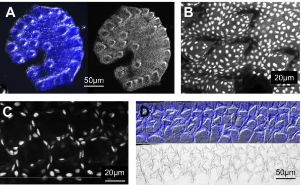
Sexually dimorphic pattern of breeding tubercles in zebrafish……….. 31
Cells of the regular superficial layer got lost in tubercles and cells with unique features were exposed at the outer surface………. 31
The cap layer of breeding tubercles is shed off indicating the need for a regeneration process in breeding tubercles……… 36
Tubercles develop prior to sexual maturity in a very rapid process………. 37
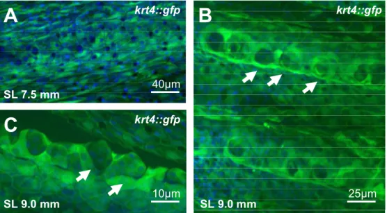
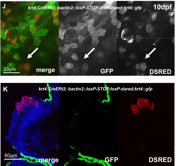
The superficial layer of adult zebrafish epidermis is gradually replaced by basal derivatives whereas the cells of the cap layer develop exclusively from the basal/intermediate layer…. 43 Breeding tubercles are rich in keratin content with differential keratin expression compared to the surrounding epidermis………. 45
Breeding tubercle ultrastructure indicated terminal cell differentiation in breeding Tubercles……… ….. 52
Proliferation is increased in breeding tubercles but also restricted to the stratum germinativum ……… 62
Non-apoptotic functions of Caspase 3 in the outer layers of breeding tubercles……….. 64
Cap cell membranes display leakiness in barrier assays pointing towards cell degradation… 66 Transglutaminase 1 is specifically expressed in the upper layers breeding tubercles………… 67
Differentiation of zebrafish keratinocytes in breeding tubercles strongly depends on Notch signalling and activation of Caspase 3………. 69
Notch signalling is activated in immature tubercles before cap layer formation starts……….. 76
Tubercle formation is compromised in TAp63 and p53 zebrafish mutants……….. 76
Ectopic over-expression of Notch rescued the diminished tubercle formation in TAp63 mutants……… . 85
Proliferation at the base of breeding tubercles is reduced in TAp63 and p53 mutant zebrafish……… …… 86
VI. Discussion………. 90
Advanced differentiation and self-renewal in breeding tubercles……….. 90
Breeding tubercles in an evolutionary context………. 94
Keratin networks in zebrafish breeding tubercles……….. 94
The linear pathway TAp63 Notch active Caspase 3 is needed for keratinocyte differentiation in zebrafish as well as mammals………. 95
The p53/TAp63 Notch active Caspase 3 pathway might be involved in regulation of shedding and renewal in breeding tubercles………. 97
Redundant functions of TAp63 and p53 might at least partially explain the lack of a severe phenotype in mouse TAp63 mutants……… 98
V. Material and Methods……… 100
VI. Abbreviations……….. 111
VII. Literature………. 112
Abstract
p63, a member of the p53 family of transcription factors, is crucial for vertebrate epidermal development. However, different isoforms of p63 are supposed to have different if not opposing functions. There is compelling genetic evidence that ∆Np63 isoforms are needed to enhance keratinocyte proliferation and stemness. However, the role of TAp63 isoforms is not fully understood, and TAp63 knockout mice display normal epidermal development. In this thesis, we describe the epidermal defects of zebrafish mutants specifically lacking TAp63 isoforms. TAp63 as well as p53 mutant zebrafish present with compromised development of breeding tubercles, epidermal appendages with more advanced stratification and keratinocyte differentiation than in regular epidermis, including continuous desquamation and renewal of superficial cells by derivatives of basal keratinocytes. Additionally, those defects are further enhanced in TAp63/p53 double mutants.
Furthermore, molecular analyses, treatments with chemical inhibitors and epistasis studies indicate the existence of a linear TAp63/p53 Notch Caspase 3 pathway required for enhanced proliferation of keratinocytes at the base of the tubercles and their subsequent differentiation in the absence of complete cell death. In summary, these studies identify the zebrafish breeding tubercles as specific epidermal structures sharing crucial features with cornified mammalian epidermis. In addition, they demonstrate essential and partially redundant roles of TAp63 and p53 to promote keratinocyte proliferation and their terminal differentiation, involving a pathway that might be conserved between fish and mammals.
Zusammenfassung
p63, ein Mitglied der p53-Familie von Transkriptionsfaktoren wird essentiell benötigt während der Entwicklung der Epidermis von Vertebraten. Andererseits scheinen verschiedene Isoformen von p63 unterschiedliche, wenn nicht sogar gegensätzliche Funktionen zu haben. Es existieren verschiedene Hinweise, dass Np63 benötigt wird um Proliferation und Stammzell-Eigenschaften in Keratinozyten anzuregen. Die Funktion der TAp63-Isoform ist dagegen nur wenig verstanden, wobei TAp63- defiziente Maus-Mutanten einen eher normalen epidermalen Phänotyp zeigen. In dieser Arbeit beschreiben wir die epidermale Defekte einer Zebrafisch-Mutante, in der spezifisch die TAp63- Isoformen von p63 fehlen. TAp63-, genauso wie p53-Mutanten zeigen eine gestörte Bildung von
„Breeding Tubercles“, epidermalen Anhangsgebilden mit weitergehender Stratifizierung und Differenzierung verglichen mit regulärer Epidermis von Fischen, einschliesslich kontinuierlicher Schuppung und Erneuerung von äusseren Zelllagen durch Nachkommen basaler Zellen. Weiterhin sind diese Defekte noch verstärkt in TAp63/p53 Doppelmutanten.
Durchgeführte molekulare Analysen, Behandlung mit chemischen Inhibitoren und Epistase-Studien deuten auf die Existenz eines linearen TAp63/p53 Notch Caspase 3 Signalübertragungsweges hin, der sowohl für Proliferation an der Basis der Tuberkel, als auch für die nachfolgende terminale Differenzierung (die allerdings ohne kompletten Zelltod einhergeht) nötig ist. Insgesamt identifiziert diese Arbeit die „Breeding Tubercels“ von Zebrafischen als epidermale Strukturen mit deutlichen Ähnlichkeiten zur verhornten Säugetier-Epidermis. Weiterhin werden teilweise redundante Funktionen von TAp63 und p53 sowohl in Bezug auf Proliferation als auch Differenzierung von Keratinozyten aufgezeigt die von einem molekularen Signalweg reguliert werden, der konserviert ist zwischen Fisch und Säugetier.
I. Introduction
Terminal differentiation of keratinocytes in land living animals
The epidermis is the outermost part of the integument that constitutes the boundary between the organism and the environment. Thereby, the epidermis has to protect against various physical, chemical and microbiological traumata. Additionally, most important for all land-living tetrapods, the epidermis provides a tight barrier against water loss and dehydration [Fuchs & Raghavan 2002, Medawar 1953]. Histologically, the epidermis of those animals has been classified as a multilayered, stratified and squamous epithelium that originates from the embryonic ectoderm [Fuchs 2007, Fuchs
& Raghavan 2002]. The epidermis also develops certain epidermal appendages, like hairs, nails or claws. The “regular” epidermis between these appendages (especially the hair producing hair follicles) is referred as interfollicular epidermis [Fuchs 2007].
The specific functions of the epidermis are achieved particularly by terminal differentiation of keratinocytes called cornification [Candi et al. 2005]. This differentiation process is a specific characteristic of mammalian epidermis but has already been present in birds, reptiles and amphibians.
In the progress of evolution, a cornified epidermis depicts an important step in the adaptation to land [Wu et al. 2004 & Alibardi 2003].
Particularly in older literature, the epidermis is only described as keratinized what means it contains high amounts of the intermediate filament keratin. However, during the last years it became clear that terminal differentiation of keratinocytes includes many more cellular hallmarks than only keratinization. All these processes together are now described as cornification. However, keratinisation and cornification are still often used as synonymous terms [Candi et al. 2005].
In mammalian - including human - epidermis different layers can be distinguished by the status of differentiation of the keratinocytes. Terminal differentiation is accompanied by differential gene expression of certain marker genes only in specific layers. Of certain importance is the so called
“keratin-code” in the epidermis: Different layers are characterized by the expression of distinct keratins [Candi et al. 2005, Fuchs & Raghavan 2002]. The anatomy of mammalian epidermis together with the expression domain of certain marker genes is summarized in figure 1.
Proliferation occurs only in the basal layer which consists of stem cells (with unlimited capacity of cell division) and transient amplifying (TA-) cells (with limited capacity of cell divisions) [Mack et al. 2005].
The proliferation rate is precisely balanced with a desquamation process of the cornified layer at the outer surface of the epidermis. Via this epidermal homeostasis process, the epidermis is constantly rejuvenated [Candi et al. 2005].
Basal keratinocytes are attached to the underlying extracellular matrix (ECM) and the basement membrane via integrins, which mediate not only attachment, but also regulate proliferation or terminal differentiation in keratinocytes [Lippens et al. 2005]. Two different cell-ECM complexes mediate the connection between the basal cells and the basement membrane: hemidesmosomes, which connect intracellularly to the keratin filaments and focal adhesion contacts, which connect to the actin cytoskeleton. Both types of contacts contain integrin dimers [Fuchs & Raghavan 2002].
Differentiation of keratinocytes starts after the cells have undergone cell cycle arrest and detached from the basement membrane [Fuchs 1995, Lippens et al. 2005]. At the molecular level, the process is initiated by unoccupied 1-integrin receptors. However, the same mechanism can also induce apoptosis or anoikis in cell culture. So far, both processes are supposed to be interdependent. The initial molecular trigger for detachment of keratinocytes and the molecular survival mechanism that prevents immediate cell death are still unidentified [Lippens et al. 2005, Levy et al. 2000, Mitra et al.
1997]. However, Notch-signalling and its target gene Hes1 are considered to play a pivotal role at the basal-suprabasal juncture to down-regulate basal genes (like K5/K14) and up-regulate spinous markers (like K1/K10) [Blanpain et al. 2006].
After detachment and initiation of terminal differentiation, keratinocytes leave the basal layer and migrate into the suprabasal compartment [Fuchs & Raghavan 2002]. This switch towards differentiation can also be seen in the transcriptional profile of the cells. While proliferating keratinocytes express keratin 5 and 14 (K5 & K14) as main intermediate filaments (bundled into KIFs
= keratin intermediate filaments) of the cytoskeleton [Fuchs & Raghavan 2002, Byrne et al. 1994], differentiating cells immediately start expressing keratins 1 and 10 (K1 & K10), which replace the old K5/K14 filament network (figure 1) [Candi et al. 2005, Fuchs & Raghavan 2002]. This switch in keratin gene expression indicates the commitment of the epidermal cell to terminal differentiation
Basement membrane
Basal layer Spinous layer Granular layer Cornified layer
K5/K14 K1/K10
Loricrin, Involucrin, Profilaggrin
TGM2 TGM1 TGM5 TGM3
Figure 1: Anatomy of mammalian epidermis. The epidermis of mammals can be divided into basal layer, spinous layer, granular layer and cornified layer. The cells above the granular layer are dead and enucleated. Cells in the epidermis differentiate while they migrate from the basal layer to the cornified layer. Certain marker genes are expressed only in specific layers (K Keratin, TGM Transglutaminase).
[Fuchs 2007]. Starting in the spinous layer, K1/K10 are expressed up to the outer cornified layer [Fuchs & Raghavan 2002, Candi et al. 2005]. The new keratin network does not only remodel the cytoskeleton of the cell to facilitate cell migration, but also regulates cell growth and cell cycle arrest via binding the adaptor protein 14-3-3 and activating the AKT/mTOR (mammalian target of rapamycin) pathway [Kellner & Coulombe 2009, Fuchs 2007, Kim et al. 2006, Watt 2002].
In the following two to four weeks, the keratinocytes migrate through all epidermal layers to the outer surface while they differentiate into dead and enucleated corneocytes [Lippens et al. 2005, Fuchs 1995]. The cornification process includes rearrangements of the cytoskeleton, formation of a cornified envelope (CE) underneath the plasma membrane and an outer lipid envelope in the extracellular space. Keratins become the main component of the cytoskeleton and form a dense intracellular network while they are bundled into large tonofibrils (keratinization) [Fuchs 2007, Candi 2005, Matoltsy 1976]. The process ends with a flattened, dead and enucleated cell at the outer surface that has lost all its organelles and is metabolically inactive. To highlight that the differentiation process is fulfilled, these cells are thus described as corneocytes [Fuchs & Raghavan 2002].
Through all layers the cells are attached to each other by cell-cell contacts. Adherens junctions connect two cells with the intracellular actin cytoskeleton and are found particularly in the lower layers of the epidermis, whereas desmosomes are in contact with the keratin intermediate filament system and can be found in all layers [Fuchs & Raghavan 2002].
Formation of the cornified envelope starts in the granular layer [Fuchs & Raghavan 2002]. Here cells acquire electron-dense keratohyalin granules which contain profilaggrin, the precursor of filaggrin that bundles keratin intermediate filaments into large macrofibrillar cables (= tonofibrils) [Fuchs 2007] and thereby promotes the collapse of the cell into a flattened shape, the characteristic of corneocytes [Fuchs & Raghavan 2002]. Keratin intermediate filaments and filaggrin together constitute between 80 and 90 % of the total cellular protein mass in the granular layer. Both proteins are heavily covalently cross-linked and build up a platform for later reinforcement and maturation of the cornified envelope [Fuchs & Raghavan 2002]. Already in the spinous layer, specific modifications at the inner side of the desmosomes had occurred that are now reinforced in the granular layer. CE-proteins like small proline-rich proteins (SPRPs), Loricrin, Involucrin and Filaggrin, which are rich in glutamine and lysine residues [Fuchs 2007] are crosslinked to the inner side of the plasma membrane and to the keratin intermediate filament system. This crosslinking occurs all over the inner side of the plasma membrane but particularly at the desmosomes [Candi 2005, Fuchs & Raghavan 2002]. The characteristic resistance and insolubility of the cornified envelope is mainly achieved by very stable covalent isopeptide bonds, whose formation is catalyzed by Ca2+ dependent transglutaminases (TGMs) [Fuchs
& Raghavan 2002]. Most important substrates for transglutaminases are type II-Keratins, Involucrin, Loricrin, Filaggrin, S100-Proteins and small proline-rich proteins (SPRPs) [Fuchs & Raghavan 2002].
Due to their main function, attaching the cornified envelope to keratin filaments, all these proteins are described as intermediate filament associated proteins (IFAPs) [Fuchs 1995]. Interestingly, most (but not all) of these CE proteins are arranged in mammalian genomes in one large gene cluster called
epidermal differentiation complex EDC (for example EDC in humans is a two Mb cluster with 45 genes on Chromosome 1q21) [Hoffjan & Stemmler 2007].
In the granular layer, the cells are tightly attached to each other by tight junctions. In cornifying epidermis, tight junctions are present only in this layer and play a crucial role in preventing transepidermal water loss [Brandner 2009, Mirota & Miyachi 2003, Tsuruta et al. 2002].
The cells of the outermost cornified layer are still tightly attached to each other. This is achieved by so-called corneodesmosomes, a modified desmosomal structure in which specific desmosomal proteins are substituted. Profound changes in desmosome morphology can be detected at the transition between the granular and the cornified layer. At the ultrastructural level, the cytoplasmic plaques of the desmosomes are integrated into the cornified envelope and a homogenous electron-dense plaque occurs in the intercellular space [Fuchs & Raghavan 2002]. This plaque is considered to consist of the transmembrane proteins Desmoglein and Desmocollin, members of the cadherin-family, and the extracellular protein Corneodesmosin, which is produced by the keratinocytes of the granular layer [Ishida-Yamamoto et al. 2011].
Additionally to the proteinacous cornified envelope, an extracellular lipid envelope is formed also starting at the granular layer. Lamellar bodies (= Odland bodies), which store complex series of lipids (e. g. ceramids, cholesterol esters or free fatty acids) originate from the Golgi apparatus and fuse with the plasma membrane to release their content into the extracellular space [Candi et al. 2005, Fuchs &
Raghavan 2002]. Outside the cell, the lipids are directly bound to the outer cell membrane to reinforce the barrier function, or aligned into extracellular lipid lamellae to prevent water loss. Together with the lipid lamellae precursors, acidic hydrolases are extruded into the intercellular space, which are responsible for changes of the lipid composition and subsequently lipid lamellae formation. At the end of this process in the upper cornified layers, the cornified envelope is embedded in a lipid envelope and the lipids are organized as stacked lamellar sheets that occupy all intercellular space between the corneocytes. An important but still unresolved field of research are interactions between the lipid envelope and the cornified envelope [Lippens et al. 2005, Fuchs & Raghavan 2002].
Simultaneously to the formation of the cornified and the lipid envelope, degradation of the DNA and cell organelles occurs. In contrast to apoptosis, no chromatin condensation or DNA laddering occurs during cornification. Finally the nucleus disappears and no chromatin remnants remain. So far, neither any DNAse responsible for this degradation process nor the processors of nuclear destruction could be identified. [Lippens et al. 2005].
Above the granular layer, the skin is comprised only of dead cells. The fully differentiated squames resemble empty “protein sacs” consisting nearly exclusively of keratins and cross-linked CE-proteins.
During the final shedding process in the uppermost layers of the stratum corneum, proteolytic degradation of cell-cell contacts allows squammation [Lippens et al. 2005, Fuchs & Raghavan 2002].
Especially during the later stages, cornification highly depends on specific proteases. Many CE protein precursors need proteolytic activation. Degradation of the nucleus and the organelles depends on proteases and finally degradation of cell-cell contacts during sloughing of cornified squames [Fuchs &
Raghavan 2002].
As already mentioned, proliferation and squammation are tightly linked and regulated processes in epidermal maintenance [Lippens et al. 2005, Fuchs 1995]. This may be achieved by organizing the epidermis into epidermal proliferating units (EPUs), columns of hexagonally packed cells as has been shown by lineage tracing experiments. All cells of an EPU originate from single basal stem cells that occasionally divide to produce cells with more restricted proliferating capacity (TA-cells), whereas all other cells differentiate [Fuchs 2007].
Cornification is the most abundant form of cell death in mammalian epidermis. Despite many similarities (e. g. loss of the nucleus and cell organelles and activation of specific proteases including effector-caspases), cornification is clearly different from apoptosis [Lippens et al. 2005, Candi et al.
2005, Fuchs & Raghavan 2002]. Apoptosis in the skin mainly occurs as a response to cell damage by UV light in proliferating basal keratinocytes, whereas cornification is a physiological process of terminal differentiation in the suprabasal layers [Lippens et al. 2005]. Characteristically, apoptosis occurs in single cells, which thereby loose their cell-cell contacts and are phagocytized afterwards by macrophages. In contrast, cornification is associated with sheet formation (cell-cell contacts remain intact) and stratification of the epidermis, meaning that the cellular changes during the differentiation process are not restricted to single cells, but occur simultaneously in all keratinocytes of the respective epithelial sheet [Lippens et al. 2005].
Interestingly, nearly all keratins as major components of differentiated keratinocytes contain conserved caspase cleavage sites. However, keratins are not considered to be cleaved during terminal differentiation. Whether they are indeed substrates of effector-caspases during apoptosis remains still unclear [Lippens et al. 2005]. The most important features of both types of cell death are summarized in table 1.
In a similar way, cornification as described above occurs in all amniotes (mammals, reptiles & birds).
For all these land-living animals protection from dehydration is crucial for survival [Alibardi 2002].
Reptiles resemble the primary amniotes from which birds and mammals derived. However, the mechanism of keratinization and cornification in reptiles is only poorly understood. In general, two different processes, - and -keratinization can be distinguished which involve - and -keratins. In - keratinized layers of the epidermis, cross-reactivity with antibodies against the CE-proteins Filaggrin and Loricrin could be detected in keratohyalin-like granules (KHLG) in the granular layer. A cornified envelope is also formed in the stratum corneum [Alibardi 2003]. These results support the hypothesis that reptilian -keratinization (which is softer and occurs often at locations exposed to movement) resembles mammalian keratinization, whereas -keratinization forms the hard scales of the reptilian skin which are folds of the complete skin. -keratins might have evolved from a single -keratin in the course of evolution, but seem to be now an independent keratin family that does not exist in mammals at all [Alibardi & Toni 2006, Alibardi 2002]. Alternating layers of - and -keratinized keratinocytes facilitate shedding [Alibardi 2003]. However, not all reptiles renew the cornified tissue via a shedding process. Continuous wearing is also very common [Alibardi & Toni 2006].
Table 1: Differences between cornification and apoptotic cell death.
Cornification Apoptosis
General features Slow process (duration ca. 2 weeks)
Occurs simultaneously in all cells of an epidermal layer
Massive synthesis of specific proteins
Fast process (48-72 hours)
Occurs in individual cells surrounded by viable neighbour cells
Limited need for protein synthesis Cell-cell contacts Remain intact Apoptotic cells loose cell-cell contacts
what causes formation of apoptotic bodies
Nuclear events Complete loss of nucleus
No chromatin condensation or DNA laddering
Responsible DNases unknown
Proteases that cause destruction of nuclear proteins unknown (except Desquamin)
Incomplete loss of nucleus
Nuclear remnants detectable
Internucleosomal DNA cleavage by caspase-activated DNases (CADs)
Caspases degrade nuclear proteins (for example Lamin)
Cytoskeletal rearrangements
Expression of different cytoskeletal components during the process
Formation of the cornified envelope and the lipid envelope
Cross-linkage between keratin filament system and cornified envelope
Dismantling of the cytoskeleton
Organelle function Organelles are completely degraded by proteases
Organelle function ceases, but organelles are not completely degraded Final fate of cell
corpses
Dead cells are regularly sloughed off into the environment as squames after proteolysis of cell-cell contacts
Apoptotic cells are phagocytosed by macrophages
Table 1 is based on Lippens et al. 2005, Candi et al. 2005 & Allombert-Blaise et al. 2003
Reptilian epidermis cornifies in a parakeratotic manner. That means that the nuclei of keratinocytes are still present in the stratum corneum. In mammals however, parakeratosis is clearly a pathological feature. Thus, cornification in reptiles may be described as a more primitive modality of cornification, where apoptosis or apoptosis-like processes appeared in conjunction with the formation of the stratum corneum [Alibardi 2003].
Both ways of keratinization are also present in birds, which are evolutionary related to reptiles. Avian epidermis comprises scaled and non-scaled areas. A soft interfollicular epidermis (apterilae) and a feathered epidermis (pterylae) can be distinguished, while scales are only present in the skin of the hind limbs [Alibardi & Toni 2006, Alibardi 2005]. Apteric epidermis is similar to mammalian epidermis but has no evident granular layer (which is the same in mammalian hard-cornified tissue). However, cross-reactivity with antibodies against mammalian cornification markers ( -keratins, Loricrin, Filaggrin, transglutaminases) has been demonstrated as well as the formation of cornified and lipid envelopes [Alibardi & Toni 2006, Alibardi 2005]. This indicates that keratinization and cornification in avian apteric epidermis roughly resembles the respective mammalian process. According to the literature, the lack of the stratum granulosum with its keratohyalin granules results in a poorly developed interkeratin matrix [Alibardi & Toni 2006] that might be responsible for the soft character of avian apteric epidermis.
Table 2: Comparison of cornification processes in amphibians, reptiles, birds and mammals.
Amphibians Reptiles Birds Mammals
Str. corneum Replacement layer -kertinization: thin -kertinization: thick (occur at the same
sites)
-kertinization: thin -kertinization: thick (occur at different
sites)
thick layer; only - kertinization
Str. granulosum No yes no yes
Shedding Moulting moulting or wearing shedding? shedding
Keratinization-Type + +
CE -- ++ ++ ++
HPRs No Loricrin
Filaggrin
Loricrin Filaggrin
Loricrin, Involucrin, Filaggrin
Parakeratosis Yes Yes ? Pathologically
Table 2 is based on Alibardi & Toni 2006, Alibardi 2005 and Alibardi 2002. Str. = Stratum, ? indicates lack of information
-keratinization occurs in the scaled regions of the epidermis and during production of the feathers. - keratins are similar to those of reptiles. Despite these similarities, the exact mechanisms of cornification in birds remains to be elucidated [Alibardi & Toni 2006].
As only anamniotes amphibian epidermis also constitutes a stratum corneum [Alibardi 2009].
Depending on the post-metamorphic adaptation, two different types of amphibian epidermis exist. In species which remain permanently aquatic, no cornified layer is formed and the epidermis is purely mucogenic. In terrestrially adapted forms, one ore two layers of cornified epidermis are formed on top of the stratified epidermis [Alibardi 2009]. Cells from the basal layer differentiate and eventually form a replacement layer that replaces the corneous layer during moult [Alibardi 2003b]. The process of moulting is under strict hormonal control (e. g. aldosterone). The dead cornified layer is shed and the granular layer underneath completes cornification [Smith 1975]. Interestingly, electron-dense granules are present in the upper spinous layer during epidermal differentiation. The smaller granules with a mucus-like content are discharged into the extracellular space, whereas the larger granules with an unknown content stay inside the keratinocytes [Farquhar & Palade 1964]. These granules are believed to contain mucus and crosslinking-proteins (that are different from the mammalian proteins) and form intracellularly a dense mucus interkeratin matrix [Alibardi 2003b, Navas et al. 1987, Bueno et al.
1981]. However, there is a lack of information about interfilamentous matrix proteins in the epidermis of non-mammalian vertebrates [Alibardi 2003b, Matoltsy 1987]. At least a cornified envelope does not seem to be formed in amphibians [Alibardi 2002].
In summary, cornification as it occurs in mammals is probably the most advanced form in terms of - keratinization, but many different - perhaps more primitive forms - can also be found throughout the animal kingdom. The main differences that are already known are listed in table 2.
Teleost epidermis
In vertebrate epidermis two different developmental pathways have been demonstrated, which are tightly linked to environmental adaptation. One pathway of this duality leads to keratinization, occurring predominantly in land-living animals, whereas the other one ends in mucogenesis and is common in amphibious or aquatic vertebrates [Henrikson & Matoltsy 1967]. Due to its aquatic environment, the epidermis has to protect fish from osmotic pressure stress, physical damage and infectious organisms including parasites [Hawkes 1974]. The epidermis of adult individuals is pluristratified and organized in three distinct strata: the basal layer on top of the basement membrane, the intermediate layer and the outer superficial layer [LeGuellec et al. 2004] (figure 2).
In contrast to mammalian epidermis, living epidermal cells are in direct contact with the environment [Hawkes 1974]. This means fish skin consists exclusively of living cells [LeGuellec et al. 2004].
Processes like keratinization and cornification that are characteristic for land-living vertebrates are absent in fish [Burgess 1956] or occur only at specific sites, as for example adhesive organs, lips or breeding tubercles. Generally, the surface of the skin is covered by mucus but not dead cornified cells [Chang & Hwang 2011,
LeGuellec et al 2004].
Ontogenetically, the cells of the basal and intermediate layer derive from the ectodermal sheet after gastrulation, whereas the origin of the superficial layer not completely understood.
It is considered to develop from the embryonic enveloping layer (EVL), which derives from the blastoderm during blastula period [Chang & Hwang 2011, LeGuellec et al.
2004].
Even if fish epidermis is also a pluristratified epithelium, the high degree of organization as seen in mammalian epidermis can
not be detected in teleost epidermis. Whereas in mammals basal, spinous, granular and horny or cornified cell layers can be distinguished, which resemble the degree of differentiation of the individual keratinocytes of the certain layers, fish epidermis consists mainly of undifferentiated cells in the
Basal layer
Intermediate layer Superficial
layer
MC
CC
Basement membrane
Figure 2: Anatomy of teleost epidermis. Three different compartments can be distinguished in the epidermis of teleosts: the basal layer, the large intermediate layer with mainly undifferentiated cells, and the flat superficial layer.
The intermediate layer also comprises certain differentiated cell types as mucous cells (MC) or club cells (CC). Ionocytes are not shown.
intermediate layer [Chang & Hwang 2011, LeGuellec et al. 2004]. However, the epidermis of fish comprises many differentiated cell types that do not exist in mammalian skin at all, for example mucous cells, club cells or ionocytes. On the other hand, those cells are structurally similar to some mesoderm- or endoderm-derived cells in mammals. For example mucous cells share similarities with mammalian goblet cells of the intestine epithelium and ionocytes have features needed for ion- and osmolarity-regulation, which are similar to mammalian epithelial kidney cells [Chang & Hwang 2011].
In mammalian skin, mitosis only occurs in basal cells. In teleost skin, autoradiographic studies with tritiated thymidine showed incorporation into cells throughout all epidermal layers indicating that in fish epidermis, mitosis is not restricted to the basal layer but occurs throughout the whole intermediate layer and the superficial layer as well [Henrikson & Matoltsy 1967].
In land-living tetrapods, epidermal cells adopt specific structural changes as they undergo terminal differentiation. They synthesize differentiation products and become transformed into protective corneocytes when they reach the distal stratum corneum. In regular fish skin, a comparable differentiation process does not seem to exist. It is assumed that density and bundling of keratin filaments do not differ in basal and intermediate layers. In mammalian skin, formation of tonofibrils is a hallmark of differentiation, whereas in teleost skin, filaments remain discrete rather than aggregate, as shown in electron microscopy studies. Most of these cytoskeletal rearrangements in mammals occur in the granular layer that is characterized by the presence of hyaline granula (keratohyalin). This cannot be found in fish skin either [Henrikson & Matoltsy 1967].
The outer surface layer, the superficial layer, is a single cell layer of flat viable cells. These cells develop microridges at the outer surface and are rich in keratins filaments. However, in contrast to mammals these cells do not get keratinized or cornified [Chang & Hwang 2011, LeGuellec et al. 2004].
The cells of the superficial layer are not shed and renewed regularly, but replaced individually after cell death [Chang & Hwang 2011].
Microridges are small protrusions (0.5 – 1 µm height) of the cell membrane, which are similar to microvilli of the intestine epithelium. They are formed by the actin cytoskeleton with the actin filaments being arranged perpendicular to the cell surface and an attachment plate at the basis of the microridge. These cellular protrusions are thought to protect fish against traumata and retain secreted mucus (which protects against various microbia) at the skin surface. Additionally, they might be needed during initial wound closure since the microridges can move by the contraction of their basal microfilaments [Bereiter-Hahn et al. 1979, Hawkes 1974].
In contrast to mammalian epidermis, tight junctions in fish epidermis are found only between the cells of the superficial layer that means in the outermost layer of the epidermis [Henrikson & Matoltsy 1967]. The origin of the superficial cells is not clear. However, they share many similarities with EVL (enveloping layer) cells which cover the embryo from blastula period on [Chang & Hwang 2011], whereas the intermediate and basal cells originate from the embryonic ectoderm [Chang & Hwang 2011, LeGuellec et al. 2004].
The large intermediate layer is composed of different cell types. The major components are relatively small basophilic cells [Henrikson & Matoltsy 1968] which resemble undifferentiated epidermal cells or keratinocytes with a low level of differentiation [Chang & Hwang 2011, LeGuellec et al. 2004]. They operate as a reservoir to replace dead cells of the epidermis.
The undifferentiated cells can divide rapidly and replace dead cells [Chang & Hwang 2011, LeGuellec et al. 2004]. The number of layers of the intermediate layer is highly variable and differs between different body regions [LeGuellec et al. 2004]. The cells are tightly connected to each other by numerous desmosomes [Henrikson & Matoltsy 1967].
The regulation of keratinocytes differentiation in fish skin remains still unclear [Chang & Hwang 2011].
Tonofibrils are described for fish keratinocytes but are only scarcely present compared to mammals and do not form a typical tight network [Hawkes 1974]. Additionally all histidine-rich proteins that are important for cornification (Involucrin, Loricrin and Filaggrin) are not existing in fish genomes and no incorporation of tritiated histidine could be detected by autoradiogaphy in contrast to mammalian or reptilian epidermis. [Alibardi 2003, www.ensembl.org]. Furthermore, no specific keratin expression has been reported in teleost epidermis so far. That means a “keratin-code” which is typical for mammalian epidermis is not known for fish epidermis so far. However, in general fish keratins are very different from mammalian keratins [Schaffeld et al. 2007].
Additionally to keratinocytes, different types of unicellular glands occur in the intermediate layer like mucous cells or club cells, sensory cells and ionocytes [Chang & Hwang 2011]. Each cell type has specific functions.
Mucous cells produce mucus substances that cover the outer surface of the fish and protect it from infections [Chang & Hwang 2011]. Mucous gland cells are basally derived cells that move upwards through the epidermis and thereby displace the boundaries of the adjacent cells [Hawkes 1974].
During their migration, the cells increase remarkably in size, until finally engorged cells reach the surface where they discharge their content into the extracellular space [Henrikson & Matoltsy 1968 b].
Club cells release alarm signal factors into the surrounding and exhibit a fright reaction. However, they are considered to have slightly different functions in different species [Chang & Hwang 2011].
Ionocytes carry specific ion transporters located at the cell membrane and are necessary for homeostasis and regulation of osmolarity of the body fluids [Chang & Hwang 2011]. This type of glands seems to develop from the same precursor population as the keratinocytes. After gastrulation, precursor cells start expressing the transcription factor Np63. In few cells, which will develop into ionocytes, the transcription factor foxi3a and deltaC are turned on whereas Np63 is downregulated.
Notch signalling in the surrounding cells together with sustained Np63 expression drives these undifferentiated cells into the keratinocytes lineage [Chang & Hwang 2011].
The basal cell layer is a single cell layer that is attached to the underlying basement membrane via hemidesmosomes [LeGuellec el al. 2004, Mittal & Whitear 1979]. The basement membrane separates the epidermis from the underlying dermis. In fish epidermis, the main function of the basal layer is the tight connection of the epidermis to the dermis [Chang & Hwang 2011, LeGuellec et al. 2004].
However, the basal cells themselves are connected to each other only by a relatively small number of
desmosomes [Mittal & Whitear 1979]. During skin development, the basal layer is involved in the production of the initial collagenous stroma of the dermis [LeGuellec et al. 2004]. In contrast to amniote epidermis, the basal layer in fish epidermis contains non-differentiated and differentiated cells [Chang & Hwang 2011].
Taken together, despite some similarities, fish epidermis and the epidermis of land-living vertebrates are quite different in architecture and composition.
Breeding tubercles
Breeding tubercles (also described as pearl organs) are spine-like epidermal structures found in at least 15 families within four orders of teleosts (especially cypriniformes) [Chen & Arratia 1996] and are thought to facilitate contact between individuals during spawning [Wiley & Colette 1970].
Accordingly, they frequently exist in body regions that come in contact with other fish, for instance head, fins or flank. Additionally, some species use them for nest or territory defence, or stimulation of females during breeding. While breeding tubercles are purely epidermal structures, analogous spikes which include also the dermis are also described in a variety of species. In contrast to breeding tubercles, these are classified as contact organs. In most cases, breeding tubercles as well as contact organs have a sexually dimorphic pattern in males and females. Often breeding tubercles are only or predominantly found in males [Wiley & Colette 1970].
Already fifty years ago, it became obvious that the cells of breeding tubercles consist of a substantial amount of keratins compared to lower levels of keratin in the remaining fish epidermis [Wiley &
Colette 1970]. Light and electron microscopy studies revealed a different composition of the epidermal cells in these tubercles including a process of keratinization in the outer cells and subsequent cell death at the outer surface [Mittal & Whitear 1979, Wiley & Colette 1970]. This is in clear contrast to the general agreement that keratinization and a terminal differentiation process are absent in fish skin.
In contrast to normal fish skin, mitosis is expected to occur in a stratum germinativum (in mammalian epidermis: basal layer + lower spinous layer) directly above the basement membrane [Mittal &
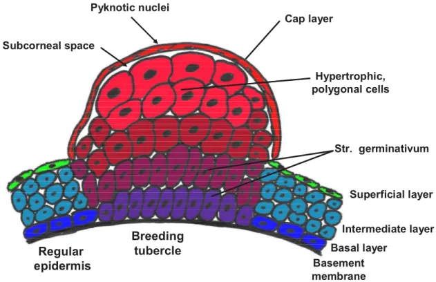
Whitear 1979, Wiley & Colette 1970]. The cells of the layers above become progressively hypertrophic and polygonal with large nuclei toward the outer surface. The outermost layer of the breeding tubercle appears keratinized with high amounts of keratin as shown by histology staining and electron microscopy [Mittal & Whitear 1979]. The transition between the hypertrophic cells and the outer keratinized cells is so abrupt that no transition zone is usually visible. In the keratinized cells, the nuclei disappear completely or persist as pyknotic remnants in the flattened, irregular cells of the keratinized layer [Mittal & Whitear 1979]. The general appearance of a breeding tubercle is summarized in figure 3.
Regular epidermis
Breeding tubercle
Basement membrane
Basal layer
Intermediate layer Superficial layer Cap layer
Str. germinativum Hypertrophic,
polygonal cells Subcorneal space
Pyknotic nuclei
In general, breeding tubercles develop prior the spawning season and reach their maximum size right before the season starts. After the spawning period is over, they break off, slough off or regress gradually. The development of the breeding tubercles seems to be affected mainly by the gonads and the pituitary gland [Wiley & Colette 1970], as gonadectomy prevents the formation of tubercles in the goldfish, and intraperitoneal injection of pituitary extract causes tubercle growth in minnows [Wiley &
Colette 1970, Ramaswami & Hasler 1955].
In amniotes and amphibia, the process of cornification is characterized not only by the presence of bundled keratin, but also involves death of cornified cells. A sloughing layer of dead but highly keratinized cells was found at the surface of certain elevations of the epidermis of the Indian catfish Bagarius bagarius [Mittal & Munshi, 1970]. These elevations showed all the features described for breeding tubercles, even if they not present with the classical breeding tubercle shape. In 1979, Mittal and Whitear used electron microscopy to further characterize the nature of keratinization and cell death in epidermal elevations compared to the non-keratinized epidermis of the furrows between the elevations [Mittal & Whitear 1979]. This study showed that basal cells below the elevations were bigger and more cubical than in the furrows. A striking feature was an abrupt transition in the appearance of keratinocytes between the fifth and sixth tier from the surface. From the fifth tier (from top) on, tonofibrils were condensed into bundles changing shape and staining behaviour of the cells.
This transition was explained by the authors by an increase in calcium content, shown by alicarin-S staining. Starting in the second tier, the cells flatten and the tonofibril bundles orientated parallel to
Figure 3: General features of breeding tubercles. The general organization and the major differences of breeding tubercles to regular epidermis are summarized here. Please refer to text for details.
the cell surface at least in the distal cytoplasm. The cells of the second tier were the most superficial living cells of the elevations. They were connected to one another by tight junctions, which normally occur only in the superficial layer in fish epidermis and to the upper tier of dead and keratinized cells by large desmosomes. A typical subcorneal space, as found in amphibians, was detected between the first and second tier in this study and large lipid inclusions were reported for the second tier [Mittal &
Whitear 1979]. The outermost cell layer showed the strongest reaction for calcium, indicating its keratinized nature. Additionally, those cells lacked all cell organelles and had an electron-dense layer of unknown material at the inner side of the plasma membrane, reminding of the cornified envelope in mammalian epidermis [Mittal & Whitear 1979].
Openings of mucous glands were never found in the keratinized regions of the elevations [Mittal &
Whitear 1979], indicating that the furrows resembled a mucogenic type of epidermis, whereas the elevations might be keratogenic. Furthermore, the keratinocytes in those regions were capable of synthesis of mucous granules [Das & Nag 2006]. In fish and amphibian epidermis, large mucous granules are expected to be involved in the clumping of keratin filaments [Fox 1986, Whitear 1986, Budtz & Larsen 1975, Parakkal & Matoltsy 1964].
Other potentially keratinized structures in fish are the horny teeth of the lamprey [Alibardi & Segalla 2011], the horny upper and lower jay sheet of Puntius sophore [Tripathi & Mittal 2011], or the organ of attachment of the catfish Garra gotyla [Das & Nag 2006], which all show many similarities on cellular or histological level. Comparison of morphology and histology of breeding tubercles among different species revealed a high number of significant differences, indicating that tubercles evolved independently in different groups [Wiley & Colette 1970]. However, the process of putative keratinization in the different described structures of fish skin seem to be basically the same or at least similar [Wiley & Colette 1970].
The p53-familiy member p63 and its role in mammalian epidermal development and maintenance
So far, three members of the p53-transcription factor family have been identified: p53, p63 and p73, all with high sequence and structural similarity, but apparently quite different functions [Koster &
Roop 2004]. All three proteins share an N-terminal transactivation domain (TAD), a central DNA binding domain (DBD) and a C-terminal oligomerization domain (OD) [Vanbokhoven et al. 2011, Candi et al. 2007, Strano et al. 2001]. Sequence similarity is particularly high between DBD of p63 and p73 but also p53 shares at least 65% similarity with other family members [Melino et al. 2002, Yang et al.
2002, De Laurenzi & Melino 2000].
The most commonly known member of the p53-family is the transcription factor p53 itself which is one of the best studied tumor suppressors and is involved in cell cycle arrest, DNA repair and leading cells into apoptosis [Riley et al. 2008]. As p53 -/- mutants (e. g. mouse mutants or zebrafish mutants)
[Molchadsky et al. 2010, Parant et al. 2010, Tyler et al. 1994] show no visible developmental defects but develop tumors later during adulthood, p53 is believed to be dispensable for development but very important to avoid tumor formation.
Different cellular stress scenarios, like DNA damage, spindle damage or hypoxia activate p53, which levels are tightly controlled via MDM2, an ubiquitin ligase, driving p53 into proteasomal degradation [Riley et al. 2008]. An activated p53 tetramer binds to p53 responsive elements (RE) in the promoter region of certain p53 target genes and thereby alternates gene expression of the cell inducing DNA repair, cell cycle arrest, cellular senescence or apoptosis [Riley et al. 2008].
Furthermore, p53 seems to be involved in asymmetric cell division of stem cells, giving rise to a dividing daughter cell and a postmitotic cell arrested in G1/S of the cell cycle [Sherley 2002]. As these results were shown mainly by in vitro experiments, the significance of this hypothesis still has to be demonstrated in vivo. However, this finding is consistent with the well-established idea that increased symmetric cell divisions increase the risk of carcinogenesis, as it was shown in p53 null animals [Sherley 2002]. Additionally, cultured explanted cells from p53 null mice do no longer show signs of senescence. The absence of senescence by cultures of p53 null tissue cells is consistent with abrogation of the asymmetric cell kinetics barrier to adult stem cell expansion by removal of p53- dependent regulation [Sherley 2002, Rambhatla et al. 2001, Merok & Sherley 2001].
On the RNA level, different p53 isoforms are transcribed by three different promoters in the p53 gene.
Additionally, alternative splicing at the 3’-end, leads to sets of -, - & - isoforms [Bourdon et al.
2005, Rohaly et al. 2005]. However, the full length protein is the most abundant isoform, and the alternative mRNAs seem to be produced especially under pathologic conditions (e. g. in tumors) [Rohaly et al. 2005]. The promoter P1 transcribes the regular p53 isoforms, whereas the alternative P1’ promoter leads to truncated 40p53 isoforms and the promoter P2 (upstream the fifth exon) to 133p53 isoforms. The functions and importance of the different isoforms remain largely unknown [Rohaly et al. 2005].
In contrast, the different isoforms of p63 and p73 are investigated in much more detail [Strano et al.
2001]. An alternative promoter upstream the third exon (in mammals) gives rise to two different types of isoforms: TA-isoforms with an N-terminal transactivation domain and N-isoforms without this domain [Strano et al. 2001, Koster & Roop 2004]. Additionally, alternative splicing at the 3`-end contributes to a more complex set of isoforms with the same 5`-end but different 3`-ends [Strano et al. 2001]. The longest set of isoforms with the same 5’-end, named -isoforms, carry an additional sterile alpha motif (SAM) domain at the 3’-end. This domain often contains protein interaction motifs frequently found in proteins that are involved in development and differentiation [Koster & Roop 2004], suggesting a role for p53-familiy members in such processes. Smaller sets of isoforms, the - and -isoforms, lack the SAM domain [Vanbokhoven et al. 2011, Candi et al. 2008, Koster & Roop 2004]. p53, on the other hand, does not have a SAM domain at all [De Laurenzo & Melino 2000].
The diversity of p63 isoforms gets even more complicated by different transcriptional and translational starts in the 5’-region. Bamberger and Schmale identified four different ATGs (AT1-4) in the TA specific region leading to four different sets of isoforms [Bamberger & Schmale 2001].
The domains of the most important isoforms of p53 and p63 are shown in figure 4.
In contrast to p53, p73 and especially p63 do not have a remarkable function as tumor suppressor, but have specific functions during development [Ramadan et al 2005, Moll & Slade 2004, Di Laurenzi
& Melino 2000]. As much more information is available for p63, the following description refers primarily to p63 which was shown to have crucial functions during formation of the epidermis.
Figure 4: Organization of p53 and p63. (A) Genomic organization of the mammalian p63 gene. p63 consists of a 3’- transactivation domain (TA, green), a DNA binding domain (DBD, blue), and an 5’-oligomerization domain (OD, red).
According to a second promoter, truncated N isoforms are generated lacking the N-terminal transactivation domain.
Alternative splicing at the 3’-end gives rise to additional sets of isoforms with the same 5’-end, named -, -, and - isoforms. Only the long -isoforms have an additional sterile alpha motif (SAM, grey) domain. Splicing variants generated by alternative splicing at the 5’-terminus are not shown. (B) Structure of p53, TAp63, and Np63 proteins. p53 and p63 share high similarity in the DBD and the OD. TAp63 and p53 both have a transactivation domain to activate specific target genes. According to alternative splicing, different truncated versions exist especially for p63, whereas those versions of p53 occur predominantly only under pathological conditions.
According to initial in vitro transactivation studies, the N-isoforms were supposed to be only dominant negative repressors of the respective TA-isoforms [Koster & Roop 2004, Koster et al. 2004, Yang et al 1998] because of the missing 5’-transactivation domain. However, newer results claim also transactivation functions for the different N isoforms possibly due to other transactivation domains [Vanbokhoven et al. 2011, Koster & Roop 2004, Wu et al. 2003, King et al. 2003, Ghioni et al. 2002, Dohn et al. 2001]. Furthermore, it is still not clear whether the dominant-negative function of the N- isoforms results from direct competition for p53-family binding sites in the promoter regions of target genes or by heterotypic dimerization [Koster & Roop 2004, Yang et al. 2002, Gaiddon et al. 2001].
The epidermis in mice (or mammals) develops from the embryonic ectoderm initially as a single- layered epithelium. Until day E9.5, this simple epithelium expresses the respective markers of simple epithelia, keratins 8 and 18. On day E9.5, the epithelial cells commit towards a stratified epithelium, which is demonstrated by a switch from K8/K18 to the basal marker keratins K5/K14. At day E10.5, the epidermis becomes bilayered. A layer of flat cells, the periderm, develops on top of the basal layer [Koster & Roop 2004b]. In contrast to the EVL in fish, the mammalian periderm originates directly from the basal layer of the epidermis. The periderm is needed for protection while the embryo is still in the amnion cavity. However, as in fish EVL, the first tight junctions of mammalian epidermis develop between cells of the new periderm at around day E11. These initial tight junctions are more similar to tight junctions of simple epithelia than to maculae occludentes in the granular layer of the mature stratified epidermis [Morita et al. 2002, M’Boneko & Merker 1988, Nakamura & Yasuda 1979].
At day E13.5, the epidermis eventually stratifies. In the following, an intermediate cell layer forms between the basal layer and the periderm. Initially this second layer proliferates as well, probably due to the rapidly growing embryo. But soon later at day E15.5, the cells in this layer become postmitotic and start to differentiate, what is reflected in the switch towards K1/K10, the keratins of suprabasal cells [Koster & Roop 2004b, Weiss & Zelickson 1975]. Calcium signalling is needed for the commitment as well as for the later differentiation of keratinocytes [Koster & Roop 2004b]. Cultured primary human keratinocytes proliferate and remain features of basal cells only when kept at low calcium concentration. When the concentration is increased, cells withdraw from cell cycle and start terminal differentiation. During epidermal development, a calcium gradient is established around the time, the spinous layer occurs [Koster & Roop 2004b, Elias et al. 1998]. In accordance with these results, Okuymama et al. demonstrated that E15.5 epidermal keratinocytes have an intrinsically higher commitment to terminal differentiation than newborn epidermal keratinocytes [Koster & Roop 2004b, Okuyama et al. 2004].
Until day E19.5, the epidermis gets multilayered, always with viable periderm cells on top. At a final step in epidermal development, the periderm cells are shed at E19.5 and are replaced by the stratum corneum with fully differentiated and dead keratinocytes [Akiyama et al. 1999, Byrne et al. 1994, Holbrook & Odland 1975]. Mammalian epidermal development is summarized in figure 5.
The expression pattern of the different p63 isoforms especially during epidermal development is still under intensive discussion. In adult mice, p63 is expressed at high levels in the basal cells of the epidermis and other stratified epithelia. In the suprabasal layers p63 is strongly reduced or completely absent [Pellegrini et al. 2001, Yang & McKeon 2000, Mills et al. 1999]. However, the generation of isoforms-specific antibodies identified only the N isoforms in the basal layer, whereas the TA isoforms were found in the suprabasal layers, indicating a switch during keratinocytes differentiation from the N to the TA isoforms [Nylander et al. 2002]. According to its expression pattern and the phenotype of p63 knock-out mice (described below), p63 was expected to have an essential role during development and/or differentiation of the epidermis [Koster & Roop 2004]. Both, TAp63 and Np63 isoforms were found to be expressed in mouse embryos and in adult mouse epidermis as shown by RT-PCR using isoforms-specific primer. In detail, this expression analysis of whole embryos (E7.5-E9.5) and embryonic epidermis (E15.5-E18.5) demonstrated the presence of TAp63 as early as E7.5, prior to commitment to stratification. In this study, Np63 transcripts were not detected prior to day E9.5, with TAp63 still being the predominant isoform until day E18.5 [King & Weinberg 2007, Koster at al. 2004]. Additionally in support of the early onset of TAp63, it was shown that ectopic expression of TAp63 but not Np63 in single layered lung epithelium was necessary and sufficient for initiation of a stratification program demonstrated by expression of K5/K14 [Koster & Roop 2004, Koster et al. 2004].
However, a different study could not find any p63 transcripts at E7, and only Np63 at E8 and E9 [King & Weinberg 2007, Laurikkala et al. 2006]. Similarly in zebrafish, Np63, but not TAp63, expression was observed and reported to be required for morphogenesis of the stratified epidermis [King & Weinberg 2007, Lee & Kimmelman 2002]. qRT-PCR data of skin samples from mouse embryos during different stages of skin development, while the epidermis is already bilayered (day E13), are available. These data revealed that 99% of the existing p63 transcripts were N isoforms, whereas only 1% was identified as TA isoform [King & Weinberg 2007, Candi et al. 2007].
To make it even more complicated, in situ hybridization studies with isoforms-specific antisense probes could show that only the N isoforms but not the TA isoforms were expressed in epithelia of mice from day from E11 to E14 [Laurikkala et al. 2006]. Additionally, isoforms-specific antibodies detected only the N isoforms but not the TA isoforms of p63 during epidermal development (E10.5- E16.5) [Romano et al. 2009].
In summary, it is still not clear if TAp63 is expressed in embryonic skin at all [Vanbokhoven et al.
2011, Livera et al. 2008, Suh et al. 2006]. It became clear, that the N-isoforms are the more abundant isoforms compared to the TA-isoforms at the protein level [Vanbokhoven et al. 2011, Koster et al. 2007]. Np63 is highly expressed in basal keratinocytes, but downregulated in suprabasal cells.
TAp63 is strongly expressed in oocytes, but expression in the epidermis is still unclear [Vanbokhoven et al. 2011, Livera et al. 2008, Suh et al. 2006].
Mutations in the human p63 gene are responsible for several ectodermal dysplasia syndromes (EDS) that are congenital disorders with abnormalities in ectodermal derived structures like hair, teeth, nails
or even craniofacial structures or digits [Vanbokhoven et al. 2011]. p63-deficient mice develop no stratified epidermis and have aberrant squamous epithelia (e. g. cervix or urothelium) [Vanbokhoven et al. 2011]. Additionally they lack epithelial appendages like mammary glands, hair follicles or teeth [Vanbokhoven et al. 2011, Koster et al. 2007]. Consequently, p63-deficient mice die shortly after birth, most probably due to dehydration because of the absent barrier function of the skin [Candi et al. 2008]. Besides the epithelial phenotype, also p63 knock-out mice have truncated limbs and cranio- facial defects [Vanbokhoven et al. 2011].
However, the generation of two independent p63-(isoform independent) knock out models led to controversial results concerning the function of p63 in epidermal development. Both strains were generated on different genetic backgrounds with different targeting strategies. On the first sight, both of them had severe defects in epidermal development and lacked epidermal appendages as described above. A subsequent more detailed analysis led to two quite different hypotheses concerning the function of p63 in the epidermis [Vanbokhoven et al. 2011]: Knock-out mice created in the Bradley laboratory were covered by a one-layered simple epithelium [Koster et al. 2007, Mills et al. 1999] that
E8.5 E9.5 E10.5 E15.5 E19.5
1
2
3
p63 K8/
K18 K5/
K14
K1/
K10
Loricrin
1 simple epithelium 2 stratification
3 cornification
Figure 5: Mammalian development of the epidermis. In mammals, the development of the epidermis starts with a simple epithelium originating from the embryonic ectoderm. After occurrence of the periderm layer, the epidermis stratifies while it still consists exclusively of living cells. At the end of the developmental process the periderm cells are shed and the cornified layer develops. The time frame of the different steps and the most important marker molecules are shown below the figure. Please see text for details. Blue: basal cells, light blue: spinous cells, red: granular cells, yellow: cornified cells, green: periderms cells.
did not stratify and lacked all specific markers of differentiation. Furthermore, the epithelial cells expressed K8 and K18, typical markers of simple epithelia [Vanbokhoven et al. 2011, Mills et al.
1999]. Consequently, it was concluded that p63 is necessary for epidermal commitment and stratification [Vanbokhoven et al. 2011]. As epithelial cells do not express K5 and K14, the epidermis might remain arrested in a premature state prior to the onset of stratification [Koster & Roop 2004b].
Similarly, it was demonstrated in 2004 that the switch in the differentiation of the Muellerian duct depends on p63. In the absence of p63, the Muellerian duct epithelium develops into a single layered uterine epithelium, while induction of p63 expression results in the differentiation into a cervicovaginal (stratified) epithelium [Koster & Roop 2004b, Kurita et al. 2004]. Additionally, it was demonstrated in this study that p63 expression in the Muellerian duct is induced via signals of the underlying mesenchyme [Kurita et al. 2004], whereas a role of the mesenchyme in epidermal p63 induction is not known so far. According to the similarities in development of the different stratified epithelia, an equivalent role for the mesenchyme in the epidermis is very likely. In support of an early function of p63 in the epidermis, p63 was found to be the first keratinocyte-specific marker in cell culture experiments [Koster & Roop 2004b, Green et al. 2003].
In contrast to the above phenotype, KO mice from the McKeon laboratory presented patches of differentiated keratinocytes that expressed terminal differentiation markers like Involucrin, Loricrin and Filaggrin, placed on an exposed dermis [Vanbokhoven et al. 2011, Koster et al. 2007, Yang & McKeon 2000, Yang et al. 1999]. In accordance with these findings, another study could show that p63 protein is enriched in putative epidermal stem cells [Koster & Roop 2004b Pellegrini et al. 2001]. This led to the interpretation that a stratified epidermis can develop initially in the absence of p63, but cannot be maintained afterwards due to a lack of proliferating cells. According to this hypothesis, p63 would be required for stem cell maintenance but not differentiation [Vanbokhoven et al. 2011].
More recent data indicate that during development and later maintenance of the epidermis, an exactly titrated balance between the different isoforms is absolutely crucial [Mack et al. 2005]. However, additionally to the above described controversial data on p63, especially the TAp63 isoform functions seem to be even more complicated. Different studies claim promoting or inhibitory functions of TAp63 during keratinocyte differentiation and cornification after commitment has occurred.
Cell culture transfection experiments identified TAp63 to directly bind to the Jagged1 promoter and thereby promoting Jagged-Notch signalling [Mack et al. 2005, Sasaki et al. 2002]. Okuyama et al.
published data in 2004 where Notch signalling activated Caspase3 in a non-apoptotic manner only during initial cornification in mouse embryonic epidermis. Blocking Notch signalling by the -secretase inhibitor DAPT also blocked activation of caspase3. Furthermore, important cornification markers like Loricrin and Filaggrin were significantly reduced in caspase3 knock-out mice [Mack et al 2005, Okuyama et al 2004]. These data claim a differentiation-driving pathway in mammalian epidermis, where TAp63 activates Jagged-Notch signalling. Then Notch enhances terminal differentiation and cornification via a non-apoptotic function of Caspase 3. However, induction of terminal differentiation of keratinocytes is characterized by withdrawal from the cell cycle and alteration of gene expression.