Vesicular trafficking dynamics enable context-dependent regulation of ErbB receptor activity and signaling
Dissertation
Zur Erlangung des akademischen Grades eines Doktors der Naturwissenschaften
(Dr. rer. nat.)
der Fakultät Chemie und Chemische Biologie der Technischen Universität Dortmund
vorgelegt von Yannick Brüggemann
Juli 2018
Vorgelegt im Juli 2018
von Yannick Brüggemann
Gutachter:
Prof. Dr. Philippe I.H. Bastiaens Prof. Dr. Andrea Musacchio
The work presented in this dissertation was performed in the laboratory of Prof. Dr.
Philippe I.H. Bastiaens at the Max Planck Institute of Molecular Physiology, Dortmund, (Germany).
Eidesstattliche Versicherung (Affidavit)
______________________________ _____________________________
Name, Vorname Matrikel-Nr.
(Surname, first name) (Enrolment number)
____________________ _________________________
Ort, Datum Unterschrift
(Place, date) (Signature)
Titel der Dissertation:
(Title of the thesis):
____________________________________________________________________________
____________________________________________________________________________
____________________________________________________________________________
*Please be aware that solely the German version of the affidavit ("Eidesstattliche Versicherung") for the PhD thesis is the official and legally binding version.
__________________________ _______________________
Ort, Datum Unterschrift
(Place, date) (Signature)
Belehrung:
Wer vorsätzlich gegen eine die Täuschung über Prü- fungsleistungen betreffende Regelung einer Hochschul- prüfungsordnung verstößt, handelt ordnungswidrig. Die Ordnungswidrigkeit kann mit einer Geldbuße von bis zu 50.000,00 € geahndet werden. Zuständige Verwaltungs- behörde für die Verfolgung und Ahndung von Ordnungs- widrigkeiten ist der Kanzler/die Kanzlerin der Techni- schen Universität Dortmund. Im Falle eines mehrfachen oder sonstigen schwerwiegenden Täuschungsversu- ches kann der Prüfling zudem exmatrikuliert werden, § 63 Abs. 5 Hochschulgesetz NRW.
Die Abgabe einer falschen Versicherung an Eides statt ist strafbar.
Wer vorsätzlich eine falsche Versicherung an Eides statt abgibt, kann mit einer Freiheitsstrafe bis zu drei Jahren oder mit Geldstrafe bestraft werden, § 156 StGB. Die fahrlässige Abgabe einer falschen Versicherung an Eides statt kann mit einer Freiheitsstrafe bis zu einem Jahr oder Geldstrafe bestraft werden, § 161 StGB.
Die oben stehende Belehrung habe ich zur Kenntnis genommen:
Official notification:
Any person who intentionally breaches any regulation of university examination regulations relating to deception in examination performance is acting improperly. This offence can be punished with a fine of up to EUR 50,000.00. The competent administrative authority for the pursuit and prosecution of offences of this type is the chancellor of the TU Dortmund University. In the case of multiple or other serious attempts at deception, the candidate can also be unenrolled, Section 63, paragraph 5 of the Universities Act of North Rhine-Westphalia.
The submission of a false affidavit is punishable.
Any person who intentionally submits a false affidavit can be punished with a prison sentence of up to three years or a fine, Section 156 of the Criminal Code. The negligent submission of a false affidavit can be punished with a prison sentence of up to one year or a fine, Section 161of the Criminal Code.
I have taken note of the above official notification.
Ich versichere hiermit an Eides statt, dass ich die vorlie- gende Dissertation mit dem Titel selbstständig und ohne unzulässige fremde Hilfe angefertigt habe. Ich habe keine anderen als die angegebenen Quellen und Hilfs- mittel benutzt sowie wörtliche und sinngemäße Zitate kenntlich gemacht.
Die Arbeit hat in gegenwärtiger oder in einer anderen Fassung weder der TU Dortmund noch einer anderen Hochschule im Zusammenhang mit einer staatlichen oder akademischen Prüfung vorgelegen.
I hereby swear that I have completed the present dissertation independently and without inadmissible external support. I have not used any sources or tools other than those indicated and have identified literal and analogous quotations.
The thesis in its current version or anotherversion has not been presented to the TU Dortmund University or another university inconnection with a state or academic examination.*
Table of Contents
Abstract ... 8
Zusammenfassung ... 9
1 Introduction ... 10
1.1 ErbB receptors ... 10
1.2 Epidermal growth factor receptor (EGFR) ... 12
1.3 EGFR structure and activation ... 12
1.4 Autonomous EGFR activation ... 13
1.5 Structural auto-inhibitory features of EGFR ... 14
1.6 Protein tyrosine phosphatases (PTP) ... 15
1.7 Endocytic trafficking of EGFR ... 15
1.8 Mitogen activated protein kinases (MAPK) ... 18
1.9 Extracellular regulated kinase (Erk) ... 19
1.10 Regulation of Erk signaling specificity ... 20
2 Objectives ... 22
3 Material and Methods ... 23
3.1 Materials ... 23
3.1.1 Chemicals and Reagents ... 23
3.1.2 Commercial Solutions ... 24
3.1.3 Buffers and Media ... 25
3.1.4 Bacterial Strains ... 26
3.1.5 Enzymes ... 26
3.1.6 Kits ... 26
3.1.7 Antibodies ... 27
3.1.8 Oligonucleotides ... 29
3.1.9 Plasmids ... 29
3.1.10 Instruments and equipment ... 30
3.1.11 Software ... 32
3.2 Molecular Biology ... 33
3.2.1 Polymerase chain reaction (PCR) ... 33
3.2.3 Restriction digestion ... 34
3.2.4 Dephosphorylation of 5’-Phosphorylated DNA fragments ... 34
3.2.5 Ligation of DNA Fragments ... 34
3.2.6 Site-Directed Mutagenesis ... 35
3.2.7 Transformation of chemically competent E. coli cells ... 35
3.2.8 Plasmid preparation ... 35
3.2.9 Determination of DNA concentration ... 35
3.2.10 DNA Sequencing ... 36
3.3 Protein biochemistry ... 36
3.3.1 Preparation of cell lysates ... 36
3.3.2 Immunoprecipitation ... 36
3.3.3 BCA assay ... 36
3.3.4 SDS PAGE ... 37
3.3.5 Western blotting ... 37
3.4 Cell biology ... 37
3.4.1 Cell culture ... 37
3.4.2 Transient Transfection ... 38
3.4.3 RNA interference ... 38
3.4.4 CRISPR/Cas9 ... 38
3.5 Immunohistochemistry ... 39
3.5.1 In-cell western assay ... 39
3.5.2 Immunofluorescence ... 39
3.6 Microscopy ... 39
3.6.1 Leica SP5 ... 39
3.6.2 Leica SP8 ... 40
3.6.3 OlympusFV1000 ... 40
3.6.4 Cell migration ... 40
3.6.5 Single cell migration ... 41
3.7 Analysis of confocal fluorescence imaging data ... 41
3.7.1 Quantification of the EGFR fraction at the RE ... 41
3.7.2 Quantification of the spatial distribution of pY845 ... 42
3.7.3 Quantification of PH-Akt translocation ... 42
3.7.4 Quantification of ErbB-mCitrine internalization ... 42
3.7.5 Quantification of pRb and CyclinD1 expression ... 42
3.7.6 Quantification of EGFR phosphorylation ... 42
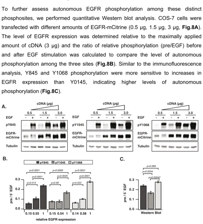
4.1 Spatial regulation of EGFR activity by vesicular trafficking ... 43
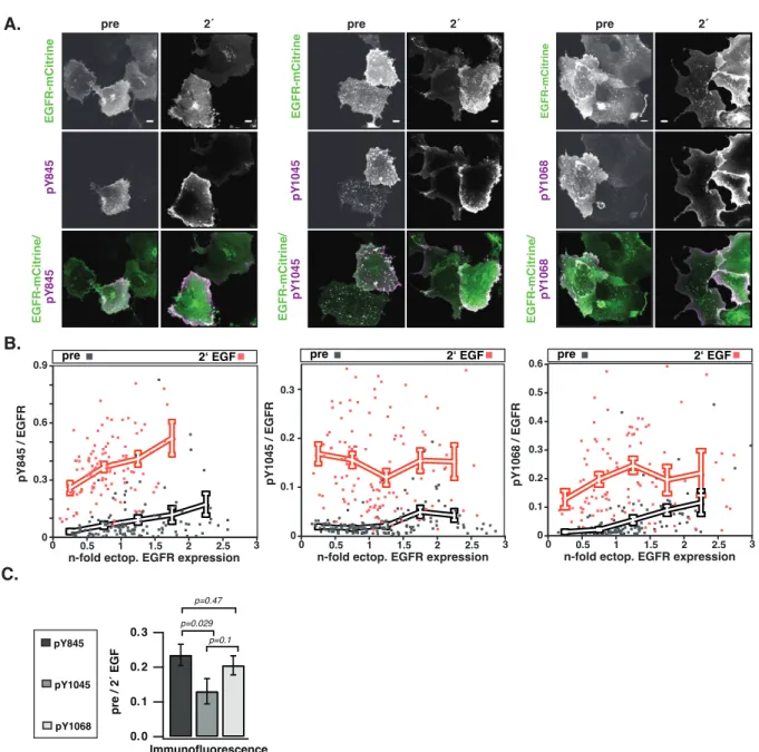
4.1.1 The dependence of spontaneous EGFR phosphorylation on its expression level ... 43
4.1.2 EGFR continuously recycles through the pericentriolar recycling endosome ... 49
4.1.3 Vesicular recycling suppresses autonomous EGFR activation ... 51
4.1.4 PTP1B dephosphorylates autonomously activated EGFR in the perinuclear area ... 54
4.1.5 EGF triggers a ubiquitin-mediated switch in vesicular trafficking of EGFR ... 57
4.2 Ligand-specific ErbB receptor trafficking determines Erk signaling specificity ... 60
4.2.1 HRG and EGF induce distinct spatial distributions of active ErbB receptors ... 60
4.2.2 KSR enhances HRG-induced Erk activation at the PM ... 65
4.2.3 EGF and HRG promote Erk-dependent cell proliferation ... 66
4.2.4 HRG stimulation promotes cellular motility ... 68
5 Discussion ... 75
5.1 Spatial regulation of EGFR activity by vesicular trafficking ... 75
5.1.1 Autonomous activation of EGFR in the absence of ligand ... 75
5.1.2 Continuous vesicular recycling suppresses autonomous EGFR activation ... 76
5.1.3 A ubiquitin mediated switch in vesicular trafficking ... 77
5.2 Ligand-specific ErbB receptor trafficking determines Erk signaling specificity ... 79
5.2.1 EGF and HRG promote Erk activation from different subcellular locations ... 80
5.2.2 KSR enhances Erk activation at the PM ... 81
5.2.3 EGF and HRG promote Erk-dependent cell proliferation ... 81
5.2.4 Membrane proximal Erk signaling promotes cellular motility ... 82
6 References ... 85
7 Abbreviations ... 99
8 Acknowlegements ... 102
9 Publications and presentations ... 103
Abstract
The ErbB signaling network comprises a complex dynamic system, which regulates a diverse set of cellular behaviors. In this thesis, we examined how regulating the distribution of ErbB receptors between the plasma membrane and different endosomal compartments enables contextual regulation of receptor activity and signaling.
In the first part of this work, we investigated how spatial regulation of EGFR controls its autocatalytic activity. We found that autonomously activated and EGF activated receptors are processed differently by the endocytic machinery. Constitutive vesicular recycling through perinuclear areas with high PTP1B phosphatase activity suppresses autonomous receptor activation in the absence of ligand, while maintaining EGFR abundance at the PM. In contrast, EGF binding enhances EGFR self-association and phosphorylation of the c-Cbl docking tyrosine Y1045, which results in receptor ubiquitination, internalization and lysosomal degradation. The coupling of EGFR self-association state, ubiquitination and vesicular dynamics thereby allows for the coexistence of a continuous safeguard cycle, while maintaining sensitivity to growth factor stimulation.
In the second part of this work, we examined how ligand-specific ErbB receptor trafficking determines Erk signaling dynamics and localization. We found that EGF or heregulin (HRG) differentially modulate ErbB receptor trafficking to generate distinct spatiotemporal patterns of receptor activities, leading to the activation of Erk from different subcellular compartments (plasma membrane and endosomes). The subcellular localization of Erk activation influences its interaction with different effector proteins and thereby generates ligand-specific cellular responses.
Proliferative Erk signals are transmitted to the nucleus, irrespective of their spatial origin, while the Erk dependent phosphorylation of pro-migratory effectors relies on membrane proximal Erk activity.
Collectively our findings highlight how vesicular dynamics enable context dependent spatial regulation of ErbB activity and downstream signaling events.
Zusammenfassung
Das ErbB-Rezeptor Signalnetzwerk umfasst ein komplexes dynamisches System, welches vielfältige zelluläre Verhaltensweisen reguliert. Im Rahmen dieser Arbeit wurde untersucht, wie die Regulation der Verteilung von ErbB-Rezeptoren zwischen der Plasmamembran und verschiedenen endosomalen Kompartimenten, eine kontextabhägige Regulation der Rezeptoraktivität und Signalweiterleitung, ermöglicht. Im ersten Teil dieser Arbeit wurde untersucht, wie die räumliche Regulation von EGFR seine autokatalytische Aktivität steuert. Es wurde gezeigt, dass autonom aktivierte und EGF-aktivierte Rezeptoren unterschiedlich durch die endozytische Maschinerie transportiert werden. Konstitutives vesikuläres Recycling durch perinukleäre Bereiche mit hoher PTP1B-Phosphatase-Aktivität unterdrückt autonome Rezeptoraktivierung in Abwesenheit von Liganden, bei gleichzeitiger Aufrechterhaltung der Membranlokalisation von EGFR. Im Gegensatz dazu verstärkt die Bindung von EGF die EGFR-Selbstassoziation und Phosphorylierung der c-Cbl- Bindestelle Y1045, was zur Ubiquitinierung, Internalisierung und zum lysosomalem Abbau des Rezeptors führt. Die Kopplung des EGFR-Selbstassoziationszustands, der Rezeptor-Ubiquitinierung und der vesikulären Dynamik, ermöglicht somit die Koexistenz eines kontinuierlichen, suppressiven vesikuläreren Transportkreislaufes, während die Empfindlichkeit für die Stimulierung mit Wachstumsfaktoren erhalten bleibt. Im zweiten Teil dieser Arbeit wurde untersucht, wie der Liganden-spezifische ErbB-Rezeptor Transport die raumzeitliche Organisation der Erk-Signalgebung steuert. Es wurde gezeigt, dass EGF oder Heregulin (HRG) den ErbB-Rezeptor Transport unterschiedlich modulieren und so unterschiedliche raumzeitliche Muster von Rezeptoraktivitäten erzeugen. Dies führt zur Aktivierung von Erk aus verschiedenen subzellulären Kompartimenten (Plasmamembran und Endosomen).
Die subzelluläre Lokalisation der Erk-Aktivierung beeinflusst die Interaktion von Erk mit verschiedenen Effektorproteinen und erzeugt dadurch unterschiedliche zelluläre Antworten. Proliferative Erk-Signale werden unabhängig von ihrem räumlichen Ursprung an den Zellkern übertragen, während die Erk-abhängige Phosphorylierung pro-migratorischer Effektoren auf membrannaher Erk-Aktivität beruht.
Zusammenfassend zeigen unsere Ergebnisse, wie die Regulation der vesikulären Dynamik von ErbB-Rezeptoren eine kontextabhängige räumliche Regulation ihrer Aktivität und Signalweiterleitung ermöglicht.
1 Introduction
Cells are constantly exposed to a wide range of extracellular cues and growth factors which they need to sense and interpret. The ability of cells to adapt their behaviour with regard to their environment is an essential aspect of tissue development and homeostasis1,2. Cells use signalling networks to process extracellular information and translate these into distinct responses (e.g. proliferation, differentiation, movement or death)3. These signalling networks are highly dynamic, often reconfigurable and can generate context-dependent function, instead of being static, linear and hardwired4–6. Although the identity of numerous central components of many signaling pathways has been determined and ample studies have addressed their inherent molecular properties, how they collectively process information to generate a defined cellular response to extracellular signals is still poorly understood. A growing number of studies supports that cells send and receive information about their environment by controlling the spatial and temporal dynamics of signaling molecules7–10. A systems level understanding how different stimuli can be encoded in spatial and temporal patterns and how these dynamics are interpreted and shape cellular behaviour could provide new insights into how cells generate higher-order emergent behavior and may reveal new pharmacological strategies for the treatment of disease11,12.
1.1 ErbB receptors
Cell surface receptors function as sensory organs, which enable cells to sense their environment and convert extracellular signals into intracellular signaling events, employing a plethora of different posttranslational modifications13. Mechanisms that regulate the spatial and temporal activity of these receptors can influence their sensitivity to stimuli, duration of activity and bias their interactions with effectors, providing different levels of regulation that can influence the cellular response to receptor activation. Based on their structure, cell surface receptors are grouped into different families.
The ErbB/HER family of cell surface receptors form a subgroup of receptor tyrosine kinases which includes four family members: EGFR (HER1/ErbB1), ErbB2 (HER2/Neu), ErbB3 (HER3) and ErbB4 (HER4)14. ErbB receptors are ubiquitously expressed in epithelial, cardiac, mesenchymal, and neuronal cells and mediate
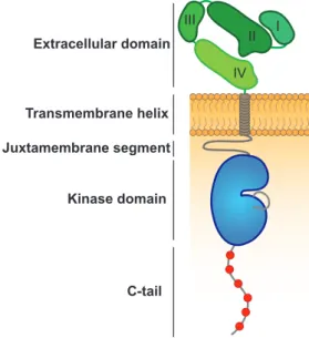
ligand-binding domain, followed by a single transmembrane helix and an intracellular module composed of a juxtamembrane segment, a tyrosine kinase domain and a C- terminal tail (Fig.1)17. The canonical scheme of ErbB receptor activation relies on ligand-induced receptor homo- or heterodimerzation, followed by allosteric activation of their intrinsic kinase activity and the phosphorylation of tyrosine residues within the C-terminal tail of the receptor which can act as effector docking sites14,17–20. Subsequent recruitment of PTB- or SH2-containing effector proteins relays the extracellular signal inside the cytoplasm21,22. Notably, the activation of ErbB2 and ErbB3 relies on the formation of receptor heterodimers, since for ErbB2 no ligands have been identified yet, while ErbB3 is kinase impaired15,23. To date, seven different ligands of EGFR have been identified: Epidermal growth factor (EGF), transforming growth factor alpha (TGF-α), betacellulin (BTC), heparin-binding EGF-like growth factor (HB-EGF), amphiregulin (ARG), epiregulin (EPR), and epigen (EGN). BTC, HB-EGF and EPR as well as neuregulins (e.g. heregulins) can also bind and activate ErbB4, whereas the latter also binds to ErbB3 and promotes its heterodimerization with ErbB216. Individual ligands can induce qualitatively and quantitatively different responses of downstream signaling molecules and thereby promote different cellular behaviours24.
Figure 1: Domain structure of ErbB receptors. The N-terminal extracellular domain is composed of four subdomains (I – IV), which participate in ligand binding and receptor dimerization. A single transmembrane (TM) spanning alpha-helix is followed by a juxtamembrane segment, the kinase domain and the C-tail containing tyrosine autophosphorylation sites (depicted in red), serving as docking sites for the recruitment of downstream signaling molecules17.
Transmembrane helix Juxtamembrane segment
Kinase domain
C-tail Extracellular domain
III I
IV II
1.2 Epidermal growth factor receptor (EGFR)
EGFR belongs to the ErbB family of receptor tyrosine kinases. Signaling initiated by EGFR is implicated in the regulation of cellular growth, survival, proliferation and differentiation and is therefore vital during development and maintenance of tissue homeostasis25,26. Since EGFR can elicit potent mitogenic responses, dysregulation of its activity due to mutations and/or overexpression has been causally linked to tumor development and progression in different of types of cancer27,28. Elevated EGFR expression/activity is a hallmark of several cancers and EGFR therefore has emerged as an attractive therapeutic target for the use of small molecule or antibody- based approaches29. EGFR regulates a complex signaling network, increasing the activity of many intracellular signaling effectors, including phosphatidylinositol 3- kinase (PI3K) and its downstream kinase Akt (PKB), the Ras/Raf/Mek/Erk1/2 signaling cascade and phospholipase C (PLCγ), among others15,30.
1.3 EGFR structure and activation
The activation of most RTKs requires trans-autophosphorylation of regulatory tyrosine residues in the activation loop, the juxtamembrane segment, and/or the C- terminal tail14. In contrast, EGFR family members are activated through the allosteric interaction between two kinase domains within a receptor dimer and do not rely on trans-autophosphorylation events17,19. The extracellular module (ECM) of EGFR is composed of four domains, denoted domains 1, 2, 3, 4. The domains 1 and 3 form the ligand binding side, while the domains 2 and 4 have regulatory functions31. Crystal structures showed that in the absence of ligand the ECM adopts an autoinhibited (“tethered’’) conformation through interactions between a β-hairpin of domain 2 (dimerization arm) with domain 4 (Fig.1)32. This configuration constrains the domains 1 and 3 to a distant orientation, which prevents an EGF molecule to contact both domains simultaneously32. Ligand binding results in conformational rearrangement and converts the ECM to an extended conformation, releasing the dimerization arm and thereby enabling `back-to-back` dimerization with the corresponding element of second a receptor molecule (Fig.2)31. On the intracellular site the kinase domains of a receptor dimer interact in an asymmetric fashion. One kinase domain (termed “activator”) allosterically locks the second kinase domain (termed “receiver”) in an activated conformation (Fig.2)19. This involves interactions
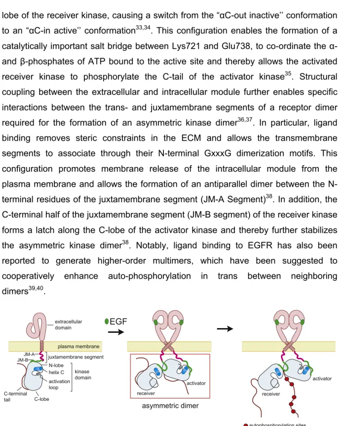
lobe of the receiver kinase, causing a switch from the “αC-out inactive’’ conformation to an “αC-in active’’ conformation33,34. This configuration enables the formation of a catalytically important salt bridge between Lys721 and Glu738, to co-ordinate the α- and β-phosphates of ATP bound to the active site and thereby allows the activated receiver kinase to phosphorylate the C-tail of the activator kinase35. Structural coupling between the extracellular and intracellular module further enables specific interactions between the trans- and juxtamembrane segments of a receptor dimer required for the formation of an asymmetric kinase dimer36,37. In particular, ligand binding removes steric constraints in the ECM and allows the transmembrane segments to associate through their N-terminal GxxxG dimerization motifs. This configuration promotes membrane release of the intracellular module from the plasma membrane and allows the formation of an antiparallel dimer between the N- terminal residues of the juxtamembrane segment (JM-A Segment)38. In addition, the C-terminal half of the juxtamembrane segment (JM-B segment) of the receiver kinase forms a latch along the C-lobe of the activator kinase and thereby further stabilizes the asymmetric kinase dimer38. Notably, ligand binding to EGFR has also been reported to generate higher-order multimers, which have been suggested to cooperatively enhance auto-phosphorylation in trans between neighboring dimers39,40.
Figure 2: Ligand-induced dimerization and allosteric activation of EGFR. Ligand binding (e.g. EGF) to the extracellular domain of EGFR promotes receptor dimerization, the formation of an asymmetric kinase dimer and subsequent trans-autophosphorylation of the C-tail of activator kinase via the allosterically activated receiver kinase (adapted from 38).
1.4 Autonomous EGFR activation
EGFR dimerization, which enables allosteric activation of its intrinsic kinase activity
factors to the ECM of the receptor. However, due to thermal fluctuations ligandless EGFR can also attain an active conformation and, via the formation of short-lived dimers, trigger spontaneous autonomous phosphorylation events14,41,42. Moreover, it has been suggested that phosphorylation of tyrosine 845 within the activation loop establishes an autocatalytic feedback by stabilizing the active conformation of the receptor and thus triggering autonomous ligand-independent signal propagation43. In accordance with this, previous studies showed that local EGF stimulation promoted lateral propagation of EGFR activation at high EGFR expression levels44,45. Growth factor stimulation further promotes the recruitment and activation of reactive oxygen species (ROS) producing enzyme systems such as NADPH oxidase to the plasma membrane46–48. Due to their short half live time, the presence of ROS remains spatially constrained to the plasma membrane. Local ROS production transiently inhibits inhibitory protein tyrosine phosphatases (PTP), through reversible oxidation of a catalytic cysteine residue, and thereby lowers the activation threshold of neighbouring receptors41,49. Coupling of EGFR activation with PTP inhibition at the plasma membrane thereby exemplifies a double-negative feedback loop, which together with the autocatalytic kinase activity, establishes a bistable reaction network with switch like response properties41,46. Analogous to a toggle switch, this reaction network converts growth factor stimuli into threshold-activated responses41,50. However, the autocatalytic kinase activity also amplifies spurious receptor activation in the absence of growth factor stimuli, necessitating additional regulatory mechanisms to control EGFR activity in the presence and absence of growth factors.
1.5 Structural auto-inhibitory features of EGFR
The activity of EGFR and its phosphorylation dynamics are regulated via different intrinsic structural features and extrinsic cellular regulatory machineries. Multiple auto-inhibitory structural mechanisms create an energy barrier for EGFR self- association and thereby antagonize its activation in the absence of growth factors.
Recent NMR and molecular dynamics simulations showed that in the ligand-free state the ECM is highly dynamic and forms an ensemble of different conformations, but generally adopts compact conformations over the extended conformation37,51. In ligand-free receptor dimers, the compact configuration of the ECM favors C-terminal over N-terminal dimerization of the transmembrane helices, dissociation and
inactive kinase dimers37. In addition, tethering of the intracellular module through electrostatic interactions between the negatively charged plasma membrane and positively charged patches obstruct the active site of the kinase domain37. The dimerization of two kinase domains is further hindered through local intrinsic disorder of the αC-helix region34. Activation loop phosphorylation of Tyr845 or the L834R mutation often found in lung cancer suppresses the disordered state, thereby enhancing receptor dimerization affinity, and suggesting a potential mechanism for autonomous, ligand-independent signal propagation34. The C-tail of EGFR has further been shown to form autoinhibitory interactions which interfere with the formation of an asymmetric kinase dimer31,38.
In addition to these intrinsic auto-inhibitory mechanisms, the endocytic machinery and protein tyrosine phosphatases (PTP) comprise extrinsic regulatory layers of EGFR activity46,52.
1.6 Protein tyrosine phosphatases (PTP)
The spatially distributed superfamily of cysteine-based PTPs regulates EGFR phosphorylation dynamics though the removal of phosphate groups from tyrosine residues. The overall level of EGFR phosphorylation is the product of a continuous and rapid cycle of phosphorylation and de-phosphorylation events46,53. Pan-specific PTP inhibition using the thiol reactive compound pervanadate has been shown to cause rapid EGFR phosphorylation in the absence of ligand, demonstrating the critical opposing role of PTPs to suppress ligand-independent EGFR activation54,55. Since the catalytic activity of certain PTPs (e.g. PTP1B and TCPTP) exceeds EGFR kinase activity by up to two orders of magnitude, spatial regulation of their activity is a prerequisite to permit EGFR phosphorylation56–58. Previous studies showed a PTP1B activity gradient emanating from the perinuclear area and declining towards the plasma membrane59. The relatively low membrane-proximal PTP activity has been suggested to be sufficient to shift the kinase-phosphatase balance in favor of the phosphatase in the absence of growth factor stimulation, while ligand binding shifts it in favor of the kinase46.
1.7 Endocytic trafficking of EGFR
The spatial localization of signaling molecules is a critical parameter determining their response properties and functional output. The endocytic machinery is composed of
functionally distinct intracellular membrane compartments and regulates the internalization, re-localization and fate (i.e. endosomal-plasma membrane recycling versus lysosomal degradation) of signaling molecules from the plasma membrane.
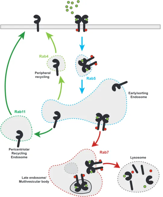
Members of the Rab small GTPase family act as multifaceted organizers of vesicular trafficking and control vesicular biogenesis, budding, delivery and fusion by the coordinated recruitment of various effector proteins60. Through hydrophobic geranylgeranyl groups, different Rab proteins are localized to distinct intracellular membranes and/or microdomains, organizing a modular system in which each Rab protein performs different functions (Fig.3)61. Rab5 participates during early stages of the endocytic pathway and mediates cargo entry and fusion of primary endocytic vesicles to form early endosomes (EE)62. The fate of endocytic cargo is determined by sorting events to subsequent endosomal compartments. Endosomal maturation from EE to late endosomes (LE) involves the replacement of Rab5 with Rab7, which mediates fusion of LE and/or multivesicular bodies with lysosomes61,63. In contrast, recycling of internalized cargo to the plasma membrane occurs via two routes: Rab4- mediated recycling directly from the EE to the plasma membrane or via Rab11- positive vesicles which mediate recycling through perinuclear recycling endosomes (RE, Fig3)64,65. Vesicular membrane dynamics enable different contextual modes of EGFR regulation by modulating its spatial distribution between the plasma membrane and different endosomal compartments66,67. The balance between internalization and recycling determines the density of EGFR at the plasma membrane. In the absence of ligand, EGFR is continuously internalized and recycled back to the plasma membrane, maintaining a steady state spatial distribution with the majority of EGFR located at the plasma membrane68,69. In contrast, ligand binding enhances EGFR internalization, resulting in its endosomal accumulation (Fig.3)67. Internalization of ligand-activated receptors extends the axial reach and the duration of plasma membrane initiated signaling events throughout the cytoplasm and can further create reaction platforms with distinct signaling repertoires46,70. Depending on the cellular context and ligand concentration, EGFR can be internalized through clathrin- mediated endocytosis (CME) or several other mechanisms (globally referred to as non-clathrin endocytosis, NCE)71. Internalization via CME in the absence or at low-to- intermediate ligand concentrations mainly results in receptor recycling to plasma membrane, thereby extending the duration of its signaling capacities72. In contrast,
ubiquitination, which targets the receptor for lysosomal degradation73. Ubiquitination of EGFR is mediated by members of the of the Cbl family of E3-ubiquitin-ligases that interact either directly with a phosphorylated tyrosine residue (pY1045) or indirectly through the adaptor protein Grb2, which binds to pY1068 and pY108674–76. Endocytosis of ligand bound receptors has further been shown to enable interaction of EGFR and the ER-bound phosphatase PTP1B, thereby contributing to the termination of receptor activity77,78.
Figure 3: Vesicular dynamics of EGFR. EGF stimulation promotes EGFR internalization via Rab5-positive endosomes. Internalized EGFR is either recycled back to the plasma membrane via the peripheral or perinuclear recycling endosomes or trafficked to the lysosome for degradation. Rab GTPases localized to distinct intracellular membranes and/or microdomains are indicated by colors.
Rab5
Rab7
Late endosome/
Mutlivesicular body
Early/sorting Endosome
Lysosome Rab11
Pericentriolar Recycling Endosome
Peripheral recycling Rab4
1.8 Mitogen activated protein kinases (MAPK)
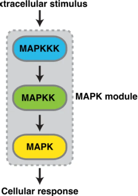
Phosphorylated tyrosine residues within the C-tail of EGFR recruit a variety of signaling molecules such as Shc or Grb2, which trigger the activation of various signal-transducing proteins15,79–81. Among these, extracellular regulated kinase 1/2 (Erk1/2) functions as an important regulator to integrate external cues into intracellular signalling events. Erk belongs to the evolutionary conserved superfamily of mitogen activated protein kinases (MAPKs), which mediate numerous cellular responses including cellular proliferation, differentiation, and survival82. MAPKs form three-tiered modules, which are activated by sequential phosphorylation events: a MAPK kinase kinase (MAPKKK) phosphorylates a MAPK kinase (MAPKK), which in turn activates a MAPK83 (Fig.4). So far six different MAPK modules have been identified in mammalian cells: Erk1/2, Erk3/4, Erk5, Erk7/8, Jun N-terminal kinase (JNK) and the p38 isoforms α/β/γ(ERK6)/δ84,85. Activated MAPKs function as proline (P) directed serine/threonine (S/T) selective protein kinases which phosphorylate a plethora of substrate proteins on selected S/T residues followed by P and thereby regulate numerous cellular responses85,86. This process is further thought to be mediated via docking interactions that occur outside of the active site between MAPKs and their substrates87. Dysregulation of MAPK activity has been implicated in various forms of human disease and developmental disorders88–90.
Figure 4: Scheme of the three-tiered MAPK module in eukaryotic cells. External signals sensed by growth factor receptors induce the sequential phosphorylation and activation of the
MAPKKK
MAPKK
MAPK
MAPK module
Cellular response Extracellular stimulus
1.9 Extracellular regulated kinase (Erk)
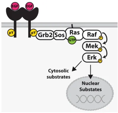
The Erk1/2 MAPK-module further involves the MAPKKK Raf and the MAPKK Mek1/2. Activation of this module is frequently initiated via Ras-family GTPases91. Plasma membrane activated growth factor receptors promote Ras activation via recruitment of the Ras-guanine nucleotide exchange factor (GEF) son-of-sevenless (SOS), which promotes the GDP to GTP exchange of Ras proteins92. Activated RasGTP in turn serves as a reactivity-enhancing recruitment factor which concentrates effector proteins such as Raf at the plasma membrane (Fig.5)93. Recruitment of Raf family kinases (A-, B-, and C-Raf) to the plasma membrane enables their allosteric activation via homo- or heterodimerization94. Activated Raf kinases further promote the phosphorylation and activation of Mek1/2, which then activate Erk1/2 via phosphorylation of threonine and tyrosine residues within their activation loop (Thr202/Tyr204 for Erk1 and Thr185/Tyr187 for Erk2)95,96. Dephosphorylation and thereby inactivation of Erk is catalysed by dual-specificity phosphatases (DUSPs), that can dephosphorylate both tyrosine or serine/threonine residues97. Erk is known to interact with over 170 proteins, including many substrates and regulators98. These interactions are further facilitated by two distinct docking domains: a common docking (CD/ED) domain which interacts with D site motifs (also called DEJL motifs), and a F Recruitment Site (FRS), which interacts with an FXF (DEF) motif on substrate proteins96. In resting cells, Erk is primarily located in the cytoplasm due to interactions with anchoring proteins; however, upon activation Erk can translocate to the nucleus where it promotes gene expression and DNA replication99. Different growth factors are thought to promote distinct cellular responses through dynamic changes of Erk interaction partners100. Different mechanisms have been suggested to cooperatively direct Erk signals to different downstream targets, including the duration and signal strength of Erk activation, its interaction with scaffold proteins, spatial compartmentalization, and crosstalk with other signalling molecules (Fig.6)101. Aberrant regulation of Erk activity further contributes to various forms of cancer and other human diseases, most commonly due to mutations in EGFR, RAS, BRAF, CRAF, or MEK1/2102.
Figure 5: EGFR mediated Erk activation at the plasma membrane. EGF induced activation of EGFR induces the recruitment of a GEF complex (Grb2-SOS) to the plasma membrane to activate the membrane-bound GTPase Ras to recruit Raf to the plasma membrane. Raf promotes Mek phosphorylation, which subsequently catalyzes Erk phosphorylation. Activated Erk phosphorylates various cytosolic effectors and also translocates to the nucleus, where it phosphorylates different transcription factors to regulate gene expression92.
1.10 Regulation of Erk signaling specificity
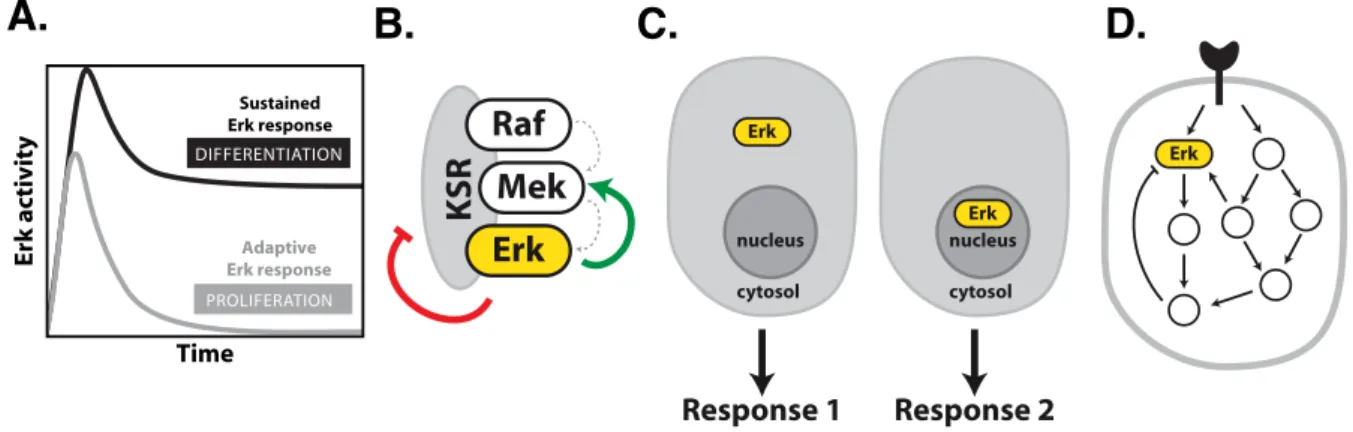
Given its ubiquitous role during numerous different and even opposing cellular responses, Erk acts as multifunctional signalling hub, which receives and integrates inputs from various stimuli. This functional plasticity represents a conundrum in regard to how the promiscuous activation of a shared signalling molecule can generate a specific cellular response to a given stimulus. The spatial and temporal organization of Erk activity are considered mechanisms that can encode variable extracellular signals into distinct cellular responses (Fig.6A, C)101,103,104. Different growth factors have been shown to induce distinct temporal patterns of Erk activation, which have been linked to different cellular fates105. Network motifs such as feed-forward loops allow decoding of Erk signal duration and thereby the induction of specific gene expression patterns that drive different cellular responses106–108. Furthermore, recent studies using constitutively active Ras mutants artificially targeted to specific subcellular locations established that the subcellular localization of Erk activation can influence its substrate pool104,109,110. Since Erk interacts with a
the spatial and temporal dynamics of Erk activity are determined by the underlying causal interactions within the MAPK signalling network (Fig.6D)3. The topology of the network depends on the cellular context and the resulting dynamics of Erk govern the cellular response. In PC12 cells, for example, the signal-dependent wiring of causal interactions within the network has been shown to result in different temporal Erk dynamics which determine cellular fate: proliferation (EGF, transient Erk response) and differentiation (NGF, sustained Erk response, Fig.6A)4. In this regard, it has been demonstrated that cellular responses can be rewired through targeted perturbation of causal interactions governing Erk dynamics4.
Figure 6: Mechanisms determining Erk signaling specificity and signaling dynamics. (A) Duration and strength of Erk activation. (B) Assembling the molecular components on scaffold proteins can facilitate their interaction and further provide positive (green arrow) and negative (red arrow) mechanisms of regulation, tuning the response dynamics. (C) The subcellular localization of Erk (nucleus or cytoplasm) can promote different cellular responses. (D) Crosstalk and conditional causal interactions can manifest in different dynamics of Erk activity3,9,111,112.
nucleus cytosol Erk nucleus
cytosol Erk
Response 1 Response 2
Erk
A. B. C. D.
Raf Mek
KSR Erk
Time
Erk activity
Sustained Erk response
Adaptive Erk response PROLIFERATION DIFFERENTIATION
2 Objectives
Vesicular trafficking allows the cell to dynamically regulate the spatial distribution of ErbB receptors between the plasma membrane (PM) and different endosomal compartments, however the signaling repertoire of endosomal and plasma membrane localized receptors can differ if biochemical components of the signal transduction machinery are not equally accessible within the different compartments.
Changes in vesicular trafficking dynamics can alter the spatial distribution of ErbB receptors and therefore form an important layer of regulation of receptor activity and signaling that could enable context-dependent regulation. The ensuing study is divided into two parts, addressing the following questions:
Autocatalytic amplification of ligand-induced EGFR activation coupled to local PTP inhibition establishes a bistable reaction network with switch like response properties.
While this feature ensures robust receptor activation to extracellular signals, it also creates the potential to amplify spontaneous phosphorylation events in the absence of growth factors41,46. Thus, the question arises of how the cell suppresses uncontrolled autonomous EGFR activation, while maintaining responsiveness to ligand stimulation. Endocytosis of ligand-activated EGFR has been shown to promote its interaction with spatially distributed PTPs to suppress its activity78. In this thesis, the role of EGFR vesicular dynamics in the regulation of autonomous receptor activity will be examined, as well as how endosomal trafficking differentially controls the activity of autonomously and ligand-activated EGFR.
Intriguingly, different ErbB ligands can induce distinct cell fates within a single cell type, despite the promiscuous activation of shared signal transducing proteins, such as the Ras/Erk mitogen-activated protein kinase (MAPK) cascade. In this regard, it has been suggested that dynamic changes in Erk-interacting proteins represent a key mechanism specifying the cellular response. Although receptor endocytosis attenuates receptor activation and signaling at the plasma membrane, active EGFR can continue to signal from endosomes. The second part of this thesis will examine how differences in the spatial and temporal distribution of active ErbB receptors within the endosomal system influence the subcellular localization of Erk activity, thereby modulating its interaction with different pools of substrates to generate
3 Material and Methods 3.1 Materials
3.1.1 Chemicals and Reagents
2-Mercapto-ethanol SERVA Electrophoresis GmbH
2’-deoxyadenosine-5’-triphosphate (dATP) InvitrogenTM Life Technologies 2’-deoxycytidine-5’-triphosphate (dCTP) InvitrogenTM Life Technologies 2’-deoxyguanosine-5’-triphosphate (dGTP) InvitrogenTM Life Technologies 2’-deoxythymidine-5’-triphosphate (dTTP) InvitrogenTM Life Technologies
40 % (29:1) Carl Roth GmbH
Ammonium persulfate (APS) SERVA Electrophoresis GmbH Ampicillin sodium salt SERVA Electrophoresis GmbH
Bromophenolblue Sigma-Aldrich®
Cell Lysis buffer, 10x Cell Signaling Technology
Cocktail Tablets Roche
cOmplete, Mini, EDTA-free Protease Inhibitor
Dimethyl sulfoxide (DMSO) SERVA Electrophoresis GmbH
Dithiothreitol (DTT) Fluka® Analytical
Epidermal growth factor (EGF) Sigma-Aldrich®
Erlotinib Selleckchem
Ethanol J.T.Baker
Ethylenediaminetetracetic acid (EDTA) Fluka® Analytical
Glycerol GERBU Biotechnik GmbH
Glycine Carl Roth GmbH
Heregulin-β3 Millipore, Merck KGaA
Histofix Carl Roth GmbH
Hydrogenperoxid (H2O2) Merck
Isopropanol J.T.Baker
Kanamycin sulfate GERBU Biotechnik GmbH
Lapatinib Selleckchem
Magnesium chloride (MgCl2) Merck KG/J.T.Baker
Methanol AppliChem GmbH
N,N,N’,N’-Tetramethylene-diamine (TEMED) Sigma-Aldrich®
PD0325901 Selleckchem
Phosphatase Inhibitor Cocktail 2 Sigma-Aldrich®
Phosphatase Inhibitor Cocktail 3 Sigma-Aldrich®
Poly-L-Lysine Sigma-Aldrich®
RedSafe, DNA Stain Chembio Ltd, Hertfordshire, UK Rotiphorese® NF-acrylamide/bis-solution
Sodium chloride (NaCl) Fluka® Analytical
Sodium dodecyl sulfate (SDS) SERVA Electrophoresis GmbH
sodium orthovanadate Sigma-Aldrich®
Tris-base Carl Roth GmbH
Tris-HCl J.T.Baker
Triton X-100 SERVA Electrophoresis GmbH
Tween 20 SERVA Electrophoresis GmbH
UltraPureTM Agarose InvitrogenTM Life Technologies 3.1.2 Commercial Solutions
100 mM dNTP mix InvitrogenTM Life Technologies
100x BSA New England Biolabs Inc.
10x Lysis Buffer Cell Signaling Technology
2-log DNA ladder New England Biolabs Inc.
50 mM Magnesium Sulfate InvitrogenTM Life Technologies 5x T4 DNA Ligation Buffer InvitrogenTM Life Technologies
CutSmart™ Buffer New England Biolabs Inc.
DPBS PANTM Biotech GmbH
Dulbecco’s Modified Eagle’s Medium (DMEM) PANTM Biotech GmbH Fetal calf serum (FCS) PANTM Biotech GmbH Fugene® 6 transfection reagent Roche Applied Science
Imaging medium PANTM Biotech GmbH
L-Glutamine GIBCO®/InvitrogenTM
LipofectamineTM 2000. InvitrogenTM Odyssey Infrared Imaging System
Precision Plus ProteinTM standards Bio-Rad Laboratories, Inc.
T4 DNA ligase reaction buffer, 10× New England BioLabs 3.1.3 Buffers and Media
LB medium 10 g/l Bacto-Trypton, 5 g/l bacto-yeast extract, 10 g/l NaCl and finally autoclave
LB agar plates add 15 g agar per litre LB medium, pour plates when the autoclaved medium has approximately 55 °C, add antibiotic of choice at desired concentration
SOC medium 20 g/l Bacto-Trypton, 5 g/l bacto-yeast extract, 0.5 g/l NaCl, 2.5 mM KCl, 10 mM MgCl2 (SOB medium), autoclave and before usage add 20 mM glucose to obtain SOC
1x PBS (pH 7.4) 138 mM NaCl, 10 mM Na2HPO4, 2.7 mM KCl, 1.8 mM KH2PO4
5x SDS sample buffer 60 mM Tris-HCl (pH 6.8), 25 % glycerol, 2 % SDS, 14.4 mM 2-mercapto-ethanol, 0.1 % bromo-phenolblue
SDS running buffer 25 mM Tris-base, 192 mM glycine, 0.1 % SDS Separating gel buffer 1 M Tris-HCl (pH 8.8)
Stacking gel buffer 0.375 M Tris-HCl (pH 6.8)
1x TAE buffer 40 mM Tris/Acetate (pH 7.5), 20 mM NaOAc, 1 mM EDTA 1x TBS 100 mM Tris-HCl (pH 7.4), 150 mM NaCl
1x TBST 100 mM Tris-HCl (pH 7.4), 150 mM NaCl, 0.1 % (v/v) Tween
Transfer buffer 25 mM Tris-base, 192 mM glycine, 20 % (v/v) methanol.
Modified RIPA buffer 50 mM Tris–HCl, 150 mM NaCl, 1 mM EDTA, 1 mM EDTA, 1 % Triton X-100, 1 % sodium deoxycholate, 1 % SDS
3.1.4 Bacterial Strains
XL10 Gold Stratagene (regrown in-house) 3.1.5 Enzymes
Calf Intestinal Phosphatase (10,000 U/ml) New England Biolabs Inc.
High-Fidelity (HF®) Restriction Endonucleases New England Biolabs Inc.
PlasmidSafe ATP-dependent DNase Epicentre
Q5® High-Fidelity DNA Polymerase New England Biolabs Inc.
T4-DNA ligase InvitrogenTM Life Technologies
3.1.6 Kits
High Pure endotoxin-freeNucleoBond®
Micro BCA™ Protein Assay Kit Thermo Scientific
Q5® Site-Directed Mutagenesis Kit New England Biolabs Inc.
Roti®-Prep Plasmid MINI Carl Roth GmbH
SiiR-DNA kit Spirochrome
Xtra Midi EF MACHEREY-NAGEL
Zymoclean™ Gel DNA Recovery Kit Zymo Research
3.1.7 Antibodies
Primary antibodies
tRFP (AB234) Evrogen
Akt (pan) (2920) Cell Signaling Technology
anti-EGF Receptor (pY845) (558381) BD Biosciences
c-Fos (9F6) (2250) Cell Signaling Technology
Cbl Antibody (C-15) (SC-170) Santa Cruz Biotechnology Cyclin D1 (92G2) (2978) Cell Signaling Technology EGF Receptor (D38B1) (4267) Cell Signaling Technology
EGFR/ErbB1 (AF231) R&D Systems
EphA2 (AF-3035) R&D Systems
ErbB-3 (C-17) (sc-285) Santa Cruz Biotechnology
ErbB2 (9G6) (sc-08) Santa Cruz Biotechnology
Erk1/2 (L34F12) (4696) Cell Signaling Technology
Erk1/2 [9B3] (ab36991) Abcam
GAPDH (6C5) (CB1001) Calbiochem, Merck
HA-antibody (MMS-101P) Covance Inc.
HER2/ErbB2 (29D8) (2165) Cell Signaling Technology HER4/ErbB4 (111B2) (4795) Cell Signaling Technology
KSR1 [EPR2421Y] (ab68483) Abcam
KSR2 (K75) (sc-100421) Santa Cruz Biotechnology Living colors® GFP AB (632381) Clontech
Living colors® GFP AB (632593) Clontech
Paxillin (610051) BD Biosciences
Phospho- Erk1/2 (Thr202/Tyr204) (4370) Cell Signaling Technology Phospho- Erk1/2 (Thr202/Tyr204) (9101) Cell Signaling Technology Phospho-Akt (Ser473) (9271) Cell Signaling Technology
Phospho-EGF Receptor (Tyr1068) (2234) Cell Signaling Technology Phospho-EGFR (Tyr1045) (2237) Cell Signaling Technology Phospho-EGFR (Tyr845) (558381) BD Biosciences
Phospho-EGFR(Tyr1068) (D7A5) (3777) Cell Signaling Technology Phospho-EphA2 (Ser897) (D9A1) (6347) Cell Signaling Technology Phospho-HER2/ErbB2 (Tyr1221/1222) Santa Cruz Biotechnology Phospho-HER3/ErbB3 (Tyr1289) (21D3) (4791) Cell Signaling Technology Phospho-Rb (Ser807/811) (D20B12) (8516) Cell Signaling Technology
Rab11 Antibody (3539) Cell Signaling Technology
Rab11A (ab65200) Abcam
Rab11a Antibody (2413) Cell Signaling Technology
WAVE2 (3659) Cell Signaling Technology
α-Tubulin (T6074) Sigma Aldrich
Secondary antibodies for Western blots and In Cell Western
IRDye 680 donkey anti-mouse IgG LI-COR Biosciences IRDye 800 donkey anti-rabbit IgG LI-COR Biosciences IRDye 800 donkey anti-mouse IgG LI-COR Biosciences IRDye 680 donkey anti-rabbit IgG LI-COR Biosciences IRDye 680 donkey anti-goat IgG LI-COR Biosciences Secondary antibodies for immunofluorescence
Alexa Fluor® 647 chicken anti-rabbit IgG Life Technologies Alexa Fluor® 647 donkey anti-mouse IgG Life Technologies Alexa Fluor® 555 donkey anti-goat IgG Life Technologies Alexa Fluor® 488 donkey anti-mouse IgG Life Technologies Alexa Fluor® 546 goat anti- rabbit IgG Life Technologies Alexa Fluor® 546 goat anti-mouse IgG Life Technologies
3.1.8 Oligonucleotides
siRNA
Rab11a 5 ́(AATGTCAGACAGACGCGAAAA)-3 ́ Qiagen Rab11b 5 ́(AAGCACCTGACCTATGAGAAC)-3 ́ Qiagen
Control siRNA (1027281) Qiagen
Rab11a siRNA (sc-36340) Santa Cruz Biotechnology
Control siRNA (sc-37007) Santa Cruz Biotechnology
ErbB2 siRNA (L-003126-00-0005) Dharmacon
ErbB3 siRNA (L-003127-00-0005) Dharmacon
ErbB4siRNA (L-003128-00-0005) Dharmacon
DNA
Desalted DNA-oligonucleotides were purchased from Sigma-Aldrich Chemie GmbH.
3.1.9 Plasmids
EGFR-mCitrine Encoding human EGFR with C-terminally fused mCitrine
EGFR-mCherry Encoding human EGFR with C-terminally fused mCherry
EGFR-Y1045F-mCitrine Encoding human EGFR-Y1045F with C- terminally fused mCitrine
EGFR-Y1045/1068/1086F-mCitrine Encoding human EGFR-Y1045/1068/1086F with C-terminally fused mCitrine
EGFR-L834R-mCitrine Encoding human EGFR-L834R with C- terminally fused mCitrine
ErbB2-mCitrine Encoding human ErbB2 with C-terminally fused mCitrine
ErbB3-mCitrine Encoding human ErbB3 with C-terminally fused mCitrine
pcDNA3.1+ Empty vector
tagBFP-Rab11a Encoding human Rab11a with N-terminally fused tagBFP
c-Cbl-mCherry Encoding human c-Cbl with C-terminally fused mCherry
mCherry-PTP1B-D181A Encoding PTP1B D181A mutant with N- terminally fused mCherry
EGFR-QG-mCitrine Encoding human EGFR with linker inserted mCitrine between Q958 and G959
pHR SFFVp PHAkt Cerulean Encoding PH domain of Akt with C- terminally fused mCerulean
pSpCas9(BB)-2A-GFP (PX458) Encoding Cas9 from S. pyogenes with 2A- EGFP
tagBFP-PTP1B Encoding human PTP1B with N-terminally fused tagBFP
PTP1B-RFP-tKRas Encoding human PTP1B with C-terminally fused RFP and tKRas
EGFR-paGFP Encoding human EGFR with C-terminally
fused paGFP
PTB-mCherry Encoding PTB domain from Shc with C-
terminal mCherry
HA-Ubiquitin Encoding HA-Ubiquitin
eGFP-ERK Encoding Rat ERK2 N-terminally fused to eGFP
3.1.10 Instruments and equipment
Eppendorf Research® plus pipettes Eppendorf
Falcon tubes (15/50 ml) BD FalconTM
Eppendorf safe lock tubes (0.5/1.5/2/5 ml) Eppendorf
Heatable magnetic stirrer “IKMAG®RCT” IKA®Labortechnik
Heating block ”QBD4” Grant Instruments
Nanodrop® ND-1000 spectrophotometer Peqlab Biotechnologie GmbH
Parafilm® Pechiney Plastic Packaging
PFE powder-free latex exam gloves (S) Kimberly-Clark
Safegrip® nitril gloves (S) Süd-Laborbedarf GmbH Sarstedt serological pipettes (5/10/25 ml) Sarstedt
Surgical disposable scalpel (No. 11, No.21) Braun Melsungen AG
Thermomixer comfort Eppendorf
“Vortex Genie 1” touch mixer Scientific Industries
BioRad Power Pac 300 Bio-Rad Laboratories, Inc.
BioRad Power Pac HC Bio-Rad Laboratories, Inc.
Centrifuge 5415R Eppendorf
Centrifuge 5810R Eppendorf
1.5 mm cassettes for western blots InvitrogenTM Life Technologies 1.5 mm 10-well combs InvitrogenTM Life Technologies 1.5 mm 15-well combs InvitrogenTM Life Technologies Incubation box for western blots Li-Cor® Biosciences
Incubator Shaker Series I26 New Brunswick Scientific Odyssey Infrared Imager Li-Cor® Biosciences
Immobilon-FL PVDF Millipore, Merck KGaA
Sonicator needle (MS 73) Bandelin Electronic GmbH
Ultraschall HD 2200 Bandelin Electronic GmbH
XCell SureLock™ Mini-Cell
Electrophoresis System InvitrogenTM Life Technologies XCell II™ Blot Module InvitrogenTM Life Technologies
Cell scraper BD FalconTM
75 mm filter unit Nalge Nunc International
Glass Pasteur pipettes (150 mm, 230 mm) Brand GmbH & Co. KG 4-well LabTek® chambers No. 1.0 Nalge Nunc International 8-well LabTek® chambers No. 1.0 Nalge Nunc International 35-mm MatTek petri dishes No. 1.5 MatTek Corporation
cabinet
Tissue culture plate (6 well) BD FalconTM
Vacusafe comfort Integra Biosciences
AutoFlow NU-4750 Water Jacket CO2 Incubator Integra Biosciences
96 well dishes Corning, Merck
3.1.11 Software
Adobe Illustrator Adobe Systems Inc.
FV10-ASW Fluoview Software Olympus
Fiji Schindelin et al., 2012113.
Microsoft Office 2011 Microsoft Corporation Odyssey® Infrared Imaging System LI-COR Biosciences GmbH
Leica LAS-AF Leica Microsystems
GraphPad Prism 6 GraphPad Software, Inc.
Leica Application Suite X Leica MICROSYSTEMS
Python Python Software Foundation
CellProfiler Kamentsky et al., 2011114.
ImageJ64 v1.44 http://rsbweb.nih.gov/
NanoDrop ND-3300 2.6.0 Coleman Technology Inc.
DNASTAR Navigator v2.2.1.1 DNASTAR Inc.
Quantity One 4.6.9 Bio-Rad Laboratories, Inc.
Application software Version 2.1
Multiscan Ascent Software Version 2.6 ThermoElectron Coorperation
IgorPRo v6.12 WaveMetrics
3.2 Molecular Biology
3.2.1 Polymerase chain reaction (PCR)
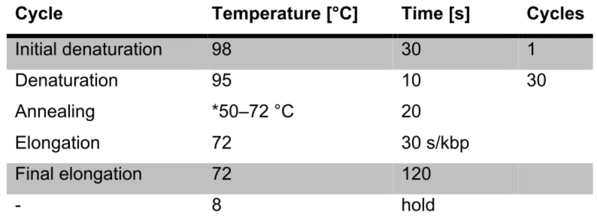
PCR reactions to amplify target DNA sequences using specific primer pairs were performed using the Q5® High-Fidelity DNA Polymerase. The following reaction setup and thermocycling Conditions were used:
Component Volume [µl]
5X Q5 Reaction Buffer 10
10 mM dNTPs 1
FW Primer (10 µM) 2.5
BW Primer (10 µM) 2.5
1-10 ng template DNA X
Q5 High-Fidelity DNA Polymerase 0.5
H2O 30.75 – X
Table 1: 50 µl PCR batch for Q5® High-Fidelity DNA Polymerase. The volume X of the template naturally depends on its concentration.
Cycle Temperature [°C] Time [s] Cycles
Initial denaturation 98 30 1
Denaturation Annealing Elongation
95
*50–72 °C 72
10 20 30 s/kbp
30
Final elongation 72 120
- 8 hold
Table 2: PCR-protocol for Q5® High-Fidelity DNA Polymerase. *The annealing temperature, depends on the used primers.
3.2.2 Agarose gel electrophoresis
DNA strands were separated according to their size using agarose gels with concentrations in the range of 1.2 – 2.0 % (w/v) as matrix. Gels were prepared with TAE buffer supplemented with the DNA binding dye RedSafeTM (50 µl/l). The size of the observed strands was determined by comparison with marker fragments of known size using the GelDoc XR System. Designated DNA-fragments were cut out of the gel and purified using the “ZymocleanTM Gel DNA Recovery Kit” from Zymo Research following the manufacturer’s instructions.
3.2.3 Restriction digestion
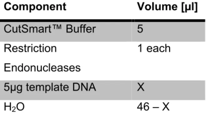
Restriction digestion was performed using High-Fidelity (HF®) Restriction Endonucleases (New England Biolabs). Restriction batches of 50 µl were prepared and incubated at 37 °C for 3 h.
Component Volume [µl]
CutSmart™ Buffer 5 Restriction
Endonucleases
1 each
5µg template DNA X
H2O 46 – X
Table 3: 50 µl restriction batch. The volume X of the template naturally depends on its concentration.
3.2.4 Dephosphorylation of 5’-Phosphorylated DNA fragments
In order to prevent self-ligation resulting in false positive vector background, linearized plasmids were treated with 1 µl calf intestinal phosphatase (CIP) for 15 min at 37 °C.
3.2.5 Ligation of DNA Fragments
Ligation of DNA fragments and plasmids, was performed using the Quick Ligation™
Kit (New England Biolabs). The starting ligation conditions required 50 ng vector DNA and threefold excess of the insert DNA. The vector/insert ratio was calculated according to the following equation:
𝑛𝑔 vector ∙ 𝑘𝐵(insert)
𝑘𝐵(vector) ∙3= 𝑛𝑔(insert)
The calculated amount of vector/insert DNA was supplemented with 10 µl 2x Quick Ligation Buffer, 1 µl of Quick T4 DNA Ligase and H2O to a total reaction volume of 20 µl. The mixture was incubated for 1 h at room temperature and afterwards for 10 min at 65 °C to inactivate the ligase
3.2.6 Site-Directed Mutagenesis
Site-specific mutagenesis of double-stranded plasmid DNA was performed using the Q5® Site-Directed Mutagenesis Kit according to the manufactures instructions.
Custom mutagenic primers to create insertions, deletions and substitutions were designed using the NEBaseChanger tool (https://nebasechanger.neb.com/).
3.2.7 Transformation of chemically competent E. coli cells
For transformation 50 µl of chemical competent XL 10-Gold cells were used. The cells were thawed on ice and 1.75 µl of 2.25 M DTT and 5 µl ligation-reaction mix or 1 µl plasmid for re-transformation were added and left on ice for 30 min. Afterwards cells were heat-shocked for 30 - 60 s at 42 °C and immediately placed on ice for 2 min. 125 µl SOC medium were added and the cells were incubated at 37 °C for 1 h while shaking. In case of a ligation mix, the whole cell suspension was plated on LB agar plates containing an appropriate antibiotic for selection. For re-transformation 50 µl cell suspension were used for plating.
3.2.8 Plasmid preparation
Single colonies from LB agar plates were transferred into 5 ml LB-medium containing the appropriate antibiotic and incubated overnight at 37 °C and 225 rpm. For MIDI preps a pre-culture of 5 ml was grown for approximately 6 h and afterwards used to inoculate an overnight culture of 200 ml. Plasmids were purified using the Using Roti®-Prep Plasmid MINI kit (Carl Roth GmbH) or the High Pure endotoxin- freeNucleoBond® Xtra Midi EF kit (MACHEREY-NAGEL) according to the manufacturers’ instructions.
3.2.9 Determination of DNA concentration
The DNA concentration was determined with the Nanodrop® ND-1000 spectrophotometer from Peqlab by measuring the absorption at 260 nm. The concentration was calculated using the approximation that an OD260 of 1 corresponds to a double stranded DNA concentration of 50 ng/µl.