Interleukin 6 trans-signaling in normal and malignant stem and progenitor cells
Dissertation
zur Erlangung des Doktorgrades der Naturwissenschaften (Dr. rer. nat.) der Fakultät für Biologie und Vorklinische Medizin der
Universität Regensburg
vorgelegt von
Milan MS Obradović
aus Belgrad, Serbien 2013
Das Promotionsgesuch wurde eingereicht am 05. Februar 2013 Die Arbeit wurde angeleitet von Herrn Prof. Dr. Christoph A. Klein Prüfungsausschuss:
Vorsitzender: Prof. Dr. Richard Warth 1. Gutachter: Prof. Dr. Ralph Witzgall 2. Gutachter: Prof. Dr. Christoph Klein 3. Prüfer: Prof. Dr. Gunter Meister
Milan MS Obradović
Dedicated to my parents, Miroslav and Smilja Obradović.
All I can do is mention your names with all the gratitude for your endless love.
Table of contents
1. Introduction______________________________________________________ 1 1.1. Cellular composition and physiology of human mammary gland _____________ 1 1.2. The mammary gland development ______________________________________ 3 1.3. Cellular hierarchy in the normal mammary gland __________________________ 5 1.4. Isolation of the mammary stem and progenitor cells _______________________ 6 1.4.1. Isolation of the mammary stem and progenitor cells based on marker expression _____ 7 1.4.2. Mammospheres- enriched population of adult mammary stem and progenitor cells___ 8 1.4.3. Isolation of the stem and progenitor cell fractions by functional properties ___________ 9 1.5. Differentiation ability of mammary stem and progenitor cells ______________ 10
1.5.1. In vitro differentiation of mammary stem and progenitor cells _____________________10 1.5.2. In vivo models for mammary stem and progenitor cell differentiation_______________11 1.6. Breast cancer _______________________________________________________ 12 1.7. Cancer stem cell concept______________________________________________ 13 1.8. Interleukin 6 and breast cancer ________________________________________ 14 1.9. The aim of the work__________________________________________________ 17 2. Materials and Methods ___________________________________________ 18
2.1. Materials ___________________________________________________________ 18 2.1.1. Reagents, solutions and cell culture media_______________________________________18 2.1.2. Antibodies ___________________________________________________________________22 2.1.3. The composition of prepared buffers, media and solutions ________________________22 2.1.4. Cell lines_____________________________________________________________________24 2.1.5. Devices ______________________________________________________________________25 2.1.6. Software ____________________________________________________________________27 2.1.7. Primers used for the PCR amplification __________________________________________27 2.2. Methods ___________________________________________________________ 29
2.2.1. Methods for in vitro cell propagation and characterization ________________________29 2.2.1.1. In vitro cell line propagation under conventional 2D conditions_________________29 2.2.1.2. In vitro cell line propagation under anchorage independent (3D) condition ______29 2.2.1.2.1. Preparation of poly-HEMA plates _______________________________________30 2.2.1.3. In vitro differentiation of mammary cells ____________________________________30 2.2.2. In vitro cell propagation of the donor’s tissue specimens __________________________31 2.2.2.1. Tissue collective __________________________________________________________31 2.2.2.2. Mammary tissue digestion and cell isolation _________________________________31 2.2.2.3. Mammosphere protocol ___________________________________________________33 2.2.3. In vivo human mammary stem and progenitor cells differentiation _________________33 2.2.3.1. In vivo human mammary stem and progenitor cells differentiation _____________34 2.2.3.1.1. Preparation of human immortalized fibroblasts used for xenotransplantation procedure and injection of human mammary stem and progenitor cells _______________________35
2.2.3.1.2. Preparation of C3H10T1/2 fibroblasts for orthotopic xenotransplantation ___35
2.2.3.2. Orthotopic xenotransplantation ____________________________________________36 2.2.3.3. In vivo breast cancer mouse model __________________________________________38 2.2.3.4. Mice dissection ___________________________________________________________38 2.2.3.4.1. Paraffin embedding of mice tissue samples ______________________________38 2.2.3.4.2. Preparation of the bone marrow _______________________________________39 2.2.3.4.3. Paraffin tissue sectioning, H&E staining and micro-dissection ______________39 2.2.4. Generation of GFP labeled cells lines by lentiviral vector-mediated gene transfer ____40 2.2.4.1. Construction of the lentiviral particles - Transfection of the HEK-293T c ells _______40 2.2.4.2. Determination of virus titer ________________________________________________41 2.2.4.3. The selective propagation of the transduced cells_____________________________41 2.2.5. Gene specific PCR_____________________________________________________________42 2.2.5.1. Agarose gel electrophoresis ________________________________________________43 2.2.5.2. Total mRNA reverse transcription and cDNA amplification from single or few cells44 2.2.6. Unspecific labeling of cell membrane- PKH26 staining_____________________________44 2.2.7. Quantification of Interleukin 6 and soluble Interleukin 6 receptor __________________44 2.2.8. Flow cytometry_______________________________________________________________45
3. Results _________________________________________________________ 46 3.1. Overview of the research rationale _____________________________________ 46 3.2. Development and modification of the protocols used for the study of IL6
signaling influence in the mammary gland_____________________________________________ 46 3.2.1. Improvement of the c ell isolation protocol ______________________________________47 3.2.2. Improvement of the mammosphere culture protocol_____________________________50 3.2.3. Establishment of in vitro differentiation on a panel of HME cell lines - In vitro differentiation of the selected mammary cell lines________________________________________________52
3.2.4. In vivo engraftment and propagation of the human mammary cells ________________54 3.2.4.1. In vivo engraftment and growth of human mammospher es in NSG mice_________54 3.2.4.2. In vivo engraftment and growth of human mammospher es in NSG mice- protocol modification 57
3.3. IL6 signaling in normal mammary cells __________________________________ 60 3.3.1. The activation of the IL6 signaling promotes survival and proliferation of the mammary cells under anchorage independent conditions ___________________________________________________60
3.3.2. IL6 signals in mammary cells via trans -signaling __________________________________61 3.3.3. Mammary cell lines produce and secrete IL6 and sIL6R ____________________________64 3.4. The activation of the PI3K/Akt signaling pathway inhibits IL6 and IL6R expression in mammary cells__________________________________________________________________ 65 3.5. IL6 signaling in the adult human mammary gland _________________________ 67 3.5.1. Mammary gland does not contain cellular population with membrane bound IL6R ___67 3.5.2. Mammary gland contains cellular populations which enable IL6 trans -signaling ______68 3.5.3. IL6 trans-signaling in adult mammary stem and progenitor cells ____________________70 3.5.3.1. IL6 signaling induces proliferation of adult mammary stem and progenitor cells __70 3.5.3.2. IL6 trans-signaling induces proliferation of adult mammary stem and progenitor
cells 72
3.6. IL6 trans-signaling preserves and promotes the functional phenotype of stem and progenitor cells________________________________________________________________ 73
3.6.1. IL6 trans-signaling triggers mammospheres self-renewal __________________________74 3.6.2. Activation of the IL6 trans -signaling does not reduce ability of the mammary stem and progenitor cells to differentiate ________________________________________________________________75
3.6.2.1. Ability of HIL6-treated cells to differentiate in vitro ___________________________75 3.6.2.2. Activation of IL6 trans-signaling in mammospheres does not influence their differentiation ability in animal hosts _________________________________________________________76
3.6.3. IL6 trans-signaling does not influence asymmetric cell division _____________________77 3.7. Activation of IL6 trans-signaling induces mammosphere forming ability of nLRC
79
3.7.1. nLRC are not able to form mammospheres unless IL6 trans -signaling is activated ____79 3.8. IL6 trans-signaling in breast cancer _____________________________________ 81
3.8.1. Breast cancer cells do not contain membrane-bound IL6R _________________________81 3.8.2. MDA-MB-231 cells express IL6 and IL6R _________________________________________82 3.8.3. IL6 trans-signaling stimulates tumor formation of MDA-MB-231 cells _______________84 3.8.4. Activation of IL6 (trans)-signaling does not induce proliferation of MCF7 derived CSC _86
4. Discussion ______________________________________________________ 88 5. Summary _______________________________________________________ 97 6. Literature_______________________________________________________ 97 7. List of abbreviations _____________________________________________ 102 8. Acknowledgments ______________________________________________ 105
1
1. Introduction
1.1. Cellular composition and physiology of human mammary gland
The mammary gland is positioned between the pectoralis major muscle and the nipple (Henrikson et al., 1997). The mammary gland is a paired, tubuloalveolar, exocrine gland which produces milk in females (Henrikson et al., 1997; Linzell and Peaker, 1971). The produced milk is collected in the nipple sinus and excreted in a response of infant’s suckling (Linzell and Peaker, 1971). The nipples are surrounded with sebaceous glands rich areola and represent the convergent point of mammary lobes, functional units of the adult mammary gland. It has been suggested that sebaceous glands positioned within the nipple areola and/or sweet glands might represent the evolutionary origin of the mammary glands (Oftedal, 2002). In human, primitive sebaceous glands and/or sweet glands evolved in 15-20 epithelial lobes which produce milk (Henrikson et al., 1997; Oftedal, 2002). During the course of mammary gland ontogeny, the mammary lobes arise from a primitive anlage, which undergoes series of morphological changes mainly postnatal (Radisky and
Hartmann, 2009). The mammary gland reaches functional maturity at pregnancy during the process known as lactation when the mammary gland is composed of secretory epithelia (Borellini and Oka, 1989).
The adult mammary gland is an inhomogeneous organ composed of epithelial derived ducts surrounded by connective tissue and immersed in the adipose tissue lobes (Figure 1 a) (Sheffield, 1988). Although the milk production and excretion in the mammary gland is accomplished by epithelial derived cells, various biological
processes (i.e. mammary gland development, hypertrophy, involution) in the adult and developing mammary gland are regulated trough inter -cellular and -tissue interactions (Maller et al., 2010).
The ratio between mammary cells of the epithelial and stromal origin is changing during the life time due to the continuous cycles of proliferation, differentiation,
2
lactation and regression of mammary gland epithelial component (Parmar and
Cunha, 2004). During the puberty, the first changes occur in the growth of the stromal tissue. It has been suggested that stromal proliferation serves to inhibit the growth of the epithelial compartment (Howard and Gusterson, 2000). While the mammary fat- pad of non-pregnant females is largely assembled of the adipose tissue, the adipose compartment is gradually substituted by epithelial tissue, blood vessel and
connective tissue during the pregnancy (Borellini and Oka, 1989; Russo and Russo, 2004) .
Moreover, the adult mammary gland is well-vascularized organ and supplied with wide lymph drainage, which is relevant to oncology because the breast cancer metastases develop on the distant sites due to the dissemination of cancer cells via blood and lymph vessels (Howard and Gusterson 2000; Eccles, Paon et al. 2007;
Andres and Djonov 2010; Vermeulen, van Golen et al. 2010).
Figure 1. Human breast anatomy and the cellular organization of a TDLU. (A) Human breast is a complex organ composed of tissue of diverse origin. 1) Chest wall; 2) Pectoralis muscles; 3) Mammary gland ducts and lobules; 4) Nipple; 5) Areola mammae; 6) Lactiferous duct; 7) Adipose tissue; 8) Skin.
(B) Mammary gland duct is composed of myoepithelial cells, luminal cells, secreting (cap) cells and mammary stem and progenitor cells. Picture modified from (Tiede and Kang, 2011; Wikipedia, 2012).
3
Deregulation of the both stromal and epithelial cell growth may cause breast hypertrophy, which is manifested in the abnormal breast sizes (macromastia or gigantomastia). The hypertrophy usually develops during puberty or at menopause (Dancey et al., 2008; Dehner et al., 1999). Following the medical care policy, the patients diagnosed with macromastia or gigantomastia undergo breast reduction surgery due to the physical, aesthetic or psychophysical difficulties (Nguyen et al., 2008). Tissue specimens of patients experiencing breast reduction can be used for the experimental purposes (Dontu et al., 2003a).
Mammary lobes of an adult gland consist of a collection of acini arising from terminal ducts embedded in intralobular stromal tissue (Parmar and Cunha, 2004). Terminal duct lobular units (TDLU) are considered as the functional units of the mammary gland. Each duct is composed of the two major cell types : myoepithelial and luminal cells (Figure 1 b). A layer of myoepithelial cells is found positioned directly below the basal membrane. Myoepithelial cells are characterized by the expression of the alpha smooth muscular actin and cytokeratins 5 and 14 (Stingl et al., 2005). Contractions of the myoepithelal cells enable milk excretion. Above the myoepithelal cell layer
towards the lumen is the inner layer of luminal cells subdivided into ductal luminal cells, which line inside of the ducts, and alveolar luminal cells, which arise during the pregnancy and secrete milk (Visvader and Lindeman, 2011). The majority of the cells (>90%) found within the mammary ducts are differentiated luminal and myoepithelial cells (Chepko and Smith, 1997; Stingl et al., 2005).
1.2. The mammary gland development
The mammary gland development occurs in three distinct and differentially regulated stages: embryonic, pubertal and adult (Gjorevski and Nelson, 2011; Howard and Gusterson, 2000). Embryonic development and extensive proliferation and
differentiation during each pregnancy cycle are enabled by the presence of mammary stem cells (Dontu et al., 2005; Van Keymeulen et al., 2011). Moreover, the regulation of developmental and differentiation processes involve an ample variety of hormones and growth factors such as estrogen, progesterone, and prolactin that drive primitive mammary stem cells towards differentiated functional cells (Henrikson et al., 1997;
Stingl, 2011; Tiede and Kang, 2011).
4
Development of the human mammary gland begins at week 5 of the embryonic development. The first developmental sign is the formation of the milk streak,
thickening in the ectoderm, extending from the axilla to the groin. During week 6 and 7 the milk streak develops to the mammary crest which later develops into the
epithelial bud (Gusterson and Stein, 2012; Howard and Gusterson, 2000). Cells expressing some of the myoepithelial cell markers are observed in this period of embryonic development (Van Keymeulen et al., 2011). Embryonic development ends with a formation of a series of blind -ended tubes, with bulbous tips, well defined lobules and terminal duct lobular units, similar to those observed in the adult
mammary gland. Shortly after birth, the mammary gland undergoes involution similar to the observed process in the post-menopausal breast (Anbazhagan et al., 1991;
Gusterson and Stein, 2012).
The infant’s mammary gland development after involution process at birth follows the overall body development until puberty when the swift development in females starts.
Nevertheless, while the knowledge about the embryonic human mammary gland development is still incomplete due to the lack of tissue specimens, the
developmental stages and processes in the animal models increase significantly our knowledge during the last decade pinpointing key steps of early development and involved signaling networks (Hens and Wysolmerski, 2005; Van Keymeulen et al., 2011).
The mammary gland development in human is hormonally regulated as many other processes of the pubertal maturation (Sternlicht et al., 2006; Stingl, 2011). The hormonal regulation starts before the first menstrual cycle when estrogen receptors are detectable in the low percent of the luminal cells (Gusterson and Stein, 2012;
Sternlicht et al., 2006). Development of the TDLU is characterized by the development of the end buds and lateral buds (Russo and Russo, 2004).
Unfortunately, the knowledge about the mammary gland development during pre - pubertal and pubertal age is not yet sufficient to prove any of the proposed models due to the low number of the analyzed samples. Moreover, little is known about the molecular mechanism of the stromal influence during development while it has become clear that stromal cells actively influence and shape many of the mammary
5
gland abnormalities related to the developmental processes (Bonafe et al., 2012; Cirri and Chiarugi, 2012).
1.3. Cellular hierarchy in the normal mammary gland
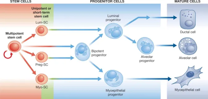
The majority of cells found within adult mammary ducts are differentiated luminal and myoepithelial cells (Chepko and Smith, 1997; Stingl et al., 2005). Myoepithelial cells are in direct contact with the basal membrane and they are cells responsible for the ductal contraction and milk circulation in a response to oxytocin (Sternlicht et al., 2006). Luminal cells are orientated towards lumen and build TDLU. During lactation, a sub-type of luminal cells located in the alveoli and hence named alveolar cells produce milk. In addition to these differentiated cell types, the adult mammary gland contains stem cells and progenitor cells (Figure 2). Thus, the mammary gland is a hierarchically structured organ (Stingl et al., 2005; Visvader and Lindeman, 2011).
The presence of the undifferentiated cells within the adult gland enables mammary gland maturation and cyclic differentiation processes during the adult life (Stingl et al., 2006).
Figure 2. Hierarchical organization of the mammary gland. The adult mammary gland contains primitive adult stem cells which are a ble to rec onstitute functional mammary gland. Figure is modified from the ref. (Vis vader and Lindeman, 2011).
6
The adult mammary gland reaches functional maturity at pregnancy when branching TDLU are formed trough multi- step sequences of proliferation and differentiation processes (Sternlicht et al., 2006). Each pregnancy cycle ends with controlled reduction of the epithelial cell compartment by apoptosis (Radisky and Hartmann, 2009). These cyclical bursts of proliferation and subsequent apoptosis recur also during each menstrual cycle (Russo and Russo, 2004) and are enabled by the existence of the cells with differentiation and self-renewal ability, adult stem and progenitor cells (Dontu et al., 2003b).
The existence of a multipotent cellular type within the adult mammary gland able to reconstitute functional adult mammary gland has been shown by numerous
independent experiments. The first mammary transplantation studies in the late 1950’ies by DeOme and colleagues suggested that the adult mammary gland
contains a cell type able to reconstitute the mammary gland of the adult mice (Daniel et al., 1971; Deome et al., 1959). Furthermore, the engraftment of human tissue pieces into pre-cleared mammary fat-pads of immunodeficient mice suggested the existence of cells able to reconstitute the functional human mammary gland
(Kuperwasser et al., 2004). The presence of the adult mammary stem cells was proven by Shakelton and colleagues who were able to reconstitute a functional mouse mammary gland form a single mammary cell (Shackleton et al., 2006).
However, although today we have experimental evidences of adult mammary stem cells existence, the main problem for the study of mammary gland development and pathophysiology remained the isolation and molecular and functional characterization of the adult mammary stem and progenitor cells.
Although several subpopulations of mammary cells have been described, the existence and precise molecular profile of adult mammary stem cells is still intensively discussed (Keller et al., 2011; Stingl et al., 2005).
1.4. Isolation of the mammary stem and progenitor cells
Stem cells are defined as cells capable for multi-lineage differentiation and self- renewal (Luo et al., 2010; Smalley and Ashworth, 2003). It has been suggested that understanding mammary gland biology, development and pathophysiology mostly
7
depends on our ability to isolate, gro w and manipulate undifferentiated cell types (Dontu et al., 2003a; Smalley and Ashworth, 2003). Therefore, the enormous efforts have been invested in the development of different strategies for the isolation and characterization of adult stem and progenitor cells.
1.4.1. Isolation of the mammary stem and progenitor cells based on marker expression
Scientists in mammary stem cell research were initially inspired by studies in the field of hematopoiesis and tried to develop various assays that help enrichment of the mammary stem and progenitor cells (Alvi et al., 2002; Eirew et al., 2008; Stingl et al., 2005).
One of the first methods for the isolation of stem and progenitor cells was based on the observation that due to over-expression of transmembrane transporter proteins stem cells are able to exclude vital marker dyes (Smalley and Ashworth, 2003). The so-called “side-population” remained unstained by DNA-binding dyes due to its ability to pump out the dye while more differentiated progenies were not able to exclude the DNA-binding dye (Charafe-Jauffret et al., 2009). Whereas the first reports
convincingly demonstrated enrichment of stem cells and cancer stem cells (Alvi et al., 2002), later reports indicated many inconsistencies which hindered the experimental reproduction of the results and their validation (Montanaro et al., 2004).
On the other hand, empirical testing of surface marker combination enabled the definition of markers used for Fluorescence Activated Cell Sorting (FACS)
methodology to identify and isolated adult stem and progenitor cells (Charafe-Jauffret et al., 2009; Ginestier et al., 2007a; Stingl et al., 2005). Thus, the cellular types
defined by their functional characteristics, luminal and myoepithelial cells are
described by the following cellular markers: 1) differentiated luminal cells are CK14- /CK18+/CK19+ and MUC1+ cells, while 2) myoepithelial cells are characterized as CK14+/SMA+ and CD10+ cells. The luminal progenitor cells are defined as as MUC1+/CD133+/EpCAM+/CD49f+/CD10-/THY-, while the myoepithelial progenitors express the marker following marker combination MUC1-/CD133-
/EpCAM+/CD49f+/CD10+/THY+ (Bartek et al., 1990; S nedeker et al., 1991; Stingl et
8
al., 1998; Stingl et al., 2001; Stingl et al., 2005). It is important to say that the function of these markers is often unclear. Unfortunately, many attempts to define adult stem and progenitor cells based on marker expression led to inconsistent conclusions.
Keller et al. used healthy mammary tissue specimens to define cellular populations within the adult mammary gland. They found that both, EpCAM+ and CD10+
population contain differentiation potential (Keller et al., 2011). On the other hand, the mentioned study (Keller et al., 2011) is opposed to the observation that mammary repopulating units were found to reside in EpCAM-/low phenotype (Eirew et al., 2008).
Taken together, our inability to isolate adult stem and progenitor cells by marker expression hampered the attempts for their perspective characterization and manipulation. Nevertheless, the alternative approaches demonstrated that the isolation and characterization of the adult mammary stem and progenitor cells is possible.
1.4.2. Mammospheres- enriched population of adult mammary stem and progenitor cells
Based on the neural stem cell research assays, Gabriela Dontu and colleagues applied a cell culture system for neural stem cells to propagate undifferentiated cells isolated from the adult mammary specimens (Dontu et al., 2003a). Basis of the mammosphere assay was the observation that rare undifferentiated cells survive anchorage independent conditions and proliferate to form multi-cellular spheroids while most of the cells isolated from mammoplasty tissue specimens underwent anoikis when grown under ultra-low attachment conditions. It was suggested that inability of differentiated cells surviving anchorage independent conditions underlies the selective propagation of adult stem and progenitor cells (Dontu and Wicha, 2005).
Mammospheres are highly enriched in undifferentiated cells, as demonstrated by the ability of single cells propagated as mammospheres to generate multi-lineage
colonies, which is not the case in the presence of serum or when propagated on a collagen substratum (Dontu et al., 2003a; Liu et al., 2008). The primary
mammospheres contain eight times more bi-lineage progenitor cells compared to the
9
tissue of origin. The secondary and later-passaged mammospheres consist of virtually 100% bi-potent progenitors (Dontu et al., 2003b; Dontu and Wicha, 2005).
Moreover, mammospheres form complex structures in reconstituted 3-D culture systems in Matrigel©, resembling the observed morphology of the functional adult mammary gland (Dontu et al., 2003a).
In vivo experiments indicated that the secondary mammospheres are able to differentiate in complex ducto -acinar structures comparable to the TDLU when inoculated in NOD/scid mice, immunodeficient mice (Liu et al., 2006; Liu et al., 2008;
Pece et al., 2010).
Growth and enrichment of the mammary stem and progenitor cells as spherical colonies was shown to be currently the most efficient way for the enrichment and propagation of the mammary stem and progenitor cells (Dontu et al., 2003a; Luo et al., 2010).
1.4.3. Isolation of the stem and progenitor cell fractions by functional properties
In 2010, Pece and colleagues developed another experimental strategy for stem cell enrichment of a pre-selected population of the mammary stem and progenitor cells.
The mammary stem and progenitor cells were propagated under anchorage
independent conditions. After the secondary mammospheres were formed stem and progenitor cells were selected on a basis on their functional characteristics (Pece et al., 2010).
The mammary stem cells are asymmetrically and slow dividing cells , which upon cell division give rise to two daughter cells; one is the self-renewed stem cell, while the other cell represents a progenitor cell (Harmes and DiRenzo, 2009; Smalley and Ashworth, 2003). The discrimination between two daughter cells is based on their cell division rates, stem cells are slower dividing cells compared to progenitor cells (Pece et al., 2010).
It was shown that single mammospheres can reconstitute the functional
mammary gland in vivo and that mammospheres arise from single cells. Thus, the mammospheres are groups of cells, which arise from a single adult stem cell (Dontu
10
et al., 2003a; Shackleton et al., 2006). The isolation of the adult stem cells by Pece and colleagues relies on the following approach: If the cellular membrane of the cells propagated under anchorage independent conditions is labeled by a fluorescent marker, and the dye is equally distributed to the daughter cells upon each cellular division, then due to the slow cycling frequency of the stem cells, the label retaining cells represent the mammosphere stem cell, while the fast cycling cells within a mammosphere become quickly unlabeled and represent daughter cells (Lanzkron et al., 1999). Therefore, during the growth of a mammosphere, the rare quiescent/slowly dividing mammary stem cells retain dye, while the bulk of population derived from the proliferation of the progenitors progressively lose it by dilutions (Pece et al., 2010).
1.5. Differentiation ability of mammary stem and progenitor cells
Stem cells are characterized by the ability to self-renew and generate daughter cells that can form all the differentiated cell types found within the mature tissue (Smalley and Ashworth, 2003). Differentiation ability of the adult mammary stem and
progenitor cells can be analyzed under in vitro and in vivo differentiation conditions (Eirew et al., 2008; Liu et al., 2008; Weaver and Bissell, 1999).
1.5.1. In vitro differentiation of mammary stem and progenitor cells
The human mammary gland develops and functions in a complex micro-environment composed of different cell types and an intricate network of extracellular molecules. It has been suggested that micro-environment influences epithelial cell homeostasis and differentiation (Nelson and Bissell, 2006) and therefore in the previous years different experimental strategies have been applied to unravel the micro-
environmental influences on the cell fate of mammary cells (Campbell et al., 2011;
Weaver and Bissell, 1999).
The most frequently applied matrix to study in vitro differentiation is Matrigel©, a reconstituted basement membrane of mice developing Engelbreth-Holm-Swarm sarcomas (Lee et al., 2007). The differentiation of adult human mammary stem and progenitor cells in such Matrigel© matrices results in a formation of (i) acinar
structures resembling terminal end-buds of human mammary glands; (ii) TDLU-
11
complex terminal ductal-lobular structures and (iii) single cells which are not able to propagate (Dontu et al., 2003a). Therefore, the in vitro differentiation strategies in Matrigel© provide possibility to study development in a relevant micro-environment.
Moreover, the Matrigel© is often used in cancer biology. One of the most important feature of malignancies is cellular invasion which enables tumor cell dissemination from the primary site and subsequent metastasis development (Hanahan and Weinberg, 2011). Cell propagation in Matrgel© can give information about the tumorigenic ability of the analyzed cells in a relevant milieu because it has been observed that the extracellular matrix can control the function of cells with aberrant genotype to some extent (Lee et al., 2007; Nelson and Bissell, 2006).
1.5.2. In vivo models for mammary stem and progenitor cell differentiation
Definitive evidence for the existence of adult stem cells within mammary gland is given by in vivo studies (Eirew et al., 2008).
The presence of stem cells within adult mouse mammary gland was demonstrated by reconstitution of normal mammary gland by a single cell (Shackleton et al., 2006), but the presence of adult stem cells within human mammary gland could not be shown due to the technical obstacles.
The first attempts to propagate normal human cells in mouse mammary fat -pad were not successful (Outzen and Custer, 1975) mainly because of the inability of normal human mammary cells to survive in recipient mice due to (i) the host immune system and (ii) micro-environmental differences between human and murine mammary gland (Howard and Gusterson, 2000; Proia and Kuperwasser, 2006). The problem of the immune rejection was circumvent by utilization and development of immune-deficient mice such as NOD.CB17-Prkdcscid/J (Kuperwasser et al., 2004). The problem of the micro-environmental difference between human and murine mammary gland is in some reports overcame by engraftment of collagen plugs containing human mammary stem and progenitor cells subcutaneously or beneath the renal capsule (Eirew et al., 2008; Parmar et al., 2002), but such approaches do not consider the endocrine signaling of the adult mammary gland which is important for the gland development and regulation (Borellini and Oka, 1989; Kuperwasser et al., 2004) .
12
Development of the orthotopic xenograft mouse models was enabled by
“humanization” of the mouse mammary fat-pad by human immortalized fibroblasts (Kuperwasser et al., 2004; Proia and Kuperwasser, 2006). The recent progress made in isolation and propagation of human mammary stem and progenitor cells (i.e
mammosphere culture) enabled study of normal and malignant mammary cells development and growth in immune-deficient mice (Liu et al., 2006; Liu et al., 2008;
Pece et al., 2010). However, reconstitution of the human mammary gland in mice by
“humanization” protocol (Liu et al., 2006; Proia and Kuperwasser, 2006) has shown many technical difficulties and risks for reproducibility due to sensitivity of the
experimental approach what imposed the need for the development of more efficient orthotopic xenograft models.
1.6. Breast cancer
Breast cancer accounts as the most frequent cancer type among women. In 2008, breast cancer caused 458 503 deaths worldwide (WHO, 2008). The main culprit of the associated mortality is metastasis development at secondary, distant sites (Jemal et al., 2008).
Currently applied therapies against breast cancer depend on pathophysiological features determined by various methods (Downs-Holmes and Silverman, 2012). The frequently applied TNM classification subdivides breast cancer based on tumor size (T), number of lymph nodes containing infiltrated tumor cells (N) and presence of distant metastasis (M). The TNM classification is often supplemented by the
morphological criteria, such as the differentiation grade of the observed tumor which indicates the probability of tumor recurrence (Bundred, 2001). The tumors may display characteristics of the differentiated mammary gland and therefore be subdivided into ductal carcinomas or lobular carcinomas. The grade is a summary score of values given to the mitotic index, percentage of tubular structures and nuclear pleomorphism (Elston and Ellis, 1991). Tumors with scores from 3 to 5 are well differentiated (grade 1), from 6 to 7 are moderately differentiated (grade 2), and 8 to 9 (grade 3) are poorly differentiated (Cianfrocca and Goldstein, 2004; Elston and Ellis, 1991). Grade 2 tumors make 50- 75% of all diagnosed breast tumors (Elston and Ellis, 1993; Parham et al., 1992) and show characteristics between differentiated
13
and poorly differentiated histological grades 1 or 3 status (with a low or high risk of recurrence, respectively) and therefore, to increase precision of cancer grades, further classification of the grade 2 tumors was preformed based on their gene expression. The applied re-classification divided the patients into two groups, groups with high versus low risk of recurrence (Sotiriou et al., 2006).
Poorly differentiated tumors exhibit the characteristics of the undifferentiated
mammary cells. Hence, it has been suggested that poor differentiation of mammary cancers reflects the expression of stem-like traits (Ben-Porath et al., 2008). The reasoning raised the interest in studies of the mammary stem cell biology.
1.7. Cancer stem cell concept
Hyper-proliferation of cancer cells combined with genetic instability in the primary tumors results in their cellular heterogeneity (Hanahan and Weinberg, 2011).
However, the cellular heterogeneity of the primary tumor is not only reflected in genetic differences among tumor clones, but also in functional hierarchy within the tumor cells (Ginestier et al., 2007a; Mani et al., 2008).
Cancer stem cells (CSC) are defined as tumor clones able to grow tumors in animal hosts and differentiate into non- CSC (Clarke et al., 2006). Rare but potent, CSC give rise to all other cancer cell types detected within the tumor and contribute to an invasive phenotype observed in metastatic breast tumors (Rudland, 1987;
Sheridan et al., 2006). The current CSC concept indicates that disease relapses and later progression is largely due to the intrinsic therapy resistance of CSC (Gupta et al., 2009). The CSC concept does not suggest that the target cell of malignant transformation is a mammary stem cell, but the CSC possess characteristics of the normal counterparts (Bjerkvig et al., 2005).
The CSC concept further holds that successful eradication of a cancer is only possible if the applied therapies are able to target CSC. The analysis of CSC
expression profiles showed that CSC utilize molecular pathways that are frequently a part of a stem cell program and moreover are correlated with the histopathological grading (Palmer et al., 2012). Therefore, it may be a plausible hypothesis that knowledge about the signaling networks governing the stem cell phenotype will also
14
improve our understanding of tumor biology and help to design novel targeted therapies.
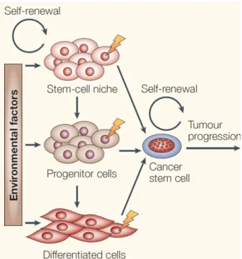
Figure 3. The adult mammary stem cells and breast cancer stem cells share certain
characteristics. The cancer stem cells might arise either from specific adult stem or progenitor cells, but stem-like phenotype acquisition by the differentiated cells should be considered (Bjerk vig et al., 2005).
1.8. Interleukin 6 and breast cancer
Interleukin 6 (IL6), a multifunctional cytokine, plays a role in p ro- and anti-
inflammatory response and has a primary function in the pathophysiology of many diseases such as rheumatoid arthritis, Castleman’s dieses and cancer (Grivennikov and Karin, 2008; Kishimoto, 2005).
IL6 signaling is mediated via IL-6 binding to IL6 receptor (IL6R) which induces homo- dimerization of the signal transducing receptor gp130 which leads to activation of multiple signaling networks (Keller et al., 2002). IL6 signaling is responsible for regulation of various biological processes as cell survival, apoptosis and proliferation in murine hematopoietic stem cells, hepatocytes and MCF7 breast cancer cell line (Gotze et al., 2001; Peters et al., 1998a; Sansone et al., 2007).
IL6 signals via a heterodimeric IL6/IL6R/gp130 complex, whose engagement triggers activation of Janus (JAK) kinases, and the downstream effectors STAT3, SHP-2/Ras,
15
NF-κB and PI3K/Akt (Kishimoto, 2005). The IL6/IL6R/gp130 complex can be build by either utilization of the membrane bound IL6R or the soluble form of IL6R (sIL6R) in the process known as IL6 trans-signaling (Peters et al., 1998b; Taga et al., 1989).
The effect of the IL6 signaling on breast cancer was elaborated in various systems (Iliopoulos et al., 2011; Korkaya et al., 2012; Sansone et al., 2007), but in all of the mentioned reports it is not clear whether the IL6 signaling is maintained via
membrane bound or soluble IL6R which is of high therapy design importance because high levels of sIL6R can be found in human sera.
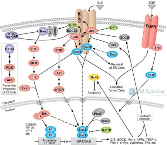
Figure 4. IL6 signaling pathway. IL6 signaling pathway is triggered by dimerization of the IL6 and IL6R. IL6-IL6R complex binds to gp130 and form a complex of 6 members complex (2 x IL6, 2x IL6R and 2x gp130). The complex triggers the signaling pat hway via intracellular domain of gp130. IL6 signaling is madiated via complex signaling networks (i.e. JAK/STA T3, P I3K/Akt/mTOR, Raf/Mek/Erk).
The figure is modified from the reference (CellsignalingTechnology, 2012).
Increased levels of IL6 in sera of breast cancer patients correlate with poor disease outcome and reduced prognosis (Bachelot et al., 2003). This might be a
consequence of an overall IL6 influence on the growth rate of the primary tumor (Korkaya et al., 2012), but the exact mechanisms of these action is not yet
16
elucidated. Possible mechanism of action might be via CSC as deduced from the studies in the MCF7 cell line. Activation of the IL6 signaling induced self- renewal, hypoxia survival, and invasiveness of MCF7 cell line derived mammospheres
(Sansone et al., 2007). Additionally, IL6 signaling has been proposed to reg ulate the conversion of the non- stem cancer cells into cancer stem cells (Iliopoulos et al., 2011) but this observation needs further clarification.
Figure 5. IL6 signaling in cancer cell. IL6 signaling in the cancer cells is maintained via IL6 secretion by various non-epithelial cells types. The IL6 signaling influences various biological processes
enabling cancer cell survival, proliferation and self-renewal (Grivennikov and Karin, 2008; Korkaya et al., 2011).
Present knowledge suggests that IL6 signaling in cancer is maintained trough IL6 expression by cancer associated fibroblasts, macrophages and various different cell types (Bonafe et al., 2012) while it is not much known about the effect and sources of the sIL6R.
Having in mind the importance of the IL6 signaling the aim of this work was to further elaborate the effect of the IL6 trans -signaling in normal and malignant breast stem and progenitor cells.
17 1.9. The aim of the work
The mammary gland is hierarchically structured organ containing cellular types of various differentiation stages. The mammary gland development and extensive proliferation and differentiation during each pregnancy cycle are enabled by the presence of the adult mammary stem cells which phenotype is regulated by
activation of stem signaling pathways. These signaling pathways are activated as a result of the dynamic interaction between mammary cells and micro-environment.
Deregulation of stem cell signaling pathways has been suggested to drive breast cancer by maintaining cancer stem cells (CSC), cells which give rise to all other cancer cell types detected within the tumor and contribute to an invasive phenotype observed in metastatic breast tumors. As normal stem cells, CSC interact with micro- environment and these interactions involve inflammatory cytokines such as
Interleukin 6 (IL6).
The aims of the proposed PhD thesis were:
1) To address how IL6 signals in normal mammary stem and progenitor cells , 2) To assess the effects of IL6 signaling in regulation of normal mammary stem and progenitor cell phenotype,
3) To assess the ability of cellular cooperation in triggering IL6 trans-signaling in mammary gland,
4) To evaluate the effects of the IL6 trans-signaling in CSC phenotype.
18
2. Materials and Methods
2.1. Materials
2.1.1. Reagents, solutions and cell culture media
Reagents, solution and cell culture media used in this work are listed in the following tables.
Reagents Company
1 AB-Serum Bio-Rad, Dreieich
2 Acetic acid Merck, Darmstadt
3 Agarose Sigma-Aldrich, USA
4 Ampicillin AppliChem GmbH, Darmstadt
5 B27 Invitrogen, USA
6 BCIP/NBT (AP color reagent) BioRad, Munich 7 Basic fibroblast growth factor (bFGF) Sigma-Aldrich, USA
8 bio-dUTP, 1 mM Roche, Penzberg
9 Bromphenol blue Sigma-Aldrich, USA
10 Bovine serum albumin (BSA), 20 mg/ml Roche, Penzberg, Mannheim 11 Bovine serum albumin (BSA), 30% Biotest, Dreieich
12 Chloroform Sigma-Aldrich, USA
13 Chloroquine Sigma-Aldrich, USA
14 Cholera toxin Sigma-Aldrich, USA
15 Collagenase Sigma-Aldrich, USA
16 DAPI Roche, Penzberg
17 dATP, 100 mM GE Healthcare, UK
18 dCTP, 100 mM GE Healthcare, UK
19 DEPC-H2O Invitrogen, USA
20 dGTP, 100 mM GE Healthcare, UK
19 21 Dichlorodimethylsilane, 2% in 1,1,1-
trichlorethane
Merck, Darmstadt
22 Diluent C Sigma-Aldrich, USA
23 Disodium phosphate Sigma-Aldrich, USA
24 Ditiotreitol (DTT), 0.1 M Invitrogen, USA
25 DMEM Pan-Biotech, Aidenbach
26 DMEM/F12 Pan-Biotech, Aidenbach
27 DNA ligase T4, 5 U/μl Roche, Penzberg
28 DNA polymerase PanTaq, 5 U/μl Pan Biotech, Aidenbach 29 DNA polymerase Taq, 5 U/μl Roche, Penzberg
30 DNA polymerase
Thermo Sequenase (TS), 32 U/μl
GE Healthcare, UK
31 DNA-Ladder 1kb Invitrogen, USA
32 DNase I, 2000 U/mg Roche, Penzberg
33 dNTPs, 100 mM GE Healthcare, UK
34 DSL peptide Research Genetics, USA
35 dTTP, 100 mM GE Healthcare, UK
36 EB buffer – QIAquick PCR Purification Kit
Qiagen, Hilden
37 EDTA Sigma-Aldrich, USA
38 Eosin Sigma-Aldrich, USA
39 Epidermal growth factor (EGF) Sigma-Aldrich, USA
40 Ethanol, absolute J.T.Baker, Griesheim
41 Ethidium-bromide, 1% Fluka, Sigma-Aldrich, USA
42 Eukitt O. Kindler GmbH, Freiburg
43 Fetal bovine serum (FBS) Sigma-Aldrich, USA 44 Fetal bovine serum (FBS) Pan Biotech, Aidenbach
45 Formaldehyde Merck, Darmstadt
46 Formamide Merck, Darmstadt
47 Formamide, deionized Sigma-Aldrich, USA
48 Gentamicin Sigma-Aldrich, USA
20
49 H2O, for HPLC Merck, Darmstadt
50 Hank’s salt solution Biochrom, Berlin
51 Hematoxylin Ventana, USA
52 Heparin Sigma-Aldrich, USA
53 HEPES, 1M Sigma-Aldrich, USA
54 Hyaluronidase Sigma-Aldrich, USA
55 Hydrochloric acid, 37% J.T. Baker, Griesheim
56 Hydrocortisone Sigma-Aldrich, USA
57 Igepal CA-630 Sigma-Aldrich, USA
58 Insulin Sigma-Aldrich, USA
59 Interleukin 6 Sigma-Aldrich, USA
60 Isopropanol Fluka, Sigma-Aldrich, USA
61 L-Glutamine, 200 mM Pan-Biotech, Aidenbach
62 Magnesium-chloride Sigma-Aldrich, USA
63 Matrigel© BD Biosciences, Heidelberg
64 MEBM Lonza, USA
65 Methanol Merck, Darmstadt
66 Mineral oil Sigma-Aldrich, USA
67 Mouse serum DAKO, Hamburg
68 mTRAPtm kit Active Motif, Japan
69 Sodium acetate, 2M pH 4 Calbiochem, Hamburg
70 Sodium citrate Applichem, Darmstadt
71 NEB buffer 1 New England Biolabs, USA
72 NEB buffer 2 New England Biolabs, USA
73 NEB buffer 3 New England Biolabs, USA
74 NEB buffer 4 New England Biolabs, USA
75 N-Laurylsarcosin Sigma-Aldrich, USA
76 Parablast embedding medium (Paraffin) Sigma-Aldrich, USA 77 Penicillin/Streptomycin, 10 U/μl Pan-Biotech, Aidenbach
78 Percoll GE Healthcare, UK
79 Peroxidase Blocking Solution DAKO, USA
21
80 Phytohemagglutinin, M form (PHA-M) Invitrogen, USA 81 PolMix polymerase – Expand long
template PCR system, 5 U/μl
Roche, Penzberg
82 Polybrene Sigma-Aldrich, USA
83 Poly-HEMA Sigma-Aldrich, USA
84 Potassium acetate, 5 M Sigma-Aldrich, USA 85 Potassium chloride (KCl) Sigma-Aldrich, USA 86 Potassium chloride (KCl), 1 M Fluka, Sigma-Aldrich, USA 87 Potassium digydrogen phospate
(KH2PO4)
Merck, Darmstadt
88 Propidiumiodid Sigma-Aldrich, USA
89 Puromycin Sigma-Aldrich, USA
90 Roti-Aqua-Phenol Carl Roth GmbH, Karlsruhe
91 RPMI 1640 Pan-Biotech, Aidenbach
92 β-Mercaptoethanol Carl Roth GmbH, Karlsruhe 93 Trisodium citrate dihydrate Merck, Darmstadt
94 Trypan blue, 0.4% Sigma-Aldrich, USA
95 Tris(hydroxymethyl)-aminomethan (TRIS)
AppliChem, Darmstadt
96 Tris-acetate Sigma-Aldrich, USA
97 Tris-HCl (pH 8), 1M Sigma-Aldrich, USA
98 tRNA, 100 mg Roche, Penzberg
99 Trypsin neutralization solution Sigma-Aldrich, USA 100 Trypsin/EDTA, 10x Trypsin, 0.5% +
EDTA, 0.2% in 1x PBS
PAA, Austria
101 Tween 20 Sigma-Aldrich, USA
102 Ultra Puretm Herring sperm DNA solution
Invitrogen, USA
103 UltraPureTM DEPC- Water Invitrogen, USA
104 Xylol Roth, Karlsruhe
105 Zeocin Invitrogen, USA
22 2.1.2. Antibodies
Antibody Company Clone Concentration 1 Biotin anti-human
CD126 (IL-6Rα)
Biolegend, USA UV4 0,5 mg/ml
2 Mouse IgG1, κ- Isotype control
Biolegend, USA MOPC- 21
0,5 mg/ml
3 Monoclonal anti- human IL6 blocking
antibody
Sigma-Aldrich, USA
6708.11 1,5 µg/ml
4 Anti-human cytokeratin 18
Chemicon CK2 20 µg/ml
5 Anti-IL6R (FITC) Abcam B-R6 1 mg/ml
2.1.3. The composition of prepared buffers, media and solutions
Solutions, Buffers and Media Composition
1 Carnoy Fixative 7,5 ml methanol
2,5 ml acetic acid 2 Cell culture medium RPMI medium without L-
glutamine 10% FCS 200 U/mL penicillin 200 U/mL streptomycin
2 mM L-glutamine
3 DAPI solution 10 µg/ml DAPI
4x SSC / 0,2% Tween-20
4 Hematoxylin solution 2 g hematoxylin
0,4 g sodium iodide 100 g potassium aluminium
23
sulfate
100 g chloral hydrate 2 g citric acid
5 LB (liquid broth) 1% NaCl
1% tryptone 5% yeast extract
pH 7,0
6 Mammosphere medium 49 ml MEBM
1x B27 10 ng/ml EGF 10 ng/ml bFGF 4 µg/ml heparin
7 Medium for the HME cell lines DMEM/F12 10% FCS 200 U/ml penicillin 200 U/ml streptomycin
10 ng/ml EGF 0,5 µg/mL hydrocortisone
10 µg/mL insulin 8 Medium for the MCF7 and MDA-MB-
231 cell lines
DMEM/F12 10% FCS 200 U/ml penicillin 200 U/ml streptomycin
2 mM L- glutamine 9 Mammary tissue digestion medium DMEM/F12
1% HEPES 200 U/ml penicillin 200 U/ml streptomycin
24
2 mM L- glutamine 2% BSA
10 mg/ml hyaluronidase (100 U/ml)
33 µg/ml collagenase (300 U/ml) 10 µg/mL insulin
0,5 µg/mL hydrocortisone 100 ng/ml cholera toxin 10 PBS (Phosphate Buffer Saline) 8,5 mM Na2HPO4
2 mM KH2PO4 NaCl 150 mM NaCl
pH 7,4
11 PCR-Buffer + dNTPs 100 mM Tris-HCl
500 mM KCl 10 mM MgCl2
1mM nucleotids
12 TE-Puffer 10 mM TRIS-HCl
1 mM EDTA pH 7,4
2.1.4. Cell lines
Identity of used cell lines was regularly controlled by the ATCC recommended DNA fingerprinting.
Cell line Description
1 C3H10T 1/2 Mouse embryonic fibroblasts 2 hTERT-HME1 Non-tumorigenic breast epithelial cell line 3 hTERT-HME1 BRAF hTERT-HME1 cell line carrying mutation in BRAF
gene
4 hTERT-HME1 EFGR hTERT-HME1 cell line over-expressing EGFR receptor
25 5 hTERT-HME1
PI3KCA ex. 20
hTERT-HME1 cell line carrying mutation in exon 20 of PI3KCA gene
6 hTERT-HME1 shPTEN
hTERT-HME1 cell line expressing reduced levels of PTEN
7 hTERT fibroblasts Immortalized human fibroblasts 8 MCF10A Non-tumorigenic breast epithelial cell line 9 MCF7 Breast cancer cell line- metastatic pleural effusion
cells
10 MDA-MB-231 Breast cancer cell line- metastatic pleural effusion cells
11 MDA-MB-231 1833 Breast cancer cell line- metastatic pleural effusion cells
12 SK-BR-3 Luminal breast cancer cell line 13 HEK-293T Human embryonic kidney cell line
2.1.5. Devices
Device Company
1 Axio Imager Z1 Fluorescence microscope
Zeiss, Göttingen
2 Balance Kern, Balingen
3 BenchMark Ultra Ventana, USA
4 Capillary holder for micromanipulation Eppendorf, Hamburg
5 Cell culture incubator Heraeus, Hanau
6 Cell culture incubator Heraeus, Hanau
7 Cell culture laminar flow Heraeus, Hanau
8 Centrifuge Heraeus, Hanau
9 Centrifuge Eppendorf, Hamburg
10 Centrifuge, tabletop Grant Bio, USA
11 Centrifuge, tabletop Eppendorf, Hamburg
12 Cytospine Centrifuge Hettich, USA
26
13 DM RXA Fluorescence microscope Leica, USA
14 Cauterizer Fine Science Tools,
Hedelberg
15 FACS Canto II BD Biosciences, USA
16 Electrophoresis gel chamber Biostep, Jahnsdorf 17 Photometer, GeneQuant II Pharmacia Biotech,
USA
18 Laser micro-dissection microscope P.A.L.M, Bernried 19 LSR II flow cytometer BD Bioscience, USA
20 Magnetic stirrer VELP Scientifica
20 MJ Research Peltier Thermal Cycler PTC-200
Bio-Rad, USA
21 MJ Research Peltier Thermal Cycler Tetrad
Bio-Rad, USA
22 Multipipette Stream Eppendorf, Hamburg
23 Neubauer- Cell counter Schubert und Weiß, Munich
24 Optical microscope Optech, Canada
25 Pipette controller Brand, Wertheim
26 pH-meter Eutech Instruments,
The Netherlands 27 Pipettes (2 µL, 20 µL, 200 µL, 1000 µL) Eppendorf, Hamburg 28 Power Supply for gel chamber
electrophoresis
MRC, Israel
29 Pump KNF, Freiburg
30 StuartTM Scientific roller mixer Stuart Scientific, UK
31 Thermo mixer Eppendorf, Hamburg
32 UV illuminator Intas, Göttingen
33 Vortex mixers VELP Scientifica, Italy
34 Water bath Memmert, Schwabach
27 2.1.6. Software
Software Company
AxioVision 4.5 Zeiss, Göttingen
FACS Diva 6.1.1 BD Biosciences, USA
FlowJo 8.8.6 TreeStar, Inc.USA
NEBcutter V2.0 New England Biolabs, USA
Vector NTI Invitrogen, USA
GraphPad Prism Graph Pad software, USA http://faculty.vassar.edu/lowry/VassarStats.html Website for statistical
computation
2.1.7. Primers used for the PCR amplification
Gene of interest
Oligonucleotide sequence
β-Actin Forward 5'-GTG ACA GCA TTG CTT CTG TG-3' Reverse 5'-TCT CAA GTC AGT GTA CAG GC-3' EF1-α Forward 5'-CCA GTT ATG TGG CAA GAC GTT-3'
` Reverse 5'-TCT GGG GAG AAT GGG TAG C-3'
GAPDH Forward 5'- AAT CCC ATC ACC ATC TTC CAG-3' Reverse 5' GCC ATC ACG CCA CAG TTT CC -3' IL6 Forward 5'- GAG AAG GCT GAG ATA AAA GGA GA -3'
Reverse 5'- CAT GAT ATA GAC GTT GTG GCT G -3' IL6R Forward 5'- GCG ACA AGC CTC CCA GGT TC -3'
Reverse 5'-GTG CCA CCC AGC CAG CTA TC -3'
28
m-dio Forward 5'-GCT CCT TAC AGT GAC TGC AG-3' Reverse 5'-TCA ATG GTC ATA TTG CAG CC-3' m-IVL Forward 5'-GAA GCA GGT AGG TGT GCA G-3' Reverse 5'-GCC CTA CTC AAC CTG AGA G-3' P53 exon
2/3
Forward 5'-AGG ACC TGA TTT CCT TAC TGC-3'
Reverse 5'-GAG GTC CCA AGA CTT AGT AC-3' Pseudo
CK 19
Forward 5'-GAA GAT CCG CGA CTG GTA C-3'
Reverse 5'-TTC ATG CTC AGC TGT GAC TG-3' CFL15CT
24
5'-CCCCCCCCCCCCCCCGTCTAGA TTTTTTTTTTTTTTTTTTTTTTTTVN -3'
CFL15C N8
5'-CCCCCCCCCCCCCCCGTCTAGANNNNNNNN-3'
CP2 5'-TCAGAATTCATGCCCCCCCCCCCCCCC-3'
CP2-BGL 5'-TCAGAATTCATGCCGCCCCCCCGGCCC-3'
Lib1 5'-AGTGGGATTCCTGCTGTCAGT-3'
ddMse11 5'-TAACTGACAGddC-3'
29 2.2. Methods
2.2.1. Methods for in vitro cell propagation and characterization
The list of used cell lines is shown in the chapter 2.1.4. The identity of the used cell lines was controlled on a regularly basis by ATCC recommended DNA fingerprinting.
2.2.1.1. In vitro cell line propagation under conventional 2D conditions
Cell lines were preserved in liquid nitrogen in appropriate medium containing 5%
DMSO. Cell lines were propagated until 70% confluence apart from the cases when the experimental strategy did not imply differently. Cell lines were de-attached using Trypsin/EDTA and re-plated in an appropriate ce ll density. The medium was changed following the recommendation of the ATCC.
2.2.1.2. In vitro cell line propagation under anchorage independent (3D) condition
The aim of 3D cell propagation system is to enrich mammary stem and progenitor cell populations.
Mammary cell lines were propagated in 2D condition until the cells reached the 70%
confluence. Propagated cells were de-attached and washed in PBS at least two times. The washing steps were introduced to remove traces of serum used in the 2D cell line propagation culture strategy. Cell lines were propagated in ultra -low
attachment plates in cell culture incubator at 37 °C, 5% CO2. The cell lines were propagated at a seeding density of 10 000 cells/ml.
Mammosphere forming ability was assessed 7 days post-plating. During this period, medium was not changed. All cell lines were propagated in the same mammosphere medium, as listed in the chapter 2.1.2.
30
2.2.1.2.1. Preparation of poly-HEMA plates
The mammary stem and progenitor cells are able to survive anchorage independent conditions while more differentiated progenies are undergoing anoikis. Cells are not able to anchorage to the dish bottom if the dishes are covered by poly-HEMA, an organic substance soluble in ethanol. Poly-HEMA ethanol suspension was applied to the culture dishes 24 h before the cells are plated. On the next day, a fine layer of poly-HEMA covers the culture dish and prevents the cells to adhere. Poly-HEMA (2,4 g) was dissolved in 95% ethanol (20 ml) at 65 ⁰C. Eight hours later ethanol dissolved poly-HEMA was diluted 10x in 95% ethanol and added to the cell culture dishes at the final concentration of 12 mg/ml. Poly-HEMA coated plates were sterilized by the UV light in prior to cell seeding. The ethanol traces were removed by washing with PBS.
2.2.1.3. In vitro differentiation of mammary cells
Cell lines propagated in 3D conditions or the secondary mammospheres obtained from the donor’s tissue specimens were used for the in vitro differentiation in Matrigel©. Matrigel© is a gelatinous protein mixture secreted by Engelbreth-Holm- Swarm mouse sarcoma cells. This mixture resembles the complex extracellular environment found in many tissues. Different differentiation strategies were tested and as a results Matrigel© “sandwich” strategy was adopted as the most efficient.
Cells were propagated in Ham’s F-12 medium supplemented with 5% FBS, 5 μg/ml insulin, 1μg/ml hydrocortisone, 10 μg/ml cholera toxin, 10 ng/ml EGF, and 1×
Pen/Strep Mix.
Briefly, Matrigel© was diluted 1:1 with differentiation media and placed in a
differentiation dish for 15 minutes at 37 ⁰C. On the top of the stiff gel, the mammary stem and progenitor cells were placed in an incubator at 37 ⁰C for 30 minutes. The cells were covered with an additional Matrigel© layer and left for additional 15 minutes at 37 ⁰C. Differentiation medium was added at the end of the embedding procedure.
Differentiation medium was changed regularly each fourth day.
Cells propagated in Matrigel© were examined 3-4 weeks post embedding for the development of the complex duct and acinar structures.
31
2.2.2. In vitro cell propagation of the donor’s tissue specimens
2.2.2.1. Tissue collective
Mammary tissues were obtained from women undergoing mammary reduction surgeries at Caritas-Krankenhaus St. Josef, Regensburg in collaboration with Dr.
Claus Lattrich and Dr. Norbert Heine. Mammary tissues were examined by
pathologists at Institut für Pathologie, Universitätsklinikum Regensburg, Regensburg.
The tissues showing the signs of breast cancer were excluded from the study and not used for this work (Figure 1).
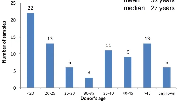
Eighty seven healthy mammary tissues were received in the period 2007-2012.
The median age of the cohort was 27. Due to hormone changes during menopause and proposed hormonal influence in mammary stem a nd progenitor cells, tissue were selected based on an arbitrary age limit (45 years). However, the samples received from the donors older than 45 were used for the modifications of the mammosphere protocol presented in result section.
Figure 1. The overview of the mammary tissue s received in the period 2007-2012.
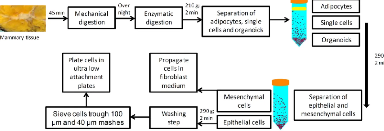
2.2.2.2. Mammary tissue digestion and cell isolation
32
Mammary tissue was minced and dissociated in Ham’s F12/Dulbecco’s modified Eagle’s medium [F12:DMEM; 1:1 (v:v)] supplemented with 10 mM HEPES, 2%
bovine serum albumin (BSA; Fraction V), 5 µg/ml insulin, 0.5 µg/ml hydrocortisone, 10 ng/ml cholera toxin, 300 U/ml collagenase and 100 U/ml hyaluronidase at 37°C for 18 h (Figure 2).
Figure 2. Digestion of the tissue specimen and cell isolation.
At the next day, the digested cell suspension was centrifuged at 210 g for 2 minutes at room temperature. S upernatant from the first centrifugation step contained single mammary epithelial and stromal cells (fibroblasts), while in the pellet were undigested tissue pieces, known as organoids. Single epithelial cells were subjected to an additional centrifugation step (290 g; 2 minutes; room temperature) and the epithelial cells found in pellet were re-suspended in the basal medium, washed and propagated in mammosphere medium in ultra low attachment plates. Mammary gland fibroblasts were obtained by centrifugation of the supernatant (500 g; 5
minutes; room temperature). Fibroblasts were propagated in standard tissue culture flasks in DMEM medium supplemented by 10% fetal bovine serum (FBS).
The organoids obtained in the first centrifugation step were either processed immediately for single cell isolation or preserved by freezing ( -80 ⁰C) in organoid freezing medium (DMEM supplemented with 10% DMSO and 20% FCS).
If obtained number of epithelial cells was not sufficient, organoids would be
processed immediately by further digestion steps in DMEM/F12 supplemented with 5 U/ml dispase and 0,25% trypsin. Trypsin was inactivated by trypsin inactivation
33
solution (TNS) or medium supplemented with serum. Digested organoids were centrifuged (300 g; 3 min at RT) and the epithelial cells found in the pellet were re- suspended in mammosphere medium.
2.2.2.3. Mammosphere protocol
Single cells obtained from digested tissues were plated in ultra-low attachment plates at a density of 20,000 cells/ml. The number of plated cells was later modified
according to the results shown in the chapter 3.2.3.
Cells were grown in a serum-free mammary epithelial growth medium (MEBM) supplemented with B27, 20 ng/ml EGF and 20 ng/ml bFGF and 4 μg/ml heparin.
Mammospheres were collected by gentle centrifugation (100 g) after 7 days and dissociated enzymatically (10 min in 0.05% trypsin, 0.53 mM EDTA-4Na).
The obtained secondary mammosphere were used for the most of the downstream assays elaborated in this work (Figure 2).
Figure 3. Mammosphere propagation.
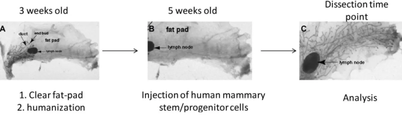
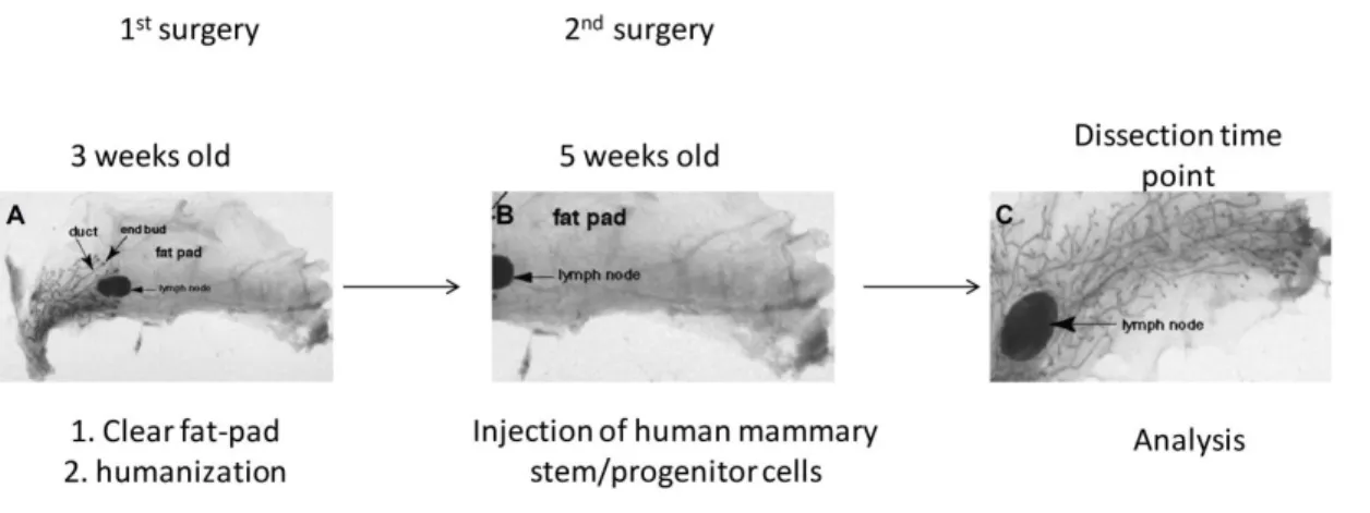
2.2.3. In vivo human mammary stem and progenitor cells differentiation
In vivo experiments were undertaken in the animal facility of the University Hospital Regensburg. All the experiments considering NOD.Cg-Prkdcscid Il2rgtm1Wjl/SzJ (NSG) mice were done in accordance with animal application 54-2532.1-15/09 Government of Oberpfalz (54-2532.1-15/09; Antrag auf Genehmigung eines Versuchsvorhabens mit Wirbeltieren; Riegirung der Oberpfalz).