2010/220
Amniotic fluid volume in intra-amniotic inflammation with and without culture-proven amniotic fluid infection in preterm premature rupture of membranes*
Si Eun Lee1, Roberto Romero2, Seung Mi Lee1 and Bo Hyun Yoon1,**
1
Department of Obstetrics Gynecology, Seoul National University College of Medicine, Seoul, Republic of Korea
2
Perinatology Research Branch,
Eunice Kennedy ShriverNational Institute of Child Health and Human
Development, NIH, DHHS, Detroit, Michigan, USA
Abstract
Objective:
Previous studies reported that the clinical signif- icance of intra-amniotic inflammation with a negative amni- otic fluid (AF) culture is similar to that of intra-amniotic inflammation with microbiologically-proven AF infection.
However, the magnitude of the fetal inflammatory response in these two conditions is different as gauged by umbilical cord C-reactive protein (CRP) concentrations. We undertook this study to determine if the frequency of oligohydramnios is different in these two conditions.
Methods:
The amniotic fluid index (AFI) was measured in 205 patients with preterm premature rupture of membranes (PROM) (
F35 weeks). AF was cultured for aerobic and anaerobic bacteria and genital mycoplasmas. Intra-amniotic inflammation was defined as an elevated AF matrix metal- loproteinase-8 (MMP-8) concentration (
)23 ng/mL).
Patients were divided into three groups according to the results of AF culture and the presence or absence of intra- amniotic inflammation: 1) without intra-amniotic inflamma- tion and a negative culture (n
s109); 2) with intra-amniotic inflammation and a negative culture (n
s44); and 3) a pos- itive culture (n
s52).
*This study was presented at the 28thAnnual Clinical Meeting of the Society for Maternal–Fetal Medicine, Dallas, TX, January 28–February 2, 2008.
This work was supported in part by Korea Science and Engineering Foundation (KOSEF) grant funded by the Korea government (MOST) (No. R01-2006-000-10607-0) and in part by the Intramural Research Program of theEunice Kennedy ShriverNational Institute of Child Health and Human Development, NIH, DHHS.
**Corresponding author:
Bo Hyun Yoon, MD, PhD
Department of Obstetrics Gynecology College of Medicine
Seoul National University Seoul 110-744
Korea
Tel.:q82-2-2702-2826 Fax:q82-2-765-3002 E-mail: Yoonbh@snu.ac.kr
Results:
Patients with a positive culture had a higher fre- quency of oligohydramnios and a lower median AFI than those with a negative culture but with intra-amniotic inflam- mation (P
-0.01). However, there was no significant differ- ence in the median AFI or in the frequency of oligo- hydramnios according to the presence or absence of intra- amniotic inflammation among patients with a negative cul- ture (P
)0.1).
Conclusion:
Oligohydramnios was more frequent in patients with culture-proven AF infection than in those with intra- amniotic inflammation and a negative AF culture.
Keywords:
Amniotic fluid index (AFI); amniotic fluid infec- tion; fetal inflammatory response syndrome (FIRS); intra- amniotic inflammation; oligohydramnios; rupture of membranes.
Introduction
Microbial invasion of the amniotic cavity is present in one third of patients with preterm premature rupture of mem- branes (PROM) and is strongly associated with impending preterm delivery, adverse pregnancy and neonatal outcome
w4, 24, 25
x. Previous studies have reported that the clinical significance of intra-amniotic inflammation with a negative culture for microorganisms is similar to that of microbiolog- ically-proven amniotic fluid (AF) infection
w25, 33
x. How- ever, proven AF infection, which is severe enough to yield a positive culture, may reflect a higher microbial burden and elicit a more intense fetal inflammatory response than intra- amniotic inflammation with a negative culture. A previous study documented that fetuses born to mothers with culture- proven AF infection had a higher umbilical cord plasma C- reactive protein (CRP) than those born to mothers with culture-negative intra-amniotic inflammation
w10
x. This could have different implications for fetal target organs, including the kidney.
Oligohydramnios is frequently present in preterm PROM
w18, 27, 29
x. Though the reduced AF volume is partly attrib-
uted to the escape of fluid through the site of membrane
rupture, microbial invasion of the amniotic cavity and the
subsequent fetal inflammatory response syndrome (FIRS) is
known to be associated with oligohydramnios, which has
been attributed to decreased fetal urine production
w18, 30
x.
The purpose of this study was to determine if proven AF
infection is associated with changes in AF volume.
Materials and methods
Study design
The relationship between AF volume and intra-amniotic inflam- mation and/or AF infection was examined in 205 singleton preg- nancies admitted to our university hospital with the diagnosis of preterm PROM who met the following criteria: (1) preterm preg- nancy wgestational age (GA) F35 weeksx; (2) AF obtained for microbiologic studies by transabdominal amniocentesis or at the time of cesarean delivery; and (3) amniotic fluid index (AFI) deter- mined before AF retrieval according to the method described by Phelan et al.w19x. Oligohydramnios was defined as AFIF5 cm.
Patients were divided into three groups according to the presence or absence of intra-amniotic inflammation and AF culture results:
Group 1: patients without inflammation, without infection (a neg- ative AF culture; ns109); Group 2: patients with intra-amniotic inflammation, without infection (a negative AF culture; ns44); and Group 3: patients with a positive AF culture (ns52). Retrieval of AF was performed after written informed consent was obtained. The Institutional Review Board of the participating institution approved the collection and use of these samples and information for research purposes.
AF studies
AF was cultured for aerobic and anaerobic bacteria, as well as gen- ital mycoplasmas (Mycoplasma hominisandUreaplasma urealyti- cum). An aliquot of AF was transported to the laboratory and examined in a hemocytometer chamber to determine the white blood cell (WBC) count. The remaining fluid was centrifuged and stored in polypropylene tubes at –708C. Matrix metalloproteinase-8 (MMP-8) concentration was measured with a commercially availa- ble enzyme-linked immunosorbent assay (Amersham Pharmacia Biotech, Inc, Bucks, UK). The sensitivity of the test was 0.3 ng/mL. Intra- and inter-assay coefficients of variation were-10%, respectively. MMP-8 was used to assess the presence of intra-amni- otic inflammation because previous studies indicated that it is a sensitive and specific index of inflammationw1, 12, 17x. Intra-amni- otic inflammation was defined as an elevated AF MMP-8 concen- tration ()23 ng/mL), as previously reportedw17x.
Diagnosis of chorioamnionitis
Clinical chorioamnionitis was diagnosed in the presence of maternal temperature ofG37.88C and two or more of the following criteria:
(1) uterine tenderness; (2) malodorous vaginal discharge; (3) mater- nal leukocytosis (WBC count of)15,000 cells/mm3); (4) maternal tachycardia ()100 beats/min); and (5) fetal tachycardia ()160 beats/min)w3x. Acute histologic chorioamnionitis was diagnosed if acute inflammatory changes were present on examination of the extra-placental membranes or the chorionic plate of the placenta, according to criteria previously publishedw31x. Funisitis was diag- nosed in the presence of neutrophil infiltration into the umbilical vessel walls or into Wharton’s jelly.
Statistical analysis
Proportions were compared with the Fisher’s exact test. A Kruskal- Wallis analysis of variance test was used for comparison of contin- uous variables among groups. Multiple comparisons between groups were performed with the Mann-WhitneyU-test. The amniocentesis- to-delivery interval was compared using the generalized Wilcoxon
test for survival analysis. The interval-to-delivery of patients deliv- ered for maternal or fetal indications was treated as a censored observation, with a censoring time equal to the amniocentesis-to- delivery interval. A P-0.05 was considered significant.
Results
The frequency of oligohydramnios (AFI
F5 cm) was 29%
(59/205). The prevalence of a positive AF culture was 25%
(52/205). Microorganisms isolated from the AF included
Ureaplasma urealyticum(n
s37), Candida species (n
s4), Streptococcus species (n
s4),
Escherichia coli(n
s3), coag- ulase-negative Staphylococcus (n
s3),
Mycoplasma hominis(n
s3), and one isolate each of Peptostreptococcus species, Corynebacterium species,
Acinatobacter baumanii,Burkhol- deria cepalia,Staphylococcus hominis,and
Staphylococcus epidermidis. Six patients had polymicrobial infections (4patients with 2 species, and 2 patients with 3 species of microorganisms).
Table 1 describes the clinical characteristics and pregnancy outcomes of the study population according to the results of AF culture and MMP-8 concentrations. Patients with intra- amniotic inflammation but a negative AF culture (Group 2) had a significantly lower median GA at delivery, and birth weight, and a higher rate of histologic chorioamnionitis and funisitis than those without intra-amniotic inflammation and a negative AF culture (Group 1). However, there was no significant difference in the frequency of oligohydramnios between groups 1 and 2 (26% vs. 16%; P
s0.2).
There were no significant differences in the clinical char- acteristics and pregnancy outcomes between patients with intra-amniotic inflammation and a negative AF culture (Group 2) and those with a positive AF culture (Group 3), except for the frequency of oligohydramnios, which was higher in group 3 than in group 2 (46% vs. 16%; P
-0.005).
Figure 1 shows that patients with a positive AF culture had a lower median AFI than those with a negative culture but with intra-amniotic inflammation (median, 5.1
wrange 0–17.5
xvs. median 8.4
wrange 0–21.9
x; P
-0.01). However, there was no significant difference in the median AFI accord- ing to the presence or absence of intra-amniotic inflammation in patients with a negative AF culture (median, 8.1
wrange 0–31.0
xvs. median 8.4
wrange 0–21.9
x; P
s0.8).
Forty-six percent (24/52) of patients with a positive AF culture had oligohydramnios. Among patients with a positive AF culture, the median GA at amniocentesis and delivery, birth weight, and WBC count in AF, and the frequency of clinical chorioamnionitis (29% vs. 11%), histologic cho- rioamnionitis (100% vs. 78%), and funisitis (77% vs. 74%) were not significantly different in patients with or without oligohydramnios. However, patients with oligohydramnios had a shorter interval-to-delivery than those without oligo- hydramnios (median, 38 h
wrange 0–285 h
xvs. median 95 h
wrange 7–748 h
x; P
-0.001, Figure 2).
Cox proportional hazards model analysis indicated that the
presence of oligohydramnios (AFI
F5 cm) was an inde-
pendent predictor of the interval-to-delivery after adjustment
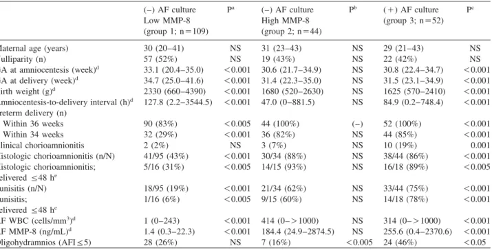
Table 1 Clinical characteristics and pregnancy outcomes of the study population according to the results of AF culture and MMP-8 concentrations.
(–) AF culture Pa (–) AF culture Pb (q) AF culture Pc
Low MMP-8 High MMP-8 (group 3; ns52)
(group 1; ns109) (group 2; ns44)
Maternal age (years) 30 (20–41) NS 31 (23–43) NS 29 (21–43) NS
Nulliparity (n) 57 (52%) NS 19 (43%) NS 22 (42%) NS
GA at amniocentesis (week)d 33.1 (20.4–35.0) -0.001 30.6 (21.7–34.9) NS 30.8 (22.4–34.7) -0.001 GA at delivery (week)d 34.7 (25.0–41.6) -0.001 31.4 (22.3–35.0) NS 31.5 (23.1–34.9) -0.001 Birth weight (g)d 2330 (660–4390) -0.001 1680 (520–2630) NS 1625 (570–2410) -0.001 Amniocentesis-to-delivery interval (h)d 127.8 (2.2–3544.5) -0.001 47.0 (0–881.5) NS 84.9 (0.2–748.4) -0.001 Preterm delivery (n)
Within 36 weeks 90 (83%) -0.005 44 (100%) (–) 52 (100%) -0.001
Within 34 weeks 32 (29%) -0.001 36 (82%) NS 44 (85%) -0.001
Clinical chorioamnionitis 2 (2%) NS 3 (7%) NS 10 (19%) 0.001
Histologic chorioamnionitis (n/N) 41/95 (43%) -0.001 30/34 (88%) NS 38/44 (86%) -0.001
Histologic chorioamnionitis; 5/16 (31%) -0.005 14/15 (93%) NS 16/18 (89%) -0.005
deliveredF48 he
Funisitis (n/N) 18/95 (19%) -0.001 21/34 (62%) NS 33/44 (75%) -0.001
Funisitis; 1/16 (6%) -0.005 9/15 (60%) NS 14/18 (78%) -0.001
deliveredF48 he
AF WBC (cells/mm3)d 1 (0–243) -0.001 414 (0–)1000) NS 314 (0–)1000) -0.001
AF MMP-8 (ng/mL)d 1.4 (0.3–22.3) -0.001 184.4 (24.9–2874.5) NS 255.6 (0.4–2370.6) -0.001
Oligohydramnios (AFIF5) 28 (26%) NS 7 (16%) -0.005 24 (46%) -0.05
Values are medians and ranges.
aComparison between groups 1 and 2.
bComparison between groups 2 and 3.
cComparison between groups 3 and 1.
dP-0.05 by Kruskal-Wallis ANOVA test.
eTo preserve a meaningful temporal relationship between the results of AF studies and histologic findings of the placenta obtained at delivery, cases delivered within 48 h of amniocentesis were considered for the analysis.
AFsamniotic fluid, MMP-8smatrix metalloproteinase-8, Low MMP-8smatrix metalloproteinase-8-23 ng/mL, High MMP-8smatrix metalloproteinase-8)23 ng/mL, GAsgestational age, NSsnot significant, WBCswhite blood cell, AFIsamniotic fluid index.
Figure 1 Amniotic fluid index (AFI) according to the presence or absence of intra-amniotic inflammation and AF culture results: the median AFI in patients with intra-amniotic inflammation and a neg- ative AF culture was significantly higher than that in patients with a positive AF culture, but was not different from that in patients without intra-amniotic inflammation and with a negative AF culture.
for GA and the AF culture results
whazards ratio (HR), 1.8;
95% Confidence interval (CI), (1.3–2.6)
x.
Comment
Principal findings of this study
1) Oligohydramnios is associated with microbiologically- proven AF infection; 2) among cases with proven AF infection, those with oligohydramnios had a shorter amnio- centesis-to-delivery interval than those with an AFI
)5 cm.
AF infection, FIRS and oligohydramnios
Oligohydramnios is often associated with microbial invasion
of the amniotic cavity
w18, 27–30
x. Microorganisms in the
AF can gain access to the fetus and elicit a systemic FIRS
w5, 23, 26
x. Pro-inflammatory cytokines released during FIRS
can have effects on multiple fetal organ systems such as the
brain and heart, leading to blood flow redistribution and car-
diac dysfunction
w22, 30, 34
x. Reduction of renal blood flow
during FIRS can result in the decrease of fetal urine produc-
tion and the subsequent oligohydramnios. Indeed, a previous
Figure 2 Survival analysis of interval-to-delivery among patients with preterm premature rupture of membranes and amniotic fluid infection according to the presence or absence of oligohydramnios (median, 38 h; range, 0–285 h vs. median, 95 h; range, 7–748 h;
P-0.001).
study indicated that umbilical cord plasma concentrations of interleukin-6, which is the hallmark of FIRS, are signifi- cantly elevated in fetuses with oligohydramnios and preterm PROM compared with those without oligohydramnios
w30
x. Therefore, there is solid evidence linking microbial invasion of the amniotic cavity, FIRS and oligohydramnios.
Intra-amniotic inflammation in the absence of proven AF infection
A novel finding of this study is that among patients with intra-amniotic inflammation, those with proven AF infection had a decreased AF volume, whereas those with a negative AF culture did not. Substantial evidence indicates that intra- amniotic inflammation is a risk factor for adverse pregnancy and neonatal outcome, regardless of the presence or absence of a positive AF culture
w2, 7–9, 16, 25, 33, 34
x. However, the data presented herein demonstrate that a culture-proven infection has clinical implications beyond the presence of intra-amniotic inflammation with a negative AF culture in preterm PROM. This finding suggests that FIRS, in cases with intra-amniotic inflammation with a negative culture, may be milder than the one associated with a positive AF culture. Milder cases of FIRS may not be severe enough to cause a reduction of fetal urine production, which appears to occur in cases with proven AF infection and more severe FIRS. Indeed, studies in adults indicate that there is disease progression which begins with a systemic inflammatory response and advances to sepsis, severe sepsis and septic shock
w11, 20
x.
We propose that fetuses with a positive AF culture have a more severe systemic inflammatory response compared to those with intra-amniotic inflammation with a negative cul- ture. Support for this hypothesis is derived from a prior report indicating that among patients with intra-amniotic inflammation, those with proven infection had a higher con- centration of umbilical cord plasma CRP than those with a negative AF culture
w10
x.
Intensity of FIRS
This study suggests that fetuses with positive AF cultures have a greater degree of systemic fetal inflammation com- pared to those with intra-amniotic inflammation and negative AF cultures. Why the difference? Intra-amniotic inflamma- tion in the absence of proven AF infection is commonly con- sidered to be attributed to infection in which organisms escaped detection by traditional microbiological methods
w15, 32
x. It is possible that the microbial burden of patients with a positive culture is greater than that of those with a negative culture. Small inoculum size of microorganisms probably contributed to the failure of cultivation. It is also possible that microorganisms recovered with culture tech- niques may be more virulent than those who resist cultivation in the laboratory.
FIRS in proven AF infection
AF infection is associated with immune responses
w6, 13, 21
x, and FIRS has been also understood as an immune response, which can signal the initiation of parturition for the fetus to exit a hostile intrauterine environment
w23
x. Of interest, our data showed that among cases with proven AF infection, patients with oligohydramnios had an increased risk for impending delivery compared to those with normal AF volume. This suggests that fetuses with oligohydramnios in the presence of proven AF infection probably have an advanced stage of FIRS. However, this cannot be addressed with current data because the difference in the magnitude of FIRS between cases with and without oligohydramnios was not examined. Such issue requires a large cohort consisting of fetuses whose umbilical cord blood was retrieved at the time of AFI determination.
Unanswered questions, limitations and further considerations
Further studies are required to determine if oligohydramnios in the presence of proven AF culture is associated with a worse neonatal outcome. This question cannot be addressed with the sample size in this study.
Clinical implication in this study
This investigation indicated that oligohydramnios is associ-
ated with microbiologically proven AF infection, and that
among cases with proven AF infection, patients with oligo-
hydramnios had an increased risk for impending delivery
compared with those with a normal AFI. Indeed, several
studies have reported a higher frequency of sepsis in neo- nates born to mothers with oligohydramnios
w14, 28–30
x. Therefore, we propose that the presence of oligohydramnios combined with the results of AF culture should be considered when planning the management of patients presenting pre- term PROM.
References
w1x Angus SR, Segel SY, Hsu CD, Locksmith GJ, Clark P, Sam- mel MD, et al. Amniotic fluid matrix metalloproteinase-8 indicates intra-amniotic infection. Am J Obstet Gynecol.
2001;185:1232–8.
w2x Erez O, Romero R. Hoppensteadt D, Fareed J. Chaiwora- pongsa T, Kusanovic JP, et al. Premature labor: a state of platelet activation? J Perinat Med. 2008;36:377–87.
w3x Gibbs RS, Blanco JD, St Clair PJ, Castaneda YS. Quantita- tive bacteriology of amniotic fluid from women with clinical intra-amniotic infection at term. J Infect Dis. 1982;145:1–8.
w4x Gibbs RS, Romero R, Hillier SL, Eschenbach DA, Sweet RL.
A review of premature birth and subclinical infection. Am J Obstet Gynecol. 1992;166:1515–28.
w5x Gomez R, Romero R, Ghezzi F, Yoon BH, Mazor M, Berry SM. The fetal inflammatory response syndrome. Am J Obstet Gynecol. 1998;179:194–202.
w6x Hamill N, Romero R, Gotsch F, Pedro Kusanovic J, Edwin S, Erez O, et al. Exodus-1 (CCL20): evidence for the partic- ipation of this chemokine in spontaneous labor at term, pre- term labor, and intrauterine infection. J Perinat Med. 2008;
36:217–27.
w7x Kim KW, Romero R, Park HS, Park CW, Shim SS, Jun JK, et al. A rapid matrix metalloproteinase-8 bedside test for the detection of intraamniotic inflammation in women with pre- term premature rupture of membranes. Am J Obstet Gynecol.
2007;197:292.e1–5.
w8x Kusanovic JP, Romero R, Mazaki-Tovi S, Chaiworapongsa T, Mittal P, Gotsch F, et al. Resistin in amniotic fluid and its association with intra-amniotic infection and inflammation. J Matern Fetal Neonatal Med. 2008 Dec;21:902–16.
w9x Lee SE, Han BD, Park IS, Romero R, Yoon BH. Evidence supporting proteolytic cleavage of insulin-like growth factor binding protein-1 (IGFBP-1) protein in amniotic fluid. J Peri- nat Med. 2008;36:316–23.
w10x Lee SE, Romero R, Jung H, Park CW, Park JS, Yoon BH.
The intensity of the fetal inflammatory response in intra- amniotic inflammation with and without microbial invasion of the amniotic cavity. Am J Obstet Gynecol. 2007;197:
294.e1–6.
w11x Levy MM, Fink MP, Marshall JC, Abraham E, Angus D, Cook D, et al. 2001 SCCM/ESICM/ACCP/ATS/SIS Inter- national Sepsis Definitions Conference. Crit Care Med. 2003;
31:1250–6.
w12x Maymon E, Romero R, Chaiworapongsa T, Kim JC, Berman S, Gomez R, et al. Value of amniotic fluid neutrophil collag- enase concentrations in preterm premature rupture of mem- branes. Am J Obstet Gynecol. 2001;185:1143–8.
w13x Mazaki-Tovi S, Romero R, Kusanovic JP, Erez O, Gotsch F, Mittal P, et al. Visfatin/Pre-B cell colony-enhancing factor in amniotic fluid in normal pregnancy, spontaneous labor at term, preterm labor and prelabor rupture of membranes: an
association with subclinical intrauterine infection in preterm parturition. J Perinat Med. 2008;36:485–96.
w14x Mercer BM, Rabello YA, Thurnau GR, Miodovnik M, Gol- denberg RL, Das AF, et al. The NICHD-MFMU antibiotic treatment of preterm PROM study: impact of initial amniotic fludi volume on pregnancy outcome. Am J Obstet Gynecol.
2006;194:438–45.
w15x Miralles R, Hodge R, McParland PC, Field DJ, Bell SC, Tay- lor DJ, et al. Relationship between antenatal inflammation and antenatal infection identified by detection of microbial genes by polymerase chain reaction. Pediatr Res. 2005;57:
570–7.
w16x Park CW, Lee SM, Park JS, Jun JK, Romero R, Yoon BH.
The antenatal identification of funisitis with a rapid MMP-8 bedside test. J Perinat Med. 2008;36:497–502.
w17x Park JS, Romero R, Yoon BH, Moon JB, Oh SY, Han SY, et al. The relationship between amniotic fluid matrix metallo- proteinase-8 and funisitis. Am J Obstet Gynecol. 2001;185:
1156–61.
w18x Park JS, Yoon BH, Romero R, Moon JB, Oh SY, Kim JC, et al. The relationship between oligohydramnios and the onset of preterm labor in preterm premature rupture of membranes.
Am J Obstet Gynecol. 2001;184:459–62.
w19x Phelan JP, Ahn MO, Smith CV, Rutherford SE, Anderson E.
Amniotic fluid index measurements during pregnancy. J Reprod Med. 1987;32:601–4.
w20x Rangel-Frausto MS, Pittet D, Costigan M, Hwang T, Davis CS, Wenzel RP. The natural history of the systemic inflam- matory response syndrome (SIRS). A prospective study. J Am Med Assoc. 1995;273:117–23.
w21x Romero R, Espinoza J, Hassan S, Gotsch F, Kusanovic JP, Avila C, et al. Soluble receptor for advanced glycation end products (sRAGE) and endogenous secretory RAGE (es- RAGE) in amniotic fluid: modulation by infection and inflammation. J Perinat Med. 2008;36:388–98.
w22x Romero R, Espinoza J, Goncalves LF, Gomez R, Medina L,¸ Silva M, et al. Fetal cardiac dysfunction in preterm premature rupture of membranes. J Matern Fetal Neonatal Med. 2004;
16:146–57.
w23x Romero R, Gomez R, Ghezzi F, Yoon BH, Mazor M, Edwin SS, et al. A fetal systemic inflammatory response is followed by the spontaneous onset of preterm parturition. Am J Obstet Gynecol. 1998;179:186–93.
w24x Romero R, Quintero R, Oyarzun E, Wu YK, Sabo V, Mazor M, et al. Intra-amniotic infection and the onset of labor in preterm premature rupture of the membranes. Am J Obstet Gynecol. 1988;159:661–6.
w25x Shim SS, Romero R, Hong JS, Park CW, Jun JK, Kim BI, et al. Clinical significance of intra-amniotic inflammation in patients with preterm premature rupture of membranes. Am J Obstet Gynecol. 2004;191:1339–45.
w26x Smulian JC, Vintzileos AM, Lai YL, Santiago J, Shen- Schwarz S, Campbell WA. Maternal chorioamnionitis and umbilical vein interleukin-6 levels for identifying early neo- natal sepsis. J Matern Fetal Med. 1999;8:88–94.
w27x Vintzileos AM, Campbell WA, Nochimson DJ, Connolly ME, Fuenfer MM, Hoehn GJ. The fetal biophysical profile in patients with premature rupture of the membranes—an early predictor of fetal infection. Am J Obstet Gynecol. 1985;152:
510–6.
w28x Vintzileos AM, Campbell WA, Nochimson DJ, Weinbaum PJ. Degree of oligohydramnios and pregnancy outcome in
patients with premature rupture of the membranes. Obstet Gynecol. 1985;66:162–7.
w29x Vintzileos AM, Campbell WA, Nochimson DJ, Weinbaum PJ, Escoto DT, Mirochnick MH. Qualitative amniotic fluid volume versus amniocentesis in predicting infection in pre- term premature rupture of the membranes. Obstet Gynecol.
1986;67:579–83.
w30x Yoon BH, Kim YA, Romero R, Kim JC, Park KH, Kim MH, et al. Association of oligohydramnios in women with preterm premature rupture of membranes with an inflammatory response in fetal, amniotic and maternal compartments. Am J Obstet Gynecol. 1999;181:784–8.
w31x Yoon BH, Romero R, Kim CJ, Jun JK, Gomez R, Choi JH, et al. Amniotic fluid interleukin-6: a sensitive test for ante- natal diagnosis of acute inflammatory lesions of preterm placenta and prediction of perinatal morbidity. Am J Obstet Gynecol. 1995;172:960–70.
w32x Yoon BH, Romero R, Kim M, Kim EC, Kim T, Park JS, et al. Clinical implications of detection of Ureaplasma urealy- ticum in the amniotic cavity with the polymerase chain reac- tion. Am J Obstet Gynecol. 2000;183:1130–7.
w33x Yoon BH, Romero R, Moon JB, Shim SS, Kim M, Kim G, et al. Clinical significance of intra-amniotic inflammation in patients with preterm labor and intact membranes. Am J Obs- tet Gynecol. 2001;185:1130–6.
w34x Yoon BH, Romero R, Park JS, Kim CJ, Kim SH, Choi JH, et al. Fetal exposure to an intra-amniotic inflammation and the development of cerebral palsy at the age of three years.
Am J Obstet Gynecol. 2000;182:675–81.
The authors stated that there are no conflicts of interest regarding the publication of this article.
Received December 4, 2008. Revised May 29, 2009. Accepted June 5, 2009. Previously published online August 27, 2009.