Large graft tectonic penetrating keratoplasty in a case of severe aspergillus keratitis
Abstract
Penetrating keratoplasty is indicated for cases of severe microbial ker- atitis, particularly if associated with impending corneal perforation. The
Albert John Bromeo
1Elenor Reina
Aquino-Alegre
1case report details a 45-year-old male farmer who consulted for blurring of vision in the left eye after an incident wherein mud was flung onto
Ruben Lim Bon Siong
1his eye during farming. He noted eye redness and a growing opacity on his left eye. He was initially treated with topical antimicrobial and cor- ticosteroid medication which did not resolve his symptoms. He
1 Department of
Ophthalmology and Visual presented with a visual acuity of hand movement on the affected eye.
Slit lamp examination showed a large protruding mound-like plaque, Sciences, Sentro occupying almost the entire corneal surface of the left eye, with associ- Oftalmologico Jose Rizal, ated scleritis. The ocular ultrasound was unremarkable. The patient Philippine General Hospital, was diagnosed with fungal keratitis, which culture from corneal scraping University of the Philippines,
Manila, Philippines showed to be from an infection withAspergillus. A tectonic penetrating
keratoplasty with 360-degree iridectomy, lens extraction, and anterior vitrectomy was immediately done, and a regimen consisting of topical natamycin was started. Despite the severe presentation of the fungal corneal infection, the eye was fortunately salvaged.
Introduction
Fungal keratitis remains a challenge for ophthalmologists, particularly in developing countries. It has been shown to be more virulent than most cases of bacterial keratitis and often requires penetrating keratoplasty [1]. Clinical signs which help distinguish fungal keratitis from bacterial keratitis include characteristic feathery border of ulcers, presence of ring infiltrates, satellite lesions, and propen- sity for plaque formation [2]. Severe cases usually result in penetration of the fungi into the anterior chamber, leading to anterior endophthalmitis and eventually en- dophthalmitis. These cases inevitably have a significantly poorer prognosis [2].
The following case report discusses a case of an unusual presentation of fungal keratitis forming a very large mound-like plaque occupying almost the entire corneal surface.
Case description
A 45-year-old male farmer consulted with a chief com- plaint of blurred vision in the left eye. The history started about 2 months prior when mud was flung onto his left eye while farming. He washed his eye with tap water and carried on. About 2 weeks later, he noted development of redness, blurred vision, mild pain, and a small white opacity on the left eye. He consulted with a private physi- cian who assessed him to have an eye infection, and he was started on topical terramycin ointment. Despite treatment, there was persistence of eye redness and enlargement of the whitish opacity. The patient sought a
second opinion with another physician, who prescribed him a regimen consisting of tobramycin-dexamethasone drops, again with no improvement in the patient’s symptoms.
The patient eventually noted loss of useful vision in his left eye. The persistence of symptoms prompted consult.
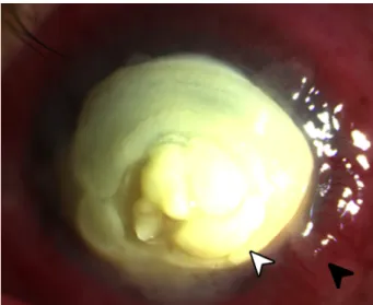
Figure 1: Slit lamp photograph of eye showing a large yellowish plaque (white arrowhead) occupying almost the entire cornea
with surrounding scleritis (black arrowhead).
On presentation, the visual acuity of the left eye was hand movement with poor light projection. There was good color perception. A reverse relative afferent pupillary de- fect was not noted. On slit lamp examination, there was a very thick yellow plaque consisting of a rounded mass projecting above the entire corneal surface, similar to a mound, measuring 9x9.5 mm with a depth of 2 mm at its thickest point located centrally (Figure 1). Despite the
1/3 GMS Ophthalmology Cases 2020, Vol. 10, ISSN 2193-1496
Case Report
OPEN ACCESS
large plaque, there seemed to be no signs of prolapse of intraocular contents. There was associated scleritis. The ocular ultrasound showed only few vitreous cellularities.
The right was grossly unremarkable with a visual acuity of 20/20.
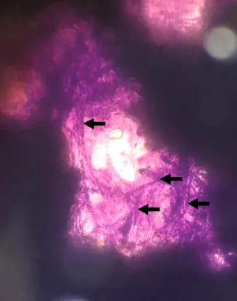
The plaque did not stain with fluorescein, lissamine green, and rose bengal dyes. Careful scraping of the mass re- vealed it to be semi-solid, albeit very friable in consis- tency. There was no bleeding on scraping. Giemsa stain of the corneal scraping showed numerous hyphal ele- ments (Figure 2). Culture of the plaque later revealed to be positive forAspergillussp.
Figure 2: Giemsa stain of scrapings obtained from the corneal plaque showing an extensive amount of hyphal elements (black
arrows).
The patient was admitted and initiated on topical natamy- cin drops, and topical moxifloxacin drops were added as prophylaxis for any superimposed bacterial infection.
Topical atropine drops were also started for relief of ciliary pain. The patient was started on oral itraconazole as well.
A therapeutic keratoplasty with a tectonic corneal graft was performed under retrobulbar anesthesia. The recipi- ent bed was sized by measuring the fungal plaque and adding a 2 mm border. The corneal graft was sized to a 12 mm diameter, necessitating manual cutting using corneal scissors. After the host cornea was excised, it was revealed that the fungus had invaded the anterior chamber, and the iris had become necrotic. A 360-degree iridectomy was performed. Lens extraction and anterior vitrectomy were done. After reforming the anterior chamber with an ophthalmic viscoelastic device, the tectonic corneal graft was placed and sutured with
16 interrupted nylon 10-0 sutures (Figure 3). The surgery was uncomplicated.
Figure 3: Slit lamp photograph of eye at the first day postoperatively showing a tectonic graft held in place with #16
interrupted nylon 10-0 sutures.
The patient was maintained on a regimen of topical natamyin, topical moxifloxacin, and topical atropine drops for the first 2 weeks after surgery. He also completed a 2-week course of oral itraconazole. The postoperative course was relatively unremarkable with decreasing signs of inflammation and no recurrence of infection. At the 2ndweek postoperatively, topical prednisolone drops were added. The patient’s vision improved to counting fingers at 6 inches. He was advised that a second procedure consisting of an optical penetrating keratoplasty and secondary intraocular lens implantation as well as pos- sible interventions for secondary glaucoma would be needed in the future.
Discussion
Fungal keratitis is a fungal infection of the cornea. It is often considered a disease associated with rural farming areas, since it is traditionally associated with a history of trauma with vegetative material. However, contact lens use seems to now be the major risk factor in developed countries [2]. It has been described to occur from infections from both filamentous (Aspergillus sp. and Fusariumsp.) and yeast (Candidasp.) fungi [2].
It remains a challenge for ophthalmologists since initial presentation may mimic bacterial keratitis. Antimicrobial agents used in bacterial keratitis are not effective for fungal keratitis, thus continued fungal growth occurs be- fore the ophthalmologist recognizes failure of response.
Some cases may be misdiagnosed as immunologic reac- tions and treated with corticosteroids, which decrease immune response, thus promoting fungal growth. In ad- dition, since the disease is traditionally associated with farmers living in rural communities, significant delays in diagnosis often occur before appropriate treatment [3].
2/3 GMS Ophthalmology Cases 2020, Vol. 10, ISSN 2193-1496
Bromeo et al.: Large graft tectonic penetrating keratoplasty in a ...
The case presented showed an unusual growth pattern ofAspergilluskeratitis wherein the fungal plaque became an exuberant mound-like structure. Plaques associated with fungal keratitis are usually planar, and often still follow the contour of the host cornea [2]. Factors associ- ated with the unusual growth pattern presented in the cases include a significant delay before initiation of ap- propriate treatment (2 weeks before diagnosis and an additional 6 weeks of inappropriate antimicrobial treat- ment) and initiation of topical medication containing a corticosteroid component. It is also unusual that despite the exaggerated exophytic growth of the infection, the fungi were only able to invade up to the anterior chamber.
Indeed, the patient was fortunate since invasion of the posterior segment will portend a significantly poorer pro- gnosis [1].
A penetrating keratoplasty was done to decrease fungal load for greater efficacy of topical antifungal medications, as well to prevent further growth of the fungi into the globe. In this procedure, the diseased portion of the cornea is excised and replaced with a donor corneal graft.
The procedure has been shown to be effective in cases of fungal keratitis [4], [5]. The major risk of performing keratoplasty in cases of fungal keratitis is recurrence of the fungal infection. Thus, a regimen consisting of topical antifungal drops with topical antimicrobrial drops to cover for secondary bacterial infection is continued for a pro- longed period of time following successful keratoplasty.
Natamycin is frequently used as initial therapy due to its availability. Voriconazole and amphotericin B are also emerging as promising first-line therapy in select cases [2], [5]. In addition, topical corticosteroids are withheld for at least 2 weeks in the early postoperative period [3].
A combination of expedient penetrating keratoplasty and initiation of appropriate culture-guided antifungal medi- cations was able to salvage this patient’s eye, and recov- ery of useful vision in the future will be possible.
Conclusion
Fungal keratitis is a fungal infection of the cornea caused by both filamentous and yeast fungi. Risk factors include trauma with vegetative material and contact lens use. It is associated with ulcers with feathery borders, ring infil- trates, plaque formation, and penetration into the anterior chamber. The cornerstone of management of fungal keratitis is initiation of culture-guided antifungal therapy.
Penetrating keratoplasty may be needed in severe cases.
Corticosteroids should never be given in cases of active fungal keratitis.
Notes
Competing interests
The authors declare that they have no competing in- terests.
Ethical statement
The case report is a minimal-risk study which was con- ducted in full compliance with the principles of the Decla- ration of Helsinki and Good Clinical Practice. All identifying patient information was kept confidential. Informed con- sent was obtained from the family of the patient.
References
1. Wong TY, Ng TP, Fong KS, Tan DT. Risk factors and clinical outcomes between fungal and bacterial keratitis: a comparative study. CLAO J. 1997 Oct;23(4):275-81.
2. Tuli S. Fungal keratitis. Clin Ophthalmol. 2011;5:275-9. DOI:
10.2147/OPTH.S10819
3. Lim Bon Siong R, Cua I. External Disease and Cornea Handbook.
Manila: St. Luke’s Medical Center; 2016.
4. Xie L, Zhai H, Shi W. Penetrating keratoplasty for corneal perforations in fungal keratitis. Cornea. 2007 Feb;26(2):158-62.
DOI: 10.1097/01.ico.0000248381.24519.0d
5. FlorCruz NV, Peczon IV, Evans JR. Medical interventions for fungal keratitis. Cochrane Database Syst Rev. 2012 Feb
15;(2):CD004241. DOI: 10.1002/14651858.CD004241.pub3
Corresponding author:
Albert John Bromeo
Department of Ophthalmology and Visual Sciences, Sentro Oftalmologico Jose Rizal, Philippine General Hospital, University of the Philippines, 1000 Taft Avenue, Manila, Philippines
albert.bromeo@gmail.com
Please cite as
Bromeo AJ, Aquino-Alegre ER, Lim Bon Siong R. Large graft tectonic penetrating keratoplasty in a case of severe aspergillus keratitis. GMS Ophthalmol Cases. 2020;10:Doc17.
DOI: 10.3205/oc000144, URN: urn:nbn:de:0183-oc0001440
This article is freely available from
https://www.egms.de/en/journals/oc/2020-10/oc000144.shtml Published:2020-04-02
Copyright
©2020 Bromeo et al. This is an Open Access article distributed under the terms of the Creative Commons Attribution 4.0 License. See license information at http://creativecommons.org/licenses/by/4.0/.
3/3 GMS Ophthalmology Cases 2020, Vol. 10, ISSN 2193-1496
Bromeo et al.: Large graft tectonic penetrating keratoplasty in a ...