1. INTRODUCTION
The brown shrimp Crangon crangon (Linnaeus, 1758) is one of the most ecologically and economi- cally important invertebrate species along the North Atlantic coast of Europe. It forms a key component in the marine food web and is commercially har- vested throughout most of its distribution range, with a yield of more than 35 000 t yr−1(Revill & Holst 2004, Beu kema & Dekker 2005, ICES 2015). Several ex - ploited crustaceans, such as the brown shrimp, the
edible crab Cancer pagurus, or the American lobster Homarus americanus, can be affected by shell dis- ease (Smolowitz et al. 2005, Shields 2013), with significant commercial implications. For example, for the US fishing industries, shell disease in the Ameri- can lobster has caused economic losses reaching millions of dollars, since it directly renders affected individuals unmarketable as a live product and in - directly affects reproduction and recruitment (Ayres
& Edwards 1982, Getchell 1991, Wahle et al. 2009, Lafferty et al. 2015).
© Inter-Research 2020 · www.int-res.com
*Corresponding author:
alexandra.segelken-voigt@uni-oldenburg.de
Shell disease does not affect biochemical profiles of the North Sea brown shrimp Crangon crangon
Alexandra Segelken-Voigt
1, 6,*, Wilhelm Hagen
2, Gabriele Gerlach
1, 3, 4, Julia Strahl
1, 3, 51Institute of Biology and Environmental Sciences, Carl von Ossietzky University Oldenburg, Carl von Ossietzky Str. 9−11, 26111 Oldenburg, Germany
2BreMarE − Bremen Marine Ecology, Marine Zoology, University of Bremen, PO Box 330440, 28334 Bremen, Germany
3Helmholtz-Institute for Functional Marine Biodiversity at the University of Oldenburg (HIFMB), Ammerländer Heerstrasse 231, 26129 Oldenburg, Germany
4Centre of Excellence for Coral Reef Studies and School of Marine and Tropical Biology, James Cook University, 4811 Queensland, Australia
5Alfred Wegener Institute Helmholtz Centre for Polar and Marine Research, Am Handelshafen 12, 27570 Bremerhaven, Germany
6Present address: Institute of Fisheries, State Research Center of Agriculture and Fisheries Mecklenburg-Vorpommern, 18375 Born, Germany
ABSTRACT: The brown shrimp Crangon crangonis a key component of the North Atlantic coastal food web and an important target species for the fishery economy. As the brown shrimp contains large amounts of protein and essential fatty acids, its consumption makes it a beneficial choice for humans. Commercially harvested crustaceans like C. crangonare frequently affected by bacterial shell disease, with necrotizing erosions and ulcerations of the cuticle. To determine whether shell disease influences the nutritional value of C. crangon, total protein and lipid contents, as well as fatty acid compositions of muscle tissue and hepatopancreas, together with the hepatosomatic index, were examined in healthy and affected individuals. The biochemical composition of the tis- sues did not differ significantly between the 2 groups. Also, the hepatosomatic index, as an indi- cator of energy reserves in shrimps, was similar between healthy and affected animals. Our results indicate that the nutritional value of C. crangonis not affected by shell disease, as long as it remains superficial as in the present study.
KEY WORDS: Black spot disease · Lipids · Fatty acids · Omega-3 · EPA · DHA · Hepatosomatic index
Resale or republication not permitted without written consent of the publisher
The term ‘shell disease’ includes both enzootic and epizootic shell disease and describes different forms of necrotic lesions and disintegrations of the cuticle of crustaceans resulting from distinct etiologies and causative agents (Dyrynda 1998, Vogan & Rowley 2002, Shields 2013). Enzootic or classical shell dis- ease induces irregularly shaped lesions of the cuticle that are commonly observed in lobsters, crabs, and shrimps (Smolowitz et al. 1992, Vogan et al. 1999). In contrast, the more aggressive epizootic shell disease progresses inwards through the carapace and occurs mainly in the American lobster (Castro & Angell 2000, Chistoserdov et al. 2005, Smolowitz et al. 2005).
In both disease forms, the characteristic black ulcer- ations may be caused by a pathogenic shift in the bacterial shell community. In this context, a multi- tude of different chitinolytic and lipolytic bacteria strains have been observed, including species in genera such as Aquimarina, Pseudomonas, and Vib- rio (Rosen 1967, Meres et al. 2012, Feinman et al.
2017).
Some of these chitinolytic and lipolytic bacteria are also present on the cuticle of healthy crus- taceans (Meres et al. 2012, Kraemer et al. 2020).
Before the symbiosis becomes pathogenic, and chitin degradation by bacteria can occur, a portal of entry is required in the form of destruction of the outermost non-chitinous epicuticle by mechanical (Vogan et al. 1999) or chemical abrasion (Schlotfeldt 1972), or predatory or cannibalistic attacks (Dyryn - da 1998). In addition, toxins, pollution, and higher temperatures may further accelerate bacterial growth and reduce the resistance of the host (Young & Pearce 1975, Glenn & Pugh 2006, Laufer et al. 2012).
Superficial early stages of shell disease are usually not fatal, and the affected individual may overcome the condition by molting (Smolowitz et al. 1992, Bar- ris et al. 2018, Segelken-Voigt et al. 2018). Neverthe- less, shell disease can also cause death, either directly through invading pathogens into the under- lying soft tissues, leading to internal infections, or indirectly, through impaired feeding function or loco- motion, due to lesions on the appendages (Smolowitz et al. 1992, Vogan et al. 2001). Additionally, shell disease can interfere with immunocompetence (Chen et al. 1995, Tlusty et al. 2007) and thus reduce the survival chance of affected crustaceans (Smolowitz et al. 2005, Hoenig et al. 2017, Segelken-Voigt et al.
2018). Likewise, affected crustaceans may be ener- getically compromised or produce elevated levels of ecdysone due to altered gene expressions and hormone levels, resulting in changes in the molting
behavior and cellular stress response (Laufer et al.
2005, Castro et al. 2006, Tarrant et al. 2012).
In marine invertebrates, proteins and lipids play an important role as the main energy reserves or as cell membrane components (Kattner et al. 1994, García et al. 2002, Lee et al. 2006, Jimen´ ez & Kinsey 2015).
During stressful environmental conditions (e.g. food scarcity, heavy metal or pathogen exposure), energy reserves can be mobilized to maintain physiological functions and increase the resilience of an organism (Floreto et al. 2000, Verslycke et al. 2003, Sacristán et al. 2017). In decapods, lipids are primarily stored in the hepatopancreas (midgut gland), which synthe- sizes enzymes and lipids (Chang & O’Connor 1983, Diaz et al. 2010, Martínez-Alarcón et al. 2019). Phys- iological impairments and diseases directly affect this organ (Johnson 1980), and a low hepatosomatic index (HSI) could indicate poor health or reduced food intake. For example, H. americanusaffected by shell disease have significantly lower lipid levels, including omega-3 fatty acids, in their hepatopan- creas compared to healthy specimens, as well as a lower HSI, indicating a reduced lipid-storage capac- ity (Floreto et al. 2000).
Our study organism, C. crangon, contributes im - portant nutritional components to the human diet. It contains high amounts of essential omega-3 fatty acids such as eicosapentaenoic acid (EPA, 20:5(n-3)) and docosahexaenoic acid (DHA, 22:6(n-3)), which can improve human health status and reduce the risk of cancer, cardiovascular diseases, and diabetes (Heu et al. 2003, Harper & Jacobson 2005, Vaughan et al. 2013, Calder 2017). The biochemical composition (e.g. contents of proteins and lipids) of shrimp and other edible crustaceans may reflect their physiolog- ical state of health and, thus, their nutritional value for human consumption. To investigate a possible relationship between shell disease and the nutri- tional value of C. crangon, total protein and lipid con- tents and the fatty acid composition were analyzed in 2 tissue types of healthy and affected brown shrimp.
The hepatopancreas served as an indicator of the metabolic state of the shrimp, whereas muscle tissue reflected the nutritional value of the animal.
2. MATERIALS AND METHODS 2.1. Sampling and preparation of
Crangon crangon
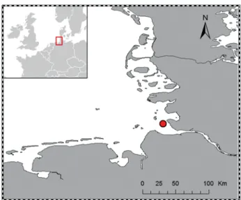
In July 2017, C. crangonspecimens were collected at depths of 8−10 m by a beam trawl of RV ‘Uthörn’ in
the Weser estuary in the German Bight, North Sea (53° 38.90' N, 8° 25.19' E) (Fig. 1). After sampling, the shrimp were immediately weighed (fresh mass) and measured (total length from end of the rostrum to end of the tail). In total, 41 adult males and non-egg-bear- ing females were analyzed for signs of shell disease (see Fig. 2). The affected individuals were photo - graphed with a Nikon P7000, and the size of the black ulcerations on the shell was measured using ImageJ software (http://rsb.info.nih.gov/ij/) (Table 1). To cal- culate the relative size of black ulcerations in per- centage coverage, the size of the necrotic lesions was normalized to the body length of the individual. In total, 18 healthy (0% shell-disease coverage) and 23 affected shrimp (> 5% shell-disease coverage) were used for subsequent biochemical analyses. The indi- viduals were snap-frozen in liquid nitrogen after measurement and dissected at a later stage for tissue sampling. The hepatopancreas and a piece of the muscle from the second abdominal segment were stored at −80°C until further biochemical analyses. As an indicator of the energetic status of the shrimp, the hepatopancreas was weighed to determine the HSI (% of fresh mass). This value was computed as HSI % = HW / BW × 100, where HW is the wet mass of the hepatopancreas (mg), and BW is the wet mass of the brown shrimp (mg). All animal work was con- ducted according to the German animal protection law (approval no. 33.19-42502-05-15A542).
2.2. Total protein content
The total protein content was determined follow- ing the protocol of Lowry et al. (1951) and using the DCTMProtein Assay Kit (BIO RAD Laboratories) with bovine serum albumin (BSA) standard. The standard curve consisted of 5 dilution standards of known pro- tein concentration (0, 0.19, 0.38, 0.76, 1.52 mg ml−1) mixed in 1 M NaOH (sodium hydroxide). Tissue sam- ples of muscle and hepatopancreas were diluted 1:200 (m:v) in 1 M NaOH. Small glass beads (∅
0.1 mm) were added to the samples. Standards and samples were homogenized by vortexing (Vortex Genie 2, Scientific Industry) for 30 s and incubated on a rotating heating block (Thermomixer comfort, Eppendorf) for 20 min at 50°C and 200 × g. These steps were repeated 3 times. Samples were centrifuged for 10 min at room temperature and 1500 × g. Aliquots of 5 μl of the supernatants and the protein standards, respectively, were transferred (in duplicate) into individual wells of a 96-well microtiter plate. Ali - quots of 25 μl alkaline copper tartrate solution (Bio-
Rad DC Reagent A) and 200 μl Folin reagent (Bio- Rad DC Reagent B) were added to each sample and standard. After 15 min of incubation in the dark, the samples were measured spectrophotometrically in a microplate reader (Infinite 200 Pro, Tecan) at a wave- length of 750 nm. Total protein content was calcu- lated based on the standard curve and expressed in
% of dry mass (% DM).
2.3. Total lipid content
The total lipid content was determined following the methods of Folch et al. (1957) and Hagen et al.
(2000). Muscle and hepatopancreas samples were lyophilized for 48 h and transferred to an evacuated desiccator for 30 min. The pulverized tissue samples were weighed and homogenized with Potter ho - mogenization and ultrasonic disruption, and then total lipids were ex tracted with dichloromethane/
metha nol (DCM/MeOH, 2:1 per volume). This proce- dure was repeated, and each time the supernatant was transferred into a clean test tube. After adding 2 ml KCl (potassium chloride) solution (0.88%), the samples were centrifuged at 1495 × g(10 min at 2°C).
After phase separation, the lipid phase was pipetted into a clean glass vial and the solvents evaporated under a stream of nitrogen. The pure, extracted lipids were weighed and the total lipid contents of muscle and of hepatopancreas were calculated in % DM. For subsequent fatty acid analyses, the extracted lipids were redissolved in DCM/MeOH and stored in nitrogen at −80°C.
Fig. 1. Sampling location of brown shrimp Crangon crangon in the Weser estuary in the German Bight, North Sea
(53° 38.90' N, 8° 25.19' E)
2.4. Fatty acid composition
A subsample of the redissolved lipid samples was hydrolyzed to convert the fatty acids to methylester derivatives (FAMEs). This trans-esterification was performed by heating the samples with 3% H2SO4
(sulfuric acid) in methanol and 250 μl hexane for 4 h at 80°C (Kattner & Fricke 1986). FAMEs were dis- solved in 1 ml hexane and their composition ana- lyzed in a gas chromatograph (HP 6890; Agilent Technologies) equipped with a DB-FFAP column (30 m length, 0.25 mm inner diameter, 0.25 μm film
thickness) operating with an auto- mated temperature vaporizer injector and with helium as carrier gas. Fatty acid peaks were identified by compar- ing their retention times with 2 stan- dards of known composition (copepod lipids and menhaden fish oil). The quantities of the individual fatty acids were ex pressed as mass percentage of total fatty acids (% TFA).
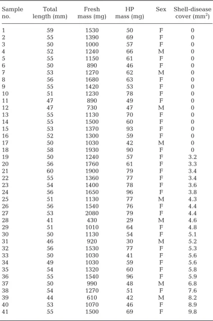
2.5. Statistical analysis Before running the statistical tests in SPSS 24, the data were checked for normality and homogeneity of vari- ances. Initially, we separated the shrimp into 3 groups to examine po - tential differences between healthy (0% shell-disease coverage), moder- ately (5−10% shell-disease coverage, Fig. 2A,B), and severely affected shrimp (>10% shell-disease coverage, Fig. 2C−F).
The biochemical profiles (total lipid and protein levels plus fatty acid com- positions) in these 3 groups, as well as separately in females and males, were then visualized by non-metric multidi- mensional scaling (nMDS) based on Bray-Curtis similarity, after a square- root-transformation of the data (Krus - kal & Wish 1978) using PRIMER 6.1.6 (Clarke & Gorley 2006). Additionally, a permutational multivariate analysis of variance (PERMANOVA, Anderson 2001) was performed to test for dif - ferences in biochemical composition (total amount of lipids and proteins as well as fatty acid composition) of mus- cle tissue and hepatopancreas with the factors ‘shell- disease coverage’ (fixed at 3 levels = healthy, moder- ately, and severely affected) and ‘sex’ (fixed at 2 levels = female and male) based on the Bray-Curtis similarity matrix of square-root-transformed data.
The analysis was run using the permutation of resid- uals under a reduced model, with 9999 permutations and type III sums of squares.
As no differences were observed between moder- ately and severely affected shrimp, we pooled these individuals into 1 group (see also Fig. 2). To test for significant differences in the biochemical parameters
Sample Total Fresh HP Sex Shell-disease
no. length (mm) mass (mg) mass (mg) cover (mm2)
1 59 1530 50 F 0
2 55 1390 69 F 0
3 50 1000 57 F 0
4 52 1240 66 M 0
5 55 1150 61 F 0
6 50 890 46 F 0
7 53 1270 62 M 0
8 56 1680 63 F 0
9 55 1420 53 F 0
10 51 1230 78 F 0
11 47 890 49 F 0
12 47 730 47 M 0
13 55 1130 70 F 0
14 55 1500 60 F 0
15 53 1370 93 F 0
16 52 1300 59 F 0
17 50 1030 42 M 0
18 58 1930 90 F 0
19 50 1240 57 F 3.2
20 56 1760 61 F 3.3
21 60 1900 79 F 3.4
22 55 1360 77 F 3.4
23 54 1400 78 F 3.6
24 56 1650 96 F 3.8
25 51 1130 77 M 4.3
26 56 1540 76 F 4.4
27 53 2080 79 F 4.4
28 41 430 29 M 4.6
29 51 1010 64 F 4.8
30 50 1130 54 F 5.1
31 46 920 30 M 5.2
32 56 1530 77 F 5.3
33 50 1030 41 F 5.6
34 49 1030 59 F 5.6
35 54 1320 60 F 5.8
36 55 1540 96 F 5.9
37 50 990 48 M 6.8
38 54 1270 51 F 7.6
39 44 610 42 M 8.2
40 53 1070 46 F 8.9
41 55 1500 69 F 9.8
Table 1. List of Crangon crangonspecimens sampled for biochemical analyses with total length, fresh masses of the individual and the hepatopancreas (HP),
sex (F: female; M: male), and shell-disease coverage
and the HSI between healthy and shell-disease af - fected animals, a t-test or a 1-way ANOVA was ap - plied (depending on the number of groups). The sig- nificance level was set to α= 0.05.
3. RESULTS
There were no apparent differences in the bio- chemical profiles of the hepatopancreas and muscle of healthy vs. affected shrimp. The nMDS plot (Fig. 3) displaying the composition of fatty acids as well as the amounts of lipids and proteins in the mus- cle (Fig. 3A) and in hepatopancreas (Fig. 3B) re - vealed no clustering for healthy, moderately, and severely affected brown shrimp. In the muscle tissue and hepatopancreas (Table 2), the PERMANOVA showed neither specific differences between the 3 groups, nor between sexes. In the following, all indi- viduals were therefore classified as either healthy or shell-disease affected.
Fresh mass, total length, and HSI did not differ significantly between healthy and shell-disease affected brown shrimp. Mean ± SD fresh mass and total length of Crangon crangonamounted to 1260 ± 300 mg and 53 ± 4 mm in healthy individuals and
Source df Pseudo-F p (perm)
Muscle
Shell disease 2 0.8268 0.577
Sex 1 1.9516 0.102
Shell disease × sex 2 0.7159 0.701
Residuals 32
Total 37
Hepatopancreas
Shell disease 2 0.4912 0.7849
Sex 1 0.6097 0.5567
Shell disease × sex 1 0.9694 0.3369
Residuals 17
Total 21
Table 2. Results of PERMANOVA testing for differences in biochemical composition of muscle and hepatopancreas of Crangon crangonbetween the 3 health categories (healthy, moderate, and severely shell-disease affected), and be- tween the 2 sexes (female and male); p (perm) refers to the
significance level assessed by a permutation test Fig. 3. Non-metric multidimensional scale ordination of Bray-Curtis similarity from square-root transformed data of biochemical compositions of (A) muscle and (B) hepatopan- creas of Crangon crangonbetween the 3 health categories.
Open circles represent healthy shrimp, black squares repre- sent severely affected shrimp, and black triangles represent moderately affected shrimp. Individuals are further differ-
entiated by sex (F: female; M: male) Fig. 2. Shell disease in Crangon crangonwith varying lesion
sizes: (A) 3.6, (B) 4.8, (C) 5.8, (D) 6.8, (E) 8.9, (F) 9.8 mm2. Scale bar = 2.5 mm
1280 ± 400 mg and 52 ± 5 mm in the affected animals.
The mean hepatopancreas mass was 61 ± 18 mg in healthy and 62 ± 22 mg in affected shrimp. This resulted in an HSI of 5.0 ± 1.0% in healthy and 4.9 ± 1.0% in affected shrimp (t= −0.060, p = 0.953).
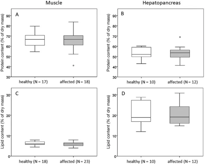
Mean total protein and lipid contents in muscle and in hepatopancreas tissue did not differ significantly between healthy and affected C. crangon. The total protein content in the muscle accounted for 65.9 ± 6.8% DM in the healthy and 65.3 ± 9.7% DM in the affected brown shrimp (t = −0.245, p = 0.808, Fig. 4A). In the hepatopancreas, the total protein content averaged 52.6 ± 11.3% DM in the healthy and 53.4 ± 7.1% DM in the affected shrimp (t = 0.188, p = 0.853, Fig. 4B). The total lipid content of the mus- cle was 6.3 ± 0.9% DM in healthy and 5.8 ± 1.0% DM in affected C. crangon (t= −1.486, p = 0.146, Fig. 4C).
The hepatopancreas had a mean total lipid content of
20.9 ± 6.1% DM in healthy animals, and 20.5 ± 5.2%
DM in affected shrimp (t= −0.158, p = 0.876, Fig. 4D).
In total, 16 fatty acids were identified, each ex - ceeding 1% of TFAs (Table 3). Major fatty acids in both tissue types of healthy and affected animals were palmitic acid (16:0), oleic acid (18:1(n-9)), eico - sapentaenoic acid (EPA, 20:5(n-3)), and docosa- hexaenoic acid (DHA, 22:6(n-3)). Fatty acid composi- tions of the muscle and of the hepatopancreas did not differ significantly between the 2 C. crangongroups.
In the muscle, all omega-3 fatty acids (Σn-3) ac - counted for 46.2 ± 1.7% of TFAs in the healthy and 46.1 ± 2.6% of TFAs in the affected shrimp (t = 0.108, p = 0.914). The omega-3 fatty acids in the hepatopan- creas amounted to 43.1 ± 5.8% in healthy and 43.5 ± 4.5% in affected individuals (t= −0.188, p = 0.853).
The compositions of saturated (SFAs), monounsat- urated (MUFAs), and polyunsaturated fatty acids
Fig. 4. Total protein contents in (A) muscle tissue and (B) hepatopancreas, and total lipid contents in (C) muscle and (D) hepatopancreas of healthy and shell-disease affected Crangon crangon. The center line in the box plot represents the median
with upper and lower quartiles, whiskers indicate minima and maxima, and open circles are outliers
(PUFAs) between affected and healthy shrimp dif- fered neither in the muscle (SFAs: t = 0.240, p = 0.811; MUFAs: t= −0.308, p = 0.760; PUFAs: t= 0.020, p = 0.984) nor in the hepatopancreas (SFAs: t= 0.221, p = 0.828; MUFAs: t= −0.047, p = 0.963; PUFAs: t=
−0.090, p = 0.930) (Table 3).
4. DISCUSSION
In this study, we examined the biochemical profiles of healthy and shell-disease affected Crangon cran- gonto assess possible effects of the classical shell dis- ease on the nutritional composition of this food prod- uct. We detected no differences in total protein or lipid contents, or in the fatty acid compositions, be - tween healthy and affected brown shrimp, pointing to a similar nutritional value of both groups. The muscle tissue of all C. crangon was rich in PUFAs, especially in the omega-3 fatty acids EPA and DHA.
These are the most common fatty acids found in seafood and have been associated with many health benefits for humans (Harper & Jacobson 2005, Vaughan et al. 2013, Calder 2017). The amounts of these dominant fatty acids are in accordance with previous findings for C. crangonand other de capods, such as the whiteleg shrimp Litopenaeus vanna -
mei and the giant freshwater prawn Macrobrachium ro senbergii (Bragag- nolo & Rodriguez-Amaya 2001, Perez- Velaz quez et al. 2003, Martínez-Alar- cón et al. 2019). Mika et al. (2014) and Turan et al. (2011) also reported simi- lar compositions, with principal fatty acids such as 16:0, 18:1(n-9), 20:5(n-3), and 22:6(n-3) in C. crangon from the White Sea and Black Sea.
In contrast to our findings, muscle tissues of the American lobster Ho - marus americanus affected by shell disease revealed slightly higher ratios of 20:4(n-6) and 22:6(n-3) compared to healthy lobsters (Floreto et al. 2000). In addition, a lower HIS, as well as signi - ficantly lower lipid levels, were meas- ured in the hepatopancreas of affected H. americanus.The authors concluded that lobsters affected by shell disease have either a reduced capability to store lipids, or exhibit a higher ener- getic requirement compared to heal - thy individuals. As in our study, the constitution and biochemistry of the lobsters may not be affected by large, superficial black ulcers, but rather by lesions that have already penetrated the cuticle into the soft tissue. In our study, in spite of shell disease, affected C. crangondo not use more of their energy stores when compared to healthy shrimp, pointing towards a stable physio- logical state in terms of biochemical composition.
Hence, our results suggest that in C. crangon,classi- cal shell disease predominantly generates superficial symptoms, as indicated by the black necrotic lesions on the cuticle. Other studies suggest that all layers of the cuticle, as well as the soft underlying tissue, of brown shrimp may be affected by shell disease (Schlotfeldt 1972, Segelken-Voigt et al. 2018).
In our study, no signs of necrotic lesions were ob - served on the epidermis of C. crangon specimens, thus the lesions were apparently not severe enough to penetrate the different shell layers and to impair the vitality of C. crangon. It has previously been stated for C. crangon that necrotic lesions remain superficial on the epicuticle or develop at the end of a molting cycle and that brown shrimp can diminish or even eliminate black spots via ecdy sis (Smolowitz et al. 1992, Segelken-Voigt et al. 2018).
Our data and literature studies suggest that the high molting frequencies in C. crangon minimize the progression of the disease symptoms into
FA Muscle Hepatopancreas
Healthy Affected Healthy Affected
(N = 15) (N = 23) (N = 10) (N = 12)
14:0 1.0 ± 0.1 1.1 ± 0.2 1.8 ± 0.7 1.8 ± 0.6
16:0 20.5 ± 0.9 20.2 ± 1.4 15.2 ± 1.9 15.0 ± 1.4 16:1(n-7) 4.2 ± 0.8 4.0 ± 0.7 5.7 ± 2.0 5.5 ± 1.5 16:3(n-4) 0.6 ± 0.2 0.7 ± 0.2 0.9 ± 0.3 0.9 ± 0.2
17:0 1.0 ± 0.1 1.0 ± 0.1 0.7 ± 0.2 0.8 ± 0.1
18:0 4.1 ± 0.7 4.0 ± 0.6 3.6 ± 0.7 3.5 ± 0.6
18:1(n-9) 9.3 ± 0.8 9.3 ± 1.2 10.0 ± 2.1 10.1 ± 1.8 18:1(n-7) 5.0 ± 0.5 5.1 ± 0.6 5.6 ± 0.6 5.6 ± 0.9 18:2(n-6) 1.2 ± 0.1 1.2 ± 0.2 1.2 ± 0.3 1.2 ± 0.2 20:1(n-11) 0.3 ± 0.1 0.2 ± 0.1 0.7 ± 0.5 0.9 ± 0.4 20:1(n-9) 0.3 ± 0.1 0.3 ± 0.3 1.0 ± 0.4 0.8 ± 0.3 20:1(n-7) 0.3 ± 0.1 0.4 ± 0.2 1.1 ± 0.6 1.2 ± 0.3 20:4(n-6) 3.6 ± 0.4 3.6 ± 0.4 3.8 ± 0.7 3.7 ± 0.8 20:5(n-3) 22.6 ± 1.0 23.0 ± 1.3 21.1 ± 3.1 21.3 ± 2.2 22:5(n-3) 1.2 ± 0.2 1.3 ± 0.3 1.6 ± 0.7 2.1 ± 0.7 22:6(n-3) 21.6 ± 2.0 21.2 ± 2.1 18.3 ± 4.6 18.4 ± 3.5 ΣSFA 28.1 ± 1.5 27.9 ± 2.1 23.6 ± 2.8 23.4 ± 2.4 ΣMUFA 19.7 ± 1.5 19.9 ± 1.8 25.1 ± 3.7 25.2 ± 2.9 ΣPUFA 52.1 ± 1.6 52.1 ± 2.7 50.9 ± 5.7 51.1 ± 4.7 Σn-3 46.2 ± 1.7 46.1 ± 2.6 43.1 ± 5.8 43.5 ± 4.5 Table 3. Fatty acid (FA) compositions (mass % of total FAs greater than 1%) of muscle tissue and of hepatopancreas of healthy and shell-disease affected Crangon crangon. Values represent means ± SD. SFA: saturated FA; MUFA:
monounsaturated FA; PUFA: polyunsaturated FA; n-3: omega-3 FA
deeper layers of the cuticle, internal organs, and tissues. Brown shrimp have a fast molting rate of approximately 3 wk (Hufnagl & Temming 2011) and a short intermolt period. Hence, these shrimp have little time to develop shell- disease-induced lesions compared to adult American lobsters that have a molting rate of approximately once per year (Hughes & Matthiessen 1962).
Despite the fact that brown shrimp can heal itself of the black necrotic lesions by molting, we cannot ex - clude an effect of shell disease on biochemical and physiological processes in C. crangon.For example, an infestation with black spots can reduce the respi- ratory performance of affected shrimp (Segelken- Voigt et al. 2018), and, in the worst case, the bacteria may cause a sepsis, possibly leading to death (Smolo - witz et al. 1992, Vogan et al. 1999, Vogan & Rowley 2002). Consequently, we cannot exclude the pos - sibility that the microbiological quality of shrimp as a consumer product will be affected by shell dis- ease, while the nutritional value remains at a high level.
Shell disease can also cause significant alterations in the immune system of crustaceans, by a reduction of serum protein, via low concentrations of circulat- ing hemocytes in the hemocoel, by destruction of blood cells through phagocytosis (Prince 1997), or by an enhancement of hemocytes in the hemo - lymph by increased immune activity in affected indi - viduals (White & Ratcliffe 1982, Vogan & Rowley 2002). Therefore, future studies should address the immune-system responses of affected brown shrimp and examine the biochemical composition of C. cran- gonbearing lesions that have already penetrated the epidermis.
5. CONCLUSIONS
Our findings show that as long as the black ulcers are superficial, shell disease in Crangon crangon does not affect its energy stores or bio- chemical profiles. No significant alterations in the biochemical composition or the HSI were detected in shrimp affected by shell disease compared to healthy specimens. Total protein and lipid con- tents, as well as fatty acid compositions in muscle and hepatopancreas, remained similar between healthy and affected shrimp. We thus conclude that the nutritional value of superficially affected brown shrimp is maintained, including the levels of essential omega-3 fatty acids such as EPA and DHA.
Acknowledgements. Special thanks to Sabrina Dorschner for technical assistance during biochemical analyses in the laboratory. We also thank the captain and crew of RV
‘Uthörn’ for their support during sampling of the brown shrimp. HIFMB is a collaboration between the Alfred Wegener Institute Helmholtz-Center for Polar and Marine Research and the Carl-von-Ossietzky University Olden- burg, initially funded by the Ministry for Science and Cul- ture of Lower Saxony and the Volkswagen Foundation through the ‘Niedersächsisches Vorab’ grant program (grant number ZN3285). English-language services were provided by stels-ol.de.
LITERATURE CITED
Anderson MJ (2001) A new method for non-parametric mul- tivariate analysis of variance. Austral Ecol 26: 32−46 Ayres P, Edwards E (1982) Notes on the distribution of ‘black
spot’ shell disease in crustacean fisheries. Chem Ecol 1:
125−130
Barris BN, Shields JD, Small HJ, Huchin-Mian JP and others (2018) Laboratory studies on the effect of temperature on epizootic shell disease in the American lobster Homarus americanus.Bull Mar Sci 94: 887−902
Beukema JJ, Dekker R (2005) Decline of recruitment suc- cess in cockles and other bivalves in the Wadden Sea:
possible role of climate change, predation on postlarvae and fisheries. Mar Ecol Prog Ser 287: 149−167
Bragagnolo N, Rodriguez-Amaya D (2001) Total lipid, cho- lesterol, and fatty acids of farmed freshwater prawn (Macrobrachium rosenbergii) and wild marine shrimp (Penaeus brasiliensis, Penaeus schmitti, Xiphopenaeus kroyeri). J Food Compos Anal 14: 359−369
Calder PC (2017) New evidence that omega-3 fatty acids have a role in primary prevention of coronary heart dis- ease. J Public Health Emerg 1: 35
Castro K, Angell T (2000) Prevalence and progression of shell disease in American lobster, Homarus americanus, from Rhode Island waters and the offshore canyons.
J Shellfish Res 19: 691−700
Castro KM, Factor JR, Angell T, Landers DF Jr (2006) The conceptual approach to lobster shell disease revisited.
J Crustac Biol 26: 646−660
Chang ES, O’Connor JD (1983) Metabolism and transport of carbohydrates and lipids. In: Mantel LH (ed) The biology of Crustacea, Vol 5. Internal anatomy and physiological regulation. Academic Press, New York, NY, p 263−287
Chen JC, Lin MN, Ting YY, Lin JN (1995) Survival, hae mo - lymph osmolality and tissue water of Penaeus chinensis juveniles acclimated to different salinity and tempera- ture levels. Comp Biochem Physiol A Physiol 110:
253−258
Chistoserdov A, Smolowitz R, Mirasol R, Hsu A (2005) Cul- ture-dependent characterization of the microbial com- munity associated with epizootic shell disease in Ameri- can lobster, Homarus americanus. J Shellfish Res 24:
741−747
Clarke KR, Gorley RN (2006) PRIMER V6: user manual- tutorial. Primer-E, Plymouth
Diaz C, Sousa L, Petriella A (2010) Functional cytology of the hepatopancreas of Palaemonetes argentinus(Crustacea, Decapoda, Caridea) under osmotic stress. Braz Arch Biol Technol 53: 599−608
Dyrynda E (1998) Shell disease in the common shrimp Cran- gon crangon: variations within an enclosed estuarine system. Mar Biol 132: 445−452
Feinman SG, Martínez AU, Bowen JL, Tlusty MF (2017) Fine-scale transition to lower bacterial diversity and altered community composition precedes shell disease in laboratory-reared juvenile American lobster. Dis Aquat Org 124: 41−54
Floreto EA, Prince DL, Brown PB, Bayer RC (2000) The bio- chemical profiles of shell-diseased American lobsters, Homarus americanusMilne Edwards. Aquaculture 188:
247−262
Folch J, Lees M, Sloane-Stanley G (1957) A simple method for the isolation and purification of total lipids from ani- mal tissues. J Biol Chem 226: 497−509
García F, González-Baró M, Pollero R (2002) Transfer of lipids between hemolymph and hepatopancreas in the shrimp Macrobrachium borellii.Lipids 37: 581−585 Getchell R (1991) Lobster shell disease survey. J Shellfish
Res 10: 489−490
Glenn RP, Pugh TL (2006) Epizootic shell disease in American lobster (Homarus americanus)in Massachusetts coastal waters: interactions of temperature, maturity, and inter- molt duration. J Crustac Biol 26: 639−645
Hagen W, Kattner G, Friedrich C (2000) The lipid composi- tions of high-Antarctic notothenioid fish species with dif- ferent life strategies. Polar Biol 23: 785−791
Harper CR, Jacobson TA (2005) Usefulness of omega-3 fatty acids and the prevention of coronary heart disease. Am J Cardiol 96: 1521−1529
Heu MS, Kim JS, Shahidi F (2003) Components and nutri- tional quality of shrimp processing by-products. Food Chem 82: 235−242
Hoenig JM, Groner ML, Smith MW, Vogelbein WK and oth- ers (2017) Impact of disease on the survival of three com- mercially fished species. Ecol Appl 27: 2116−2127 Hufnagl M, Temming A (2011) Growth in the brown shrimp
Crangon crangon. I. Effects of food, temperature, size, gen- der, moulting, and cohort. Mar Ecol Prog Ser 435: 141−154 Hughes JT, Matthiessen GC (1962) Observations on the biology of the American lobster, Homarus americanus.
Limnol Oceanogr 7: 414−421
ICES (2015) Report of the Working Group on CrangonFish- eries and Life History (WGCRAN), 18−20 May 2015, Ijmuiden, the Netherlands. ICES Document CM 2015/
SSGEPD: 07
Jiménez AG, Kinsey ST (2015) Energetics and metabolic regulation. In: Chang ES, Thiel M (eds) The natural his- tory of the Crustacea, Vol 4. Physiology. Oxford Univer- sity Press, New York, NY, p 391−419
Johnson G (1980) Energy migration and transfer in molecu- larly doped polymers. Macromolecules 13: 145−152 Kattner G, Fricke HS (1986) Simple gas-liquid chromato-
graphic method for the simultaneous determination of fatty acids and alcohols in wax esters of marine organ- isms. J Chromatogr A 361: 263−268
Kattner G, Wehrtmann I, Merck T (1994) Interannual varia- tions of lipids and fatty acids during larval development of Crangonspp. in the German Bight, North Sea. Comp Biochem Physiol B Comp Biochem 107: 103−110
Kraemer P, Gerlach G, Brinkhoff T (2020) Diversity and flex- ibility of the bacterial communities on Cancer pagurusat different temperatures. J Sea Res 156: 101827
Kruskal JB, Wish M (1978) Multidimensional scaling. Sage, Beverly Hills, CA
Lafferty KD, Harvell CD, Conrad JM, Friedman CS and others (2015) Infectious diseases affect marine fisheries and aquaculture economics. Annu Rev Mar Sci 7:
471−496
Laufer H, Demir N, Biggers WJ (2005) Response of the American lobster to the stress of shell disease. J Shellfish Res 24: 757−760
Laufer H, Chen M, Johnson M, Demir N, Bobbitt JM (2012) The effect of alkylphenols on lobster shell hardening.
J Shellfish Res 31: 555−562
Lee RF, Hagen W, Kattner G (2006) Lipid storage in marine zooplankton. Mar Ecol Prog Ser 307: 273−306
Lowry OH, Rosebrough NJ, Farr AL, Randall RJ (1951) Pro- tein measurement with the Folin phenol reagent. J Biol Chem 193: 265−275
Martínez-Alarcón D, Saborowski R, Melis E, Hagen W (2019) Seasonal lipid dynamics of the shrimps Crangon crangon and Pandalus montagui in the German Bight (North Sea). Mar Ecol Prog Ser 625: 41−52
Meres NJ, Ajuzie CC, Sikaroodi M, Vemulapalli M, Shields JD, Gillevet PM (2012) Dysbiosis in epizootic shell dis- ease of the American lobster (Homarus americanus).
J Shellfish Res 31: 463−472
Mika A, Gołębiowski M, Skorkowski E, Stepnowski P (2014) Lipids of adult brown shrimp, Crangon crangon: sea- sonal variations in fatty acids class composition. J Mar Biol Assoc UK 94: 993−1000
Perez-Velazquez M, González-Félix ML, Lawrence AL, Gatlin DM (2003) Changes in lipid class and fatty acid composition of adult male Litopenaeus vannamei(Boone) in response to culture temperature and food deprivation.
Aquacult Res 34: 1205−1213
Prince DL (1997) Studies on the etiology and pathogenesis of shell disease in the American lobster, (Homarus ameri- canus). PhD thesis, University of Maine, Orono, ME Revill AS, Holst R (2004) Reducing discards of North Sea
brown shrimp (C. crangon) by trawl modification. Fish Res 68: 113−122
Rosen B (1967) Shell disease of the blue crab, Callinectes sapidus.J Invertebr Pathol 9: 348−353
Sacristán HJ, Rodríguez YE, De los Angeles Pereira N, Greco LSL, Lovrich GA, Gimenez AVF (2017) Energy reserves mobilization: strategies of three decapod spe- cies. PLOS ONE 12: e0184060
Schlotfeldt H (1972) Jahreszeitliche Abhängigkeit der
‘Schwarzfleckenkrankheit’ bei der Garnele, Crangon crangon (L). Ber Dtschn Wiss Komm Meeresforsch 22:
397−399
Segelken-Voigt A, Miller MG, Gerlach G (2018) Shell dis- ease in Crangon crangon (Linnaeus, 1758): links be - tween elevated temperature on the progression of black spots and the life history characteristics of the host. J Exp Mar Biol Ecol 500: 105−111
Shields JD (2013) Complex etiologies of emerging diseases in lobsters (Homarus americanus) from Long Island Sound. Can J Fish Aquat Sci 70: 1576−1587
Smolowitz RM, Bullis RA, Abt DA (1992) Pathologic cuticu - lar changes of winter impoundment shell disease pre- ceding and during intermolt in the American lobster, Homarus americanus. Biol Bull (Woods Hole) 183:
99−112
Smolowitz R, Chistoserdov AY, Hsu A (2005) A description of the pathology of epizootic shell disease in the Ameri- can lobster, Homarus americanus, H Milne Edwards 1837. J Shellfish Res 24: 749−756
Tarrant AM, Franks DG, Verslycke T (2012) Gene expres- sion in American lobster (Homarus americanus) with epi- zootic shell disease. J Shellfish Res 31: 505−513
Tlusty MF, Smolowitz RM, Halvorson HO, DeVito SE (2007) Host susceptibility hypothesis for shell disease in Ameri- can lobsters. J Aquat Anim Health 19: 215−225
Turan H, Kaya Y, Erdem ME (2011) Proximate composition, cholesterol, and fatty acid content of brown shrimp (Crangon crangonL 1758) from Sinop region, Black Sea.
J Aquat Food Prod Technol 20: 100−107
Vaughan VC, Hassing MR, Lewandowski PA (2013) Marine polyunsaturated fatty acids and cancer therapy. Br J Cancer 108: 486−492
Verslycke T, Vercauteren J, Devos C, Moens L, Sandra P, Janssen CR (2003) Cellular energy allocation in the estu- arine mysid shrimp Neomysis integer(Crustacea: Mysi- dacea) following tributyltin exposure. J Exp Mar Biol Ecol 288: 167−179
Vogan CL, Rowley AF (2002) Effects of shell disease syn-
drome on the haemocytes and humoral defences of the edible crab, Cancer pagurus. Aquaculture 205:
237−252
Vogan CL, Llewellyn PJ, Rowley AF (1999) Epidemiology and dynamics of shell disease in the edible crab Cancer pagurus: a preliminary study of Langland Bay, Swansea, UK. Dis Aquat Org 35: 81−87
Vogan CL, Costa-Ramos C, Rowley AF (2001) A histological study of shell disease syndrome in the edible crab Can- cer pagurus.Dis Aquat Org 47: 209−217
Wahle RA, Gibson M, Fogarty M (2009) Distinguishing dis- ease impacts from larval supply effects in a lobster fish- ery collapse. Mar Ecol Prog Ser 376: 185−192
White K, Ratcliffe N (1982) The segregation and elimination of radio and fluorescent-belled marine bacteria from the haemolymph of the shore crab, Carcinus maenas.J Mar Biol Assoc UK 62: 819−833
Young JS, Pearce JB (1975) Shell disease in crabs and lob- sters from New York Bight. Mar Pollut Bull 6: 101−105
Editorial responsibility: Jeffrey Shields, Gloucester Point, Virginia; USA
Submitted: June 20, 2020; Accepted: July 17, 2020 Proofs received from author(s): September 20, 2020