Disease Models & Mechanisms • DMM • Advance article
© 2016. Published by The Company of Biologists Ltd.
This is an Open Access article distributed under the terms of the Creative Commons Attribution License (http://creativecommons.org/licenses/by/3.0), which permits unrestricted use, distribution and reproduction in any medium provided that the original work is properly attributed.
Identification of gene expression patterns critically involved in experimental autoimmune encephalomyelitis and multiple sclerosis
Martin M. Herrmann1, Silvia Barth1, Bernhard Greve1, Kathrin M. Schumann1, Andrea Bartels1, and Robert Weissert1,2
1Department of General Neurology, Hertie Institute for Clinical Brain Research, University of Tuebingen, Tuebingen, Germany
2Department of Neurology, University of Regensburg, Regensburg, Germany
Corresponding author:
Prof. Robert Weissert, MD PhD Department of Neurology University of Regensburg Universitaetsstrasse 84 93053 Regensburg Germany
Phone: +49-941-9418721 Fax: +49-941-9413015
E-mail: robert.weissert@ukr.de
Key words: Experimental autoimmune encephalomyelitis, multiple sclerosis, central nervous system, cellular traffic, T cell, adjuvant
http://dmm.biologists.org/lookup/doi/10.1242/dmm.025536 Access the most recent version at
DMM Advance Online Articles. Posted 12 August 2016 as doi: 10.1242/dmm.025536
Disease Models & Mechanisms • DMM • Advance article ABSTRACT
After encounter with central nervous system (CNS)- derived autoantigen, lymphocytes leave the lymph nodes and enter the CNS. This event leads only rarely to subsequent tissue damage. Genes relevant in CNS- infiltrating cells leading to subsequent CNS pathology are largely undefined. Myelin-oligodendrocyte-glycoprotein (MOG)- induced experimental autoimmune encephalomyelitis (EAE) is an animal model of multiple sclerosis (MS), a chronic autoimmune disease of the central nervous system (CNS), resulting in disability. To assess genes which are involved in encephalitogenicity and subsequent tissue damage mediated by CNS infiltrating cells we performed a DNA microarray analysis from cells derived from lymph nodes and eluted from CNS in LEW.1AV1 (RT1av1) rats immunized with MOG 91-108. The data was compared to immunizations with adjuvant alone or naïve rats and to immunizations with the immunogenic but not encephalitogenic MOG 73-90 peptide. Here we show involvement of Cd38, Cxcr4 and Akt and confirm these findings employing CD38 knock-out (B6.129P2-Cd38tm1Lnd/J) mice, S1P-receptor modulation during EAE and quantitative expression analysis in patients with MS. The hereby defined underlying pathways indicate cellular activation and migration pathways mediated by G-protein coupled receptors as critical events in CNS tissue damage. These pathways can be further explored for novel therapeutic interventions.
Disease Models & Mechanisms • DMM • Advance article INTRODUCTION
Multiple sclerosis (MS) is a disease of the central nervous system (CNS) which leads to chronic inflammation, demyelination, axonal and neuronal loss resulting in disability (Noseworthy et al., 2000, Weissert, 2013). Myelin-oligodendrocyte-glycoprotein (MOG)- induced experimental autoimmune encephalomyelitis (EAE) in rats reproduces major aspects of the human pathology (Weissert et al., 1998b, Storch et al., 1998, Kornek et al., 2000, Weissert, 2016). MOG is expressed on the outer surface of the myelin sheath. In contrast to merely T cell-mediated animal models, the pathogenesis of MOG-induced EAE in the rat involves the combined action of T and B cells, antibodies as well as macrophages, mimicking type II lesions in MS (Genain et al., 1995, Mathey et al., 2004, Lucchinetti et al., 2000).
Encephalitogenic peptides presented on MHC class II molecules to T cells lead to a program which forces lymphocytes to be activated and migrate towards the CNS (Riedhammer and Weissert, 2015). Adjuvant contributes by affecting multiple signalling pathways in lymphocytes as well as in organ resident cells like the CNS. We have previously demonstrated that MOG 91-108 is the major determinant to trigger disease in rats expressing RT1av1 or RT1n haplotypes (Weissert et al., 2001). Interestingly, the capacity of MOG 91-108 to induce EAE was dissociated in regard to Th1 or Th2 cytokine expression in lymphoid tissue compared to CNS. Moreover, different MOG 1-125- derived peptides, such as MOG 73-90, were immunogenic showing strong Th1 responses but were not encephalitogenic. The induction of active EAE in LEW MHC congenic rat strains and DA (RT1av1) rats does not require the application of pertussis toxin like in mice. This is an advantage since the exact role of pertussis toxin in EAE induction is not clear so far. Pertussis toxin inhibits Gi proteins and influences by this multiple cellular processes and pathways (Dumas et al., 2014). Active EAE in susceptible rat strains is induced by immunization with an encephalitogenic peptide mixed with mineral oil (incomplete Freund’s adjuvant) with the addition of heat-inactivated
Disease Models & Mechanisms • DMM • Advance article mycobacterium tuberculosis (MT) as adjuvant (complete Freund’s adjuvant). MT leads by
binding and signalling through Toll-like receptors (TLR) to an activation program in a number of cell types and is also a systemic ‘danger signal’ (Mills, 2011).
In regard to susceptibility to EAE and MS gene expression profiling studies were performed to elucidate genes which are involved in disease pathogenesis. A number of interesting genes were described like osteopontin (Hur et al., 2007). In no study a systematic comparison of gene expression profiles was performed in EAE in which the influence of adjuvant and antigen was systematically compared on the expression profile of lymph node derived cells or cells eluted from CNS of diseased animals. In this study we systematically compared the gene expression profiles of cells from draining lymph nodes and CNS infiltrating cells which were eluted in LEW.1AV1 (RT1av1) rats immunized with MOG91-108 in CFA, CFA alone and naïve rats. Moreover we compared the gene expression profile of rats immunized with the encephalitogenic MOG peptide 91-108 with rats immunized with the non- encephalitogenic MOG peptide 73-90. We found differentially expressed genes which are of major importance for encephalitogenicity. The influence of these genes was subsequently verified by different means.
Disease Models & Mechanisms • DMM • Advance article RESULTS
Gene expression after immunization with encephalitogenic and non-encephalitogenic peptides
One of the important questions in MS and other inflammatory diseases of the CNS is to understand the requisites of autoantigenic peptides to induce CNS inflammation (Riedhammer and Weissert, 2015). Beside presentation of autoantigen-derived peptides on MHC molecules and the availability of reactive T cell and B cell repertoires as well as the presence of the target antigen in the CNS, pathways of cellular activation exist that allow disease development. These pathways are presently only partly elucidated. We used MOG- induced EAE in LEW.1AV1 (RT1av1) rats as a model system for CNS inflammation. In this EAE model the determinant MOG 91-108 is immunogenic and encephalitogenic. In contrast the determinant MOG 73-90 is immunogenic but not encephalitogenic. We assessed the gene expression profiles by gene arrays of lymphocytes from draining lymph nodes and from lymphocytes eluted from the CNS.
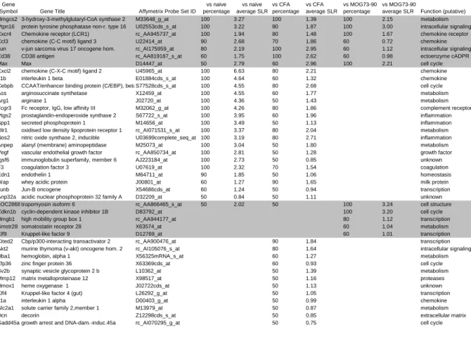
To focus on genes which are truly relevant to encephalitogenicity and not simply involved in general inflammatory responses, we compared gene arrays of LEW.1AV1 (RT1av1) rats immunized with the encephalitogenic MOG stretch MOG 91-108 to naïve LEW.1AV1 (RT1av1) rats and rats immunized with the adjuvant CFA alone as well as rats immunized with the non-encephalitogenic MOG 73-90 determinant. We analyzed ten comparisons for each of the naïve and CFA groups versus MOG 91-108 and five comparisons for MOG 73-90 versus MOG 91-108. The number of comparisons in which a given gene had a signal log ratio (SLR) of above 1 were counted. In table 1, we show genes which are upregulated in at least half of the comparisons (=50%). Besides Cxcr4 and Cd38, which were subsequently analysed in greater detail, many genes with a known function in EAE and MS pathology were found to have an increased expression in MOG 91-108 immunized rats as compared to
Disease Models & Mechanisms • DMM • Advance article controls. This validates our gene list and supports the relevance of the genes not previously
described in EAE. In table S1, genes with decreased expression in EAE are listed. In this analysis, the variability between gene arrays was much higher and less genes were found to be regulated with a clear pattern according to our criteria.
Comparisons of microarrays of CNS-infiltrating lymphocytes derived from LEW.1AV1 (RT1av1) rats after immunization with MOG 91-108, MOG 73-90 and CFA alone resulted in many more genes being differentially expressed as compared to the analysis of lymph node cells (Table S2 and Table S3). This could mirror the influx of different cell populations into the CNS during an inflammatory attack. Similar to the analysis of lymph node cells we found that Cd38 and Cxcr4 mRNA was strongly increased in CNS-infiltrating cells.
Subsequently, we analysed purified CD4+ cells from lymph nodes and CNS of MOG 91-108 and MOG 73-90 immunized rats. To some extent similar gene expression profiles were found in the purified CD4+ cell population compared to non-separated lymph node cells (Table 1, Table S4, Table S5).
Due to their strong expression in MOG 91-108-immunized rats compared to naïve, CFA- immunized and MOG 73-90-immunized rats, we chose Cxcr4 and Cd38 for further analysis.
Cxcr4 and Cd38 in EAE in LEW.1AV1 (RT1av1) rats
Confirming our microarray results by quantitative PCR we found a significant upregulation of Cxcr4 expression in lymph node cells of MOG 91-108-immunized rats (n=8) as compared to CFA-immunized (n=8, ANOVA, P<0.0001) and naïve (n=6, ANOVA, P<0.0001) LEW.1AV1 (RT1av1) rats (Fig. 1A). Also an increased expression of Cd38 was measured (MOG 91-108-immunized rats [n=8] as compared to CFA-immunized [n=8, ANOVA, P<0.05] and naïve [n=6, ANOVA, P<0.05] LEW.1AV1 [RT1av1] rats).
Disease Models & Mechanisms • DMM • Advance article In cells eluted from CNS we found upregulation of Cxcr4 (Fig. 1B) and CD38 (Fig. 1C) in
MOG 91-108 immunized LEW.1AV1 (RT1av1) rats (n=6) compared to rats immunized with MOG 73-90 (n=6, ANOVA Cxcr4 and Cd38 each P<0.001) or CFA alone (n=6, ANOVA, Cxcr4 and Cd38 each P<0.001).
Cxcr4 and Cxcl12 expression in spinal cord of DA (RT1av1) rats
Next we assessed the mRNA expression of Cxcr4 (Fig. 2A) and its ligand Cxcl12 (Fig. 2B) in spinal cord of either naïve DA (RT1av1) rats or DA (RT1av1) rats immunized with IFA or CFA alone or MOG 1-125 in IFA or MOG 1-125 in CFA (each n=4). Upregulation of both Cxcl12 and Cxcr4 mRNA expression was observed in CFA and MOG 1-125 in CFA immunized DA (RT1av1) rats in spinal cord compared to naïve rats, IFA injected or MOG 1-125 in IFA immunized DA (RT1av1) rats (ANOVA, P<0.001). Increased Cxcl12 and Cxcr4 mRNA expression was observed in MOG 1-125 in CFA compared to CFA immunized DA (RT1av1) rats (ANOVA, P<0.01).
Cxcr4 and CXCL12 in patients with MS
Upregulation of mRNA of Cxcr4 was also observed in white blood cells of MS patients with a relapsing-remitting disease course (RRMS, n=32) and a secondary chronic progressive disease course (SPMS, n=22) compared to controls (n=25, ANOVA for RRMS and SPMS each P<0.05) (Fig. 3A, Table S6, Table S7). Also, we detected increased protein CXCL-12 serum levels in both patients with RRMS (n=24) and SPMS (n=28) compared to controls (n=21, ANOVA for RRMS and SPMS each P<0.05) (Fig. 3B, Table S6, Table S7).
Disease Models & Mechanisms • DMM • Advance article EAE in B6.129P2-Cd38tm1Lnd/J mice
Cd38 was strongly upregulated in encephalitogenic lymph node cells. To functionally validate our data and to elucidate the role of CD38 in EAE, we induced disease with the extracellular domain of MOG (MOG 1-125) in CD38 knock out (B6.129P2-Cd38tm1Lnd/J) mice and appropriate controls. We found reduced disease severity in MOG 1-125-immunized B6.129P2-Cd38tm1Lnd/J mice (n=22) compared to wild type control mice (n=21, t-test, cumulative disease score P<0.01) (Fig. 4A). Next we determined the height of the antibody response to MOG 1-125. Reduced anti-MOG IgG autoantibody responses in B6.129P2- Cd38tm1Lnd/J mice (n=4) compared to wild type control mice (n=4, ANOVA, P<0.05)) after immunization with MOG 1-125 were seen (Fig. 4B) on day 12 p.i. Furthermore, also T cell responses upon restimulation with MOG 1-125 were reduced in the B6.129P2-Cd38tm1Lnd/J mice (n=4) on day 12 p.i. compared to control mice (n=4, t-test for stimulation with 50µg/ml MOG 1-125, P<0.05) as measured in a proliferation assay indicating in addition a T cell priming or expansion defect in B6.129P2-Cd38tm1Lnd/J mice (Fig. 4C).
Targeting cellular migration by FTY720
We found an upregulation of Akt in our differential gene expression studies (Table 1).
FTY720 is a S1P-receptor modulator known to influence T cell trafficking by an Akt- dependent mechanism, as does CXCR4 (Mandala et al., 2002, Cyster, 2005, Lee et al., 2001, Brinkmann et al., 2002, Matloubian et al., 2004). We evaluated inhibition of Akt-dependent cell trafficking in MOG 91-108-immunized LEW.1AV1 (RT1av1) rats. Disease was completely inhibited in rats treated from day 0 p.i. with FTY720 (n=10) as compared to the vehicle treated controls (n=10, t-test, cumulative disease score P<0.0001) (Fig. 5A). Next, we tested the efficacy of FTY720 to treat established relapsing-remitting MOG 1-125-induced
Disease Models & Mechanisms • DMM • Advance article EAE in the DA (RT1av1) rat. FTY720 treatment was started on day 21 p.i., after the first bout
of disease (n=8), and showed a significant effect on the disease course as compared to vehicle treatment (n=8, t-test, cumulative disease score day 21-44 p.i., P<0.01) (Fig. 5B). To further examine the beneficial effect of FTY720 treatment on EAE, we boosted the rats with MOG 1-125 on day 44 after the first immunization. Although a relapse was induced in both groups, the DA rats under FTY720 treatment had a better clinical outcome compared to the vehicle treated animals (t-test, cumulative disease score day 45-56 p.i. P<0.0001). On day 14 p.i.
FTY720 treatment led to an increase of the relative size of the CD4 T cell compartment (ANOVA, P<0.01) in the treated LEW.1AV1 (RT1av1) rats (n=10) compared to controls (n=9) concurrent with a decrease in the CD8 T cell (ANOVA, P<0.01) and the B cell (ANOVA, P<0.01) compartment (Fig. 5C; Fig. S1). FTY720 treatment (n=9) compared to controls (n=9) led to down-regulation of mRNA expression of its receptor S1p1 (ANOVA, P<0.0001) and of Akt2 (ANOVA, P<0.0001), one of the genes involved in the intracellular signalling cascade connected to S1P1 and CXCR4 on day 14 p.i. Treatment also had a negative effect on the expression levels of Cd38, (ANOVA, P<0.0001). In contrast, the expression of Cxcr4 was not altered (ANOVA, not significant) (Fig. 5D).
Disease Models & Mechanisms • DMM • Advance article DISCUSSION
In this study we identified gene networks that are critically involved not only in raising an autoantigen- specific immune response but also constituting encephalitogenicity. We analysed the expression and functional relevance of genes and their products expressed on lymphocytes after immunization with the encephalitogenic MOG91-108 peptide in adjuvant, the non-encephalitogenic MOG73-90 peptide in adjuvant, adjuvant alone or naïve rats. Most interestingly in the comparison of MOG91-108 in adjuvant to MOG73-90 in adjuvant immunized rats, compared to the other analyses, only a small number of genes were differentially expressed. These genes seem to be of major importance since they are genes involved in the encephalitogenic response leading to disease manifestation. From the overall expression data, we selected three genes which were upregulated in many comparison: Cxcr4, Cd38 and Akt. We performed functional studies regarding these genes in EAE and analysed tissue samples from MS patients.
CXCR4 (CD184) is a seven transmembrane G-coupled receptor expressed by a number of tissues including cells of the immune system (Campbell et al., 2003). CXCR4 knock-out mice die in utero or perinatally and do not only have defects in the hematopoietic system (impairment of myeloid and B cell generation, reduced proliferation of triple-negative and double-positive lymphocytes), but also in the circulatory system and in the CNS (Zou et al., 1998, Tachibana et al., 1998). Overexpression of CXCR4 on T cells induces their accumulation in the bone marrow and reduction of these cells in the peripheral blood.
CXCR4 signalling leads to a prolonged protein kinase B (AKT) and extracellular signal- regulated kinase 2 activation in T cells. AKT activation promotes cell survival and can act as costimulation for T cell activation (Tilton et al., 2000). CXCR4 has only one known cognate ligand which is CXCL12. CXCL12 is constitutively produced by stromal and endothelial cells. CXCL12 activates numerous signalling pathways like receptor-associated trimeric G
Disease Models & Mechanisms • DMM • Advance article proteins, phospholipase C, PI3K and small G proteins (Pawig et al., 2015). Signalling
through these receptors leads to an increase in the intracellular calcium concentration, cytoskeleton reorganization and cellular migration. Several modulating factors such as phosphatases, regulator of G-protein signalling, adaptor proteins and ubiquitin may affect signalling and/or chemotactic response of CXCR4 to its ligand. An important function of CXCR4/CXCL12 is the regulation of bone-marrow homeostasis and lymphocyte trafficking.
Chemotaxis and integrin-mediated adhesion are the main cellular responses to CXCL12.
CXCL12 knock-out mice display the same phenotype as CXCR4 knock-out mice (Nagasawa et al., 1996).
In autoimmunity there are indications that the interaction of CXCR4 and CXCL12 could be important. CXCL12 recruits B cells to inflamed glomeruli in which these cells my produce autoantibodies (Balabanian et al., 2003). Also in rheumatoid arthritis CXCR4 and CXCL12 have been proposed to be important in the disease precipitation (Zhang et al., 2005). A role of CXCR4 and CXCL12 in EAE (Meiron et al., 2008, Kohler et al., 2008, McCandless et al., 2006) as well as in MS has been described (Azin et al., 2012, Krumbholz et al., 2006).
Our data indicates that also in MOG-induced EAE and possibly MS the interaction of CXCR4 and CXCL12 is of paramount importance for disease development. We found specific upregulation of Cxcr4 on cells derived from lymph nodes and eluted from CNS of rats immunized with encephalitogenic MOG91-108 peptide in comparison to controls. In addition, we measured upregulation of Cxcl12 and Cxcr4 in spinal cords in EAE compared to controls. CXCL12 is upregulated in the CNS of MS patients (Krumbholz et al. 2006 and own unpublished observations). Together the presented data would argue for a scenario in which lymphnode- derived cells are in the context of an encounter with an encephalitogenic antigen activated and migrate towards CXCL12 in the CNS. This is further underscored by the fact
Disease Models & Mechanisms • DMM • Advance article that nitric oxide enhances LPS-induced expression of CXCR4 and migration towards
CXCL12 (Giordano et al., 2006).
CD38 is a membrane associated type II glycoprotein which acts both as an receptor and enzyme (Cockayne et al., 1998, Kato et al., 1999, Salmi and Jalkanen, 2005). As an enzyme it catalyzes NAD+ into cyclic ADP-ribose and further into ADP-ribose. It also regulates Ca2+
levels from ryanodine receptor stores. CD38 is expressed on a variety of myeloid and lymphoid cells. CD38 ligation in B cells leads to tyrosine phosphorylation of several intracellular proteins such as Syk, p85 of phophatidylinositol-3 kinase and phospholipase C-
(Silvennoinen et al., 1996). CD38 ligation in T cells results in phosphorylation of the Raf- 1/MAP kinase and CD3-/ZAP-70 signalling pathway (Zubiaur et al., 1997). Furthermore it is involved in dendritic cell migration and adhesion between lymphocytes and endothelial cells (Partida-Sanchez et al., 2004). B6.129P2-Cd38tm1Lnd/J mice show slight reduction of antibody titers to T cell-dependent antigens (Cockayne et al., 1998). Additionally, these mice had increased susceptibility to bacterial infections, which is thought to be caused by a defective chemotactic response of neutrophils towards bacteria underscoring a role in innate immunity as well (Partida-Sanchez et al., 2001).
We induced EAE in CD38 knock out mice (B6.129P2-Cd38tm1Lnd/J) and controls. CD38 knock out mice had a reduced disease severity and lower autoantibody and T cell responses as compared to the controls. These finding show the importance of CD38 in EAE and possibly MS. This work lies the basis for further analysis of the involved cellular compartments and regulation of human disease (Mayo et al., 2008, Lischke et al., 2013).
FTY720 is a potent drug that affects lymphocyte trafficking and homing. Our studies and studies by others (Brinkmann et al., 2002, Balatoni et al., 2007) showed a strong beneficial effect of FTY720 on EAE under various experimental settings. The drug was approved for treatment of MS (Kappos et al., 2010). We demonstrate that treatment of MOG-EAE with
Disease Models & Mechanisms • DMM • Advance article FTY720 impacts the gene expression of the genes that are involved in encephalitogenic
immune responses. This further validates our approach. We measured changes in expression of S1P1, Akt2 and Cd38. Interestingly, no changes in expression of Cxcr4 was observed. The reason for this is not fully understood and deserves further investigations.
In conclusion in this study we have identified genes which are involved not only in raising an autoantigen-specific immune responses but constitute encephalitogenicity. The immunization with encephalitogenic peptides induces a network of genes involved in activation and migration of lymphocytes. Based on this platform we have established the paramount importance of G-coupled proteins in encephalitogenicity of adaptive immune responses. We speculate that similar involvement might operate also in other autoimmune diseases and possibly transplant rejection thereby establishing common mechanisms. These pathways might be valuable targets of therapeutic approaches as we have shown attenuation of EAE after treatment with FTY720 or reduced EAE severity in CD38-deficient mice. These findings not only validate our gene expression data, but also underscore the importance of the rat EAE model in translational medicine.
Disease Models & Mechanisms • DMM • Advance article MATERIALS AND METHODS
Animals and EAE induction
Female rats or mice, 10-14 weeks of age, were used in all experiments. LEW.1AV1 (RT1av1) rats were obtained from Hans Hedrich (Central Animal Laboratory, Hannover Medical School, Hannover, Germany) and DA (RT1av1) rats were obtained from Harlan Winkelmann (Borchen, Germany). Female B6.129P2-CD38tm1Lnd/J mice and the appropriate controls C57BL/6J 000664 were purchased from the Jackson Laboratory (Bar Harbor, USA).
Animals were bredand kept under specific pathogen-free conditions. Animals were injected intradermally at the base of the tail (rats) or both flanks (mice) with 100 µg of MOG 91–108 (rats) or 50 µg of rat recombinant MOG 1-125 (rats) or 100 µg MOG 35-55 (mice) or 20 µg rat recombinant MOG 1-125 (mice). The antigens in a total volume of 100 µl were mixed with 100 µl of CFA (1:1). Atotal of 100 µl of CFA consisted of IFA (Sigma-Aldrich, St.
Louis, MO) and 500 µg for rats or 400 µg for mice of heat-inactivated Mycobacterium tuberculosis (strain H37 RA; Difco Laboratories, Detroit, MI) (Weissert, 2016). Mice additionally received 100 ng of pertussis toxin (Calbiochem, Darmstadt, Germany) on day 0 and 2 intraveniously. Some groups of rats were also injected with 100 µl IFA mixed with 100 µl MOG 1-125 in PBS (50 µg) without the addition of Mycobacterium tuberculosis or with IFA or CFA alone. The clinical scoring was as follows:0 = no illness; 1 = tail weakness or paralysis; 2 = hind legparaparesis or hemiparesis; 3 = hind leg paralysis or hemiparalysis;4 = tetraparesis or moribund. All experiments were approved by the regional board in Tübingen, Germany.
Disease Models & Mechanisms • DMM • Advance article Human samples
Blood samples were obtained after consent from patients with MS and controls. The characteristics of the MS patients and controls are indicated in Table S6 and Table S7. The research was approved by the Ethics committee of the University of Tuebingen in Germany (Permission 125/2001).
Isolation of CNS infiltrating cells
Infiltrating cells from the CNS were prepared as described before (Weissert et al., 1998a, Weissert et al., 2001). In brief, rats were perfused with cold PBS, and brains and spinal cords weredissected out on day 12 p.i. Subsequently, brains and spinal cordswere homogenized in 10 ml 50% Percoll/0.1% BSA/1% glucose (AmershamPharmacia Biotech) containing 500 U DNase type I (Life Technologies). Ten ml of 50% Percollwere added to each sample after homogenization. A discontinuous Percollgradient was obtained by adding seven ml of 63%
Percoll below and 20ml of 30% Percoll above the sample. Samples were centrifugedfor 40 min at 1000 x g at 4°C. Lymphocytes were collected fromthe 63/50% Percoll interface. The cells were subsequently washed twicein 15–25 ml PBS with centrifugation at 600 x g for 15 min at 4°C.
Isolation of mononuclear cells (MNC) from lymph nodes (LN) and spleens
Draining inguinal LN and spleens were dissected out under deep anaesthesia. LN were disrupted and MNC washed twice in Dulbecco’s modified eagle medium (DMEM, Life Technologies, Paisley, U.K.), resuspended in complete medium (CM) containing DMEM supplemented with 5% fetal calf serum (PAA Laboratories Linz, Austria), 1%
penicillin/streptomycin (Life Technologies), 1% glutamine (Life Technologies), and 50 µM
Disease Models & Mechanisms • DMM • Advance article 2-mercaptoethanol (Life Technologies) and flushed through a 70-µm plastic strainer (Falcon;
BD Biosciences, Franklin Lakes, NJ). MNC from spleen were prepared in the same way as from LN with the difference that red blood cells (RBC) were lysed with lysis buffer consisting of 0.15 M NH4Cl, 10 mM KHCO3, and 0.1 mM Na2 EDTA adjusted to pH 7.4.
CD4+ cells were isolated by anti- rat CD4 microbeads using MACS technology (Miltenyi Biotech, Bergisch Gladbach, Germany) according to the manufacturers instruction.
CD4+ cell purification
CD4+ cells from the lymph nodes were purified by MACS (Miltenyi Biotech, Bergisch Gladbach, Germany) and subsequently analyzed by Affymetrix gene array for differential gene expression.
Spinal cord tissue
From PBS- perfused naïve or in either IFA, CFA, MOG 1-125 in IFA or MOG 1-125 in CFA DA immunized rats, spinal cord tissues was dissected and homogenized and subsequently assessed for mRNA expression.
RNA preparation
Total RNA of brain infiltrating leukocytes or lymphocytes or spinal cord tissue was isolated by using a RNeasy kit (Qiagen, Hilden, Germany) according to the manufacturer’s instructions. The RNA quality was analysed with a Bioanalyser 2100 (Agilent, Palo Alto, USA).
Disease Models & Mechanisms • DMM • Advance article Microarrays
Affymetrix microarrays of the type RG U34 A (Affymetrix Inc., Santa Clara, USA) representing approximately 7000 full length genes and 1000 EST clusters were used. For the purified CD4+ cells the rat expression set 230A (Affymetrix) containing about 30.000 features was used. For each array, samples from at least three rats were pooled. Biotin labelled cRNA was prepared and hybridized to the arrays. In brief, double stranded cDNA was synthesized from whole RNA using a superscript choice kit (Invitrogen) with a T7- (dT)24 primer (Metabion) and in vitro transcribed into biotin-labelled cRNA. After hybridization gene arrays were washed and stained by a fluidics station (Affymetrix) and scanned by a confocal laser scanning microscope (Agilent).
The data was analyzed using the microarray suite software, micro DB, and data mining tool (Affymetrix). Only genes and EST were included in further analysis which were “present”
and gave a difference call of either “increase” or “decrease” according to the Affymetrix software. The extend of differential expression is expressed as signal log ratio (SLR). For the tables the cut off was set to a SLR of 1 signifying an 2-fold change in expression. Doubles, ESTs, and sequences not corresponding to a gene were not included in the tables.
Real time PCR
To avoid amplification/detection of contaminating genomic DNA, extracted RNA was treated with Rnase free DNase (Promega, Madison, WI). Subsequently, cDNA was synthesized by reverse transcription with Moloney murine leukemia virus reverse transcriptase and random pdN6 primers in the presence of RNase inhibitor (Promega). Amplification was performed on an Applied Biosystems Prism 7000 Sequence Detection System (Applied Biosystems, Foster
Disease Models & Mechanisms • DMM • Advance article City, CA) using a SYBR green protocol. Results were expressed as 2- CT values. The
primer sequences 5´ to 3´ were as follows:
Rat primers
Gapdh: forward: GGTTGTCTCCTGTGACTTCAA
reverse: CATACCAGGAAATGAGCTTCAC
S1p1: forward: TAGCCGCAGCAAATCAGAC reverse: GCAGCAGTGGAGAAAGAGAGA Cxcr4: forward: GATGGTGGTGTTCCAGTTCC
reverse: CAGCTTGGAGATGATGATGC Cxcl12: forward: CGATTCTTTGAGAGCCATGT reverse: AGGGCACAGTTTGGAGTGTT Cd38: forward: AGGACACACTGCTGGGCTAT
reverse: CAGGGTTGTTGGGACAATTT Akt2: forward: GAAGACTGAGAGGCCACGAC
reverse: GGGAGCCACACTTGTAATCC
Human primers
h18s: forward: CGGCTACCACATCCAAGGAA reverse: GCTGGAATTACCGCGGCT Cxcr4: forward: CATCAGTCTGGACCGCTACC,
reverse: GGATCCAGACGCCAACATAG.
Disease Models & Mechanisms • DMM • Advance article FACS
FITC conjugated mAb against CD45RA (OX-33) and TCRAB (R73) and PE conjugated mAb against CD4 (OX-35) and MHC II (OX-6) and appropriate isotype controls were purchased from Becton Dickinson (Heidelberg, Germany). Flow cytometry was performed on a FACScalibur running with Cellquest software (Becton Dickinson). Cells were gated on the lymphocyte population in the FSC-SSC dot plot. For data analysis also Flowing Software 2.5.1 (Turku Center for Biotechnology, Finland) was used.
ELISA
Serum taken at the time point of euthanasia was subject to an anti-MOG autoantibody ELISA. ELISA plates (96 well; Nunc, Roskilde, Denmark) were coated with 2.5 µg/ml (100 µl/well) MOG 1-125 overnight at 4°C. Plates were washed with PBS/0.05% Tween 20 and blocked with milk powder for 1 h at room temperature. After washing, diluted serum samples were added and plates were incubated for 1 h at room temperature. Then, plates were washed and rabbit anti mouse antiserum (IgG, IgG1, IgG2a, IgG2b, IgG3, IgM Nordic, Tilburg, The Netherlands) was added and incubated for 1 h at room temperature. Plates were washed prior to the addition of peroxidase conjugated goat anti rabbit antiserum (Nordic) diluted in PBS/0.05% Tween 20. After 30 min incubation, plates were washed and bound Abs were visualized by addition of ABTS (Roche Diagnostics Mannheim, Germany). After 15 minutes of incubation optical density was read at 405 nm.
ELISA with human serum was performed according to the instructions of the manufacturer.
Disease Models & Mechanisms • DMM • Advance article Proliferation assay
Cells from draining LN were prepared as described above and cultured in the presence of rrMOG in 96 well plates as described in the Elispot section. Cells were cultured for 48h and pulsed with 1µCi [3H]thymidine for the last 18 h. The incorporation of [3H]thymidine was measured using a beta-scintillation counter.
FTY720 treatment
Rats were treated with FTY720 (generous gift of Novartis AG, Switzerland) and received a daily dose of 0,4 mg/kg in sterile water by oral gavage as described (Balatoni et al., 2007).
Disease Models & Mechanisms • DMM • Advance article Acknowledgements
We thank Thomas Weiss and Kerstin Stuck for excellent technical assistance.
Competing interests
There are no competing interests
Author contribution
M.M.H. performed the gene arrays, data analysis, the target validation in rats and revised the paper. S.B. performed the gene arrays, data analysis and the target validation. B.G. did the work on CD38 in mice. K.M.S. performed target validation studies in rats and analysed the data. A.B. performed studies in MS patients and controls. R.W. designed the study, raised the funding, performed the data analysis and wrote and revised the paper.
Funding
This study was supported by grants from the Interdisciplinary Centre for Clinical Research (Fö. 01KS9602), and German Research Foundation to R.W. (DFG We 1947).
Disease Models & Mechanisms • DMM • Advance article REFERENCES
AZIN, H., VAZIRINEJAD, R., AHMADABADI, B. N., KHORRAMDELAZAD, H., ZARANDI, E. R., ARABABADI, M. K., KARIMABAD, M. N., SHAMSIZADEH, A., RAFATPANAH, H. & HASSANSHAHI, G. 2012. The SDF-1 3'a genetic variation of the chemokine SDF-1alpha (CXCL12) in parallel with its increased circulating levels is associated with susceptibility to MS: a study on Iranian multiple sclerosis patients. J Mol Neurosci, 47, 431-6.
BALABANIAN, K., COUDERC, J., BOUCHET-DELBOS, L., AMARA, A., BERREBI, D., FOUSSAT, A., BALEUX, F., PORTIER, A., DURAND-GASSELIN, I., COFFMAN, R. L., GALANAUD, P., PEUCHMAUR, M. & EMILIE, D. 2003.
Role of the chemokine stromal cell-derived factor 1 in autoantibody production and nephritis in murine lupus. J Immunol, 170, 3392-400.
BALATONI, B., STORCH, M. K., SWOBODA, E. M., SCHONBORN, V., KOZIEL, A., LAMBROU, G. N., HIESTAND, P. C., WEISSERT, R. & FOSTER, C. A.
2007. FTY720 sustains and restores neuronal function in the DA rat model of MOG-induced experimental autoimmune encephalomyelitis. Brain Res Bull, 74, 307-16.
BRINKMANN, V., DAVIS, M. D., HEISE, C. E., ALBERT, R., COTTENS, S., HOF, R., BRUNS, C., PRIESCHL, E., BAUMRUKER, T., HIESTAND, P., FOSTER, C. A., ZOLLINGER, M. & LYNCH, K. R. 2002. The immune modulator FTY720 targets sphingosine 1-phosphate receptors. J Biol Chem, 277, 21453-7.
CAMPBELL, D. J., KIM, C. H. & BUTCHER, E. C. 2003. Chemokines in the systemic organization of immunity. Immunol Rev, 195, 58-71.
COCKAYNE, D. A., MUCHAMUEL, T., GRIMALDI, J. C., MULLER-STEFFNER, H., RANDALL, T. D., LUND, F. E., MURRAY, R., SCHUBER, F. & HOWARD, M. C. 1998. Mice deficient for the ecto-nicotinamide adenine dinucleotide glycohydrolase CD38 exhibit altered humoral immune responses. Blood, 92, 1324-33.
CYSTER, J. G. 2005. Chemokines, sphingosine-1-phosphate, and cell migration in secondary lymphoid organs. Annu Rev Immunol, 23, 127-59.
DUMAS, A., AMIABLE, N., DE RIVERO VACCARI, J. P., CHAE, J. J., KEANE, R.
W., LACROIX, S. & VALLIERES, L. 2014. The inflammasome pyrin contributes to pertussis toxin-induced IL-1beta synthesis, neutrophil intravascular crawling and autoimmune encephalomyelitis. PLoS Pathog, 10, e1004150.
GENAIN, C. P., NGUYEN, M. H., LETVIN, N. L., PEARL, R., DAVIS, R. L., ADELMAN, M., LEES, M. B., LININGTON, C. & HAUSER, S. L. 1995.
Antibody facilitation of multiple sclerosis-like lesions in a nonhuman primate. J Clin Invest, 96, 2966-74.
GIORDANO, D., MAGALETTI, D. M. & CLARK, E. A. 2006. Nitric oxide and cGMP protein kinase (cGK) regulate dendritic-cell migration toward the lymph-node- directing chemokine CCL19. Blood, 107, 1537-45.
HUR, E. M., YOUSSEF, S., HAWS, M. E., ZHANG, S. Y., SOBEL, R. A. &
STEINMAN, L. 2007. Osteopontin-induced relapse and progression of autoimmune brain disease through enhanced survival of activated T cells. Nat Immunol, 8, 74-83.
KAPPOS, L., RADUE, E. W., O'CONNOR, P., POLMAN, C., HOHLFELD, R., CALABRESI, P., SELMAJ, K., AGOROPOULOU, C., LEYK, M., ZHANG-
Disease Models & Mechanisms • DMM • Advance article AUBERSON, L., BURTIN, P. & GROUP, F. S. 2010. A placebo-controlled trial
of oral fingolimod in relapsing multiple sclerosis. N Engl J Med, 362, 387-401.
KATO, I., YAMAMOTO, Y., FUJIMURA, M., NOGUCHI, N., TAKASAWA, S. &
OKAMOTO, H. 1999. CD38 disruption impairs glucose-induced increases in cyclic ADP-ribose, [Ca2+]i, and insulin secretion. J Biol Chem, 274, 1869-72.
KOHLER, R. E., COMERFORD, I., TOWNLEY, S., HAYLOCK-JACOBS, S., CLARK-LEWIS, I. & MCCOLL, S. R. 2008. Antagonism of the chemokine receptors CXCR3 and CXCR4 reduces the pathology of experimental autoimmune encephalomyelitis. Brain Pathol, 18, 504-16.
KORNEK, B., STORCH, M. K., WEISSERT, R., WALLSTROEM, E., STEFFERL, A., OLSSON, T., LININGTON, C., SCHMIDBAUER, M. & LASSMANN, H. 2000.
Multiple sclerosis and chronic autoimmune encephalomyelitis: a comparative quantitative study of axonal injury in active, inactive, and remyelinated lesions.
Am J Pathol, 157, 267-76.
KRUMBHOLZ, M., THEIL, D., CEPOK, S., HEMMER, B., KIVISAKK, P., RANSOHOFF, R. M., HOFBAUER, M., FARINA, C., DERFUSS, T., HARTLE, C., NEWCOMBE, J., HOHLFELD, R. & MEINL, E. 2006. Chemokines in multiple sclerosis: CXCL12 and CXCL13 up-regulation is differentially linked to CNS immune cell recruitment. Brain, 129, 200-11.
LEE, M. J., THANGADA, S., PAIK, J. H., SAPKOTA, G. P., ANCELLIN, N., CHAE, S. S., WU, M., MORALES-RUIZ, M., SESSA, W. C., ALESSI, D. R. & HLA, T.
2001. Akt-mediated phosphorylation of the G protein-coupled receptor EDG-1 is required for endothelial cell chemotaxis. Mol Cell, 8, 693-704.
LISCHKE, T., HEESCH, K., SCHUMACHER, V., SCHNEIDER, M., HAAG, F., KOCH-NOLTE, F. & MITTRUCKER, H. W. 2013. CD38 controls the innate immune response against Listeria monocytogenes. Infect Immun, 81, 4091-9.
LUCCHINETTI, C., BRUCK, W., PARISI, J., SCHEITHAUER, B., RODRIGUEZ, M.
& LASSMANN, H. 2000. Heterogeneity of multiple sclerosis lesions: implications for the pathogenesis of demyelination. Ann Neurol, 47, 707-17.
MANDALA, S., HAJDU, R., BERGSTROM, J., QUACKENBUSH, E., XIE, J., MILLIGAN, J., THORNTON, R., SHEI, G. J., CARD, D., KEOHANE, C., ROSENBACH, M., HALE, J., LYNCH, C. L., RUPPRECHT, K., PARSONS, W.
& ROSEN, H. 2002. Alteration of lymphocyte trafficking by sphingosine-1- phosphate receptor agonists. Science, 296, 346-9.
MATHEY, E., BREITHAUPT, C., SCHUBART, A. S. & LININGTON, C. 2004.
Commentary: Sorting the wheat from the chaff: identifying demyelinating components of the myelin oligodendrocyte glycoprotein (MOG)-specific autoantibody repertoire. Eur J Immunol, 34, 2065-71.
MATLOUBIAN, M., LO, C. G., CINAMON, G., LESNESKI, M. J., XU, Y., BRINKMANN, V., ALLENDE, M. L., PROIA, R. L. & CYSTER, J. G. 2004.
Lymphocyte egress from thymus and peripheral lymphoid organs is dependent on S1P receptor 1. Nature, 427, 355-60.
MAYO, L., JACOB-HIRSCH, J., AMARIGLIO, N., RECHAVI, G., MOUTIN, M. J., LUND, F. E. & STEIN, R. 2008. Dual role of CD38 in microglial activation and activation-induced cell death. J Immunol, 181, 92-103.
MCCANDLESS, E. E., WANG, Q., WOERNER, B. M., HARPER, J. M. & KLEIN, R.
S. 2006. CXCL12 limits inflammation by localizing mononuclear infiltrates to the perivascular space during experimental autoimmune encephalomyelitis. J Immunol, 177, 8053-64.
Disease Models & Mechanisms • DMM • Advance article MEIRON, M., ZOHAR, Y., ANUNU, R., WILDBAUM, G. & KARIN, N. 2008.
CXCL12 (SDF-1alpha) suppresses ongoing experimental autoimmune encephalomyelitis by selecting antigen-specific regulatory T cells. J Exp Med, 205, 2643-55.
MILLS, K. H. 2011. TLR-dependent T cell activation in autoimmunity. Nat Rev Immunol, 11, 807-22.
NAGASAWA, T., NAKAJIMA, T., TACHIBANA, K., IIZASA, H., BLEUL, C. C., YOSHIE, O., MATSUSHIMA, K., YOSHIDA, N., SPRINGER, T. A. &
KISHIMOTO, T. 1996. Molecular cloning and characterization of a murine pre- B-cell growth-stimulating factor/stromal cell-derived factor 1 receptor, a murine homolog of the human immunodeficiency virus 1 entry coreceptor fusin. Proc Natl Acad Sci U S A, 93, 14726-9.
NOSEWORTHY, J. H., LUCCHINETTI, C., RODRIGUEZ, M. & WEINSHENKER, B. G. 2000. Multiple sclerosis. N Engl J Med, 343, 938-52.
PARTIDA-SANCHEZ, S., COCKAYNE, D. A., MONARD, S., JACOBSON, E. L., OPPENHEIMER, N., GARVY, B., KUSSER, K., GOODRICH, S., HOWARD, M., HARMSEN, A., RANDALL, T. D. & LUND, F. E. 2001. Cyclic ADP-ribose production by CD38 regulates intracellular calcium release, extracellular calcium influx and chemotaxis in neutrophils and is required for bacterial clearance in vivo. Nat Med, 7, 1209-16.
PARTIDA-SANCHEZ, S., GOODRICH, S., KUSSER, K., OPPENHEIMER, N., RANDALL, T. D. & LUND, F. E. 2004. Regulation of dendritic cell trafficking by the ADP-ribosyl cyclase CD38: impact on the development of humoral immunity. Immunity, 20, 279-91.
PAWIG, L., KLASEN, C., WEBER, C., BERNHAGEN, J. & NOELS, H. 2015.
Diversity and Inter-Connections in the CXCR4 Chemokine Receptor/Ligand Family: Molecular Perspectives. Front Immunol, 6, 429.
RIEDHAMMER, C. & WEISSERT, R. 2015. Antigen Presentation, Autoantigens, and Immune Regulation in Multiple Sclerosis and Other Autoimmune Diseases.
Front Immunol, 6, 322.
SALMI, M. & JALKANEN, S. 2005. Cell-surface enzymes in control of leukocyte trafficking. Nat Rev Immunol, 5, 760-71.
SILVENNOINEN, O., NISHIGAKI, H., KITANAKA, A., KUMAGAI, M., ITO, C., MALAVASI, F., LIN, Q., CONLEY, M. E. & CAMPANA, D. 1996. CD38 signal transduction in human B cell precursors. Rapid induction of tyrosine phosphorylation, activation of syk tyrosine kinase, and phosphorylation of phospholipase C-gamma and phosphatidylinositol 3-kinase. J Immunol, 156, 100- 7.
STORCH, M. K., STEFFERL, A., BREHM, U., WEISSERT, R., WALLSTROM, E., KERSCHENSTEINER, M., OLSSON, T., LININGTON, C. & LASSMANN, H.
1998. Autoimmunity to myelin oligodendrocyte glycoprotein in rats mimics the spectrum of multiple sclerosis pathology. Brain Pathol, 8, 681-94.
TACHIBANA, K., HIROTA, S., IIZASA, H., YOSHIDA, H., KAWABATA, K., KATAOKA, Y., KITAMURA, Y., MATSUSHIMA, K., YOSHIDA, N., NISHIKAWA, S., KISHIMOTO, T. & NAGASAWA, T. 1998. The chemokine receptor CXCR4 is essential for vascularization of the gastrointestinal tract.
Nature, 393, 591-4.
TILTON, B., HO, L., OBERLIN, E., LOETSCHER, P., BALEUX, F., CLARK-LEWIS, I. & THELEN, M. 2000. Signal transduction by CXC chemokine receptor 4.
Stromal cell-derived factor 1 stimulates prolonged protein kinase B and
Disease Models & Mechanisms • DMM • Advance article extracellular signal-regulated kinase 2 activation in T lymphocytes. J Exp Med,
192, 313-24.
WEISSERT, R. 2013. The immune pathogenesis of multiple sclerosis. J Neuroimmune Pharmacol, 8, 857-66.
WEISSERT, R. 2016. Actively Induced Experimental Autoimmune Encephalomyelitis in Rats. Methods Mol Biol, 1304, 161-9.
WEISSERT, R., DE GRAAF, K. L., STORCH, M. K., BARTH, S., LININGTON, C., LASSMANN, H. & OLSSON, T. 2001. MHC class II-regulated central nervous system autoaggression and T cell responses in peripheral lymphoid tissues are dissociated in myelin oligodendrocyte glycoprotein-induced experimental autoimmune encephalomyelitis. J Immunol, 166, 7588-99.
WEISSERT, R., SVENNINGSSON, A., LOBELL, A., DE GRAAF, K. L., ANDERSSON, R. & OLSSON, T. 1998a. Molecular and genetic requirements for preferential recruitment of TCRBV8S2+ T cells in Lewis rat experimental autoimmune encephalomyelitis. J Immunol, 160, 681-90.
WEISSERT, R., WALLSTROM, E., STORCH, M. K., STEFFERL, A., LORENTZEN, J., LASSMANN, H., LININGTON, C. & OLSSON, T. 1998b. MHC haplotype- dependent regulation of MOG-induced EAE in rats. J Clin Invest, 102, 1265-73.
ZHANG, X., NAKAJIMA, T., GORONZY, J. J. & WEYAND, C. M. 2005. Tissue trafficking patterns of effector memory CD4+ T cells in rheumatoid arthritis.
Arthritis Rheum, 52, 3839-49.
ZOU, Y. R., KOTTMANN, A. H., KURODA, M., TANIUCHI, I. & LITTMAN, D. R.
1998. Function of the chemokine receptor CXCR4 in haematopoiesis and in cerebellar development. Nature, 393, 595-9.
ZUBIAUR, M., IZQUIERDO, M., TERHORST, C., MALAVASI, F. & SANCHO, J.
1997. CD38 ligation results in activation of the Raf-1/mitogen-activated protein kinase and the CD3-zeta/zeta-associated protein-70 signaling pathways in Jurkat T lymphocytes. J Immunol, 159, 193-205.
Disease Models & Mechanisms • DMM • Advance article Figures
Disease Models & Mechanisms • DMM • Advance article Fig. 1. Cxcr4 and Cd38 expression in lymph node cells and CNS.
A, Quantitative SYBR green real time PCR was performed for Cxcr4 and Cd38 in lymph node cells from naïve (black bars, n=6), CFA (grey bars, n=8) or MOG 91-108 in CFA (white bars, n=8)- immunized LEW.1AV1 (RT1av1) rats on day 12 p.i.. Increased Cxcr4 (*ANOVA, P<0.0001) and Cd38 (*ANOVA, P<0.05) expression was found in MOG 91-108 in CFA- immunized rats compared to naïve and CFA- alone- immunized rats. B, Quantitative expression of Cxcr4 in cells eluted from CNS of CFA (white bars, n=6), MOG 73-90 in CFA (grey bars, n=6) and MOG 91-108 in CFA (n=6) immunized LEW.1AV1 (RT1av1) rats.
Cxcr4 was upregulated in MOG 91-108 in CFA-immunized rats compared to the other groups (*ANOVA, P<0.001) on day 12 p.i.. C, Quantitative expression of Cd38 of lymphocytes eluted from CNS of CFA (white bars, n=6), MOG 73-90 in CFA (grey bars, n=6) and MOG 91-108 in CFA (n=6) immunized LEW.1AV1 (RT1av1) rats on day 12 p.i.
Cd38 was upregulated in MOG 91-108 in CFA-immunized rats compared to the other groups (*ANOVA, P<0.001). Results are expressed as 2-ΔΔCT values. Numbers are mean +/- SEM.
Disease Models & Mechanisms • DMM • Advance article
Disease Models & Mechanisms • DMM • Advance article Fig 2. Cxr4 and Cxcl12 expression in spinal cord of DA (RT1av1) rats.
Quantitative SYBR green real time PCR was performed for Cxcr4 (A) and Cxcl12 (B) from PBS- perfused spinal cord tissue of naïve DA (RT1av1) rats (n=4), rats immunized with IFA (n=4) or CFA alone (n=4) and DA rats immunized with MOG 1-125 in IFA (n=4) or MOG 1- 125 in CFA (n=4) on day 12 p.i. Increased Cxcr4 and Cxcl12 expression was observed in spinal cord in DA (RT1av1) rats immunized with CFA and MOG1-125 in CFA (*ANOVA, P<0.001). There was an upregulation of Cxcr4 and Cxcl12 mRNA in DA (RT1av1) rats immunized with MOG 1-125 in CFA compared to DA (RT1av1) rats immunized with CFA alone (*ANOVA, P<0.01). Quantitative PCR results are expressed as 2-ΔΔ CT values.
Numbers are mean +/- SEM.
Disease Models & Mechanisms • DMM • Advance article Fig. 3. Cxcr4 expression in white blood cells and CXCL12 protein in serum of patients
with MS.
A, Cxcr4 mRNA was quantified by real time PCR from white blood cells of patients with relapsing remitting MS (grey bars, n=32), secondary chronic- progressive MS (black bars, n=22) and controls (white bars, n=25). Upregulation of Cxcr4 in both patient groups compared to controls was observed (*ANOVA, P<0.05). B, CXCL12 serum levels were
Disease Models & Mechanisms • DMM • Advance article assessed by ELISA in patients with relapsing-remitting MS (grey bars, n=24), secondary
chronic progressive MS (black bars, n=28) and controls (white bars, n=21). Increased serum levels of CXCL12 were measured in both MS groups compared to controls (*ANOVA, P<0.05). Quantitative PCR results are expressed as 2-ΔΔ CT values. Numbers are mean +/- SEM.
Disease Models & Mechanisms • DMM • Advance article
Disease Models & Mechanisms • DMM • Advance article Fig. 4. EAE in B6.129P2-Cd38tm1Lnd/J mice.
A, EAE was induced in female B6.129P2-Cd38tm1Lnd/J mice (open triangles) and C57BL/6J 000664 controls (closed triangles) with MOG 1-125 in CFA. On day 0 and 2 p.i. mice received an i.v. injection of 150 ng pertussis toxin. EAE was scored as follows: 0, no disease;
1 tail paralysis; 2, paraparesis; 3, paraplegia; 4, tetraparalysis; 5, moribund or dead.
Immunization with the extracellular domain of MOG, MOG 1-125 lead to disease reduction in B6.129P2-Cd38tm1Lnd/J (n=22) compared to C57BL/6J 000664 controls (n=21) mice (t-test, cumulative disease score, P=0.01). B, Antibodies against MOG 1-125 were measured by ELISA as described (Weissert et al., 2001). B6.129P2-Cd38tm1Lnd/J (white bars, n=4) mice had reduced IgG and IgG1 antibodies compared to C57BL/6J 000664 mice (black bars, n=4) (*ANOVA, P<0.05) on day 12 p.i. c, T cell responses upon restimulation against MOG 1-125 from draining lymph nodes on day 12 p.i. were reduced in B6.129P2-Cd38tm1Lnd/J (open triangles, n=4) compared to C57BL/6J 000664 (closed triangles, n=4) mice (*t-test for stimulation with 50µg/ml MOG 1-125, P=0.05). Numbers are mean +/- SEM.
Disease Models & Mechanisms • DMM • Advance article Fig. 5. Influence of FTY720 in EAE.
A, Female LEW.1AV1 (RT1av1) rats were immunized with MOG 1-125 in CFA. EAE was scored as follows: 0, no disease; 1 tail paralysis; 2, paraparesis; 3, paraplegia; 4, tetraparalysis; 5, moribund or dead. FTY720 completely inhibited EAE in rats treated orally with 0.4 mg/kg from day 0 p.i. (open triangles, n=10) as compared to vehicle (PBS)- treated controls (closed triangles, n=10) (t-test, cumulative disease score P<0.0001). B, Female DA (RT1av1) rats treated orally with 0.4 mg/kg FTY720 starting on day 21 p.i. after a first bout of disease (open triangles, n=8) showed a disease course compared to vehicle (PBS)- treated controls (closed triangles, n=8) (t-test, cumulative disease score day 21-44 p.i., P=0.01). On day 44 p.i. rats were boosted with MOG 1-125 in CFA. Both groups relapsed but with a better outcome of the FTY720 treated group (t-test, cumulative disease score day 45-56 p.i., P<0.0001). C, FACS analysis of cells from draining lymph nodes demonstrated an increase in relative size of the CD4 T cell compartment and a decrease in the size of the CD8 and B cell compartment in FTY720 treated (white bars, n=10) rats compared to controls (black bars,
Disease Models & Mechanisms • DMM • Advance article n=9) (*ANOVA, P<0.01) on day 14 p.i.. D, Expression of S1p1 (Edg1), Akt2, Cxcr4 and
Cd38 was assessed in FTY720 (open bars, n=9) compared to vehicle- treated controls (black bars, n=9) by quantitative PCR. FTY720 treatment lead to downregulation of S1p1 and Akt2 as well as Cd38 but not Cxcr4 (*ANOVA for all except Cxcr4 P< 0.0001) on day 14 p.i.
from cells derived from lymph nodes. Results are expressed as 2-ΔΔ CTvalues. Numbers are mean +/- SEM.
Disease Models & Mechanisms • DMM • Advance article
Gene
Symbol Gene Title Affymetrix Probe Set ID
MOG 91-108 vs naive percentage
MOG 91-108 vs naive average SLR
MOG 91-108 vs CFA percentage
MOG 91-108 vs CFA average SLR
MOG 91-108 vs MOG73-90 percentage
MOG 91-108 vs MOG73-90
average SLR Function (putative)
Hmgcs2 3-hydroxy-3-methylglutaryl-CoA synthase 2 M33648_g_at 100 3.27 100 1.39 100 2.15 metabolism
Ptpn16 protein tyrosine phosphatase non-r. type 16 U02553cds_s_at 100 3.22 80 1.87 100 3.00 intracellular signaling
Cxcr4 Chemokine receptor (LCR1) rc_AA945737_at 100 1.94 80 1.48 100 1.67 chemokine receptor
Ccl3 chemokine (C-C motif) ligand 3 U22414_at 90 2.68 70 1.86 60 0.72 chemokine
Jun v-jun sarcoma virus 17 oncogene hom. rc_AI175959_at 80 2.19 100 2.95 60 1.12 intracellular signaling
Cd38 CD38 antigen rc_AA819187_s_at 60 1.75 100 2.62 60 0.98 ectoenzyme cADPR
Max Max D14447_at 50 2.79 60 2.96 100 2.21 cell cycle
Cxcl2 chemokine (C-X-C motif) ligand 2 U45965_at 100 6.63 80 2.21 chemokine
Il1b interleukin 1 beta E01884cds_s_at 100 4.64 60 1.32 chemokine
Cebpb CCAAT/enhancer binding protein (C/EBP), betaS77528cds_s_at 100 4.55 80 2.68 cell cycle
Ass arginosuccinate synthetase X12459_at 100 4.55 60 1.77 metabolism
Arg1 arginase 1 J02720_at 100 4.36 50 1.43 metabolism
Fcgr3 Fc receptor, IgG, low affinity III M32062_g_at 100 4.26 80 1.86 complement receptor
Ptgs2 prostaglandin-endoperoxide synthase 2 S67722_s_at 100 3.95 60 1.96 inflammation
Spp1 secreted phosphoprotein 1 M14656_at 100 3.49 50 1.13 inflammation
Olr1 oxidised low density lipoprotein receptor 1 rc_AI071531_s_at 100 3.37 80 2.04 metabolism
Nos2 nitric oxide synthase 2, inducible U03699complete_seq_at 100 3.19 80 2.71 inflammation
Anpep alanyl (membrane) aminopeptidase M25073_at 100 3.04 50 1.80 metabolism
Vegf vascular endothelial growth factor rc_AA850734_at 100 2.81 50 1.28 growth factor
Igsf6 immunoglobulin superfamily, member 6 AJ223184_at 100 2.73 50 0.85 unknown
F3 coagulation factor 3 U07619_at 100 2.32 70 1.54 coagulation
Edn1 endothelin 1 M64711_at 90 1.85 50 1.06 homeostasis
Wap whey acidic protein J00801_at 60 1.27 90 1.65 milk protein
Junb Jun-B oncogene X54686cds_at 60 1.24 50 0.94 transcription
Anp32a acidic nuclear phosphoprotein 32 family A D32209_at 50 0.84 50 1.11 unknown
LOC286890tropomyosin isoform 6 rc_AA866465_s_at 50 2.02 50 100 3.24 cell structure
Cdkn1b cyclin-dependent kinase inhibitor 1B D83792_at 100 3.20 cell cycle
Hmgb1 high mobility group box 1 rc_AA944177_at 80 1.12 transcription
Smstr28 somatostatin receptor 28 X63574_at 60 1.04 metabolism
Klf9 Kruppel-like factor 9 D12769_at 60 1.01 transcription
Cited2 Cbp/p300-interacting transactivator 2 rc_AA900476_at 90 1.84 transcription
Akt2 murine thymoma (v-akt) oncogene hom. 2 rc_AI105076_s_at 80 1.64 intracellular signaling
Hba1 hemoglobin, alpha 1 X56325mRNA_s_at 60 1.27 metabolism
Zfp36 zinc finger protein 36 X63369cds_at 60 0.93 cell cycle
Sv2b synaptic vesicle glycoprotein 2 b L10362_at 50 1.39 metabolism
Mmp12 matrix metalloproteinase 12 X98517_at 50 1.16 proteases
Hmox1 heme oxygenase 1 J02722cds_at 50 1.13 unknown
Klf4 Kruppel-like factor 4 (gut) L26292_g_at 50 1.05 transcription
Il1a interleukin 1 alpha D00403_g_at 50 0.99 chemokine
Slc2a1 solute carrier family 2,member 1 M13979_at 50 0.87 metabolism
Dcn decorin Z12298cds_s_at 50 0.85 extracellular matrix
Gadd45a growth arrest and DNA-dam.-induc.45a rc_AI070295_g_at 50 0.75 cell cycle
Table 1. Genes with increased expression in lymph node cells of LEW.1AV1 (RT1av1) rats
LEW.1AV1 (RT1av1) rats were immunized with MOG 91-108 in CFA, MOG 73-90 in CFA and CFA alone. Gene expression in lymph node cells was analysed using Affymetrix gene arrays. Gene arrays from rats immunized with MOG 91-108 in CFA (pooled samples of at least n=3 rats, n=5 gene chips) were compared to gene arrays from naive rats (pooled samples, n=2 chips), CFA immunized rats (pooled samples, n=2 chips) and MOG 73-90 in CFA immunized rats (pooled samples, n=1 chip) resulting in each ten comparisons for CFA and naïve to MOG 91-108-immunized rats and five for MOG 73-90 to MOG 91-108- immunized rats. The number of comparisons in which a given gene had a SLR above 1 were counted. If a gene had a SLR above 1 in 50% of the comparisons it was included in the analysis. ESTs and sequences not corresponding to a gene and duplicates were removed from
Disease Models & Mechanisms • DMM • Advance article the table. Genes which fulfilled the criteria in all of the three comparisons are displayed on
the top of the table. Genes which fulfilled the criteria only in naive versus MOG 91-108 in CFA comparisons are not displayed here. Lymph node cells were prepared as described (Weissert et al., 2001).