Dynamics, regulation and function of macrophages in skin repair
Inaugural-Dissertation
zur
Erlangung des Doktorgrades
der Mathematisch-Naturwissenschaftlichen Fakultät der Universität zu Köln
vorgelegt von
Tina Lucas aus Krefeld
Köln, 2011
Berichterstatter: Prof. Dr. Matthias Hammerschmidt
PD Dr. Roswitha Nischt
Tag der mündlichen Prüfung: 05. Juli 2011
Tissue repair is a highly dynamic process comprising the sequential phases of inflammation, tissue formation, and maturation. The mechanisms that orchestrate the natural sequence of the wound healing response remain elusive. Influx of macrophages plays a crucial role in tissue repair. However, the precise function of macrophages during the healing response has remained a subject of debate due to their functional dichotomy as effectors of both, tissue injury and repair. In this study the hypothesis was examined whether macrophages recruited during the diverse phases of skin repair after mechanical injury exert specific functions to restore tissue integrity. For this purpose a mouse model was developed that allows conditional depletion of macrophages during the sequential stages of the repair response by using the inducible diphtheria toxin receptor mouse model in combination with a myeloid cell- specific Cre mouse line. Depletion of macrophages restricted to the early stage of the repair response (inflammatory phase) significantly reduced the formation of a vascularized granulation tissue and showed impaired re-epithelialization. However, recruitment of macrophages during the mid phase of repair, after macrophage depletion was stopped, rescued the impaired healing response and resulted in minimized scar formation. In contrast, depletion of macrophages restricted to the mid stage of the repair response (phase of tissue formation) resulted in severe hemorrhages within the wound tissue. Under these conditions, transition into the subsequent phase of tissue maturation and wound closure did not occur.
Finally, macrophage depletion restricted to the late stage of repair (phase of tissue maturation) did not significantly impact the outcome of the repair response. Taken together, these results demonstrate that macrophages exert distinct functions during the different phases of skin repair, which are crucial to control the natural sequence of repair events.
Furthermore, the effect of macrophages on endothelial cell function and wound angiogenesis appeared to be critical. Therefore the impact of macrophage-derived vascular endothelial growth factor-A (VEGF-A) on the outcome of the wound healing response was analyzed, by using conditional gene targeting to specifically deplete VEGF-A expression in myeloid cells. It could be shown that during the early phase of repair, myeloid cell-derived VEGF-A is essential to induce the angiogenic response, in contrast, at later stages of the wound healing response epidermal-derived VEGF-A controls vascular growth. We further showed that myeloid cell-derived VEGF-A is critical for tip cell formation, a process fundamental for vascular sprouting. Collectively, our findings propose novel mechanistic insights on macrophage-mediated repair events after skin injury and potentially might identify new therapeutic targets that can promote wound angiogenesis in impaired wound healing conditions.
Wundheilung ist ein komplexer und dynamischer Prozess, der mehrere Phasen umfasst:
Entzündung, Gewebebildung und -reifung. Die Mechanismen, die diese Abfolge der Wundheilung kontrollieren sind bisher wenig verstanden. Auf der Grundlage bereits bekannter Untersuchungen ist davon auszugehen, dass Makrophagen eine wichtige Funktion im Heilungsprozess übernehmen. Dennoch ist ihre genaue Aufgabe noch ungeklärt. Zum einen sind Makrophagen bedeutend für die Immunabwehr in offenen kutanen Wunden, zum anderen spielen sie aber auch eine entscheidende Rolle bei der Gewebeheilung. In dieser Arbeit wurde der Hypothese nachgegangen, dass Makrophagen in den individuellen Phasen der Wundheilung unterschiedliche Funktionen ausüben. Um dieser Fragestellung nachgehen zu können, wurde ein Mausmodel entwickelt, in dem spezifisch und induzierbar Makrophagen depletiert werden können. Dazu wurde eine transgene Mauslinie verwendet in der der humane Diphterietoxinrezeptor nur in myeloiden Zellen exprimiert wird. Makrophagendepletion in der frühen Entzündungsphase der Wundheilung resultierte in einer signifikant verringerten Bildung von Granulationsgewebe und einer verzögerten Reepithelisierung. Dahingegen bewirkte eine Makrophagendepletion in der folgenden Phase der Gewebeneubildung massive Hämorrhagien im Wundgewebe, so dass eine Reifung der Wunde zu einem stabilen Narbengewebe nicht stattfinden konnte und es zu keinem Wundschluss kam. Eine Makrophagendepletion in der späten Phase der Wundheilung, der Phase der Gewebereifung, hatte keinen wesentlichen Effekt auf den Verlauf der Heilung. Die bisherigen Ergebnisse zeigen deutlich, dass Makrophagen unterschiedliche Funktionen in den individuellen Phasen der Wundheilung ausüben, welche für den physiologischen Ablauf der Heilung entscheidend sind. Darüber hinaus scheinen Makrophagen einen wichtigen Einfluss auf die Funktion von Endothelzellen auszuüben. Aus diesem Grund wurde die Bedeutung von Makrophagen-spezifischem vaskulären endothelialem Wachstumsfaktor-A (VEGF-A) in der Wundheilung analysiert, indem VEGF-A spezifisch in myeloiden Zellen deletiert wurde. Es konnte gezeigt werden, dass speziell von myeloiden Zellen sezerniertes VEGF-A in der frühen Phase der Wundheilung die Angiogenese stimuliert. Im Gegenzug gewinnt epidermales VEGF-A in den späteren Phasen der Wundheilung an Bedeutung. Darüber hinaus erscheint myeloid Zell-spezifisches VEGF-A wichtig für die Bildung von neusprießenden Gefäßen in der frühen Phase der Wundheilung zu sein, ein fundamentaler Prozess der Wundangiogenese. Zusammenfassend liefern diese Daten neue mechanistische Einblicke in die Makrophagen-vermittelte Wundheilung und bieten möglicherweise neue therapeutische Angriffspunkte zur Unterstützung der Wundangiogenese bei Ischämie und chronischen Wundheilungsstörungen.
1 Introduction ... 1
1.1 Skin morphology and function ... 1
1.2 Physiological skin repair ... 2
1.2.1 The inflammatory phase ... 2
1.2.2 The tissue formation phase ... 3
1.2.3 The tissue maturation phase ... 4
1.3 Macrophages ... 5
1.3.1 Macrophage origin ... 5
1.3.2 Macrophage activation and function ... 7
1.3.3 The role of macrophages in wound healing ... 8
1.4 Vascular endothelial growth factor-A ... 9
1.4.1 VEGF function in angiogenesis ... 11
1.4.2 The role of VEGF and macrophage-derived VEGF in wound repair ... 13
2 Specific aims ... 15
3 Results ... 16
3.1 Cell type specific and timely restricted depletion of macrophages in LysMCre/iDTR mice ...16
3.2 Macrophage function during the different stages of repair ...19
3.2.1 Macrophage function during the early stage of repair ... 19
3.2.1.1 Macrophages recruited during the inflammatory phase of repair induce granulation tissue formation, which results in scar formation ... 19
3.2.1.2 Macrophage depletion in wounds of LysMCre/iDTR mice receiving DT injections following regimen A ... 24
3.2.1.3 Wound vascularization and contraction is controlled by macrophage influx during the inflammatory phase of repair ... 26
3.2.1.4 Macrophage recruitment during the inflammatory phase of repair promotes alternative activation ... 28
3.2.2 Analysis of macrophage depletion during the mid stage of repair ... 30
3.2.2.1 Macrophages recruited during the inflammatory phase of tissue formation control vascular stability and the transition of granulation tissue into scar tissue ... 30
3.2.2.2 Endothelial cell damage and apoptosis in macrophage-depleted granulation tissue ... 33
3.2.3 Analysis of macrophage depletion during the late stage of repair ... 35
3.2.3.1 Macrophages present at the late stage of repair do not impact tissue maturation ... 35
3.3 The impact of myeloid cell-derived VEGF-A on the outcome of the wound healing response ...38
3.3.1 Macrophages are the prevailing VEGF-A source in the early phase of tissue repair ... 38
3.3.2 VEGF expressing macrophages reveal a pro-inflammatory M1 phenotype ... 41
3.3.3 Efficient VEGF gene deletion in macrophages... 41
3.3.4 VEGF synthesis by myeloid cells is critical for the induction of wound angiogenesis and tissue growth during the early phase of repair ... 45
3.3.5 Epidermal-derived VEGF is critical for wound angiogenesis during the late phase of tissue repair ... 48
3.3.6 Myeloid cell-derived VEGF controls tip cell formation and the spatial association between macrophages and sprouting vessels during the early phase of tissue repair ... 55
4 Discussion ... 58
4.2 Macrophage functions during the early phase of repair ...59
4.2.1 Macrophages recruited during the inflammatory phase of repair induce a highly vascularized granulation tissue, which results in scar formation ... 59
4.2.2 Myeloid cell-derived VEGF initiates the angiogenic response in the early phase of the wound healing response ... 60
4.2.2.1 Myeloid cell-derived VEGF controls tip cell formation and the spatial association between macrophages and sprouting vessels during the early phase of tissue repair ... 62
4.2.3 Macrophage depletion during the early phase of repair attenuates alternative activation ... 63
4.3 Macrophage depletion during the mid stage of repair abrogates transition into scar tissue and causes vessel instability ...65
4.4 Macrophages present at the late stage of repair do not impact tissue maturation ...67
4.5 Model: Macrophages as sentinels directing the quality of skin repair ...67
5 Material and Methods ... 69
5.1 Material ...69
5.1.1 Chemicals and enzymes ... 69
5.1.2 Buffers used... 69
5.1.3 Kits ... 69
5.1.4 Oligonucleotides ... 70
5.1.5 Antibodies ... 71
5.1.6 Special technical equipment ... 72
5.1.7 Software ... 72
5.2 Standard molecular biology methods ...72
5.2.1 RNA extraction, RT PCR and quantitative real time PCR ... 72
5.2.1.1 Mouse angiogenesis real time PCR array ... 73
5.2.2 RNA quantification ... 73
5.2.3 Isolation of genomic DNA ... 73
5.2.4 Polymerase chain reaction (PCR) ... 74
5.2.5 VEGF-specific ELISA ... 74
5.3 Mice ...74
5.3.1 Mouse strains ... 74
5.3.2 Genotyping ... 75
5.3.3 Administration of diphtheria toxin ... 75
5.3.4 Thioglycolate-induced peritonitis ... 76
4.3.5 Wounding... 76
5.4 Flow cytometrie ...77
5.4.1 Single cell suspensions ... 77
5.4.1.1 Peritoneal lavage ... 77
5.4.1.2 Blood leukocyte cell suspension ... 77
5.4.1.3 Wound cell suspension ... 77
5.4.2 FACS staining ... 78
5.4.2.1 FDG staining ... 78
5.5 Cultivation of macrophages ...79
5.6 Histology ...79
5.6.1 Histochemistry ... 79
5.6.4 Morphometric analysis ... 82
5.6.4.1 Quantification of wound healing parameters ... 82
5.6.4.2 Quantification of histochemical stainings ... 83
5.6.4.3 Quantification of immunohistochemical stainings... 83
5.7 Statistical analysis ...83
6 References ... 84
7 Abbreviations ... 89
1
1 Introduction
1.1 Skin morphology and function
The skin is the largest organ in mammals and protects the organism from the surrounding environment. This anatomical barrier protects the inside from pathogens and UV-damage, but it is also a water resistant barrier protecting from liquid and nutrient loss. Furthermore, this organ is an important storage compartment for water and lipids, and the place of vitamin D synthesis. The skin contains nerve endings sensing heat and cold, touch, pressure and pain and protects the organism in this way passively from mechanical damage. It is highly vascularized and therefore important in thermoregulation by controlled vascular contraction and dilatation, respectively.
Structurally, the skin is divided into three layers, the cell-rich epidermis, the collagen-rich dermis and the subcutis, consisting of fat tissue. The epidermis is a stratified squamous epithelium and consists mainly of keratinocytes, but also hosts Langerhans cells, melanocytes and merkel cells. The epidermis is renewed constantly [1]. Keratinocytes originate from stem cells in the basal cell layer and differentiate on their way through the different epidermal layers until they reach the stratum corneum, where they undergo apoptosis and build up this layer. The layers between the stratum basale and the stratum corneum are named stratum spinosum and stratum granulosum and are characterized by the different differentiation states and a specific expression pattern of keratinocytes. The epidermis itself is anchored via hemidesmosomes to the basement membrane, which is a thin sheet of fibers and mediates the contact to the dermis (for review see [2]). The dermis predominantly consists of connective tissue characterized by a strong tensile strength. Its major constituent are cross-linked collagen bundles, with collagen type I being the major collagen found, and elastin fibers. Embedded in this network the most common cell type found in the dermis is the fibroblast, but also cells from the innate immune system, such as mast cells or macrophages are located there. Both collagen and elastin fibers as well as the cells are embedded in a basic substance consisting of glycosaminoglycans and proteoglycans, which are produced by fibroblasts [3]. Furthermore, although coming from and anchored in the epidermis, the dermis hosts appendages of the skin, namely hair follicles, sebaceous glands and sweat glands. It also carries many blood and lymphatic vessels. Without a defined border the dermis merges into the subcutis. This subcutaneous fat layer is less elastic, but rich in blood vessels and lipocytes and is directly lying on the muscle fascia. A cartoon illustrating skin structure is shown in figure 1 A.
2
1.2 Physiological skin repair
Restoration of skin integrity and homeostasis following injury is a vital process, because the skin serves as a protective barrier against pathogens and water loss and any break in it must be rapidly and efficiently mended [4]. Across different species this event requires a complex and dynamic interplay of epithelial and mesenchymal cells in concert with tissue resident and recruited hematopoetic cells to accomplish the sequential phases of the repair response:
inflammation, tissue formation and maturation [5-8].
1.2.1 The inflammatory phase
Skin injury causes blood vessel damage. To prevent blood loss, a clot is formed which consists of platelets embedded in a fibrin-fibronectin network. The clot further serves as a provisional matrix over and into which cells can migrate, and in the same time it is also a reservoir for cytokines and growth factors released by activated platelets [4]. These factors are important to recruit inflammatory cells from the circulation to the site of injury, which initiate the subsequent wound healing steps. Within a few hours post injury, polymorphonuclear leukocytes (PMN, neutrophils) transmigrate across the endothelial cell wall of blood vessels by adhesion to P- and E-selectin as well as to the inter-cellular adhesion molecules 1 and 2 (ICAM-1 and -2). Bacterial compounds such as lipopolysaccharides (LPS) and formyl-methionyl peptides can accelerate the directed neutrophil locomotion. Recruited neutrophils begin the debridement of devitalized tissue and phagocytosis of infectious agents. For this purpose they release reactive oxygen species (ROS) and a cocktail of different proteases such as elastase, proteinase 3 and cathepsin G [9]. Under physiological situations, neutrophils normally disappear after a few days of healing as they become phagocytosed by macrophages, appearing at the wound site around two days post injury. Besides some tissue-resident macrophages already present at the site of injury, the main portion of macrophages is recruited from the blood. Macrophage infiltration is regulated by gradients of different chemotactic factors, such as macrophage inflammatory protein-1α (MIP-1α) and chemokine (C-C motif) ligand 2 (CCL2, also known as monocyte chemotactic protein-1, MCP-1) [10, 11], which are secreted by platelets, hyperproliferative keratinocytes, fibroblasts and leukocyte-subsets themselves. Monocytes leave the blood stream via adhesion to selectins, ICAMs or integrins, which are expressed by endothelial cells. After leaving the circulation, they differentiate in the wound environment to become mature and activated tissue macrophages due to different stimuli. Besides their immunological functions as antigen presenting cells and phagocytes, they are important sources of growth factors, such as transforming growth factor-ß (TGF-ß), basic fibroblast growth factor (bFGF, FGF2), platelet-derived growth factor (PDGF) and vascular endothelial
3
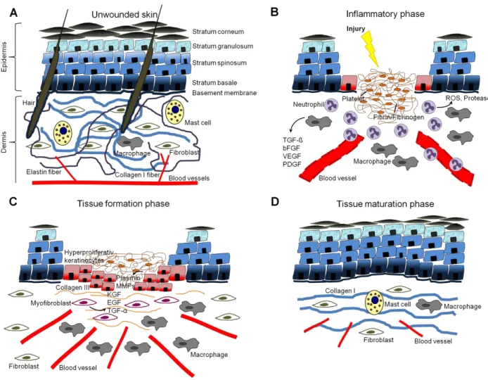
growth factor-A (VEGF-A), which promote directly or indirectly angiogenesis, cell proliferation and the synthesis of extracellular matrix (ECM) molecules by resident skin cells [12]. In contrast to neutrophils, macrophages stay at the wound site for the subsequent healing phases. The cartoon in figure 1 B illustrates the critical events of the inflammatory phase.
Figure 1: Skin morphology and the physiological sequence of wound healing. (A) Cartoon of normal unwounded skin, divided into in the keratinocyte-rich epidermis and the collagen-rich dermis.
(B) The inflammatory response three days post injury. A fibrin/fibrinogen-rich clot is formed and a lot of inflammatory cells are recruited. (C) The tissue formation phase five to seven days post injury.
Keratinocytes start to proliferate in order to reach wound closure. Myofibroblasts and new blood vessels are present to support wound healing. (D) Late phase of the tissue maturation phase resulting in a remaining scar tissue.
1.2.2 The tissue formation phase
The mid stage of the repair response consists of the phase of tissue formation, which is characterized by the development of granulation tissue, refilling the dermal wound space, and keratinocyte proliferation that closes the epidermal gap. Granulation tissue formation
4
encompasses the invasion of endothelial cells and angiogenesis, the influx of fibroblasts, differentiating into myofibroblasts, and the accumulation of additional macrophages.
During neo-angiogenesis, new vessels sprout out of existing vessels and along growth factor gradients, mainly consisting of VEGF-A and bFGF secreted by keratinocytes, macrophages, and other cell types [4]. To mediate this outgrowth, endothelial cells have to degrade the basement membrane and the surrounding ECM, which is accomplished by the expression of proteases, mainly matrix metalloproteases (MMPs), serin, and cystein proteases [13].
Furthermore, they have to alternate their integrin expression for the adhesion to the provisional ECM and successful migration [14]. Contemporaneous resident dermal fibroblasts start to proliferate and migrate from the adjacent unwounded skin area into the provisional matrix of the wound bed, in response to secreted TGF-ß. Once arrived, they produce a new collagen-rich matrix, mostly consisting of collagen type III. Besides of collagen deposition, some fibroblasts transform into myofibroblasts, which express α-smooth muscle actin (α-SMA) and promote wound contraction [15]. Wound contraction is a concerted action mediated by cell-cell and cell-matrix contacts as well as by tractional forces generated by migrating cells within the collagen matrix [16]. This contractile force supports the contemporaneously keratinocyte hyperproliferation and migration at the wound edge in order to restore the epidermal barrier, finally leading to wound closure. Growth factors which support proliferation and migration of keratinocytes are mainly epidermal growth factor (EGF), transforming growth factor-α (TGF-α) and keratinocyte growth factor (KGF) expressed by keratinocytes and dermal fibroblasts. As mentioned above in unwounded skin keratinocytes are attached to the basement membrane. This contact has to be dissolved and the integrin expression profile needs to be changed to allow crawling over the provisional fibrin-fibronectin wound matrix [17]. By expression of proteases, mainly plasmin and MMPs, keratinocytes carve path through the fibrin clot and the underlying dermal granulation tissue.
Once the wound area has been covered by a layer of keratinocytes their migration stops, and a new basal lamina is synthesized, to which both keratinocytes and fibroblasts contribute [17], followed by reestablishment of the stratified epithelium starting at the wound margins [18]. Granulation tissue formation continues until the wound space is refilled and the overlaying epidermis is restored. The critical events of the tissue formation phase are illustrated in figure 1 C.
1.2.3 The tissue maturation phase
Upon completion of the epidermal barrier, the repair response enters the last and longest stage, which is characterized by tissue maturation. During the phase of tissue maturation
5
granulation tissue transforms into scar tissue, characterized by attenuated cell proliferation, inflammation, neovascularization as well as replacement of the provisional matrix by deposition of collagen. Remaining vessels mature by recruitment of pericytes and the network re-organizes by pruning [19]. Most endothelial cells, macrophages and myofibroblasts undergo apoptosis or exit the wound, leaving a cell-poor and ECM-rich scar tissue [5]. Collagen type III, predominating in the wound matrix, is exchanged by a collagen type I network which is re-arranged, cross-linked and aligned along tension lines to increase tensile strength [20]. This process is supported by MMPs secreted by fibroblasts, macrophages and endothelial cells and strengthens the remaining scar tissue. However, tensile strength of uninjured skin is never re-established [21]. Also skin appendages like hairs and sweat glands are not regenerated. Figure 1 D illustrates the remaining scar tissue after wounding.
1.3 Macrophages
1.3.1 Macrophage origin
Organisms are exposed to many different pathogens in their environment. Besides physical barriers like the skin protecting the inner organism from pathogen infection passively, multi- cellular organisms developed additional immune host defense mechanisms against pathogens which are carried out by specialized cells and proteins. Vertebrates use two types of immune defense, first the rapid but pathogen-unspecific innate immune response and second the more effective and pathogen-specific adaptive immune response. The innate immune system is the first line of defense against invading pathogens. It is composed of different cell types including mast cells, dendritic cells (DCs), natural killer cells (NK cells), neutrophils and macrophages. Mast cells spontaneously degranulate upon infection and release different pro-inflammatory cytokines which in turn recruit other innate immune cells for phagocytosis and degradation of the invaded pathogens. Macrophages and DCs further link the innate and the adaptive immune response by presenting antigens to the corresponding T helper cells, effector cells of the adaptive immune system. Macrophages and many other leukocytes do not normally divide or reproduce by themselves. They develop from multipotent hematopoetic stem cells (HSCs) in the bone marrow. Two cell lineages originate from HSCs, the lymphoid lineage, containing T-, B-, and NK cells and the myeloid lineage, containing macrophages, neutrophilic granulocytes, and DCs among others.
The first developing stadium of macrophages is the highly proliferative monoblast which resides in the bone marrow and develops to the promonocyte stadium. The promonocytic
6
stadium is mainly found in the bone marrow as well, but can also enter the blood circulation, which is the main location for the following maturation state the monocytic stadium (Figure 2). Monocytes share already some typical features of macrophages such as phagocytic capacity and adhesion to specific endothelial cell molecules. They are a systemic reservoir for the renewal of tissue macrophages and DCs [22, 23]. Monocyte development depends on the availability of the growth factor colony-stimulating factor-1 (Csf-1, also known as macrophage colony-stimulating factor, M-CSF, and CD115) [24]. Different subpopulations of monocytes have been described in the circulation of mouse and humans, which are distinguishable by a different expression pattern of cell surface marker proteins [25, 26]. It is speculated that in mouse the Ly-6Chigh expressing monocyte subset is related to inflammatory conditions whereas the Ly-6Clow subset takes part in the renewal of tissue macrophages [26]. The majority of macrophages are stationed at critical points to police the respective organ in case of emergency. These so called tissue resident macrophages are found in skin, lung (alveolar macrophages), liver (Kupffer cells), bone (osteoclasts), neural tissue (microglia) and spleen (Figure 2). Without a specified stimulus or activation, macrophages have a vital homeostatic role by phagocytosis of erythrocytes, tissue debris and apoptotic cells. In contrast, in case of inflammation or tissue damage, monocytes adhere to endothelial cells via selectins or ICAMs. They transmigrate through the endothelial wall into the destination tissue where they mature and become activated macrophages.
Figure 2: Macrophage development and diversity. Multipotent hematopoetic stem cells in the bone marrow pass through different maturation states until they are released into the circulation as blood monocytes and differentiate into macrophages when entering the tissue. Monocytes represent a reservoir for the renewal of tissue resident macrophages, such as alveolar macrophages in the lung, Kupffer cells in the liver, osteoclasts in the bone, microglia cells in neural tissues and skin macrophages.
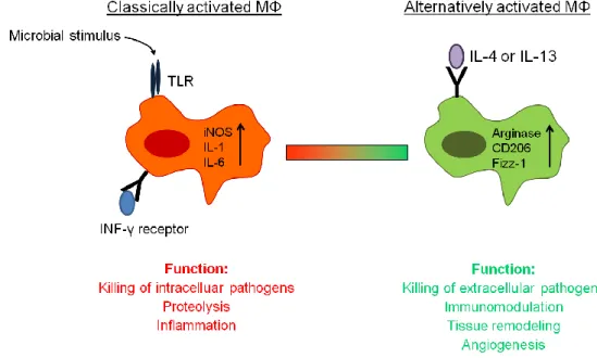
7 1.3.2 Macrophage activation and function
Classically macrophages become activated by bacterial compounds such as LPS, recognized by Toll-like receptors (TLR), and by T helper 1 cells (Th1) via interferon-γ (INF-γ) and the corresponding receptor, which both trigger a harsh pro-inflammatory response, required to kill intracellular pathogens, such as Mycobacterium tuberculosis, Leishmania spp.
or HIV [27] (Figure 3). Certainly, macrophages are plastic cells and they can adopt to different stimuli in the environment, leading to the concept of pro-inflammatory “classically activated M1” macrophages versus anti-inflammatory “alternatively activated M2”
macrophages. In contrast to the classically M1 activation state, the alternative activation state is T helper 2 cell (Th2) –mediated in response to the two cytokines interleukin-4 (IL-4) and interleukin-13 (IL-13) [28] (Figure 3). The M2 phenotype is in contrast to M1 macrophages described to be important for the immune response to extracellular pathogens, such as nematodes or helminths. In addition, M2 macrophages are thought to have an important role in tissue repair and homeostasis. They function as immunoregulator and contribute to the production of ECM [29]. But alternatively activated M2 macrophages can also be detrimental to the host when their matrix-enhancing activity is dysregulated, similarly to the dysregulated activity of classically activated macrophages in autoimmunity [29].
Besides their substantial role in innate immunity and homeostasis, macrophages are important player in tumor biology. On the one hand, they are involved in antitumor immunity (rather M1 macrophages), but on the other hand, there is substantial evidence that in the majority of tumors these tumor-associated macrophages (TAMs) enhance tumor progression to malignancy by supporting tumor-associated angiogenesis, promoting tumor cell invasion, migration, and intravasation, as well as suppressing antitumor immune responses [30]. TAMs are thought to resemble the M2 phenotype and to support tumor progression by secreting growth factors like VEGF-A, TGF-ß and PDGF [31].
M1 and M2 macrophages can be distinguished by the expression of different proteins. M1 macrophages highly up-regulate pro-inflammatory cytokines such as interleukin-6 (IL-6), interleukin-1ß (IL-1ß) and tumor necrosis factor-α (TNF-α) as well as inducible nitric oxide synthase (iNOS), regarding their function in host defense and bacterial killing. By contrast, the M2 macrophage phenotype is characterized by the expression of arginase 1 (antagonist of iNOS), mannose receptor (CD206), found in inflammatory zone-1 (Fizz1, also known as resistin like molecule-α, Relm-α), eosinophil chemotactic factor (Ecf, also known as Ym1) and selected chemokines [32] (Figure 3). The expression of the intracellular enzyme arginase is implicated in cell recruitment and granuloma formation, whereas the mannose receptor stimulates endocytosis [33]. Most of the work studying the importance and the phenotype of M2 macrophages was done in mouse and cannot be directly translated to
8
humans. Arginase 1 for instances cannot be used as a marker for human M2 macrophages because it is also induced by other pathways [28]. It is discussed that both activation stadia are not terminal and that they can switch from one to the other due to environmental stimuli.
1.3.3 The role of macrophages in wound healing
Numerous studies in the past revealed functional consequences of the innate immune response of resident cells as well as of recruited inflammatory cells during skin repair [34].
They combat invading microbes and contribute to debris scavenging, but may also critically support the repair process by releasing a spectrum of growth factors. However, due to the release of pro-inflammatory and cytotoxic mediators, the uncontrolled activity of macrophages may also be detrimental to tissue repair. Indeed, imbalanced inflammation characterized by increased numbers of macrophages is a hallmark of an attenuated repair response in human diseases including diabetes mellitus, vascular disease as well as aging [9, 35]. Neutrophils and macrophages represent the major fraction of inflammatory cells recruited to the wound site. Within a few hours post injury neutrophils transmigrate across the endothelial cell wall and begin the debridement of devitalized tissue and infectious agents as described above. Whereas the presence of neutrophils at the wound site is timely restricted to the early stage of the wound healing response, macrophages persist through all stages of the repair response. Their number increases during the phase of inflammation, peaks during the phase of tissue formation and gradually declines during the maturation phase [36].
Experiments in the 1970’s established the concept, that under sterile conditions, the influx of macrophages is essential for efficient healing of incisional skin wounds, whereas the influx of neutrophils might not be crucial [37, 38]. This dogma has been challenged by recent reports, thereby arguing against an essential role of inflammatory cells in wound repair: early fetal wounds heal with minimal scarring, which is associated with little inflammation [39].
Furthermore, wounds in the neonatal PU.1 null mouse, which lacks macrophages and neutrophils (but also B cells, mast cells, eosinophils), heal without scarring and, surprisingly, with a similar time course as wild-type siblings [40]. However, the need of macrophage and neutrophil influx for physiological repair in adults is supported by different studies using murine knock out models deficient for specific endothelial cell or leukocyte adhesion molecules (E-, P- selectins, ICAM-1, ß-1,4-galactosyltransferase, CD18) [41-44] or individual inflammatory mediators or their receptors (IL-6, CX3CR1) [11, 45]. These mouse mutants showed a dramatic delay in wound closure and a significantly reduced infiltration of neutrophils and macrophages.
9
Although these studies emphasize that leukocytes clearly affect the quality of the healing response, validity of these models is limited, because they either do not target pathways mediated exclusively by macrophages or neutrophils or they address a neonate repair response, which is known to differ from healing in the adult organism [46]. Furthermore, it is hypothesized that both macrophage phenotypes, M1 and M2 are present in wounds during physiological repair. The classically activated M1 differentiation state is thought to mediate apoptosis of damaged cells, killing of bacteria and destruction of matrix and extracellular structures, whereas the alternatively activated M2 phenotype seems to rather play an immunomodulatory role and to induce cell proliferation and angiogenesis (Figure 3). As both macrophage types are necessary at the inflamed site, the right balance between these two populations is required for healing and resolution of inflammation [47-49]. However, experiments proving this concept of the importance of M1 and M2 macrophages in skin repair are still missing.
Figure 3: Model of macrophage activation. Macrophages can either be activated by a microbial stimulus and INF-γ to function as a classically activated macrophage or by IL-4/IL-13 to act as an alternatively activated macrophage. TLR, toll-like receptor; iNOS, inducible nitric oxide synthase; IL, interleukin; INF-γ, interferon-γ; CD206, mannose receptor; Fizz-1, found in inflammatory zone; MΦ, macrophage.
1.4 Vascular endothelial growth factor-A
The vascular endothelial growth factor-A (VEGF-A) was discovered around thirty years ago, first as an inducer of vascular permeability in tumors, the reason why it is also known as
10
vascular permeability factor (VPF), and later as a strong endothelial cell specific mitogen [50, 51]. Since its discovery VEGF-A has been one of the most studied angiogenic growth factors and is thought to be of singular importance in vascular biology. The essential role of VEGF-A during developmental vasculogenesis (mobilization of bone marrow-derived endothelial stem cells) and angiogenesis (sprouting of capillaries from existing blood vessels) was shown by different knock out models, which unraveled that already a heterozygous knock out of VEGF- A is embryonically lethal [52, 53]. Further, it was shown in adults, that endothelial cells require an autocrine VEGF-A-mediated survival signal [54]. Meanwhile, the VEGF family in mammals consists of five members, VEGF-A, VEGF-B, VEGF-C, VEGF-D and placenta growth factor (PlGF). Furthermore, structurally related proteins were also found in parapoxvirus (VEGF-E) and snake venom (VEGF-F). All VEGF members are homodimeric glycoproteins and share a common structure of eight characteristically spaced cystein residues in a VEGF-homology domain. They have different physiological functions and exert them by binding in an overlapping fashion to three receptor tyrosine kinases, known as VEGFR-1 (also known as Fms-related tyrosine kinase-1, Flt-1), -2 (also known as kinase insert domain receptor, KDR or fetal liver kinase, Flk-1), and -3 (also known as Flt-4), as well as to co-receptors, including neuropillins (Nrp) and heparan sulfate proteoglycans [12, 55, 56] (Figure 4).
VEGF-A is the most prominent member of the VEGF family and is henceforth referred to as VEGF. VEGF is encoded by eight exons. Exon one to five encodes for the VEGFR-binding domains, whereas exons six and seven encode for two separate heparin binding domains [51]. Currently, eight alternative splice variants are described and named by their corresponding amino acid length (VEGF121, 145, 148, 162, 165, 183, 189, and 206). Their relative abundance varies among different tissues [57]. All VEGF isoforms differ in their capability to bind to cell surfaces, the ECM and the corresponding receptors and have therefore their own biological significance [12]. VEGF121, VEGF165 and VEGF189 are the major isoforms expressed in VEGF expressing cells. VEGF121 lacks both exons encoding for heparin binding domains and represents therefore a soluble isoform. VEGF165 comprises the heparin binding domain encoded by exon 6 which mediates a moderate binding to heparin, and VEGF189
contains both heparin binding domains and has therefore a strong affinity to ECM structures.
Due to alternate capabilities of different VEGF isoforms to bind ECM proteins VEGF gradients can be formed [12]. Among the different isoforms, VEGF165 is the major gene product found in human tissues. In mice, VEGF-isoforms are one amino acid shorter than in humans, and therefore denoted as VEGF120, VEGF164, VEGF188 and so on.
The major VEGF functions are mediated in binding to VEGFR-2 which is mostly expressed on endothelial cells but also on neuronal cells and hematopoetic stem cells [58]. VEGFR-2 is
11
a type III transmembrane receptor tyrosine kinase. The extracellular domain of the receptor comprises seven immunoglobulin-like domains, of which the second and third interact with VEGF. A single transmembrane domain connects the outer domain to the intracellular two tyrosine kinase domains (Figure 4). Binding of VEGF to VEGFR-2 leads to enhanced proliferation, migration, cell survival and permeability by the induction of various complex signaling pathways (for review see [58]). Besides, VEGF is also able to bind to VEGFR-1, but VEGFR-1 signaling is less well understood. Strong experimental evidence indicates that VEGFR-1 on the vasculature may act primarily as a ligand-binding molecule during angiogenesis, rather than as a signaling tyrosine kinase [12]. Furthermore, in vitro studies revealed that VEGFR-1 on monocytes/macrophages promotes chemotaxis [59].
Figure 4: VEGF members and their receptors. Schematic representation of interactions between VEGF ligands and their transmembrane or soluble receptors (sVEGFR-1) as well as co-receptors (Modified: Eming et al., 2007, [12]).
1.4.1 VEGF function in angiogenesis
Almost all tissues depend on blood supply, which in turn depends on endothelial cells, which form the inner linings of the blood vessels. Endothelial cells originate in the early embryo during development from bone marrow-derived stem cells. Early embryonic endothelial cells migrate, proliferate, and differentiate to form the first rudimental blood vessels in a process
12
termed vasculogenesis. Subsequent growth and branching of the vessels throughout the embryo and later in adults during tissue repair is mainly mediated by proliferation and migration of endothelial cells of pre-existing vessels, in a process termed angiogenesis. New vessels originate as a capillary sprout from the side of an existing capillary. Under physiological situations, endothelial cells are in a quiescent state, thus, they need to be activated and re-programmed during the early stages of angiogenesis [60]. Hypoxia is a strong stimulator for angiogenesis by leading to stabilization of the transcription factor hypoxia-induced factor-1α (HIF-1α) which in turn induces the expression of a wide range of pro-angiogenic mediators such as angiopoietins, TGF-ß, bFGF and VEGF (Figure 5). VEGF then stimulates endothelial cell proliferation and directed migration. For endothelial cell migration, cell-cell contacts have to be disrupted, an additional function of VEGF activity [61].
The impaired cell barrier leads furthermore to vascular leakage and extravasation of plasma proteins, forming a provisional matrix constituting an anchorage point for migrating cells via integrins. In addition, VEGF stimulates the expression of different proteases to digest the basal membrane and the surrounding matrix to allow cell invasion. The outgrowing vascular sprout is guided by a single specialized endothelial cell, distinctive by tip structures with potential functions in guidance and migration [62] (Figure 5). The endothelial cells following the tip cell, named stalk cells, are hyperproliferative, while the tip cell is not. The tip cell guides the way by expressing VEGFR-2 and recognizing extracellular VEGF gradients released by nearby oxygen-deficient tissue [63] (Figure 5). Tip cells meet and fuse, forming blood vessel loops in a process termed anastomosis, to develop a new vascular network;
though the precise mechanism remains unclear [64]. It is speculated that macrophages might function as bridge cells between two sprouting vessels in order to prepare them for fusion [65, 66]. Lateral inhibition prevents overgrow of blood vessels during angiogenesis by a precise selection of tip cells. This inhibition is mediated by delta/notch signaling. The delta like-4 ligand (Dll4) is up-regulated in tip cells and inhibits via signaling through the notch receptor, expressed by stalk cells, the same cell fate in neighboring cells [67] (Figure 5). Dll4 expression in turn is induced by VEGF, indicating not only a stimulatory, but also a modulatory role of VEGF in angiogenesis [68]. The newly formed blood vessels are highly instable, leaky and, dependent of VEGF signaling for their survival. Vessels mature by the recruitment of pericytes, which cover the outside of the vessels and help to build a novel basal membrane [69]. Besides of the pro-angiogenic effects of VEGF in developmental and postnatal angiogenesis, there is substantial evidence that implicates VEGF as a mediator of pathological angiogenesis [55]. VEGF is highly up-regulated in the vast majority of human tumors, expressed by tumor and stromal cells, in order to supply the tumor environment with blood vessels. Anti-VEGF therapy in humans and VEGF knock out mouse models could substantiate the supporting effect of VEGF on tumor growth.
13
Figure 5: Model of vascular sprouting. Induced by a VEGF gradient, specialized tip cells guide the expanding sprout by exploring the environment for tissue gradients of guiding cues. They are followed by proliferating stalk cells, which form the sprout and generate lumen. Tip cell fate is determined by lateral inhibition, mediated by Dll4/Notch signaling.
1.4.2 The role of VEGF and macrophage-derived VEGF in wound repair
Angiogenesis is a central event during wound healing because the restoration of blood flow to the site of injured tissue is necessary to mount the initial immune response to pathogens, and at the same time to initiate repair of wounded tissue. Thus, VEGF, as a key regulator of angiogenesis represents an important mediator in skin repair. VEGF expression is nearly absent in unwounded skin, but highly induced after injury, mainly by hypoxia but also in response to different pro-inflammatory cytokines and growth factors [70].
A central function of VEGF during skin wound healing is supported by different studies in which reduced expression or activity of VEGF caused severe wound healing defects [11, 70].
The first study indicating VEGF as an important factor in wound healing was done in the wound healing impaired diabetic mouse model (db/db). It was shown that VEGF amounts were dramatically decreased, directly linking the reduced angiogenic response observed in this mouse model to the subsequent impaired healing response [70]. In another study it was shown that under the pathological healing conditions in the db/db mouse model, VEGF is a target for increased proteolytic activity, probably causing in the wound healing defects in diabetic mice [71]. Furthermore, these impaired healing conditions can be rescued or improved by applying a mutant protease resistant VEGF variant [72, 73].
14
Mechanistically, during the early phase of repair release of VEGF into the wound is likely to contribute to increased vascular permeability, which mediates extravasation and extravascular deposition of plasma proteins (fibrinogen, fibronectin), a process central for the formation of the provisional wound matrix [74]. During the phase of tissue formation, VEGF provokes wound angiogenesis. Further, it is discussed that VEGF serves as a chemoattractant for monocytes and macrophages. Both express VEGFR-1 and can therefore answer to VEGF signals [75].
Initial studies of wound healing demonstrated that VEGF is mainly expressed by keratinocytes at the wound edge and by recruited macrophages [71, 74]. Some in vitro studies could further show that also platelets, mast cells, pericytes and fibroblasts are able to secrete VEGF. Additional wound healing studies with two different reporter mouse lines for VEGF expression gave contradictive results with regard to the sites of VEGF expression during repair. One model revealed VEGF expression mainly in wound fibroblasts, whereas in the other model keratinocytes were the main source for VEGF supply [12, 76, 77]. These different results can be explained by different promoter regions used and by the fact that the human VEGF promoter was used in these mouse models. However, the contribution of different cell compartments of VEGF expression during skin repair is not well understood.
The importance of one specific cellular compartment releasing VEGF in wounds was shown by a Cre-mediated knock out for VEGF in keratinocytes, which results in an impaired healing response and decreased susceptibility to chemically-induced skin carcinogenesis [78].
Surprisingly, however, in wound repair consequences of VEGF depletion in keratinocytes became only apparent after wound closure had been completed, whereas granulation tissue formation and angiogenesis during the early phase occurred normally. This study suggests that keratinocyte-derived VEGF is dispensable for the early healing response, but plays a role in the later phases, indicating that during the phase of inflammation, other or additional cells must be a source for VEGF. Macrophages for example are the dominant cell type during the early steps of repair and they express VEGF [71]. Important stimuli for macrophage activation and induction of VEGF expression are hallmarks of microenvironmental conditions found in injured tissues, including hypoxia and lactate [79].
Furthermore, the importance of macrophage-derived VEGF on angiogenesis was shown by two recently published articles. A Cre-mediated knock out for VEGF exclusively in macrophages was analyzed in a mouse model for breast cancer and lung fibrosis [80, 81]. In both cases, the macrophage-specific knock out for VEGF had fundamental influences on the outcome of the angiogenic response, which was diminished.
15
2 Specific aims
Skin injury leads to an acute phase response, which is characterized by activation of the innate immune system, resulting in the activation of various repair mechanisms.
Physiologically, when timely limited, the inflammatory response is beneficial for wound closure. However, when the acute inflammation persists and a chronic inflammatory response develops, it leads to severely impaired healing conditions. Therefore, it can be speculated that the inflammatory response is a crucial target to impact the outcome of the healing response. Currently, it is not completely understood how macrophages exactly influence the physiology of the repair response and how they may contribute to the pathology of healing in diseased conditions. Therefore, a more thorough understanding of macrophage- specific functions during the diverse phases of the repair response might broaden the understanding of this cell type in skin physiology and pathology. So far it is not examined how different cellular compartments contribute to VEGF-A supply in wounds. Macrophages could be an eminent source releasing significant amounts of this growth factor into wounds.
The specific aims of this study are:
1.) To test the hypothesis that macrophages present at the wound site during the different stages of skin repair exert specific functions. To this end, a mouse model is needed that allows the conditional depletion of macrophages in a timely restricted fashion during the distinct phases of the repair response in skin to delineate repair mechanisms dependent or independent from macrophage function.
2.) To determine the time course of VEGF-A expression in wounds and to identify different cellular compartments of VEGF-A expression by using a VEGF-lacZ reporter mouse, in which both proteins, VEGF-A and ß-galactosidase are expressed under the control of the murine VEGF-A promoter.
3.) To analyze the specific functional impact of macrophage-derived VEGF-A on the outcome of the healing response by using conditional gene targeting to specifically deplete VEGF-A expression in myeloid cells.
16
3 Results
3.1 Cell type specific and timely restricted depletion of macrophages in LysMCre/iDTR mice
To analyze the functional impact of macrophages during diverse phases of skin repair, mice were generated in which macrophages can be inducibly ablated, using a model of Cre- inducible diphtheria toxin receptor-mediated cell ablation. This system is based on a Cre- inducible human diphtheria toxin receptor transgenic mouse line (iDTR) in which Cre- mediated excision of a STOP cassette, downstream of the ubiquitous active Rosa26 promotor, renders naturally diphtheria toxin (DT)-resistant mouse cells DT sensitive (kindly provided by Ari Waisman [82]). To generate mice in which macrophages can be inducibly ablated, the iDTR mouse line was crossed to lysozyme M Cre (LysMCre) mice, reported to express the Cre recombinase in cells of myeloid origin (macrophages and neutrophils) (kindly provided by Irmgard Förster [83], Figure 6).
Figure 6: Mouse model for inducible myeloid cell depletion. (A) To generate mice in which myeloid cells can be indubly ablated the iDTR mouse line, in which the expression of the DT receptor is under control of the Rosa26 promotor after Cre-mediated excision of a stop cassette, was crossed to the LysMCre mouse line. (B) Genotyping of iDTR (left) and LysMCre (right) mice by PCR. The 300 bp fragment represents the iDTR gene, whereas the 600 bp fragment shows the wild type allele in the iDTR PCR. For LysMCre, a 700 bp fragment indicates the presence of the gene encoding the Cre recombinase. The 350 bp fragment indicates the wild type allele. bp, base pairs; DTR, diphtheria toxin receptor; LysM, lysozyme M; wt, wild type; fl, loxP site.
A
B
17
First, it was investigated how macrophage depletion can be controlled by the dose, kinetics, and route of diphtheria toxin (DT) application (intraperitoneal versus intravenous), as well as the DTR gene dose (heterozygous versus homozygous). As revealed by flow cytometry for macrophages (F4/80 and CD11b), a single intraperitoneal (i.p.) injection of DT (25 ng/g bodyweight) in LysMCre/iDTR(heterozygous) mutants resulted in complete depletion of peritoneal macrophages 24 hours later, that persisted approximately for 3 days (Figure 7 A).
In contrast, total numbers of peritoneal B cells (CD19) as identified by flow cytometry were similar in LysMCre (control) and LysMCre/iDTR(heterozygous) mice, demonstrating the specificity of macrophage depletion following DT injection. By contrast, depletion of tissue resident macrophages in skin, liver and spleen was not achieved by a single i.p. injection of DT in LysMCre/iDTR(heterozygous) mice, as revealed by immunohistochemical staining for F4/80 (Figure 7 B, mid panel). Also repetitive i.p. DT injections did not result in efficient depletion of resident macrophages in these tissues. Therefore, it was investigated whether increasing the DTR gene dose and intravenious (i.v.) DT application affects the efficacy of tissue resident macrophage depletion. In fact, repetitive i.v. injections of DT (25 ng/g bodyweight) at two consecutive days in LysMCre/iDTR(homozygous) mice resulted in efficient depletion of both peritoneal and tissue resident macrophages in skin, spleen and liver one day after DT injection (Figure 7 A, B). Furthermore, the same regimen of DT application resulted in efficient depletion of circulating monocytes (CD115, CD11b) 24 hours later (Figure 7 C). Interestingly, although the lysozyme M gene is also expressed in polymorphonuclear leukocytes, DT injection did not result in efficient depletion of neutrophils (Gr-1) in the circulation (Figure 7 C). To investigate whether myeloid cells can be efficiently depleted under inflammatory conditions, LysMCre/iDTR(homozygous) and LysMCre mice received two i.v. injections of DT at consecutive days, which 24 hours later was followed by a single i.p injection with thioglycolate. This substance is standardly used to induce a sterile peritonitis characterized by a strong infiltration of macrophages and neutrophils into the peritoneal cavity. After thioglycolate injection, mice received DT injections i.p. for 3 consecutive days, and at day 4 post thioglycolate application, peritoneal lavage cells were analyzed. As revealed by flow cytometry for macrophages (F4/80, CD11b) and neutrophils (Gr-1) only macrophages were efficiently ablated, while the treatment did not affect neutrophil populations (Figure 7 D). Whether increased neutrophil number in LysMCre mice is simply the result of reduced phagocytosis by macrophages or results from the lack of different macrophage-mediated control mechanisms, is currently unknown. For the subsequent wound healing studies LysMCre/iDTR(homozygous) and LysMCre (control) mice were used.
18
19
Figure 7: Selective depletion of macrophages in LysMCre/iDTR mice. (A) Analysis of macrophage depletion by peritoneal lavage following a single i.p. injection of DT (25 ng/g bodyweight) in LysMCre/iDTR(heterozygous) or LysMCre/iDTR(homozygous) mice resulted in efficient depletion of peritoneal macrophages (F4/80, CD11b) 24 hours after DT injection that lasted for 3 days. Specificity of macrophage depletion was identified by normal B cell numbers (CD19). DT injection in LysMCre (control) mice had no effect on macrophage numbers. (B) Tissue resident macrophages (stained by F4/80) in skin, spleen and liver were efficiently depleted in LysMCre/iDTR(homozygous) mice 24 hours following i.v. DT injections (25 ng/g bodyweight) at two consecutive days. Tissue resident macrophages were not affected by DT injection in LysMCre mice and partially depleted in LysMCre/iDTR(heterozygous) mice. (C) Efficient depletion of monocytes (CD11b, CD115) and partial depletion of neutrophils (Gr-1) in the circulation 24 hours after two i.v. DT injections at consecutive days. (D) Effective depletion of macrophages (F4/80, CD11b) but not neutrophils (Gr-1) in LysMCre/iDTR(homozygous) mice after thioglycolate induced peritonitis. e, epidermis; d, dermis; sc, subcutaneous fat layer; wp, white pulp; rp, red pulp.
3.2 Macrophage function during the different stages of repair
To analyze macrophage function during the different phases of tissue repair, the above described mouse model was used and a cell depletion scheme in which macrophages were inducibly depleted in a timely restricted manner during the different phases of repair was developed (Figure 8). To this end, injections were performed on specific days prior or post wounding to achieve macrophage depletion during the inflammation, the tissue formation or the maturation phase (Figure 8 regimen A to C).
3.2.1 Macrophage function during the early stage of repair
3.2.1.1 Macrophages recruited during the inflammatory phase of repair induce granulation tissue formation, which results in scar formation
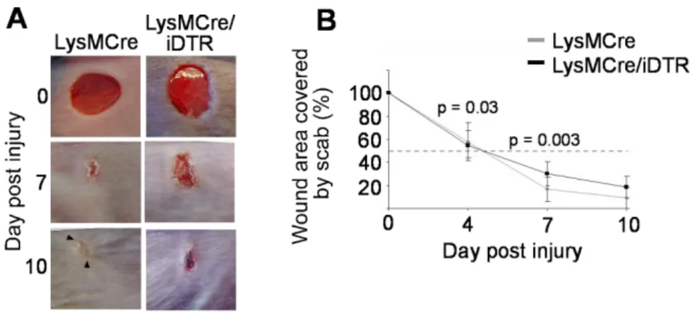
First, the impact of macrophages recruited during the early inflammation phase was analyzed by cell depletion following injection scheme A (Figure 8). For this purpose, mice were first injected with DT twice prior wounding to prevent an initial influx of macrophages after injury, which was followed by two i.p. DT injections at day two and four post injury. Macroscopic assessment of wound closure showed that depletion of macrophages during the inflammatory phase resulted in a significant delay of the early repair response compared with control (LysMCre) mice. While at day 5 post injury, the wound area was reduced to 50% of the original wound size in control mice, in macrophage-depleted wounds of LysMCre/iDTR mice the wound size was reduced by only 25%. However, at later time points, when DT
20
injections were discontinued, wounds in LysMCre/iDTR mice demonstrated rapid wound closure comparable to control wounds (Figure 9).
Figure 8: Schematic representation of DT-mediated macrophage depletion in distinct stages of the repair response. To achieve wound-phase-restricted depletion of macrophages LysMCre/iDTR and LysMCre mice were injected with DT (intraveniously, i.v. or intraperitoneally, i.p.) according to three regimens: (A) DT injection regimen A, macrophage depletion during the inflammatory phase: DT injections 2 and 1 days prior wounding as well as at day 2 and 4 post wounding; (B) DT injection regimen B, macrophage depletion during the tissue formation phase: DT injections at day 3, 4, 6 and 8 post injury; (C) DT injection regimen C, macrophage depletion during the maturation phase: DT injections at days 8, 9, 11, and 13 post injury. At time points indicated mice were sacrificed and the wound tissue was excised for analysis. MΦ, macrophages; DT, diphtheria toxin.
21
Figure 9: Macrophage depletion during the early stage of repair results in a delayed wound closure rate. (A) Macroscopic appearance of wounds in LysMCre/iDTR and LysMCre (control) mice after DT injection following regimen A; whereas wounds of control mice had already lost their scab, macrophage-depleted wounds in LysMCre/iDTR mice still carry a firmly adherent scab 7 days post wounding. (B) At the time points indicated, the wound area was determined using image analysis and expressed as percentage of the wound area immediately after injury (n = 12 wounds on 6 mice for each time point and genotype). Data are expressed as mean ± SD.
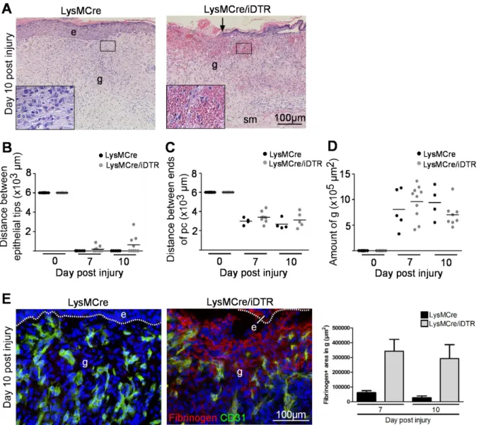
The macroscopic findings were confirmed by histological assessment. For this purpose LysMCre/iDTR and control mice were sacrificed on day 5, 10 and 14 after injury and the wound tissue (5-12 wounds on 3-6 mice per time point for each group) was excised and analyzed. For histological quantification, the amount of granulation tissue, the distance between the two ends of the epithelial tips and the distance between the ends of the panniculus carnosus were measured on H&E stained paraffin sections (see overview in Figure 10 A).
At day 5 post injury, a significantly shorter distance between the tips of the epithelial tongues, representing the longitudinal diameter of the wound, was measured for the control wounds compared to macrophage-depleted wounds (Figure 10 B, C). Furthermore, as revealed by expression of the cell proliferation marker Ki67, the epidermal margins in control wounds were hyperproliferative, whereas the epidermal wound edge in macrophage-depleted wounds was short and showed few proliferating keratinocytes (Figure 10 B, G). To analyze dermal repair the amount of granulation tissue formation in wound tissue of macrophage- depleted and control mice was determined. Differences in the quantity of granulation tissue were analyzed in H&E-stained sections and were shown to be significantly reduced at all time points in macrophage-depleted wounds compared to controls (Figure 10 E). Whereas at
22
day 5 post injury control wounds showed a highly vascularized, cellular and proliferative granulation tissue, in macrophage-depleted wounds granulation tissue was scarcely vascularized and showed few proliferating cells (Figure 10 B, G). Wound contraction was significantly reduced in LysMCre/iDTR mice when compared to controls (Figure 10 D).
At day 10 post injury, wounds in LysMCre/iDTR and control mice were still covered by eschar (Figure 9 A) and histological analysis revealed that in both a complete neo-epithelium had formed beneath the eschar (Figure 10 B). However, in LysMCre/iDTR mice, the epithelium appeared fragile, was thinner, and partially detached from the dermis, when compared to control mice, indicating immature anchorage and basement membrane formation.
Furthermore, although in these wounds the quantity of granulation tissue increased when compared to day 5 post injury, it was significantly reduced compared to control wounds (Figure 10 B, E). Also wound contraction remained reduced in LysMCre/iDTR mice when compared to controls (Figure 10 D).
At day 14 post injury both wounds in LysMCre/iDTRand control mice had lost their eschar and were similar in their macroscopic appearance (Figure 9 A). In contrast, major differences between wounds in LysMCre/iDTRand control mice became apparent regarding the extent of scar tissue formation. As revealed by H&E (Figure 10 B) and Sirius red staining analyzed in polarized light (Figure 10 F), fine collagen bundles characteristic for scar tissue were almost absent in wounds of LysMCre/iDTRmice. Morphometric quantification of scar tissue revealed a significant reduction in LysMCre/iDTR mice when compared to controls (Figure 10 E). Morphological analysis of the epidermis overlaying the scar tissue revealed a slightly hyperproliferative, closed epithelium which was similar in mutant and control wounds (Figure 10 B).
23
24
Figure 10: Macrophage depletion during the early stage of repair attenuates epithelialization, granulation tissue formation and wound contraction. (A) H&E stained wild type wound section 5 days following injury. (B) H&E stainings of wounds in LysMCre (control) and LysMCre/iDTR mice at indicated time points after injury. Whereas in LysMCre mice, the day-5 wound is filled with a vascularized granulation tissue in LysMCre/iDTR mice only scarce granulation tissue has formed (black hatched line outlines granulation tissue, white dotted line outlines hyperproliferative epithelial tongue); in day-10 wounds of LysMCre and LysMCre/iDTR mice the granulation tissue is covered by a complete epithelium; however the epithelium is detached; day-14 wounds of LysMCre and LysMCre/iDTR are closed, scar tissue is minimal in LysMCre/iDTR mice (hatched line outlines scar tissue). (C-E) Morphometric analysis of wound tissue at different time points post injury: (C) Distance between the epithelial tips; (D) Distance between the edges of the panniculus carnosus; (E) Amount of granulation tissue (day 5 and 10 post injury) or scar tissue (day 14 post injury). Each dot represents one wound (day 5: two wounds on one mouse, day 10 and 14: one wound per mouse); horizontal bar represents the mean. (F) Sirius red staining and examination with polarized light revealed increased scar formation in control wounds when compared to LysMCre/iDTR wounds 14 days post injury. (G) Left: day 5 wounds of LysMCre and LysMCre/iDTR mice stained for Ki67 (green) and propidium iodide (red), arrowheads indicate Ki67 positive cells (yellow). Right: quantification of Ki67 positive cells in the hyperproliferative epithelium. Data are expressed as mean ± SD, n = 3 wounds on 3 mice for each time point and group. Hatched line indicates basement membrane. e, epidermis; he, hypertrophic epidermal wound edge; d, dermis; sm, subcutaneous muscle layer; pc, panniculus carnosus; g, granulation tissue; st, scar tissue; arrows point to the tips of epithelial tongue, white arrowheads indicate wound edges, black arrowheads indicate edges of panniculus carnosus.
3.2.1.2 Macrophage depletion in wounds of LysMCre/iDTR mice receiving DT injections following regimen A
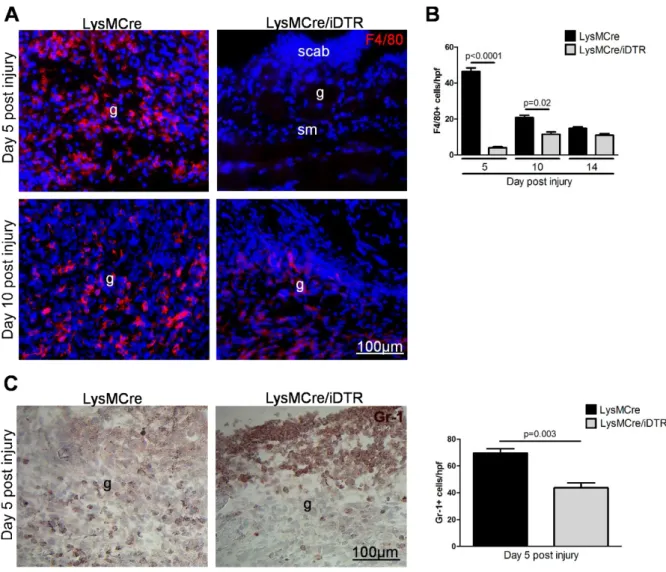
To analyze whether changes observed in the tissue repair response between LysMCre/iDTR and control (LysMCre) mice correlated with macrophage depletion, wound tissue was stained for the macrophage marker F4/80. In control wounds 5, 10 and 14 days post injury, F4/80 positive cells were present throughout the entire granulation tissue (Figure 11 A, B).
Quantification of F4/80 positive macrophages revealed that their number was highest at day 5 post injury and subsequently declined to approximately 1/3 until 14 days post injury (Figure 11 B). In contrast, in LysMCre/iDTR mice receiving DT injections following regimen A (Figure 8), F4/80 positive cells were absent at day 5 post injury but appeared at the wound site at day 10 and 14 post injury (Figure 11 A, B). Thus, in LysMCre/iDTR mice, the absence of macrophages during the early stage of repair corresponded to the time course of DT injections. Furthermore, the data revealed that after DT injections were abolished, newly generated macrophages were recruited into the wound site during the consecutive phases of tissue formation and maturation. Finally and most importantly, the results demonstrate that macrophages recruited during the inflammatory phase impact repair mechanisms not only in the early stage of the repair response, but also in the consecutive mid and late stages of repair. As revealed by staining for the neutrophil marker Gr-1 (Figure 11 C) 5 days post injury neutrophils were not effectively depleted at the wound site by DT injections in this model, which is in agreement with the data presented in figure 6 D after thioglycolate-induced

![Figure 4: VEGF members and their receptors. Schematic representation of interactions between VEGF ligands and their transmembrane or soluble receptors (sVEGFR-1) as well as co-receptors (Modified: Eming et al., 2007, [12])](https://thumb-eu.123doks.com/thumbv2/1library_info/3615759.1501473/18.892.174.712.448.866/receptors-schematic-representation-interactions-transmembrane-receptors-receptors-modified.webp)