Inaugural-Dissertation zur Erlangung des
Doktorgrades der Mathematisch-Naturwissenschaftlichen Fakultät der Universität zu Köln
Engineering AAV-2 Targeting Vectors:
A New Insertion Site and scFv Driven Vectors
vorgelegt von
Jorge Miguel Martins Bouças
aus Lissabon, Portugal
Köln, August 2008
Berichterstatter/in:
Prof. Dr. Manolis Pasparakis Prof. Dr. Herbert Pfister PD Dr. Hildegard Büning
Tag der mündlichen Prüfung: 28. Oktober 2008
Eidesstattliche Erklaerung
Ich versichere, daß ich die von mir vorgelegte Dissertation selbständig angefertigt, die benutzen Quellen und Hilfsmittel vollständig angegeben und die Stellen der Arbeit – schließlich Tabellen, Karten und Abbildungen -, die anderen Werken in Wortlaut oder dem Sinn nach entnommen sind, in jedem Einzelfall als Entlehnung kenntlich gemacht habe; daß diese Dissertation noch keiner anderen Fakultät oder Universität zur Prüfung vorgelegen hat; daß sie – abgesehen von unten angegebenen Teilpublikationen – noch nich veröffentlicht worden ist sowie, daß ich eine solche Veröffentlichung vor Abschluß des Promotionsverfahrens nicht vornehmen werde.
Die Bestimmungen dieser Promotionsordnung sind mir bekannt.
Köln, 19. August 2008 Jorge Miguel Martins Bouças
I thank PD Dr. Hildegard Büning for accepting me in her group, for constant and valuable scientific advice and for personal support during these years.
I would like to thank Prof. Dr. Michael Hallek for opening me the doors to the group of PD Dr. Büning and creating the opportunity that led to the realization of this work.
Also, I thank Prof. Manolis Pasparakis for supporting my thesis.
All this work would have not been as successful and exciting without the collaboration of all my friends and colleagues of the Büning group and of the University of Cologne.
A especial thank goes to Prof. Dr. Hinrich Abken, Prof. Dr. Dagma Moersdorf, Prof. Dr. Ulrike Protzer, Dr. Andreas Hombach, Dr. Sibille Quadt- Humme, Dr. David Kofler, Dr. Luca Perabo, Dr. Kerstin Lux, Dr. Marianna Hössel, Dr. Caro Kopecky, Dr. Venkateswara Rao Simhadri, Dr. Marcus Chmielewski, Dr.
Natascha Schuhmann, Stefanie Stahnke, Hanna Janicki, Stefan Maersch, Anke Huber, Silke Uhrig, Tobias Riet, Heike Köhler, Martin Zuther and Patrick Schmidt for stimulating discussions and practical help.
I thank all those that so kindly received me in Cologne with open arms.
Finally I thank my mother and my cousins for continuous support over a life span. I thank to all those that during the last decade treated me as brother and especially to who, without any obligation, treated me as a son.
I especially thank Mareike for the extraordinary help, understanding and patience, along the years involving this work.
Die vorliegende Arbeit wurde in der Zeit von August 2005 bis August 2008 an der Klinik I für Innere Medizin der Medizinische Fakultät der Universität zu Köln unter der Anleitung von PD Dr. Hildegard Büning verfasst.
Im Verlaufe dieser Arbeit wurden folgende Veröffentlichungen angefertigt:
Perabo L. Goldnau D., White K., Endell J., Boucas J., Humme S., Work L.M., Janicki H., Hallek M., Backer A.H. and Buning H. (2006). Heparan sulfate proteoglycan binding properties of adeno-associated virus retargeting mutants and consequences for their in vivo tropism. J Virol 80(14), 7265-9.
PCT/EP2008/004368; Buning H., Lux K., Nieland J., Boucas J., Ritter M., Hoerer M., (2008) Mutated structural protein of a parvovirus. World Intellectual Property Organization
Perabo L., Boucas J., Coutelle O., Hallek M. and Buning H. 2008. Engineering of viral capsids to evade the host immune system. In R. Herzog (ed.), Immunology of Gene Therapy. John Wiley & Sons. In press.
Boucas J., Lux K., Huber A., Schievenbusch S., Perabo L., Quadt-Humme S., Odenthal M., Hallek M., Buning H. (2008). Engineering AAV-2 based targeting vectors using a new insertion site – 453 – and single point mutations. J Gene Med, under review.
Boucas J., Hombach A., Hallek M., Abken H. and Buning H. (2008). Construction of scFv-VP2 directed AAV-2 vectors. Manuscript in preparation.
During all those years of experimentation and research, I never once made a discovery. All my work was deductive, and the results I achieved were those of invention, pure and simple.
Thomas Edison
To my mother
ZUSAMMENFASSUNG 4
SUMMARY 8
1. INTRODUCTION 11
1.1. GENE THERAPY 11
1.1.1. TYPES AND METHODS 11
1.1.2. VECTORS 12
1.1.3. STATE-OF-THE-ART AND PRESENT GOALS 13
1.2. ADENO-ASSOCIATED VIRUS 16
1.2.1. GENOMIC ORGANIZATION OF AAV 17
1.2.2. INFECTIOUS BIOLOGY OF AAV 19
1.2.3. TISSUE DISTRIBUTION OF AAV ISOLATES 25
1.2.4. IMMUNE RESPONSES TO AAV 25
1.2.5. PRODUCTION OF RECOMBINANT AAV VECTORS 26
1.3. TARGETING VECTORS 29
1.3.1. GENETIC CAPSID MODIFICATIONS 29
1.3.2. NON-GENETIC CAPSID MODIFICATIONS 36
1.3.3. COMBINING GENETIC AND NON-GENETIC CAPSID MODIFICATIONS 37
2. OBJECTIVES 38
3. MATERIALS 40
3.1. CHEMICALS, SOLUTIONS AND ENZYMES 40
3.2. PLASMIDS 41
3.3. PRIMERS 45
3.4. ANTIBODIES AND PROTEINS 47
3.5. BACTERIA STRAIN 48
3.6. EUKARYOTIC CELLS 49
3.7. DATA TREATING SOFTWARE 50
3.8. GI AND ACCESSION NUMBERS 50
3.9. LABORATORY EQUIPMENT, DISPOSABLES AND KITS 50
4. METHODS 52
4.1. VISUALIZATION OF STRUCTURES AND MOLECULAR MODELING 52
4.2. BACTERIA CULTURE 52
4.2.1. CULTIVATION OF BACTERIA 52
4.2.2. PREPARATION OF COMPETENT BACTERIA 52
4.2.3. TRANSFORMATION OF BACTERIA 53
4.3. DNA TECHNIQUES 54
4.3.1. PLASMID AMPLIFICATION AND EXTRACTION 54
4.3.2. DNA QUANTIFICATION 56
4.3.3. RESTRICTION ENZYME DIGEST 56
4.3.4. AGAROSE GEL ELECTROPHORESIS 56
4.3.5. CLONING 57
4.4. EUKARYOTIC CELL CULTURE 59
4.4.1. CULTIVATION OF CELLS 59
4.4.2. TRYPSINISATION 59
4.4.3. SEEDING / PASSAGING 59
4.4.4. FREEZING AND THAWING CELLS 60
4.4.5. COUNTING 60
4.5. PRODUCTION OF AAV-2 VECTORS 60
4.5.1.PRODUCTION OF HBS-BUFFER 60
4.5.2. AAV VECTOR PACKAGING 61
4.5.3. IODIXANOL GRADIENT PURIFICATION 62
4.6. VECTOR TITRATION 62
4.6.1. GENOMIC TITER 62
4.6.2. TRANSDUCING TITER 63
4.6.3. CAPSID TITER 64
4.7. CELL TRANSDUCTION 65
4.7.1. COMPETITION STUDIES 65
4.7.2. BINDING PARTICLES 66
4.8. PROTEIN TECHNIQUES 66
4.8.1. PROTEIN QUANTIFICATION 66
4.8.2. WESTERN BLOT 66
4.8.3. ELISA 68
4.9. IMMUNOPHENOTYPING 70
4.10. BIODISTRIBUTION STUDY IN MICE 70
5. RESULTS 71
5.1. G453 AS A NEW AAV-2 INSERTION SITE 71
5.1.1. INSERTION OF THE RGD4C PEPTIDE 72
5.1.2. INSERTION OF THE NGRI PEPTIDE 83
5.1.3. INSERTION OF AN N587 DISPLAY LIBRARY SELECTED PEPTIDE 86
5.1.4. TRANSLATION TO OTHER PARVOVIRINAE 88
5.2. FUSING SCFVS TO AAV-2’S VP2 FOR THE GENERATION OF NEW TARGETING
MUTANTS 91
5.2.1. GFP IS LOCATED ON THE OUTER SURFACE OF THE CAPSID 91
5.2.2. ACD30-GFP-VP2 92
5.2.3. ACD30NVP2 95
5.2.4. OPTIMIZING EXPRESSION IN CIS 96
5.2.5. OPTIMIZING EXPRESSION IN TRANS 97
5.2.6. FUNCTIONAL SCFV 100
5.2.7. TRANSDUCTION OF TARGET CELLS 101
5.2.8. ANTI-CA19.9 SCFV 105
5.2.9. ANTI-CEA SCFV 106
6. DISCUSSION 109
ABBREVIATIONS 122
REFERENCES 123
Zusammenfassung
Die Gentherapie ist eine alternative Strategie zur Behandlung von Gendefekten und malignen Erkrankungen, die als therapeutisches Agens eine Nukleinsäure verwendet. Essentiell für den Erfolg vieler gentherapeutischer Ansätze ist ein effizienter und Zell-spezifischer Gentransfer. Hierzu werden Zell-spezifische Vektoren, sogenannte Targeting-Vektoren, benötigt.
Für das in dieser Arbeit verwendete Vektorsystem, das auf dem nicht- pathogenen Serotyp 2 des Adeno-Assoziierten Virus (AAV-2) beruht, wird bevorzugt das direkte oder genetische targeting verwendet. Hierbei werden Peptidliganden in das AAV-Kapsid eingefügt, die dann die Interaktion mit dem gewünschten zellulären Rezeptor vermitteln. Als besonders geeignete für die Insertion und Präsentation von Peptidliganden hat sich hierbei die Aminosäureposition 587 (sowie die benachbarte Position, 588) erwiesen. In Ermangelung einer 3D-Struktur wurde zu ihrer Identifizierung ein Sequenz-Alignment mit einem verwandten Parvovirus, dem Canine Parvovirus (CPV) von dem entsprechende Strukturinformationen vorlagen, durchgeführt.
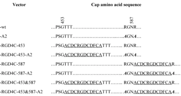
In der vorliegenden Arbeit wurde zum ersten Mal die nun vorhandene 3D- Struktur des AAV-2-Kapsid verwendet, um eine neue Klasse von Insertionsmutanten zu generieren. In silico-Analysen ermöglichten die Identifizierung der Aminosäureposition 453 als eine mögliche Insertionsstelle, die aufgrund ihrer deutlich besser exponierten Lage eine effizientere Interaktion zwischen einem hier inserierten Liganden und seinem Rezeptor ermöglichen sollte. Dies wurde mit Hilfe des Modell- Liganden RGD4C (ACDCRGDCDFCA), der an αVβ3 und αVβ5 Integrine bindet, untersucht. Hierzu wurde der Modell-Ligand genetisch in die Kapsidposition 453, 587 oder in beide Positionen gleichzeitig eingefügt. Da eine Insertion in Aminosäureposition 453 – im Gegensatz zu einer Insertion in 587 - nicht mit der Bindung des AAV-2-Kapsid an seinen Primärrezeptor Heparansulfatproteoglykan (HSPG) interferiert, wurde parallel ein zweiter Mutantensatz generiert, bei dem die Insertionsmutanten zusätzlich zwei Aminosäuresubstitutionen (R585A und R588A) enthielten. Diese Aminosäuresubstitutionen (A2) führen zum Verlust der Bindungsfähigkeit des Kapsids an HSPG.
Alle Mutanten konnten effizient hergestellt werden und waren mit Ausnahme der Doppelinsertionsmutanten zur effizienten Transduktion von HeLa-Zellen
(permissiv für AAV2) befähigt. Interessanterweise scheint die „Unfähigkeit“ zur Transduktion, die für die Doppelinsertionsmutanten beobachtet werden konnte, nicht in einer „Unfähigkeit“ zur Zell- oder Rezeptorbindung begründet zu sein, da sowohl eine Zellbindung als auch die Rezeptorbindung nachgewiesen werden konnte. In einem im Rahmen dieser Arbeit entwickelten ELISA konnte gezeigt werden, dass alle Insertionsmutanten die Fähigkeit zur Bindung des Zielrezeptors aufweisen.
Interessanterweise, zeigten alle Mutanten, die zusätzlich zur Insertion die A2- Mutation trugen, eine deutlich effizientere Rezeptorbindung als die „reinen“
Insertionsmutanten. Die effizienteste Mutante war r-RGD4C-453-A2 mit Peptidinsertion in der „neuen“ Insertionsstelle und zusätzlicher A2-Mutation. Diese Mutante war zudem deutlich effizienter als rAAV-2 (mit unmodifiziertem Kapsid) und als die übrigen Mutanten in der Transduktion der HSPG-defizienten Zelllinie CHO psA-745. Kompetitionsexperimente mit löslichen Peptiden sowie mit Heparin (lösliches Analogon des HSPG) auf verschiedenen Zelllinien sowie auf primären HUVEC (Human Umbilical Vein Endothelial cells) zeigten, dass der inserierte Ligand die Zelltransduktion vermittelt, wenn die natürliche (HSPG-) Rezeptorbindung verhindert wird. Die in Maus durchgeführte Bioverteilungsstudie machte deutlich, dass – nach Eliminierung der HSPG-Bindung – die Bioverteilung von Targetingvektoren, die eine Insertion in Position 453 oder 587 tragen, allein vom inserierten Liganden bestimmt wird.
Die Insertion eines alternativen Peptids (NGRI) in 453 führte zur Generierung des AAV-Targetingvektors r-NGRI-453, mit dem HeLa-Zellen effizienter transduziert werden konnten als durch rAAV-2. Mit diesem Peptid konnten zudem infektiöse Doppelinsertionsmutanten hergestellt werden. Ein Peptid, dass durch AAV Peptide display für die Position 587 selektioniert worden war, konnte zwar erfolgreich in die Position 453 eingebaut werden; der entsprechende Vektor wies aber keine verbesserte Transduktionseffizienz auf. Zusätzlich wurde mit Hilfe der NCBI Concerved domain database zu 453 im AAV-2 Kapsid homologe Positionen in anderen Parvoviren identifiziert, um die Anwendungsmöglichkeiten der in dieser Arbeit identifizierten und optimierten Insertionsstelle zu erweitern.
Somit konnte in diesem Teil der Arbeit gezeigt werden, dass sich die Aminosäureposition 453 zur Insertion von Tropismus-modulierenden Peptidliganden eignet. Doppelinsertionsmutanten (Peptidliganden in Position 453 und 587) können
sein. Ein weiterer interessanter Befund ist die deutliche Verbesserung der Rezeptorbindungsfähigkeit durch Kombination von RGD4C-Insertion und A2- Mutation. Dies stellt das erste Beispiel für die Bedeutung von Punktmutationen zur Optimierung von Targeting-Vektoren dar. Auf diesem Hintergrund wird die Wichtigkeit von evolutionären Ansätzen und high throughput-Selektionen für die Vektorentwicklung noch einmal deutlich. In diesen Ansätzen werden aus Bibliotheken von Kapsidmutanten für die jeweilige Anwendung optimierte Mutanten selektioniert.
Die sich mehrenden Berichte über die hohe Spezifität von Antikörperfragmente (single-chain Antikörper; scFv) motivierte das zweite Projekt dieser Arbeit. Da scFv aufgrund ihrer Größe ungeeignet für eine Insertion innerhalb von AAV-Kapsidproteinen z.B. an Position 453 oder 587 sind, wurde in dieser Arbeit der Versuch unternommen, Fusionsproteine zwischen scFv und dem N-Terminus von VP2 (zweitgrößtes AAV-Kapsidprotein) in das Kapsid von AAV einzubauen. Da wir vor kurzem zeigen konnten, dass Fusionsproteine aus GFP und VP2 in das Kapsid eingebaut werden, dass das GFP auf der Kapsidoberfläche exponiert wird, und dass GFP-markierte Viren infektiös sind, wurde zunächst ein Fusionsprotein aus anti- CD30 scFv-GFP-VP2 hergestellt. Diverse Optimierungen des Verpackungsprotokolls waren nötig, um anti-CD30 scFv-GFP-VP2 enthaltende Vektorpartikel zu generieren.
Diese Mutanten konnten HeLa-Zellen transduzieren und interagierten mit einem anti- idiotypischen Antikörper. Es konnte jedoch keine spezifische Transduktion von CD30-positiven Zellen nachgewiesen werden. Außerdem konnte weder nach einer noch nach vierstündiger Inkubation der Zellen mit Vektorlösung Vektor-DNA in oder an Zellen nachgewiesen werden. Weder die Eliminierung des GFP-Anteils noch das Einbringen von linker-Sequenzen führte zu einer Verbesserung der Bindungs- oder Transduktionseffizienz. Der Austausch des anti-CD30 scFv gegen ein anti-CEA scFv und ein anti-CA19 scFv führte zu denselben Ergebnissen, woraus geschlossen werden kann, dass die mit anti-CD30 scFV erhaltenen Ergebnisse nicht spezifisch für den anti-CD30 scFv sind.
Vergleicht man Studien, die vollständige Immunglobuline verwenden, mit dieser Studie, so könnte die geringe Vektorpartikelkonzentration einen limitierten Faktoren darstellen. Zudem finden sich pro AAV-Kapsid nur 5 VP2-Proteine, d.h. es können maximal 5 VP2-Fusionsproteine eingebaut werden. Dies ist deutlich weniger als bei Targeting-Vektoren, die einen Ligand z.B. in Position 453 inseriert enthalten.
Hier wird der Ligand in alle 60 Kapsidproteinen eingebaut und steht so deutlich häufiger für eine Rezeptorbindung zur Verfügung.
Trotzdem darf festgehalten werden, dass durch die hier durchgeführte Arbeit, das bisher größte Fusionsprotein (~ 127 kDa) in das AAV-Kapsid eingebaut werden konnte. Diese Tatsache zusammen mit der Beobachtung, dass sich sowohl das GFP als auch der anti-CD30 scFv auf der Kapsidoberfläche befinden, eröffnet viele neue Möglichkeiten der Anwendung. So könnte AAV-2 gleichzeitig als Proteincarrier (Fusion mit dem N-Terminus) und als Gencarrier (Transgen) fungieren.
Summary
Gene therapy can be defined as the introduction of nucleic acids into cells with the purpose of altering the course of a medical condition or disease. In many clinical settings, efficient and successful gene therapy relies on the delivery of genes to specific cells in the human body. Such specific delivery can only be achieved through the design of vehicles/vectors that are able to recognize the target cell – targeting.
Adeno-associated virus type 2, a non-pathogenic human virus, has received an increased amount of attention as a vector for gene therapy since it was first cloned into a bacterial plasmid in 1982. Although the first attempt to construct an AAV-2 targeting vector made use of a single chain antibody fragment, it was the insertion of small peptide ligands into the AAV-2 capsid that marked the beginning of AAV-2 vector targeting. Sequence alignment of AAV-2 with CPV led to the discovery of amino acid position 587. Several reports have shown that small peptide ligands, once inserted in this position, are able to mediate transduction of the respective target cells, and that target specificity is retained in vivo.
In this work, the available three dimensional structure of AAV-2 is used for the first time for the rational design and construction of targeting vectors. In silico analysis revealed that insertions at position 453 should result in a better exposure of the inserted peptides and, as a consequence, in a more efficient interaction with the target receptor. The RGD4C peptide – a ligand for αVβ5 and αVβ3 integrins – was inserted in position 453 and/or 587. Moreover, loss of AAV-2 wild-type (wt) tropism was achieved by R585A and R588A – A2 – mutations that abolish binding to the primary receptor – heparan sulfate proteoglycan, HSPG.
All mutants were able to efficiently package and, with the exception of the double RGD4C mutants, to transduce HeLa cells. Interestingly double insertion mutants – r-RGD4C-453&587 and r-RGD4C-453&587-A2 – were able to efficiently bind to cells. Cell free based assays, involving targeting vectors and one target receptor revealed that all insertion mutants were able to bind the target receptor.
Surprisingly, addition of A2 mutations to targeting vectors resulted in an increased amount of bound receptors. A mutant containing RGD4C at 453 and A2 mutations – r-RGD4C-453-A2 – emerged as the most efficient mutant. This mutant was also the most efficient vector for the transduction of an HSPG ko cell line – CHO pgsA-745.
Competition studies with soluble peptides verified specificity of transduction in this and other cell lines, as well as in primary Human Umbilical Vein Endothelial Cells – HUVEC, which often serve as a model for tumor endothelial cells. In vivo biodistribution studies revealed that, once HSPG binding is abolished, vector distribution is determined by the inserted peptide, independent of the insertion position and independent of the modification that led to elimination of the wt tropism.
Insertion of the NGRI peptide in position 453 – r-NGRI-453 – resulted in a vector that was more efficient than wt AAV-2 in the transduction of HeLa cells.
Furthermore, double insertion mutants remained infectious. Insertion of an N587 display library selected peptide in position 453 was not detrimental but did not result in a vector with improved transduction efficiencies. NCBI’s Conserved Domain Database was used to identify 453 homologues in other vector systems.
In summary, 453 emerged as a suitable position for the insertion of targeting peptides in AAV-2 and other vector systems. Double insertion mutants will have to be analyzed for each specific case. The increased targeting efficiency after single point mutations of residues linearly distant from the inserted peptide shows for the first time how such mutations can indeed be relevant for the design of targeting mutants.
Moreover, high-throughput selection protocols emerge as master tools and should be put into practice for the identification of similar mutants and for the optimization of targeting vectors.
The rising number of reports on the high efficiency of antibody fragments for the construction of targeting molecules motivated the fusion of single chain antibody (scFv) fragments to the capsid of AAV-2. Being most likely too large for insertions in non-terminal positions like 453 or 587, we decided to genetically fuse a scFv to the N- terminus of a viral protein - VP. Furthermore, vectors containing a GFP-VP2 fusion protein have previously been shown to be useful tools for infectious biology studies.
Our analysis of this GFP labeled vectors revealed that at least a part of the GFP molecule is present on the outer surface of the capsid. These observations led to generation of an anti-CD30 scFv fused to the N-terminus of a GFP molecule that was fused to the N-terminus of VP2.
Optimization of the packaging procedure, by increasing the amount of the fusion protein encoding plasmid, resulted in an efficient packaging of scFv-AAV-2 mutants. Mutants were able to effectively transduce HeLa cells and to bind an anti-
targeting mutants were not able to specifically transduce CD30 positive cells.
Furthermore it was not possible to detect vector DNA with target cells neither 1 nor 4 hours post-transduction. Engineering similar constructs without the GFP molecule and with or without linker sequences did not result in an improved binding or transduction efficiencies. Substitution of the anti-CD30 scFv by an anti-CEA scFv or an anti-CA19 scFv revealed that the results observed with the anti-CD30 scFV targeting mutants were not idiotype specific. When compared to studies made with whole immunoglobulins, the low vector particle concentration emerges as one of the limiting steps to the application of such targeting approaches. Moreover, while each capsid possesses sixty repeats of G453, only five VP2 proteins exist per capsid.
Despite this, the scFv-GFP-VP2 fusion represents the largest fusion (~127 kDa in total) ever assembled in an AAV capsid. This, together with the fact that both GFP and anti-CD30 scFv molecules could be recognized by respective antibodies reinforces the idea that the N-terminus of VP2 can indeed be displayed on the outer surface of the capsid. Our results open the door to many other therapeutic designs where the vector can be used as a carrier for genes and at the same time for high molecular weight proteins assembled in pentavalent forms.
1. Introduction
1.1. Gene therapy
Genes, which are carried on chromosomes, are the basic physical and functional units of heredity. Genes are specific sequences of bases that encode instructions on how to make proteins. Although genes get a lot of attention, it’s the proteins that perform most life functions and even make up the majority of cellular structures. When genes are altered so that the encoded proteins are unable to carry out their normal functions, genetic disorders can result.
The application of recombinant DNA technology and gene cloning (which started in the 1980s) and the resulting increase in genomics data during the 1990s have contributed to define some disease-causing genetic factors and to explore the potential of new therapies based on engineered genes and cells (161, 190)
Gene therapy can be defined as the introduction of nucleic acids into cells for the purpose of altering the course of a medical condition or disease. In general with some exceptions, the nucleic acids are DNA molecules encoding gene products or proteins. The original ideas were directed toward treating monogenic (single-gene) disorders, but it has become clear that the gene can be considered a new pharmaceutical agent for treating many types of diseases (96).
Gene therapy is a complex process, involving multiple steps in the human body (delivery to organs, tissue targeting, cellular tracking, regulation of gene expression level and duration, biological activity of therapeutic protein, safety of the vehicle and gene product, to name just a few) most of which are not completely understood.
1.1.1. Types and methods
Gene therapy approaches can be divided into two types: germ line therapy and somatic therapy. In the first type, a gene is inserted into the DNA of the germline cells (egg or sperm) so that the offspring of the patient will have the inserted gene. This more recent approach, theoretically, should be highly effective in counteracting genetic disorders. Somatic gene therapy involves the manipulation of gene expression
in cells that will be corrective to the patient but not inherited by the next generation (somatic cells include all the non-reproductive cells in the human body).
For the correction of faulty genes four different approaches are normally used:
a) a normal gene may be inserted into a nonspecific location within the genome to replace a nonfunctional gene; b) an abnormal gene can be swapped for a normal gene through homologous recombination; c) the abnormal gene can be repaired through selective reverse mutation, which returns the gene to its normal function; d) the regulation (the degree to which a gene is turned on or off) of a particular gene could be altered.
Transfer of the genetic material can be achieved either ex vivo or in vivo. In the first case the patient cells are harvested and cultivated in the laboratory. After incubation with carrier molecules containing a corrective or therapeutic gene the cells with the new genetic information are then harvested and transplanted into the patient.
Although normally less feasible for wide-scale application due to its complexity, this method is quite attractive due to the improved safety gained by the possibility to separate target from non-target cells. In most cases though, efficient therapy requires direct in vivo administration of the vehicle. Although, in situ administration may help avoiding the transfer of genetic material to non-target cells, the carrier molecule needs in this case to discriminate between target and non-target cells. Thus, carrier molecules are one of the masterpieces of most gene therapy designs.
1.1.2. Vectors
A carrier molecule called a vector must be used to deliver the therapeutic gene to the patient's target cells. There are two major classes of vectors: viral vectors and non-viral vectors.
Some researchers believe that viral vectors will be most successful because viruses have evolved for millions of years to become efficient vesicles for transferring genetic material into cells, whereas others believe that some of the side effects of such viral vectors and possible previous exposures to the respective viruses rendering the host resistant to transduction (gene transfer into the cell) will preclude their long-term use in gene therapy.
There is no "perfect vector" that can treat every disorder. Like any type of medical treatment, a gene therapy vector must be customized to address the unique
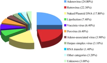
features of the disorder. Furthermore, interpatient variations can not be forgotten and must be taken into account. For such reasons, a wide variety of vectors have been used in clinical trials worldwide (Figure 1).
Figure 1. Vectors used in gene therapy clinical trials worldwide. Includes data relative to 1,347 of approved, ongoing or completed clinical trials worldwide. As of March 2008. (1)
1.1.3. State-of-the-art and present goals
Gene therapy is not a new idea. In 1963, Joshua Lederberg (1925-2008) wrote:
“We might anticipate the… interchange of chromosomes and segments. The ultimate application of molecular biology would be the direct control of nucleotide sequences in human chromosomes, coupled with recognition, selection and integration of the desired genes…It will only be a matter of time… before polynucleotide sequences can be grafted by chemical procedures onto a virus DNA.” Less than 30 years later, the first clinical study using gene transfer was reported (31).
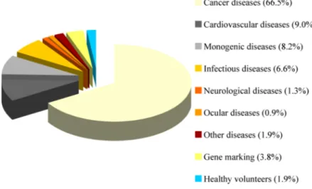
The disease states that have been approached with gene therapy are now widely diverse, including autossomal dominant disorders, many forms of cancer, HIV, and other infectious diseases, inflammatory conditions, and intractable pain (Figure 2). In most instances, the nature of the disease may dictate the vector to be used, since it may involve a specific cell type, expression level, and duration of expression that will be required for therapeutic effect. In general, genetic diseases will require long- lasting gene expression, and so may indicate the use of a gammaretrovirus, lentivirus, or AAV vector; while a tumor vaccine or cytotoxic gene approach to cancer may
require a short burst of gene expression, such as might be produced by an adeno or non viral vector.
Figure 2. Diseases addressed by gene therapy clinical trials. Includes data relative to 1,347 of approved, ongoing or completed clinical trials worldwide. As of March 2008. (1)
For a disease to be a good candidate for gene therapy, the role of the therapeutic gene in disease pathophysiology must be clearly understood. It seems likely that in future, more gene therapy targets will be identified, primarily as a result of the rapid ability to identify specific gene associations with human diseases, particularly after the completion of the Human Genome Project. This includes both single gene diseases, and, multifactorial diseases in which complex networks of gene and environmental effects are elucidated, and key genetic determinants of disease are identified within those pathways. The future remains uncertain regarding the ultimate clinical impact that gene therapy will have in each of those very distinct fields.
Indeed, clinical progress has been slow. After the first clinical trial in 1990 using a gammaretroviral vector a major setback occurred in September 1999 when a widely publicized death resulting from a gene therapy trial was reported (19, 155).
The resulting investigation concluded that the patient died from a massive immune reaction against the used Adenovirus type 5 vector. Fortunately, less than 1 year after a successful trail was published. Two children suffering from a severe combined immunodeficiency disorder (SCID-XI), which had restricted them to life in an isolated environment, were able to leave the hospital and resume normal lives after ex-vivo transduction of their lymphocytes with a gammaretrovirus vector (26). Out of the approximately 18 patients that were treated with the same type of vector 5
developed leukemia (50, 52). While in 4 of these patients the complications resulted from vector integration in a nonrandom manner near the LM02 gene, in the 5th patient unphysiologic expression of the IL2RG transgene is suspected to contribute to the development of the complications (50). Although chemotherapy already led to sustained remission in 3 of the patients with T cell leukemia, it failed in one (70). A clinical trial to correct a clotting disorder, Factor IX deficiency, by hepatic gene transfer using an AAV vector recently showed that transient correction was possible but quite limited in time because of subsequent immune reaction (116). In the end of 2007, 3 patients having an inherited blind disease with onset during childhood underwent subretinal delivery of an AAV vector. Each patient had a modest improvement in measures of retinal function on subjective tests of visual acuity (114).
Recently, a clinical setting for the treatment of ADA-SCID using a gammaretrovirus – GIADA1MLV retroviral vector – showed that ADA gene transfer is an efficacious treatment for ADA-SCID (25).
The development of versatile vectors with defined and controlled functions is without any doubt one of the present major goals in the field. Indeed, while the natural target cell of one vector might make it the best candidate for the designed therapy, the lack (or not) of integration capacity might make it inadequate. The low amount of vector preparations utilized in most trials linked to the small number of subjects involved in average in each gene therapy clinical trial reveals how important basic aspects like production are still to be optimized (121). Development of massive production and high purification methods that result in stock solutions with high titers and purity is indeed part of the present goals. A lot has been made in the vector field during the last 18 years. Transferring the knowledge obtained with one vector system to another as shown efficient results but many lessons are still to be learned.
Furthermore, the lack of more multifunctional groups working each one with a wider variety of systems and the need for more work to be done in vivo and ex vivo using adequate animal models has created a dependency on other areas of research that made the field develop in short steps.
Despite the early high expectations and the subsequent set-backs, one has to recognize that this new therapeutic modality is still in its infancy. It will neither deliver medical “miracles” (as its early prophets predicted) nor will it “disappear'”
because of a few disappointing cases (as some of its recent antagonists predict). As
“development phase” before it reaches “maturation”, when its full potential will be exploited. This in turn will offer significant opportunities to effectively target the causative factors for several disabling diseases afflicting mankind.
1.2. Adeno-associated virus
Adeno-associated virus (AAV) was discovered in 1965 when different groups described small, uniformly formed, virus like particles which were noticed during electron microscopical examination of simian adenovirus type 15 (SV15) (7). These particles were 18-20 nm in diameter and showed an icosahedral symmetry (119).
Staining with acridin orange demonstrated that these particles contained DNA, which contributed to the suggestion that those particles were viruses. The particles replicated only in cells that were coinfected with adenovirus. The authors named the particles adeno-associated virus (7).
Today, AAV has been classified as a member of the Parvoviridae. With a diameter of only 18 to 30 nm the Parvoviridae are among the smallest known viruses (latin: parvum = small). Viruses of this family contain a single-stranded DNA genome of approximately 5 kb and a non-enveloped icosahedral capsid. The family of Parvoviridae contains two subfamilies: the Densovirinae which infects invertebrates and the Parvovirinae which is specific for vertebrates. The subfamily Parvovirinae includes the genera Parvovirus, Erythrovirus and Dependovirus. Adeno-associated viruses belong to the Dependovirus genus. Parvovirus B19 is the only human pathogen within the Parvoviridae (Erythrovirus genus) and causes Erythema infectiosum, hydrops fetalis and abortion (23, 185). All other representatives of this family, including AAV, are not pathogenic for humans (17). On the contrary, AAV seems to be protective against bovine papillomavirus and adenovirus mediated cellular transformation (34, 82, 98, 118) and to have cytotoxic effects in malignant cells (154).
In contrast to the Parvovirus and Erythrovirus genus, which are autonomous Parvoviridae, AAV replication, and thus a productive infectious life cycle, depends on co-infection with unrelated helper viruses e.g. adenovirus (Ad), herpesvirus (HSV), human cytomegalovirus (HCMV), or papillomavirus (16, 129). In the absence
of a helper virus AAV establishes a latent form of infection by stably integrating its genome into the host cell genome. Helper viruses can be partially replaced by chemical or physical carcinogens (80, 204-206). This leads to the conclusion that helper viruses induce specific changes in the host cell and thereby providing competence for AAV replication.
Until now, 12 serotypes which share different levels of sequence homology, have been identified (56, 58, 126). AAV-1 to -4 and -6 have been detected as contaminants of adenoviral preparations. AAV-5 was isolated from penile condylomata lata (human wart) (11), AAV-6 seems to be a recombination between AAV-1 and -2 (199). AAV-9 was isolated from human tissues (56). AAV-7 and AAV-8 have been detected in Rhesus monkey, AAV-10 and AAV-11 in Cynomolgus monkey (58, 126). Sera epidemical studies suggest that AAV-2, AAV-3 and AAV-5 are epidemic in humans, whereas AAV-4, -7-11 are endemic in nonhuman primates (58). The natural occurrence of AAV-1, -6 and -12 is not known.
Although the other serotypes have attracted increasing attention during recent years, AAV-2 is still the best characterized serotype, being the first isolated and cloned.
1.2.1. Genomic organization of AAV
Wild type AAV-2 contains a single stranded DNA genome of 4679 nucleotides (178). The genome can be divided into three functional subunits. These are the two open reading frames (ORF) rep and cap flanked by the inverted terminal repeats (ITR) (24). It contains three promoters (p5, p19 and p40) and a common polyadenylation signal (Figure 3). The 5’-ORF rep encodes four Rep proteins, a family of multifunctional, nonstructural proteins. The different Rep proteins are named upon their molecular weight: Rep78, Rep68, Rep52 and Rep40. The larger Rep proteins are controlled from the p5, the smaller Rep proteins from the p19 promoter (112). Splicing of a common intron leads to Rep68, a splice variant from Rep78, and Rep40 from Rep52. The larger Rep proteins are important for site specific integration, control of replication and transcription (27, 142-144). The smaller Rep proteins seem to be involved in accumulation and packaging of single-stranded DNA into the preformed capsid (45, 100). The Rep proteins can act as transactivators of
transcription in the presence of helper virus functions and as repressors of the three viral promoters in absence of a helper virus (106, 142).
Figure 3. Organization of the AAV genome. The AAV genome encompasses 4680 nucleotides, divided into 100 map units. Indicated are the two inverted terminal repeats (ITRs), the three viral promoters at map position 5, 19, and 40 (p5, p19, and p40) and the polyadenylation signal at map position 96 (poly A). The open reading frames are represented by rectangles, untranslated regions by solid lines and the introns by nicks intercepting the solid lines. Large Rep proteins (Rep78 and Rep68) controlled by the p5 promoter and small Rep proteins (Rep52 and Rep40) driven by the p19 promoter exist in spliced and unspliced variants (Rep68 as a splice variant of Rep78; Rep40 as a splice variant of Rep52). The cap genes encoding the three different capsid proteins VP1, VP2, and VP3 are controlled by the p40 promoter. Figure kindly provided by Dr. N. Huttner.
The 3’-ORF cap encodes the three capsid proteins VP1, VP2 and VP3, which form the 60 subunits of the viral capsid in a 1:1:10 ratio (105). All three capsid proteins are controlled by the p40 promoter and use the same stop codon. VP2 and VP3 are N-terminal truncated variants of VP1. Synthesis of VP1 is regulated by alternative splicing whereas VP2 is initiated from an unusual translation initiation codon (ACG) (14, 15). The molecular weight of VP1, VP2 and VP3 is 90 kDa, 72 kDa and 60 kDa, respectively. VP3 alone is sufficient for capsid formation, but VP1 is required for viral infection (189). At least ex vivo VP2 seems not essential for capsids formation and infectivity (113, 189). Capsid assembly takes place inside the nucleus (193, 194).
The 145 bp long ITRs form hairpins of a T-shaped structure which contains Rep binding sites (RBS) and a terminal resolution site (TRS) which is a specific cleavage site for Rep proteins (90, 120, 176). They serve as origin of replication, are important for site-specific integration and rescue of the provirus from the human chromosome 19 (107, 122, 164).
1.2.2. Infectious biology of AAV 1.2.2.1. Virus cell contact
Of all the serotypes, AAV-2’s infectious biology is the best characterized.
However, a detailed understanding of intracellular trafficking, endosomal release and viral uncoating is still missing. Moreover, most experiments were performed within the same cell line, the human cervix carcinoma cell line HeLa. The current model of the infection process is depicted in Figure 4.
Figure 4. The AAV infection pathway. AAV touches the membrane several times before entering the cell. Attachment to its primary receptor HSPG and co-receptors such as FGFR and αvβ5 integrin is triggering a receptor-mediated endocytosis in a dynamin dependent manner into clathrin coated pits. This internalization is facilitated by the activity of Rac1. Activation of Rac1 subsequently stimulates PI3K pathways which regulate endosome trafficking along the cytoskeleton. The exact mechanism of endosomal release is not clear yet. Viral uncoating takes place before or during nuclear entry. Viral DNA enters the nucleus by an unknown mechanism. (Figure modified from Büning et al. 2003.)
Single Virus Tracing (SVT) studies characterized the motion of AAV-2 outside the cell as normal diffusion with a diffusion coefficient of D = 7.5 µm2/s. In this studies AAV-2s’ diffusion decelerated as it approached the cell membrane, and finally stopped when AAV got in contact with the cell membrane with a mean touching time of 62 ms. Most virions showed multiple contacts to the cell before entering or being finally released from the cell membrane. In average 4.4 repetitive touching events are observed. It is not clear whether these multiple touching events represent a binding and release process to viral receptors or adsorption to cellular structures (170).
Heparan sulfate proteoglycan (HSPG) has been identified as the primary receptor of AAV-2 (182). HSPG binding residues are located within the VP3 region (which is common to all capsid proteins). 5 amino acids have been identified to be involved in HSPG binding: R484, R487, K532, R585 and R588. Mutational analysis showed that especially R585 and R588 are essential for the interaction with HSPG (97, 135, 198). Even though HSPG has been described as AAV-2’s attachment receptor, it was shown that AAV-2 is able to enter some cells in the absence of HSPG (21). It was proposed that HSPG is also the primary receptor for AAV-3 whereas a 2,3-O- and 2,3-N-linked sialic acid was identified as the attachment receptor for AAV-4 and -5, respectively (76, 92, 153). For AAV-6 the situation is quite complex.
Seiler and colleagues observed that the use of sialic acid as attachment receptor for AAV-6 is dependent on cell type and cell differentiation status (169). Moreover, although AAV-6 can bind to heparin, it does not interfere with viral infection when applied together with virus onto the cell (74). The 37/67-kDa lamin receptor (LamR) was recently described as receptor or AAV-8. AAV-1 does not appear to utilize heparan sulfate, sialic acid or LamR and its receptor remains unknown (152). The same holds true for AAV-7. However, since it closely resembles AAV-1, they may share a common receptor (152). Even less is known about potential (attachment) receptors for AAV-9 to -12 (56, 57, 126, 167).
In addition to the attachment receptor, secondary receptors are required for viral infection. Five coreceptors have been described for AAV-2 so far, human fibroblast growth factor receptor I (hFGFR I), αVβ5-integrin, α5β1-integrin, hepatocyte growth factor receptor (HGFR) and LamR (6, 95, 149, 152, 181). Human FGFR I was also shown to interact with AAV-3 (18). For hFGFR I a function in enhancing the interaction of virion and HSPG was proposed (149). The function of HGFR is not known yet. Since blocking of αVβ5-integrin with antibodies can prevent internalization of rAAV-2 into HeLa cells it was suggested that binding to αVβ5- integrin mediates endocytosis (165). α5β1-integrin is thought to be an alternative coreceptor to αVβ5 (6). For AAV-5 platelet derived growth factor receptor (PDGFR) was identified as coreceptor (38). PDGFR was also discussed to act alone as a receptor for AAV-5 because it is in itself a sialo-glycoprotein (33).
1.2.2.2. Receptor mediated endocytosis of AAV
The endocytotic process was studied for AAV-2 and 5. Following receptor binding AAV-2 enters the cell by a receptor mediated endocytosis through clathrin coated pits in a dynamin dependent manner (13, 43). Single Virus Tracing measurements showed an individual viral uptake within milliseconds. AAV-5 was also predominantly localized in clathrin coated vesicles. However, in rare situations, AAV-5 was found to be endocytosed in noncoated vesicles, representing probably caveolae (10). Similar to ligand-receptor interaction, it was shown that receptor binding of AAV-2 causes intracellular signal transduction. Binding to αVβ5 activates (in addition to mediate endocytosis) Rac1, a small GTP binding protein, stimulating thereby phosphoinositol-3 kinase (PI3K) which facilitates the rearrangement of microfilaments and microtubuli (165). Treatment of infected cells with nocodazole, which leads to depolymerization of microtubules, or with cytochalasin B, which disrupts microfilaments, reduces perinuclear accumulation of AAV-2 (165).
1.2.2.3. Endosomal processing of AAV
Intracellular trafficking and endosomal processing of the virion are further complex steps which are known to be important for efficient cell transduction. These
endocytosed in polarized airway epithelial cells from the apical and the basolateral surface. Although the apical in contrast to the basolateral surface does not contain HSPG and αVβ5 integrin, only a 3- to 5-fold reduction in endocytosis was detected.
However, transduction of cells from the apical surface is reduced >200-fold, indicating that in addition “postendocytotic” barriers exist for AAV mediated gene transfer (44). It has been shown that several viruses can penetrate barrier cells (epithelia and endothelia) by transcytosis (20, 136, 195). This process is cell type and serotype specific. For AAV-5 it was shown that this transport pathway is distinct from transduction, cell type and serotype specific (37).
Studies evaluating subcellular distribution of AAV-2 following infection remain ambiguous and sometimes controversial. It was proposed that AAV-2 is released from the early endosome (200) or might traffic through late endosome compartments (42, 77). It was also shown that AAV-2 colocalizes with transferrin (43). Transferrin is known to be recycled through the perinuclear recycling endosome (PNRE) (158, 177). Therefore, it was suggested that this compartment might be involved in the processing of AAV. Recently a dose dependent trafficking was described, observing a predominant trafficking of AAV-2 to the late endosome at low multiplicities of infection (MOI, 100 genomes/cell) and trafficking of AAV-2 to the PNRE at high MOI (104 genomes/cell). In addition, dose-response curves showed that viral movement through the PNRE is more competent for transgene expression than movement through the late endosome (41). Furthermore, it was proposed that AAV-2 and -5 localize inside the golgi compartment (10, 137).
1.2.2.4. Endossomal escape of AAV
In addition, to the unsolved question when and where AAV escapes from the endosome, the mechanism of endosomal release is not known. Acidification inside the endosomes seems to be essential in priming AAV for nuclear entry. This assumption is based on the observation that microinjection of AAV-2 particles directly into the cytoplasm (instead of natural infection) did not result in gene expression (40). The same effect can be reached by the addition of inhibitors of acidification like bafilomycin A1 or ammonium chloride (13). It might be that this acidification leads to a conformational change inside the viral capsid. Interestingly, it has been shown that the N-terminal region of VP1 contains a domain that resembles a secretory
phospholipase A2 (sPLA2); a domain that was not known to exist in virus capsids (209). A mutation in the catalytic center of the PLA2 motif of AAV-2 causes a dramatic drop in infectivity (64). PLA2s catalyze the hydrolysis of phospholipid substrates at the 2-acyl ester (sn-2) position to release lysophospholipids and free fatty acids (9, 36, 104). This VP1 domain is located inside the AAV-2 capsid and was shown to be exposed after heat shock (105). This domain might be involved in endosomal escape or nuclear uptake.
1.2.2.5. Nuclear translocation of AAV
Viral translocation into the nucleus is, in contrast to endocytosis and trafficking to the perinuclear region, a slow and inefficient process. Perinuclear accumulation can be observed from 30 min p.i. on (13) and persist also many hours after gene expression has already started (200). Only very few information is available about the mechanism of nuclear import. Having a diameter of 25 nm AAV can potentially pass the nuclear pore complex (NPC). However, it is not clearly demonstrated whether AAV uses the NPC to enter the nucleus. It was shown that AAV interacts with nucleolin, a nuclear shuttle protein (150). Others have suggested a nuclear entry independent of the NPC (78). Inside the unique VP1 and VP2 regions nuclear localization sequence (NLS) are located which are important for capsid assembly (88) but it is not known whether this NLS has a function for an incoming virus. There are also controversial data about the compartment in which uncoating (release of the viral genome out of the capsid) takes place and whether an intact capsid or only the DNA is shuffled into the nucleus. Some groups detected viral capsids inside the nucleus (13, 165). Xiao and colleagues observed a significant difference in the efficiency of nuclear translocation of the viral capsid dependent on presence or absence of a helper virus (200). In the absence of adenovirus, only the viral genome seems to be transported into the nucleus, while in presence of adenovirus, a shuffling of intact viral capsids into the nucleus was observed. By Single Virus Tracing directed motion (reminding of microtubule dependent movements) of viral capsids inside the nuclear area was also observed. The authors suggested that those particles move along nuclear invaginations that are continous with the cytoplasm. Studies with nocodazole which inhibited such directed motions
intact viral capsids, but of viral genomes into the nucleus independent of the presence of a helper virus. Data were generated by taking advantage of a new confocal microscopic software which allowed a more precise localization of signals within the z axis (113). With this method the authors also detected capsids within nuclear invaginations confirming the hypothesis made by SVT studies.
Another interesting aspect of AAV infection is the enhancement of transduction by proteasome inhibitors. This was shown for several serotypes in different cells (AAV-2 (44, 207), AAV-1-4 (72), AAV-5 (207)). An effect of proteasome inhibitors was also observed for AAV-2 in vivo in some organs from AAV treated mice: proteasome inhibitors augmented the transduction efficiency in lung from 0 to 10% and liver from 0.5 to 5% whereas inhibitors had no effect on transduction efficiency in muscle and heart muscle (44). Furthermore, it was shown that denaturated capsids of AAV-2 and AAV-5 are ubiquitinated in contrast to intact capsids which were no substrate for ubiquitination (44, 207). This suggests that only AAV capsids that passed the endosomal processing, and thereby underwent a conformational change, are accessible for ubiquitination (207).
1.2.2.6. Latent or lytic cycle
Upon nuclear entry, the presence or absence of a helper virus determines whether AAV enters a lytic or latent life cycle. In the absence of helper functions AAV enters a latent cycle which leads to integration of the viral genome into chromosome 19q13.4 (46). This locus is called AAVS1 (46, 103). Before viral integration, second strand synthesis and a basal expression of the Rep proteins are activated (22, 157). A complex of Rep78 and Rep68 was shown to bind to both, the Rep binding site (RBS) in the viral ITRs and to a homologous sequence in the AAVS1 locus, mediating thereby integration (109, 191).
After super infection with a helper virus, the integrated AAV enters the lytic cycle, leading to viral gene expression, rescue and replication of the AAV genome with subsequent production of viral progeny (17). In the presence of a helper virus during AAV infection, induction of gene expression and replication takes place directly.
1.2.3. Tissue distribution of AAV isolates
The isolation of new serotypes as well as the study of Gao and colleagues led to some important findings on molecular epidemiology of AAV in primate populations (57). This study included 479 non-human primate (NHP) tissues from 258 animals of 7 different species and 259 human tissues from 250 individuals. For NHP, an average of 19% of tissue samples screened were positive for AAV, among which lymph nodes, liver, spleen and heart were the tissues where AAV sequences were most frequently found. Gao and colleagues assumed that the high frequency of AAV detection in lymphoid tissue may implicate the importance of co-infection with helper viruses such as adenovirus which often reside in these tissues. Serological analysis of NHP (Rhesus macaques, Cynomolgus macaques, Japanese and Pig-tailed macaques, Chimpanzees and Baboons) revealed a clear prevalence for AAV-7 and AAV-8.
In the case of human tissue nearly the same frequency (18%) of integrated AAV sequences was detected among the analysed tissue. In contrast to the NHP study, AAV was not detected in lymph nodes and heart. However, this may be due to the limited amount of lymph node and heart samples. Tissues with a high frequency of integrated AAV sequences were bone marrow (39%), liver (33%), spleen (30%), small bowl (22%) and colon (15%). From their results, Gao and colleagues assumed that oral transmission is one of the common routes for AAV infection in humans.
1.2.4. Immune responses to AAV
AAV vectors have been shown to stably transduce many therapeutic targets in vivo in the absence of immunological sequelae (186). Yet, in a clinical trial of liver directed gene transfer this rule did not hold true (116). Parameters that determine these and similar occurrences have been proposed to be pre-existing immunity to AAV, the route of administration, the kinetics of expression, the dose, the vector serotype and its ability to transduce antigen-presenting cells (APCs) as well as host species and nature of the specific transgene product (186).
Gene transfer vectors in general and AAV in particular are theoretically capable of activating all arms of the host immune system. For AAV most of the focus has been on its interaction with the adaptive immune system on both human and
provide an insight into one of its most important strengths as a gene therapy vector: its apparent minimal pro-inflammatory potential (210). AAV does not appear to engage pattern recognition receptors such as toll-like receptors (TLRs) known to initiate such responses (81). The pro-inflamatory context or ‘signal zero’ is known to determine the many parameters of the immunological outcome. If AAV indeed is ale to evade detection by innate immunity’s sensors, the responses of the host should be quite blunted. However, further research is necessary to determine if this holds true.
In regard to a humoral immune response, it is of notice that approximately 80% of the human population has antibodies against the AAV-2 capsid. Furthermore, 18%-35% of the population has AAV-2 neutralizing antibodies. This is indeed, one of the hurdles for AAV-2 based gene transfer (131).
1.2.5. Production of recombinant AAV vectors
The structural properties of the AAV capsid allow the production of recombinant viral particles that carry an up to approximately 4.5 kb long DNA molecule flanked by ITR sequences (183). The production protocol takes advantage of the ability of the two viral genes (rep and cap) and of the ITR sequences to accomplish their role in the replication of the viral DNA and in the packaging of mature virions even when provided to the host cell in trans on exogenous plasmids (108, 163). Therefore, transduction in a permissive cell of two plasmid species, one encoding the two viral ORFs (rep/cap plasmid), and the other coding for an exogenous DNA sequence flanked by ITR sequences (vector plasmid), leads to the production of viral particles containing a DNA molecule coding for the exogenous sequence flanked by the ITRs (Figure 5). Given the absence of the packaging sequences on the rep-cap coding plasmid, no contamination of wt virus will be present in the final viral preparation. Another requirement of this procedure is the concomitant action of a helper factor for the replication of the viral genome. This can be provided by co-infecting the cells with adenovirus. In this case however, the resulting viral preparation will contain adenovirus progeny. Adenovirus free AAV preparations can be obtained by providing the helper function as a third plasmid that contains the essential Ad helper genes (E4, VA and E2a) but lacks the Ad structural and replication genes (29, 67, 201). AAV viral progeny can be harvested 48 h p.i. by
lysing the transfected cells and purified to high titers by one of several described protocols (2, 3, 8, 29, 55, 59, 68, 91, 94, 110, 124, 125, 160, 184, 188, 201, 211).
Figure 5. Schematic representation of the rAAV production protocol. Progeny capids carry the DNA sequence comprised between the ITR sequences of the vector plasmid. The resulting vector preparation is devoid of wt AAV and of adenovirus particles. Kindly provided by Dr. Luca Perabo.
The traditional method used to purify AAV from infected or transfected cells has been density equilibrium gradients (cesium chloride, CsCl) (162). Because cesium atoms are heavy, concentrated solutions of CsCl can form density gradients after only a few hours of ultracentrifugation. This method allows the physical separation of full particles (AAV packaging a genome) from empty particles based on their differences in density but it has several disadvantages. Two or three rounds of CsCl centrifugation must be carried out to get purified AAV. Dialysis of CsCl fractions containing AAV against a physiological buffer is necessary prior to in vivo use because CsCl can exert toxic effects on animals in the study. Furthermore, it has also been shown that, even after CsCl purification, 1% of the input infectious Ad and/or Ad proteins can still be found as contaminates.
These limitations led to the development of an immunoaffinity column to purify AAV-2 particles (67, 193). This approach makes use of the monoclonal
antibody A20 which recognizes assembled AAV-2 capsids but not unassembled capsid proteins. Despite the low recovery (70%) and purity (80%) of the eluted particles, this method allows a fast and easy preparation of AAV-2 stocks from a cell homogenate.
Zolotukhin and colleagues developed a novel way of purifying rAAV from crude lysates that involved the use of iodixanol gradients and heparin column purification (211). To purify rAAV utilizing iodixanol, step gradients of 15%, 25%, 40%, and 60% were generated and the crude lysate was placed on top of the 15% step.
After an hour of centrifugation, the majority of the rAAV bands were within the 40%
density step. Approximately 75–80% of the rAAV in the crude lysate is recovered in the iodixanol fraction. For further purification after isolation of rAAV from iodixanol gradients, heparinized columns were used. HPLC chromatography utilizing UNO-S1 heparin columns was capable of yielding a rAAV product that is greater than 99%
pure based on polyacrylamide gel electrophoresis and silver staining. A five-fold higher recovery of rAAV particles and a greater than 100-fold increase in infectivity was obtained using this method of purification when compared to the traditional CsCl gradient purification. The main disadvantage to using this system of purification is that AAV must have the ability to bind heparin/HSPG.
Recently, methods have been developed that have the ability to purify all AAV serotypes by utilizing ion exchange chromatography (55, 93, 151). Of notice is the work of Qu and colleagues (151). They separated empty capsids from genome containing capsids by making use of the less anionic character of empty particles.
AAV-2 vector purification and particle separation using a cation exchange resin followed by an anion exchange resin allowed a yield of 74% with 86-fold reduction in empty capsids.
In summary, numerous methods have been developed for the production and purification of AAV that are focused on acquiring high vector yields free from contaminating cellular and helper virus proteins and are amenable to scaling up for human clinical trials. Needless to say, as better methods are developed for generating high yields of rAAV, better methods of purification must be generated to acquire them in pure form. In addition, the biotechnology industry is building upon these numerous methods to produce rAAV by scaling up production utilizing large bioreactors containing liters of media and suspension cells, resulting in log increases of vector yield over the conventional small-scale methods described above.
1.3. Targeting vectors
AAV-2’s broad host range theoretically allows application of AAV-2 vectors for a variety of diseases but is at the same time a drawback in terms of safety since tissues or organs different from the target may be transduced when, after local application viral vectors are transported away from the target tissue into the body.
Although tissue specific regulation can be achieved by using cell type specific promoters, systemic applied AAV-2 based vectors will accumulate in the liver. Thus, they will not be able to transduce other organs.
One possibility to overcome this limitation is the generation of receptor/tissue specific vectors. To do this, viral particles containing a selective receptor binding domain have to be engineered. This enables a stringent interaction with a receptor specific of the targeted cell (vector re-targeting). Besides this selective infectivity, a second advantageous aspect of targeting is the possibility to generate vectors that are able to transduce cell types which are refractory to infection with natural occurring AAVs.
1.3.1. Genetic capsid modifications
In a genetic or direct targeting approach, cell-specific targeting of the vector is mediated by a ligand that is genetically/directly inserted into the viral capsid. When the first attempts were made to target AAV, the three dimensional structure of the capsid was still unknown. Thus, it was hard to predict where to insert a peptide ligand without interfering with capsid assembly and stability while displaying the ligand on the capsid surface in order to mediate binding to the desired receptor.
Antibodies have proven to be an excellent paradigm for the design of high- affinity, protein-based binding reagents. Although the general structure of all antibodies is very similar, a small region at the tip of the protein is extremely variable, allowing millions of antibodies with slightly different tip structures to exist. This region is known as the hypervariable region (Figure 6A). Each of these variants can bind to a different target, known as an antigen. Recently, recombinant monoclonal antibodies (mAbs) have been dissected into minimal binding fragments, rebuilt into multivalent high-avidity reagents and fused with a range of molecules limited only by imagination (Figure 6B)(85).