Inaugural Dissertation zur Erlangung des Doktorgrades
der Mathematisch-Naturwissenschaftlichen Fakultät der Universität zu Köln
vorgelegt von
Amirali Sattatrzadeh Mohammadi aus Tabriz, Iran
Köln, 2006
Prüfungsvorsitzender: Prof. Dr. Wolfgang Werr Berichterstatter: Prof. Dr. Paul Schulze-Lefert
Prof. Dr. Martin Hülskamp
Tag der mündlichen Prüfung: 30 Oktober 2006
Abbreviations/acronyms
(v/v) volume per volume
(w/v) weight per volume
µ micro
a.a amino acid
Amp ampicillin
At Arabidopsis thaliana
ATP adenosine 5-triphosphate
avr avirulence
BDM 2, 3-Butanedione monoxime
Bgh Blumeria graminis f.sp. hordei
bp base pair(s)
C.C coiled-coil
cDNA complementary DNA
CFP cyan fluorescence protein
CLSM confocal laser scanning microscopy dATP deoxyadenosinetriphosphate
dCTP deoxycytidinetriphosphate ddH2O deionised and distilled water
DEPC diethylpolycarbonate
dGTP deoxyguanosinetriphosphate
dicot dicotyledonous
DIL mouse dilute locus
DMF dimethyl formamide
DMSO dimythysulfoxide
DNA deoxyribonucleic acid
DNase deoxyribonuclease
dNTP deoxynucleosidetriphosphate
dpi days post inoculation
EDS1 Enhanced Disease Susceptibility 1
EDTA ethylenediaminetetraacetic acid
ER endoplasmic reticulum
EST expressed sequence tag
EtBr ethidium bromide
EtOH ethanol
FP fluorescence protein
g gram
GFP green fluorescent protein
h hour
hpi hours post inoculation
HR hypersensitive response
Hv Hordeum vulgare
Kan Kanamycin
Kb kilobase(s)
kDa kiloDalton(s)
KO Knock-Out
l litre
m milli
M Molar
Min Minute(s)
mmol millimolar
monocot monocotyledonous
mRNA messenger ribonucleic acid
N amino
Nt Nicotiana tabacum
ORF open reading frame
Os Oryza sativa
p pico
PCR polymerase chain reaction
PEG polyethylene glycol
pH negative decimal logarithm of the H
R resistance
RFP red fluorescent protein
RNA ribonucleic acid
Rpm rounds per minute
RT room temperature
RT-PCR reverse transcription- polymerase chain reaction
sec second(s)
ssp. Species
ST Sialyl transferase
T-DNA transfer DNA
TRIS Tris-(hydroxymethyl)-aminomethane TTSS type III secretion system
U unit
UV ultraviolet
V Volt
WT wild-type
YFP yellow fluorescence protein
CONTENTS
ABBREVIATIONS/ACRONYMS... 3
1 INTRODUCTION ... 8
1.1 T
HE PLANT CYTOSKELETON AND THE INTRACELLU LAR MOTILITY MEDIATED BY MOLECULAR MOTORS...9
1.2 T
HE SUPER FAMILY OF M YOSINS...10
1.3 E
NZYMATIC ACTIVITY AND PRODUCTION OF MOVEMENT BY MYOSIN MOTORS...17
1.4 T
HE TAIL OF YEAST CLASSV
MYOSIN(M
YO2
P)
AS A MODEL FOR THE STUDY OF PLANT CLASSXI
MYOSINS...20
1.5 M
OLECULAR MOTORS IN PLANT CELLS...25
1.5.1 Myosin motors in Arabidopsis thaliana ... 25
1.5.2 Myosin motors in Zea mays... 26
1.5.3 Myosin motors in Oryza sativa... 26
1.5.4 Myosin motors in Nicotiana tabacum... 27
1.5.5 Domains in tails of plant myosins ... 27
1.5.6 Subcellular localization and function of plant myosins ... 28
1.5.7 Potential function of plant myosins ... 29
1.6 O
BJECTIVES OF THIS STUDY...31
2 MATERIALS AND METHODS... 34
2.1. M
ATERIALS...34
2.1.1 Plant Material... 34
2.1.2 Bacteria / fungi / oomycetes /yeast... 34
2.1.3 Media and Additives ... 35
2.1.4 Nucleic Acids ... 38
2.1.5 Enzymes, Buffers and Solutions ... 42
2.1.6 Chemicals and radiochemicals ... 45
2.1.7 Microscopes ... 46
2.1.8 Online Software... 47
2.2 M
ETHODS...47
2.2.1 Nucleic acids-related methods ... 47
2.2.2 Methods for the cultivation of bacteria and transformation of plants .. 56
2.2.3 Methods related to staining and microscopy ... 61
2.2.4 Methods related to Yeast transformation and two -hybrid assays ... 61
2.2.5 Methods related to fluorescence labeling of organelles ... 64
3 RESULTS... 66
3.1 C
HARACTERIZATION OF PUTATIVE VESICLE BINDING SITES IN MYOSINXI
TAIL DOMAINS...66
3.1.1 Identification of putative vesicle binding sites in myosin XI tail domains
... 66
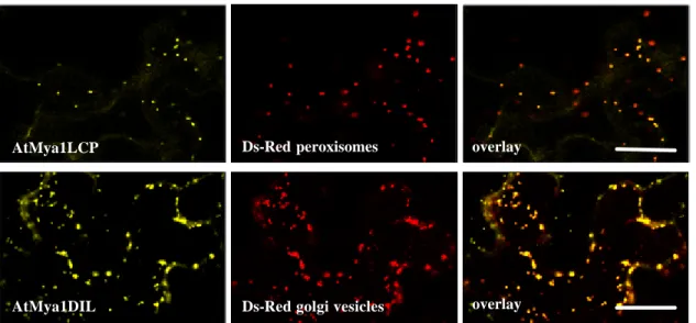
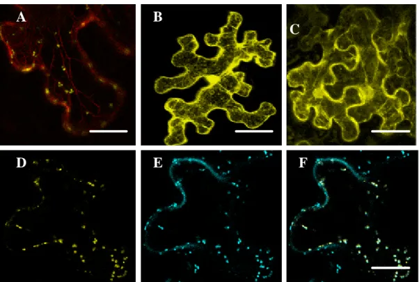
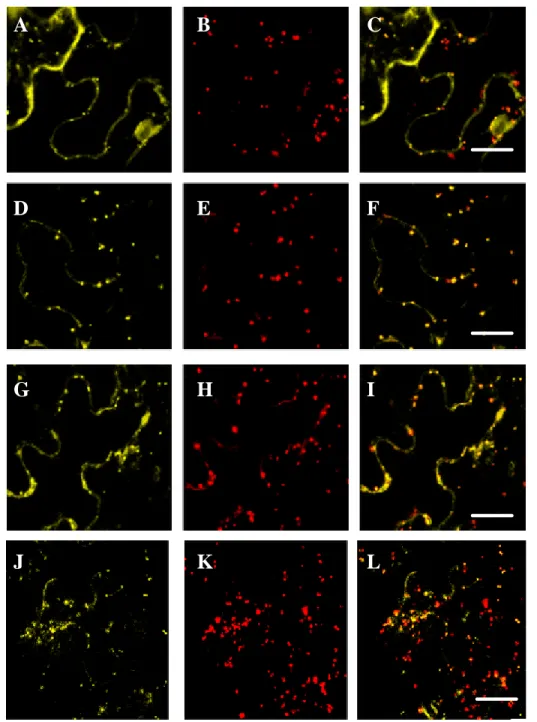
3.1.2 Subcellular localization of the DIL- and the LCP domain ... 70
3.1.3 Subcellular localization of class XI myosins from different subclasses 73 3.1.4 In planta expression of selected fragments of S. cerevisiae class V myosin, Myo2p ... 78
3.1.5 Transient expression and subcellular localization of selected fragments of AtMya1 ... 80
3.2
C
HARACTERIZATION OFA
RABIDOPSIS THALIANA CLASSVIII
MYOSINS...81
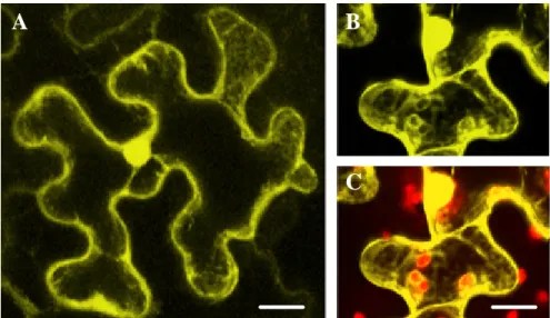
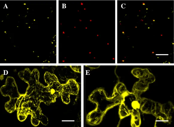
3.2.1 Subcellular localization of ATM2 ... 83
3.2.2 Subcellular localization of ATM1 ... 91
3.3 S
UBCELLULAR LOCALIZATION OF SELECTED MYOS IN CONSTRUCTS INA.
THALIANA...92
3.4 I
DENTIFICATION OF PUTATIVE MYOSIN BINDING PROTEINS BY YEAST TWO-
HYBRID SCREENING...93
3.4.1 Screening for Mya1 ... 93
3.4.2 Screening for ATM2 ... 95
3.5 A.
THALIANA MYOSIN KNOCK-
OUT LINES...96
3.5.1 Selection and identification of A. thaliana myosin knock-out lines ... 96
3.5.2 Phenotypic characterization of Arabidopsis thaliana class XI myosin knock -out lines... 97
3.5.3 Phenotypic characterization of Arabidopsis thaliana class VIII myosin knock -out lines... 99
3.6 S
TUDIES ON THE INVOLVEMENT OF MYOSINS IN PLANT DEFENCE RESPONSES...101
3.6.1 Non-host resistance (powdery mildew of barley) ... 101
3.6.2 Host resistance ... 103
3.6.3 Influence of BDM application on the defence response of barley ... 104
3.7 E
FFECTS OFBDM
ON THE SUBCELLULAR LOCALIZATION OF THEA
TM
YA1 DIL
DOMAIN...105
3.8 E
XPRESSION STUDIES OF BARLEY MYOSINS...106
3.8.1 Subcellular localization of myosins from barley... 108
4 DISCUSSION ... 110
4.1 T
RANSIENT OVEREXPRESSION OF MYOSIN FLUORESCENCE PROTEIN(FP)
FUSIONS AS ASSAY SYS TEM FOR THE IN VIVO ANALYSIS OF CARGO BINDING SITES...110
4.2 S
YSTEMATIC ANALYSIS OFA.
THALIANA MYOSIN CLASSXI
CARGO BINDING SITES...115
4.3 I
N PLANTA EXPRESSION AND LOCALIZATION OF THE YEAST CLASSV
MYOSIN(M
YO2
P)
CARGO BINDING DOMAINS...118
4.5.1 Class XI myosins ... 124
4.5.1.1 Transportation of Peroxisomes...124
4.5.1.2 Transportation of Golgi stacks/vesicles ...126
4.5.1.3 Motility of mitochondria and endoplasmic reticulum (ER) in plant cells ...128
4.5.2 Class VIII myosins ... 130
4.6 P
OTENTIAL MYOSIN INTERACTING PROTEINS...132
4.6.1 Myosin class XI ... 132
4.6.2 Myosin class VIII... 134
4.7 A
NALYSIS OF MYOSIN KNOCK OUT LINES...135
4.7.1 Class XI myosins ... 135
4.7.2 Class VIII myosins ... 137
4.8 A
PPLICATION OFBDM
AS MYOSIN INHIBITOR...137
4.9 P
OTENTIAL ROLE OF MYOSINS IN PATHOGEN DEFENCE...139
4.9.1 Effect of BDM on plant-pathogen interaction... 139
4.9.2 Involvement of plant class XI myosins in host resistance ... 140
5 SUMMARY... 142
5.1 S
UMMARY(E
NGLISH)...142
5.2 Z
USAMMENFASSUNG(D
EUTSCH) ...145
6 OUTLOOK... 148
6.1 C
LASSXI
MYOSINS: ...148
6.2 C
LASSVIII
MYOSINS...148
6.3 Y
EAST-
TWO-
HYBRID...149
7 LITERATURE ... 150
8 ACKNOWLEDGEMENTS ... 172
9 EIDESSTATLICHE ERKLÄRUNG... 174
10 LEBENSLAUF ... 175
1 Introduction
Plants play directly or indirectly an essential role as a nutritional resource in extending our life. Improvements in crop yields need a better understanding of the mechanisms underlying intracellular trafficking in respect of carbohydrate, protein transport and plant interaction with the environment (Robinson, 2003). In respect to environment, plants are sessile organisms;
therefore under stress conditions such as physical stresses like strong light, adverse temperature, dryness, wetness, high salt, or biological stresses like pathogen or herbivore attack, they can not escape but have to overcome the stress. Hence, plants evolved complicated adaptive responses at the organelle level as well as at the cell, tissue, and organ levels to avoid stresses (Nagai, 1993; Williamson, 1993). It is believed that intracellular organelle trafficking plays a crucial role in allowing plants to cope with environmental stresses and to propagate efficiently. Although each organelle type must have its own efficient mechanism for intracellular trafficking, however harmonic coordination between all these organelles is required to enable each organelle to accomplish its function (Wada and Suetsugu, 2004).
Furthermore, the increasing knowledge demonstrating that plants could
serve as excellent bioreactors for the production of a variety of valuable
proteins (Stoger et al., 2002; Fischer et al., 2004; Vitale and Pedrazzini,
2005) underlines the requirement for basic research on intracellular protein
trafficking. Although the mechanisms underlying intracellular protein
transport in animal and yeast cells are well known and many of the
must to be taken into account that the plant endomembrane system has some features which are unique, e. g. decentralized Golgi apparatus consisting of a network of motile stacks wh ich does not need fragment during mitosis, multiple vacuolar compartments including a protein storage compartment and a large central vacuole, cell- plate (phragmoplast) facilitated cytokinesis and cell division (Robinson, 2003; Hawes, 2005). Another specific feature of the intracellular transport in plant cells is cytoplasmic streaming which moves large quantities of cytoplasm (including organelles) around the cell and is most pronounced in larger, highly vacuolated cells (Nagai, 1979;
Shimmen and Yokota, 1994). Cytoplasmic streaming has been studied extensively in characean algae (Nagai, 1979; Shimmen and Yokota, 2004).
Studies of streaming events in the past have relied on visualiza tion of moving organelles with phase-contrast microscopy. The nature of the organelles was therefore mostly unknown. However it is been shown that cytoplasmic streaming is mostly driven by the acto- myosin system (Shimmen and Yokota, 2004).
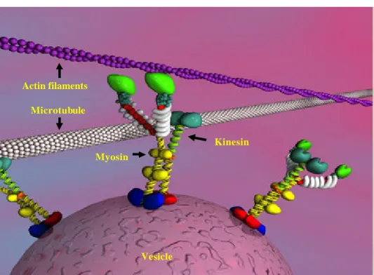
1.1 The plant cytoskeleton and the intracellular motility mediated by molecular motors
The cytoskeleton is playing an important role in a variety of cellular
functions of plant cells such as intracellular transport, signaling, cell
division and generation as well as maintenance of cell shape (Williamson,
1986; Volkmann and Baluska, 1999; Mathur et al., 2003; Mathur et al.,
2003). In eukaryotes the cytoskeleton consists of three types of filaments
called actin filaments or micro filaments, intermediate filaments and
microtubules. Notably, so far the presence of intermediate filaments has not been confirmed in plants (Meagher and Fechheimer, 2003).
Actin filaments and microtubules are polar filaments, the ends of which are referred to as plus and minus ends. Both types of filaments provide the tracks for motor based transport. Transport along microtubules occurs in both directions and is performed by kinesins and dyneins. Kinesins are generally plus-end directed (Vallee and Sheptner, 1990) motor proteins.
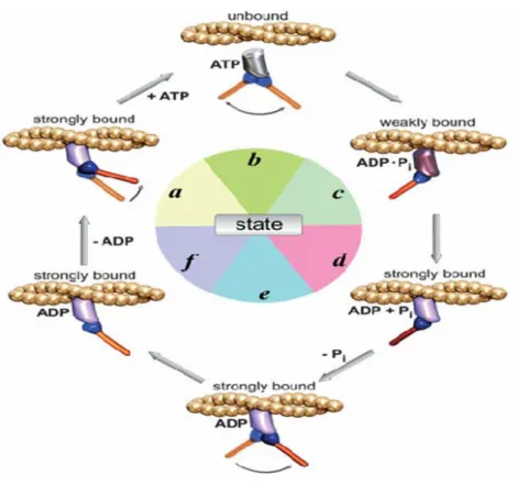
Movement along actin filaments is accomplished by members of the myosin super family (Sellers, 2000). Myosin mediated movement is mainly plus- end, although recently myosin VI (Wells et al., 1999) has been shown to move in the opposite direction. All these motors utilize energy from the hydrolysis of ATP to move in association with the cytoskeleton. ATP hydrolysis generates a small conformational change in the motor domain that is amplified and translated into movement with the assist of structural motifs ( reviewed by Schilwa and Woehlke, 2003).
1.2 The super family of myosins
Myosins comprise a large super family of proteins that share common
domains which have been shown to interact with actin, hydrolyze ATP and
produce movement in all cases examined to date. All myosins have typically
three functional domains as shown in Figure 1: (I) the motor domain which
interacts with actin and binds ATP and is responsible for force production,
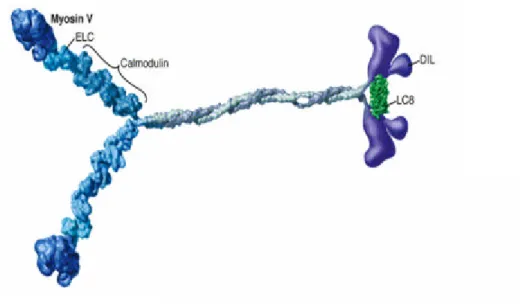
(II) the neck or regulatory domain which binds light chains or calmodulin,
and (III) the tail domain which is thought to be important for subcellular
localization and cargo binding (reviewed by Seller, 1999). However for
domains are highly conserved with the exception of several surface loops and the amino-terminus. With one known exception (Heintzelman and Schwartzman, 1997), the motor domain is linked to the C- terminal tail domain by the neck domain of variable length that contains sites for binding of regulatory light chains or calmodulin (Mermall et al., 1998). The neck consists of a characteristic helical sequence termed the IQ motif wit h a consensus sequence of IQXXXRGXXXR (Cheney and Mooseker, 1992).
The number of IQ motives present in the neck of different myosins can vary between zero and six. The tail domain is the most divergent domain among the different myosin classes varying widely in length and in sequence.
Functional motives, such as kinase domains, GTPase-activating domains, SH3, GAP, FERM and pleckstrin homology (PH) domains are sometimes found in the tails of myosins known to function in signal transduction, membrane binding, or protein–protein interactions (Mermall et al., 1998;
Oliver et al., 1999). In addition, the tails of many myosins contain coiled-
coil forming sequences which allow the molecules to dimerize and produce
two- headed molecules. In general, the tail domains of myosins are believed
to be largely responsible for class-specific functions. In Table 1.1 some of
the most important domains found in the tail of unconventional myosins are
listed.
Figure 1: Major structure features of myosin molecular motors with their corresponding track (actin filaments) are shown. Myosin head domain (green), IQ domain (white) and tail domain (yellow and blue) are shown (Fig from Cell motility and Cytesckleton 2000, 45-4).
The myosins are subdivided into an ever- increasing number of classes (17 at the last count) according to phylogenetic analyses of the motor domains (Hodge and Cope, 2000). Traditionally they have been segregated into two categories: the conventional (muscle and nonmuscle myosins, class II) and the unconventional myosins, which include all other myosins. Class II reviewed by (Seller, 2000) was discovered over 60 years ago and is found in muscles and in the cytoplasm of animal cells. Class I myosins were next discovered and the subsequent classes were numbered in order of the
Microtubule Actin filaments
Vesicle Myosin
Kinesin
all types of myosins are expressed in one organism. For example class VIII, XI and XIII are plant specific myosins and plants do not have any other myosins from other classes. However even the simplest of eukaryotes express multiple myosins (Reddy and Day, 2001). The budding yeast, Saccharomyces cerevisiae, for example has five myosin genes: one conventional class II myosin and two of each classes I and V (reviewed in Pruyne et al., 2004). The number of myosin genes present in mammals is conservatively estimated at 25–30 from classes I, II, III, V, VI, VII, IX, X and XV. In any case, all eukaryotic animal cells examined contain at least one myosin II gene and, usually, multiple myosin I genes. In addition, myosin V genes are found widely, if not universally (Mermall et al., 1998;
Oliver et al., 1999).
Although most family trees are constructed by analysis of the motor
domains, analysis of the whole molecule or of the tail domains alone
generally gives similar relationships (Cope et al., 1996). While relatively
little is known about the detailed cellular functions of most of the
unconventional myosins, their importance is highlighted by the discovery
that mutations in unconventional myosins can lead to severe defects as
deafness, blindness, seizures or even death (Table 1.1, Mermall et al., 1998).
Figure1.1: An unrooted phylogenetic tree of the myosin superfamily based on head domain protein sequences. Myosin classes are shown in roman numerals.
Sequence divergence is proportional to the length of the connecting branches. The large globular domains represent heads, smaller globular structures represent neck and tail motifs, and twisted lines denote coiled-coils (Fig from Hodge and Cope, 2000).
Generally, the tail domain structure is conserved within each class but
differs between classes (Mermall et al., 1998) . In light of the high degree of
conservation between the motor domains, the myosin tail is thought to
determine the specificity of the protein, both for interactions such as
domain, which has been extensively studied in both conventional and several of the unconventional myosins, the functions of the individual tail domains are by far less well understood (Mermall et al., 1998). There are multiple ways in which motor proteins may be regulated: regulation of enzymatic activity, regulation of affinity to the cytoskeleton, regulation of protein levels and regulation of affinity to the cargo in the cell (Howard, 2001; Schliwa and Woehlke, 2003). The regulatory pathways involved appear to be highly complex, and are just at the beginning of characterization. In a few studies it has been demonstrated that phosphorylation, G proteins, and calcium/calmodulin are signals involved in myosin regulation (Reilein et al., 2001; Legesse- Miller A et al., 2006). For instance, in case of myosin V, the closest homologue of class XI myosins, it has been shown that organelle transport by myosin V is down-regulated during mitosis by myosin- V phosphorylation (Karcher et al., 2001). Using mass spectrometry phosphopeptide mapping the authors showed that the tail of myosin- V was phosphorylated in mitotic Xenopus egg extracts on a single serine residue localized in the carboxyl-terminal organelle-binding domain which resulted in the release of the motor from the organelle.
Interestingly, the phosphorylation site matched the consensus sequence of
calcium/calmodulin dependent protein kinase II (CaMKII), and inhibitors of
CaMKII prevented myosin- V release. However, to understand how these
multiple pathways and mechanisms are coordinated to regulate a single
motor in other motor proteins remains a question to be solved in the future
(Karcher et al., 2001).
Class Function Domains Dis Diesease
I Cell growth and development, Cell movement, Endocytosis
Basic,SH3,GPA E(NP)
II cytoplasmic Cytokinesis , phagocytosis, cell shape and
polarity
N-terminal SH3 like E(NP) FHC
III Rhabdomere function (Drosophila), photoadaptation
PK, Basic Meta Deafness
IV Acanthamoeba species only. MyTH4,SH3 Ac V Vesicle transport, mRNA
transport, Chitin localization
DIL E(NP) Pigmentation disorder VI Stabilizing and anchoring,
Endocytosis
Reverse Gears Meta Deafness
VII Sensory epithelia FERM,MyTH4,SH3 Meta Deafness
VIII ? - Plants
IX Signal transduction, Leukocyte differentiation
RA, RhoGAP, DAG_PE
Meta
X Signal transduction PH,MyTH4,FERM Vert
XI Vesicle transport DIL Plants
XIII ? - Acl XIV Toxoplasma and Plasmodium
species only
- Prot
XV Auditory FREM,MyTH4,SH3 Mamm Deafness
XVI Neuronal cell migration ANK Mamm
XVII chitin synthase Pyricularia and Emiricella species only.
Chitin synthase Fungi