Devic’s syndrome in aquaporin-4 antibody negative patient. What we need to know …
Abstract
Introduction: Neuromyelitis optica (NMO) is a severe demyelinating syndrome characterized by optic neuritis (ON) and acute myelitis. The
Ana Teresa Nunes
1Ana Cláudia Fonseca
1NMO spectrum is actually recognized to typically evolve as a relapsing
Filomena Pinto
1disorder that also includes patients with atypical unilateral ON and those with index events of ON and myelitis occurring weeks or even years
apart (Jarius/Wildemann 2013). NMO was previously assumed to be a 1 Ophthalmology Department, Centro Hospitalar Lisboa variant of multiple sclerosis (MS), but the discovery of aquaporin-4 an-
tibodies in patients with neuromyelitis optica has led to this view being Norte/Hospital Santa Maria, Lisboa, Portugal
revised (Mandler 2006, Barnett/Sutton 2012, Wingerchuk et al. 2007).
The cause of the condition is still unknown, but it has been shown that the antibodies bind selectively to a water channel expressed mainly on astrocytes at the blood-brain-barrier, which has an important role in the regulation of brain volume and ion homeostasis. However, there are some patients with NMO that are antibodies negative. The diagnosis is made on the basis of case history, clinical examination, magnetic res- onance imaging (MRI) of the brain and spinal cord, analysis of cerebrospinal fluid (CSF), visual evoked potentials and a blood test with analysis of aquaporin-4 antibodies (Barnett/Sutton 2012, Wingerchuk et al. 2007, Thornton et al. 2011). This suggests that periodical revisions of established concepts and diagnostic criteria are necessary.
Purpose:The authors describe an extremely rare case of neuromyelitis optica and the aim of this paper is to call attention for the cases of NMO whith NMO-IgG negative.
Methods:The selected method is a case report.
Results:To date the patient showed partial recovery of left eye acuity and improvement of muscle strength of upper and lower limbs and does not show recurrence of the disease.
Conclusion:NMO has a distinct clinical, imaging and immunopatholo- gical features sufficient to distinguish it from MS. This distinction is essential, because the treatment and the prognosis is different.
Keywords:neuromyelitis optica, diagnostic criteria, treatment, Devic’s syndrome, aquaporin-4 antibody
transverse myelitis and many cases in which optic neur- itis and transverse myelitis are separated by months and years [5], [6].
Currently, NMO is considered as a central nervous system AQP4 channelopathy which causes variable damage predominantly to the optic nerves and spinal cord, al- though other CNS structures that highly express AQP4 may be also affected [7], [8].
Purpose
The aim of this study is to report a rare case study.
Materials and methods
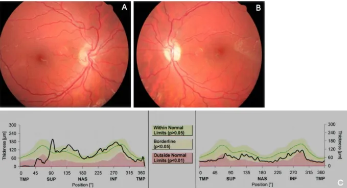
We report the case of a 20-year-old Caucasian woman who presented to the Ophthalmology Emergency room, claiming progressive, painless vision loss in the left eye with 3 days of evolution and one week after she com- plained paresthesias in the lower extremities. The patient presented a visual acuity of 10/10 in right eye and in the left eye absent luminous perception. The direct pupillary reflex in the left eye was absent. Anterior segment in both eyes was normal. The intraocular pressure was 13 mmHg in both the eyes and fundoscopy in the left eye showed edema of optic nerve and venous engorgement and tor- tuosity bilaterally (Figure 1). Ocular motility was normal.
The patient performed in the emergency room a CT and blood tests. On the same day she was admitted to the Neurology Department where she performed MRI (Figure 2, Figure 3), lumbar punction with analysis of CSF.
More specific tests and chest CT for screening of thymoma were requested. On the next day our patient was seen at the Ophthalmology Department where she made the following imaging tests: optical coherence tomography, angiography, visual fields and electrophysiolo- gical tests.
Results
The complementary exams realized in emergency room (brain and orbits CT and blood tests) were normal, except the slight increase of the inflammatory parameters.
Discussion
NMO is more frequent in women than in men (>80%).
The onset of the syndrome varies, from adolescent to young adulthood, with a median peak incidence in the late 30s [8], [9], [10].
NMO is often associated with other autoimmune disorders in the patient or in the family. NMO manifests the sole clinical syndrome, with serological markers of autoim- munity, like a positive ANA, rheumatic factor or thyro- globulin antibody. Association with infectious disorders, like tuberculosis, viral infections and immunizations have been observed.
Nowadays, all studies suggest that neuromyelitis optica is distinct from MS. This distinction is based on clinical, imaging, pathological and serological criteria and has practical implications, in the prognosis and therapies [10].
Since 2004 with the discovery of the NMO-specific anti- body and AQP4, as its targeted antigen,it is recognized as a turning point in the concept and understanding of the disease [11]. Aquaporin-4 is the most abundant water channel in the central nervous system, expressed in the foot processes of astrocytes in contact with blood vessels throughout the brain, spinal cord and optic nerves. The periventricular area, the hypothalamus and the brainstem are also considered sites of high expression of AQP-4.
Although AQP-4 predominates in the CNS, it is also found in other organs such as kidneys, stomach, airways, glands and skeletal muscle. However, the paucity of clinical ab- normalities outside the CNS remains to be explained, as well as the underlying mechanisms of AQP4-IgG seronega- tive status in some patients [10], [12], [13]. Presumed reasons include, suboptimal sensitivity of the currently available assays (specific for 91% of cases, but not very sensitive yet, 73%, of this could be explained because the substrates of the immunofluorescence assay were not human, but are from mousse brain tissues), very low serum concentration of the antibodies or their absence at some periods in the disease course, and the inhibitory effect of previous treatment with corticosteroids or im- munosuppressive agents. It is also possible that in some NMO patients other antigens may play a role in the pathogenesis of the disease. Therefore, notwithstanding
Figure 1: Retinography (day 1) – RI: tilted disc and vascular tortuosity (A); LE: ON edema, venous engorgement and vascular tortuosity (B)
Figure 2: Brain MRI (day 2) (A, B and C) showed small areas of increased signal intensity on left temporal lobe and right periventricular area in cerebral white matter; with gadolinium uptake in the left optic nerve.
Figure 3: Sagittal T2 weighted MRI of spinal cord showing swelling of the cervical segments (more than 3 contiguous segments) with high signal intensity.
1. Temporal. Acute or relapsing involvement of the optic nerves and spinal cord, coincidental or separated by months or years
2. b.Serology. Absent serum NMO-IgG antibody 3. Spatial. Myelitis can be total or partial. Optic neuritis
can be unilateral or bilateral.
4. Absence in general, of central nervous system symptoms and signs outside the spinal cord and optic nerves, with exception of hypothalamic and lower brainstem dysfunction
5. Course. The disease can either be monophasic or re- current.
6. Spinal cord MRI. Can be normal, but often T2 signal abnormalities with cavitation and sometimes swelling, extending over 3 or more consecutive segments might reinforce the criteria
7. Brain MRI. In general, normal brain MRI. No white matter abnormalities in the brain, brainstem or cere- bellum. White matter anomalies might sometimes be seen in hypothalamus and lower brainstem and rarely in the brain parenchyma.
Partial NMO
1. Clinical. Either optic neuritis or transverse myelitis 2. Serology. Positive serum NMO-IgG
This new classification with revised diagnostic criteria represent an important advance in NMO research and clinical practice. They discriminate NMO from MS and allow the diagnosis of partial forms and negative NMO- IgG forms, like our patient.
In general, NMO is more acute, sometimes fulminant.
Myelitis, like optic neuritis, can also be fulminant, with acute urinary retention, paraparesis or quadriparesis, severe tonic flexor spasms and severe back pain [9].
Supportive laboratory features in NMO are absence of CSF oligoclonal bands and pleocytorrachia.
Brain MRI generally shows no white matter abnormalities in the brain, brainstem and cerebellum. Anomalies might be found in the optic nerves. Spinal cord RMI can be supportive when continuous lesions with gadolinium en- hancement extend over 3 or more vertebral segments.
The prognosis of NMO is, in general, more guarded than that of MS patients. Patients with NMO can die from acute ventilator failure produced by necrosis of the cervical cord and its complications. Predictors of mortality in relapsing
severe attacks that worsen during corticosteroid therapy or do not demonstrate improvement. Predictors of re- sponse included early treatment initiation, male sex and preserved deep tendon reflexes.During the relapsing disease, patients with NMO can receive prednisone (1 mg/Kg/d) and azathioprine (2–3 mg/Kg/d) [2], [10], [14]. According to some studies there is a stabilization of the disease for at least 18 months. Most patients re- ceive a maintenance doses of azathioprine of 75–150 mg together with 10 mg of prednisone dose q/o/d. Other treatments like rituximab and mitoxantrone have been studied [14].
Our patient during hospitalization received methylpred- nisolone, 1,000 mg q/day (IV) for 5 days, following with steroids in the form of oral prednisone (1 mg/Kg/d). At hospital discharge (three weeks later), our patient showed partial recovery of left eye acuity and improvement of muscle strength of upper and lower limbs.
Five weeks later, the patient repeated the MRI, that showed swelling diminished and sign change became less intense and smaller (Figure 4A and B), however the left frontal lesion maintains (Figure 4C) and the vascular tortuosity and the NO edema in the fundus had improved (Figure 4F).
In the follow-up visit, four months later, the dilated fundus examination showed a sectorial optical atrophy in the left eye (Figure 5B), confirmed by OCT (Figure 5C) with a visual acuity 20/20 in each eye. At the moment she was receiving prednisone 40 mg/d and azathioprine 150 mg/d.
Conclusion
NMO has a distinct clinical, imaging, pathological and immunopathological features sufficient to distinguish it from MS. This distinction is not just academic, because the treatment and the prognosis is different too, NMO optic neuritis can be fulminant and devastating. Because a small proportion of cases nevertheless fulfill the criteria for both NMO and MS, challenges persist for clear defin- itions, especially concerning the roles of immunomodu- latory and immunosuppressive treatment [15]. Early re- cognition of the relapsing form may allow expeditious acute and long-standing treatment and prevent significant morbidity and mortality.
Figure 4: After the acute phase (5 weeks later), MRI showed swelling diminished and sign change became less intense and smaller (A and B), however the left frontal lesion maintains and the vascular tortuosity and the NO edema in the fundus had
improved (D and F).
Figure 5: Four months later, the left eye showed a sectorial optical atrophy (A and B), confirmed by OCT (C).
2. Mandler RN. Neuromyelitis optica – Devic's syndrome, update.
Autoimmun Rev. 2006 Oct;5(8):537-43. DOI:
10.1016/j.autrev.2006.02.008
3. Barnett MH, Sutton I. Neuromyelitis optica: not a multiple sclerosis variant. Curr Opin Neurol. 2012 Jun;25(3):215-20. DOI:
10.1097/WCO.0b013e3283533a3f
4. Wingerchuk DM, Lennon VA, Lucchinetti CF, Pittock SJ, Weinshenker BG. The spectrum of neuromyelitis optica. Lancet Neurol. 2007 Sep;6(9):805-15. DOI: 10.1016/S1474- 4422(07)70216-8
5. Thornton IL, Rizzo JF, Cestari DM. Neuromyelitis optica: a review.
Semin Ophthalmol. 2011 Jul-Sep;26(4-5):337-41. DOI:
10.3109/08820538.2011.588667
6. Lennon VA, Wingerchuk DM, Kryzer TJ, Pittock SJ, Lucchinetti CF, Fujihara K, Nakashima I, Weinshenker BG. A serum autoantibody marker of neuromyelitis optica: distinction from multiple sclerosis. Lancet. 2004 Dec 11-17;364(9451):2106- 12. DOI: 10.1016/S0140-6736(04)17551-X
7. Sellner J, Boggild M, Clanet M, Hintzen RQ, Illes Z, Montalban X, Du Pasquier RA, Polman CH, Sorensen PS, Hemmer B. EFNS guidelines on diagnosis and management of neuromyelitis optica.
Eur J Neurol. 2010 Aug;17(8):1019-32. DOI: 10.1111/j.1468- 1331.2010.03066.x
8. Takahashi T, Fujihara K, Nakashima I, Misu T, Miyazawa I, Nakamura M, Watanabe S, Ishii N, Itoyama Y. Establishment of a new sensitive assay for anti-human aquaporin-4 antibody in neuromyelitis optica. Tohoku J Exp Med. 2006 Dec;210(4):307- 13. DOI: 10.1620/tjem.210.307
9. Wingerchuk DM, Weinshenker BG. Neuromyelitis optica: clinical predictors of a relapsing course and survival. Neurology. 2003 Mar 11;60(5):848-53. DOI:
10.1212/01.WNL.0000049912.02954.2C
10. Wingerchuk DM, Lennon VA, Pittock SJ, Lucchinetti CF, Weinshenker BG. Revised diagnostic criteria for neuromyelitis optica. Neurology. 2006 May;66(10):1485-9. DOI:
10.1212/01.wnl.0000216139.44259.74
11. Papadopoulos MC, Verkman AS. Aquaporin 4 and neuromyelitis optica. Lancet Neurol. 2012 Jun;11(6):535-44. DOI:
10.1016/S1474-4422(12)70133-3
neuromyelitis optica: A multicentre study of 175 patients. J Neuroinflammation. 2012;9:14. DOI: 10.1186/1742-2094-9- 14
14. Mandler RN, Ahmed W, Dencoff JE. Devic’s neuromyelitis optica:
a prospective study of seven patients treated with prednisone and azathioprine. Neurology. 1998 Oct;51(4):1219-20. DOI:
10.1212/WNL.51.4.1219
15. Verkman AS. Aquaporins in clinical medicine. Annu Rev Med.
2012;63:303-16. DOI: 10.1146/annurev-med-043010-193843
Corresponding author:
Ana Teresa Nunes
Ophthalmology Department, Hospital Santa Maria, Avenida Professor Egas Moniz, 1649-035 Lisboa, Portugal nunesesegalas@gmail.com
Please cite as
Nunes AT, Fonseca AC, Pinto F. Devic’s syndrome in aquaporin-4 antibody negative patient. What we need to know …. GMS Ophthalmol Cases. 2014;4:Doc09.
DOI: 10.3205/oc000022, URN: urn:nbn:de:0183-oc0000228
This article is freely available from
http://www.egms.de/en/journals/oc/2014-4/oc000022.shtml Published:2014-12-02
Copyright
©2014 Nunes et al. This is an Open Access article distributed under the terms of the Creative Commons Attribution License
(http://creativecommons.org/licenses/by-nc-nd/3.0/deed.en). You are free: to Share — to copy, distribute and transmit the work, provided the original author and source are credited.