A rare desmoid tumor of the shoulder – excision,
implantation of brachytherapy applicators and wound closure by pedicle musculus latissimus dorsi flap
Ein seltener Desmoidtumor der Schulter – Exzision, Implantation von Brachytherapie-Applikatoren und Defektdeckung mit gestielter Muskulus Latissimus Dorsi Lappenplastik
Abstract
Desmoid tumors are non-metastatic mesenchymal tumors with an ag- gressive local growth. Depending on the anatomic location, morbidity
Peter L. Stollwerck
1Thomas Namdar
1varies. We report of a patient with a desmoid tumor of the right shoulder
Tanja Bartscher
1which was treated in our department by surgical excision, plastic-surgical
wound closure and postoperative adjuvant radiation.
Thomas Lange
1Keywords:neoplasms, aggressive fibromatosis, plastic surgery, flaps, perioperative interstitial brachytherapy
Felix H. Stang
1Peter Kujath
2Guenther Bohlen
3Zusammenfassung
Desmoidtumoren sind seltene, nicht-metastasierende mesenchymale Tumore mit einem lokal agressiv-infiltrativen Wachstum. Wir berichten
György Kovács
3Peter Mailänder
1über einen Patienten, der aufgrund eines Desmoidtumors in unserer Abteilung durch Exzision, plastisch-chirurgische Defektdeckung und einer adjuvanten Bestrahlung behandelt wurde.
1 Plastic Surgery, Hand Surgery, Burns Unit, University Hospital Schleswig- Schlüsselwörter:Neubildung, aggressive Fibromatose, Desmoidtumor,
Plastische Chirurgie, Lappenplastik, perioperative interstitielle Brachytherapie
Holstein Campus Lübeck, Germany
2 General Surgery, University Hospital Schleswig-Holstein Campus Lübeck, Germany 3 Interdisciplinary
Brachytherapy Unit, University Hospital Schleswig- Holstein Campus Lübeck, Germany
Introduction
Desmoid tumors are rare entities and belong to the group of fibromatoses, which arise from the connective tissue, fascia or aponeurosis of a muscle [1].
Even though desmoid tumors are benign lesions per definition, they show an infiltrative growth with destruction of surrounding structures and neighbouring organs, re- sulting in extensive morbidity [2]. The incidence is report- ed to be 2.4 bis 4.3 cases/1 million/year.
In the majority desmoid tumors are found intraabdomin- ally and only a small amount of 7–15% are found in the
head and neck area. As this tumor entity is rare, data giving evidence based recommendations for the optimal treatment algorithm for this disease is lacking [3].
We report the case of a patient with a desmoid tumor of the right shoulder region that was diagnosed and treated interdisciplinary in the general surgery department for tumor resection, in our department for plastic surgical wound closure and in radio-oncology for adjuvant brachy- therapy.
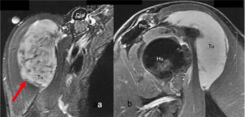
Figure 1: Preoperative MRI of the right shoulder showing the tumor a) in transverse cross-section with Tumor (Tu/arrow) 8x6x3.5 cm, Clavicle (Cla) and b) in relation to neighboring structures such as Humerus (Hu), Processus coracoideus (Prc).
Figure 2: a) Excision of the tumor and parts of deltoid muscle, b) separation from humerus (Hu) and processus coracoideus (Prc) and M. biceps brachii caput longum, c) resulting wound cavity with exposed humerus and clavicle (Cla) d) tumor mass
with overlying skin spindle after resection.
Case description
A 52-year old Caucasian German male with no past medical history had observed a rapidly growing tumor of his right shoulder and slight soreness during a period of four weeks after having performed physical exercise for several weeks in a fitness studio. He reported an allergy against contrast medium. The tumor was palpable as a firm lump of the ventral shoulder. An external magnet resonance imaging (MRI) showed an 8x4x5.3 cm soft tissue tumor of the ventral shoulder below the deltoid muscle overlying the humerus head. Furthermore, the tumor had contact to the biceps muscle and the axillary perivascular sheath (Figure 1).
After completion of preoperative staging, the tumor was excised in toto by the general surgery department. Resec- tion was achieved without harming neuro-vascular structures but part of the deltoid muscle had to be ex- cised. After resection, the resulting wound cavity was too large to be closed by plain skin suture (Figure 2 a–c).
The final histology revealed an 8 cm mean diameter crude mass with elongated spindle-shaped cells, intertwined by a large amount of collagen-fibres. The tumor expressed vimentin and actin but immuno-staining did not show pancytokeratin expression. The S100 antigen, desmin and CD117 (C-kit) were negatively tested. All resection lines were free of tumor.
In the plastic surgery department, the patient underwent surgery for wound closure by a pedicle musculus latisimus dorsi flap from his right back and a sheet skin graft from his left thigh. Intraoperatively, region of radiation target area was marked by placing several metal surgical clips, as well as eight brachytherapy applicators (plastic tubes) which were implanted parallel using one cm interspacing between each tube (Figure 3). The applicators were fixed to the tumor bed with single sutures so that latero-lateral movement was blocked but longitudinal movement was kept free. After inserting the tubes through the skin they were fixed by special buttons, which were sutured to the skin to avoid longitudinal movements. Postoperatively, a multislice CT (1 mm slice thickness) was performed in
Figure 3: a+b) Wound closure achieved by pedicle musculus latissimus dorsi flap and split skin graft and implantation of 8 afterloading applicators. (Day 1 after operation)
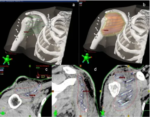
Figure 4: a–e) Brachytherapy dose distribution on the 2D and 3D level.
Figure 5: a–c) 6 Weeks after removal of afterloading tubes (Brachtherapy) stable muscle flap and skin graft as well as maturing scar of the right flank after harvest of M. latissimus dorsi. Good arm mobility with 90° shoulder abduction. d+e) stable skin
graft and muscle flap at follow-up 1 year after treatment.
order to create a virtual model of anatomy and applica- tors. Based on this model, an individual volume optimized dose distribution was calculated by using the Brachyvision software package (Varian, USA). After completing the fractionated image adapted brachytherapy treatment (IABT) the fixing buttons were removed and the applicators could be extracted without any anaesthesia. During the postoperative course, no adverse events were recorded.
Because of the high risk of local recurrences in solitary surgery and the current tumor size of eight cm, radiation therapy (RT) was recommended in the interdisciplinary tumor board and brachytherapy was initiated with a total of 30 Gy (2x2.5 Gy in 8 days, no radiation on Saturday and Sunday) two weeks after achievement of wound closure. Representative images of dose distribu- tion (Figure 4) show the optimal radiation coverage of the target area.
After completion of radiation, patient was treated by a physysiotherapist for the mobilization of the shoulder.
After 6 months a control MRI was performed with no signs of a recurrent tumor. The patient was very satisfied with the result of the muscle flap (Figure 5).
Discussion
Desmoid tumors originate from muscular connective tis- sue, fasciae and aponeurosis. Their growth typically shows to be locally invasive, yet without a metastatic tendency.
The underlying cell differentiation contains spindle shaped cells, which are intertwined by collagen fibres. The inci- dence is reported to be 2.4 bis 4.3 cases/1 million/year, whilst the majority of desmoid tumors are found intraab- dominally and only 7–15% are found in the head and neck area. When a surgeon is challenged by needing to treat the rare entity of a desmoid tumor, he needs to take the high risk of recurrence (25–65%) [4] after surgery into account.
The etiology of aggressive fibromatosis has not been sufficiently explained, but trauma, endocrine and genetic factors have been suggested to be causative [4].
In the presented case the patient reported to have under- gone extensive muscle training before the tumor occurred.
This leads to the assumption, that the growth of the tumor could have been induced by this trauma to the muscle and its tendons.
Diagnostics should be performed by ultrasound, MRI or CT-scans. Before any type of surgery is performed, the expansion of the tumor and its relation to functional structures and the neuro-vascular system need to be identified.
Successful treatment of aggressive fibromatosis can be achieved by surgical excision, radiation and with pharma- cological agents.
In the current literature, radical surgical excision with tu- mor-free margins up to 3 cm is widely accepted and re- commended [5], [6]. However, this often confronts the surgeon with the difficulty of having to sacrifice structures of functional or even vital importance. If a complete re-
section cannot be achieved because vital structures i.e.
large blood vessels and vital organs are in danger, this fact consequently leads to local recurrence with a poor outcome. Further problems arise if a large wound cavity results, which cannot be closed by direct skin suture.
Nonetheless, radical excision should not be avoided, as sufficient wound closure can be provided by plastic sur- gery as shown in the present case.
In cases of incomplete resection, the role of adjuvant ra- diation or pharmacological treatment becomes even more important to limit the rate of recurrence and need to be discussed rationally in an interdisciplinary tumor board.
There are studies, which emphasize the effectiveness of adjuvant radiation [7], [8], [9], [10], [11]. However, a re- cent 24-year retrospective study demonstrated 3-year local control rates without significant differences after surgery, radiotherapy or a combination of both. Patients were selected for treatment branches according to the size of the tumor, the expansion of the tumor and the possibility of achieving margin free resection. The actuar- ial 3-year local control rates ranged from 69.0% to 92.3%
[12].
In a further retrospective study of 115 patients with desmoid tumors Guadagnolo et al. described that the recurrence with RT alone can amount up to 32% in cases where surgery is not feasible due to gross disease. Pa- tients treated with both surgery and RT showed a lower local recurrence of their tumor of 20%. Yet in comparison the combination of surgery and RT did not prove to show different local control rates than in solitary RT. Addition- ally, in the case of positive resection margins, the combi- nation of RT and surgery, did not show to be significantly associated with a lower rate of local control [5].
Nuyttens et al. demonstrated in a literature review that RT alone or surgery in combination with RT results in significantly better local control than just with solitary surgery. Subdividing the groups into cases with free and positive margins and cases with primary and recurrent tumors showed that the best local control is achieved with RT or surgery with RT [10].
Intraoperative radiotherapy was proven to be an effective treatment with low toxicity [6] and a total dose below 56 Gy was recommended [5].
Interstitial brachytherapy offers minimal normal tissue toxicity due to the steep dose fall-off of the sources and due to the intraoperative applicator placement the target tissue for postoperative radiotherapy can be defined op- timally. Regarding total dose values, a National Patterns of Care Study on radiotherapy of desmoid tumors [11]
showed an over 80% cure rate in the adjuvant dose range of 36–65 Gy. Radiobiological modelling by the use of the linear-quadratic (LQ) model allows the calculation of the biological equivalent dose (BED) of the used fractionation schedule [13] and results in a total BED of 46.5 Gy on the reference isodose. However, tissue volumes near to the applicators will be irradiated with higher dose (for example the volume covered by the 200% isodose line with BED 93.0 Gy). The quality of the implant can be de- scribed by the dose non-homogeneity ratio (DNR), which
is the value of the 150% isodose volume divided by the covered volume of the 100% isodose line. In this case it was 0.39 – indicating a homogenous brachytherapy dose distribution.
Pharmacological treatment of desmoids tumors offers further effective possibilities, if the biological properties and the patient's characteristics are suitable. Options are cytostatic chemotherapy, and non-cytotoxic agents such as hormonal, anti-inflammatory and biological agents [14]. Unfortunately, also in the field of pharmacological treatment there is a lack of evidence due to non-sufficient patient numbers and missing prospective randomized studies.
This shows that it is more than ever necessary to establish treatment algorithms by inducing prospective multicenter studies – also in this rare entity.
Conclusion
The successful treatment of desmoid tumors requires an interdisciplinary approach. In cases of large tumors in need of wide excision, a plastic surgeon needs to be consulted to ensure radical excision and sufficient wound closure. Wound closure with viable tissue allows effective adjuvant radiation therapy. Normal tissue sparing and effective adjuvant therapy strategies are the implemen- tation of perioperative image adapted brachytherapy techniques, as well pharmacological agents such as anti- estrogens (i.e. Tamoxifen) and non-steroidal anti-inflam- matory drugs (NSAID). There is a need for prospective randomized studies with larger numbers of patients to allow the development of standardized and evidence- based treatment algorithms.
Notes
Consent
Written informed consent was obtained from the patient for publication of this case report and accompanying im- ages. A copy of the written consent is available for review by the editor-in-chief of this journal.
Authors' contributions
All authors contributed equally to this work. P. Stollwerck recruited the patient, analyzed the data, performed the photography and wrote the manuscript. P. Kujath and T. Lange and T. Bartscher performed the surgery, G. Bo- hlen and G. Kovács performed image adapted brachyther- apy as well they collected brachytherapy related radiobi- ology informations, F. Stang reviewed the current literat- ure, T. Namdar and P. Mailänder contributed in revising the manuscript. All authors read and approved the final manuscript.
Competing interests
The authors declare that they have no competing in- terests.
References
1. Goy BW, Lee SP, Eilber F, Dorey F, Eckardt J, Fu YS, Juillard GJ, Selch MT. The role of adjuvant radiotherapy in the treatment of resectable desmoid tumors. Int J Radiat Oncol Biol Phys.
1997;39(3):659-65. DOI: 10.1016/S0360-3016(97)00334-9 2. Posner MC, Shiu MH, Newsome JL, Hajdu SI, Gaynor JJ, Brennan
MF. The desmoid tumor. Not a benign disease. Arch Surg.
1989;124(2):191-6.
3. Kumar V, Khanna S, Khanna AK, Khanna R. Desmoid tumors:
experience of 32 cases and review of the literature. Indian J Cancer. 2009;46(1):34-9. DOI: 10.4103/0019-509X.48593 4. Ferenc T, Sygut J, Kopczynski J, Mayer M, Latos-Bielenska A, Dziki A, Kulig A. Aggressive fibromatosis (desmoid tumors):
definition, occurrence, pathology, diagnostic problems, clinical behavior, genetic background. Pol J Pathol. 2006;57(1):5-15.
5. Guadagnolo BA, Zagars GK, Ballo MT. Long-term outcomes for desmoid tumors treated with radiation therapy. Int J Radiat Oncol Biol Phys. 2008;71(2):441-7. DOI: 10.1016/j.ijrobp.2007.10.013 6. Roeder F, Timke C, Oertel S, Hensley FW, Bischof M, Muenter
MW, Weitz J, Buchler MW, Lehner B, Debus J, Krempien R.
Intraoperative electron radiotherapy for the management of aggressive fibromatosis. Int J Radiat Oncol Biol Phys.
2010;76(4):1154-60. DOI: 10.1016/j.ijrobp.2009.03.067 7. Bonvalot S, Rimareix F, Paumier A, Roberti E, Bouzaiene H, Le
Péchoux C. Actualisation de la strategie therapeutique locoregionale dans les sarcomes des tissus mous et les tumeurs desmoides des membres [What is new in the local approach of limb sarcomas and desmoid tumours?]. Cancer Radiother.
2010;14(6-7):455-9. DOI: 10.1016/j.canrad.2010.06.016 8. Ballo MT, Zagars GK, Pollack A, Pisters PW, Pollack RA. Desmoid
tumor: prognostic factors and outcome after surgery, radiation therapy, or combined surgery and radiation therapy. J Clin Oncol.
1999;17(1):158-67.
9. Ballo MT, Zagars GK, Pollack A. Radiation therapy in the management of desmoid tumors. Int J Radiat Oncol Biol Phys.
1998;42(5):1007-14. DOI: 10.1016/S0360-3016(98)00285-5 10. Nuyttens JJ, Rust PF, Thomas CR Jr, Turrisi AT 3rd. Surgery versus radiation therapy for patients with aggressive fibromatosis or desmoid tumors: A comparative review of 22 articles. Cancer.
2000;88(7):1517-23. DOI: 10.1002/(SICI)1097- 0142(20000401)88:7<1517::AID-CNCR3>3.0.CO;2-9 11. Micke O, Seegenschmiedt MH; German Cooperative Group on
Radiotherapy for Benign Diseases. Radiation therapy for aggressive fibromatosis (desmoid tumors): results of a national Patterns of Care Study. Int J Radiat Oncol Biol Phys.
2005;61(3):882-91. DOI: 10.1016/j.ijrobp.2004.07.705 12. Gluck I, Griffith KA, Biermann JS, Feng FY, Lucas DR, Ben-Josef
E. Role of Radiotherapy in the Management of Desmoid Tumors.
Int J Radiat Oncol Biol Phys. 2010 Jul 7. DOI:
10.1016/j.ijrobp.2010.02.053
13. Pötter R, Haie-Meder C, Van Limbergen E, Barillot I, De Brabandere M, Dimopoulos J, Dumas I, Erickson B, Lang S, Nulens A, Petrow P, Rownd J, Kirisits C; GEC ESTRO Working Group. Recommendations from gynaecological (GYN) GEC ESTRO working group (II): concepts and terms in 3D image-based treatment planning in cervix cancer brachytherapy-3D dose volume parameters and aspects of 3D image-based anatomy, radiation physics, radiobiology. Radiother Oncol. 2006;78(1):67- 77. DOI: 10.1016/j.radonc.2005.11.014
14. Janinis J, Patriki M, Vini L, Aravantinos G, Whelan JS. The pharmacological treatment of aggressive fibromatosis: a systematic review. Ann Oncol. 2003;14(2):181-90. DOI:
10.1093/annonc/mdg064
Corresponding author:
Dr. med. Peter L. Stollwerck
Plastic Surgery, Hand Surgery, Burns Unit, University Hospital Schleswig-Holstein Campus Lübeck, Ratzeburger Allee 160, 23538 Lübeck, Germany, Tel.:
+49-451-5002061, Fax: +49-451-5002190 peter.stollwerck@gmail.com
Please cite as
Stollwerck PL, Namdar T, Bartscher T, Lange T, Stang FH, Kujath P, Bohlen G, Kovács G, Mailänder P. A rare desmoid tumor of the shoulder – excision, implantation of brachytherapy applicators and wound closure by pedicle musculus latissimus dorsi flap. GMS Ger Med Sci.
2011;9:Doc04.
DOI: 10.3205/000127, URN: urn:nbn:de:0183-0001277
This article is freely available from
http://www.egms.de/en/journals/gms/2011-9/000127.shtml
Received:2010-10-16 Revised:2011-02-03 Published:2011-02-23
Copyright
©2011 Stollwerck et al. This is an Open Access article distributed under the terms of the Creative Commons Attribution License
(http://creativecommons.org/licenses/by-nc-nd/3.0/deed.en). You are free: to Share — to copy, distribute and transmit the work, provided the original author and source are credited.