Adaptation to salinity in Atlantic cod from different regions of the Baltic Sea
Agnieszka Kijewska
a,⁎ , Hanna Kalamarz-Kubiak
a, Bart ł omiej Arciszewski
b, Tatiana Guellard
a, Christoph Petereit
c, Roman Wenne
aaInstitute of Oceanology Polish Academy of Sciences, Dept. of Genetics and Marine Biotechnology, Powstańców Warszawy 55, 81-712 Sopot, Poland
bMarine Station, Institute of Oceanography, University of Gdańsk, ul. Morska 2, 84-150 Hel, Poland
cGEOMAR, Helmholtz Centre for Ocean Research Kiel, Research Division 3: Marine Ecology, Research Unit: Evolutionary Ecology of Marine Fishes, Düsternbrooker Weg 20, 24105 Kiel, Germany
a b s t r a c t a r t i c l e i n f o
Article history:
Received 22 January 2015
Received in revised form 8 February 2016 Accepted 9 February 2016
Available online xxxx
Atlantic cod (Gadus morhua) occur in marine water of different salinities: from oceanic waters at salinity of 35 to Baltic Sea waters where the lowest level of salinity reaches 5–6. The stress response to different salinities in the eastern and western Baltic cod populations was examined. Two genes of Na+, K+-ATPase 1a (atp1a) and heat shock protein 70 (hsp70) expression, plasma cortisol and osmolality were used as markers of osmotic stress to characterize the reaction profiles of two populations ofG. morhuafrom the western and eastern parts of the Baltic Sea. Atlantic cod were sampled in November 2012 from western Kiel Bight (KIEL, salinity of 18) and eastern Gdańsk Bay (GDA, salinity of 8). Livefish were transported to the Marine Station of the University of Gdańsk in Hel and were settled in tanks (3500 L). Cod were kept at 10 °C in recirculated water, which simulated the natural salinities of the geographic source region of thefish. Results showed that in the reduced and elevated salinity water of the KIEL group, we observed no change in expression ofatp1aand slightly increased expression of hsp70. In the GDA group, there were no significant changes ofhsp70expression but the level ofatp1awas significantly increased in both salinities. In both groups, concentration of cortisol increased after exposure to elevated salinity, while infish exposed to reduced salinity, a significantly higher concentration of cortisol was observed after 72 h. The high expression ofatp1athat observed in the eastern group (GDA) supports the thesis of a genetic background to the adaptation to variable salinity. This adaptation may protect this species against an osmotic stress caused by daily vertical migrations and long-distance migration to spawning areas. At the same life-time, salinity is a barrier maintaining the genetic and physiological separations betweenG. morhua stocks and affecting the structure of thisfish subpopulation in the Baltic Sea.
© 2016 The Authors. Published by Elsevier B.V. This is an open access article under the CC BY-NC-ND license (http://creativecommons.org/licenses/by-nc-nd/4.0/).
Keywords:
Gadus morhua Salinity Gene expression Stress Cortisol Osmolality
1. Introduction
Atlantic cod (Gadus morhua) is a species widely distributed in the North Atlantic Ocean. This species occurs in marine water of varying sa- linity: from oceanic waters (salinity of 35) to the Baltic Sea waters of which the lowest level of salinity reaches 5–6 in the eastern part of this sea. Fish from the Kiel Bight represent cod living in the west Baltic Sea, at salinity over 15 whilefish from Gdańsk Bay (GDA) live in the inner/east Baltic Sea where salinity is at its lowest. The Baltic Sea is an enclosed, non-tidal ecosystem with steep latitudinal and vertical salinity (Tomkiewicz et al., 1998). The source of high salinity is the inflow of oceanic waters from the North Sea through the Danish Straits.
Additionally, the central Baltic Sea is permanently stratified with a
halocline located about 30–90 m below the surface. The halocline is dy- namic due to vertical mixing. Water is mixed by number of factors such as the surface wind stress and an internal wave mixing, which erodes the halocline. Finally, water is mixed in varying intensity due to seasonal changes of temperature/thermocline (Reissmann et al., 2009). Rapid changes of salinity during vertical migration of cod and during migra- tion to spawning areas have been observed earlier byNeuenfeldt et al.
(2007, 2009). Atlantic cod from the East Baltic Sea living in low salinity waters, periodically return for the spawning season to waters of salinity over 14 (Nissling and Westin, 1991; Westin and Nissling, 1991).
Studies of Baltic cod populations have demonstrated their distinc- tiveness from Atlantic populations (Nielsen et al., 2003; O'Leary et al., 2007; Kijewska et al., 2011). Geographic and genetic data support the hypothesis of two separate subpopulations living in the Baltic Sea (Antoszek et al., 2011; Kijewska et al., 2009, 2011; Poćwierz-Kotus et al., 2015; Berg et al., 2015). Nonetheless, present knowledge about physiological adaptations to different salinities is still incomplete for this species. The hypoosmotic environment of the Baltic Sea influences
⁎ Corresponding author.
E-mail addresses:agnes@iopan.gda.pl(A. Kijewska),hkalamarz@iopan.gda.pl (H. Kalamarz-Kubiak),ocebar@ug.edu.pl(B. Arciszewski),tguellard@iopan.gda.pl (T. Guellard),cpetereit@geomar.de(C. Petereit),rwenne@iopan.gda.pl(R. Wenne).
http://dx.doi.org/10.1016/j.jembe.2016.02.003
0022-0981/© 2016 The Authors. Published by Elsevier B.V. This is an open access article under the CC BY-NC-ND license (http://creativecommons.org/licenses/by-nc-nd/4.0/).
Contents lists available atScienceDirect
Journal of Experimental Marine Biology and Ecology
j o u r n a l h o m e p a g e :w w w . e l s e v i e r . c o m / l o c a t e / j e m b e
the physiology and ecology of this species. Changes in physiology and genetics have been seen as responsible for interactions between the organism and the specific environment of the Baltic Sea and leading to anatomical adaptations including the width of the chorion and larger diameter (Nissling et al., 1994). The maintenance of homeostasis in suboptimal ranges of salinity requires a number of adaptations (such as a mechanism for sodium and chloride uptake, changes in excretory patterns and in metabolic enzyme activities) among which ion regula- tion is crucial for good condition and fertility. However, the Atlantic cod still requires salinity higher than the average in the Baltic Sea for successful reproduction (Nissling and Westin, 1997).
Salinityfluctuations and exposure offish species to different salin- ities (higher or lower) can affect the expression of genes involved in so- dium and potassium ion regulation and physiological stress.Cutler et al.
(1995)andDeane and Woo (2004)have shown that Na+, K+-ATPase 1a (atp1a) is fundamental for the osmoregulation and ion exchange and its expression increases during salinity changes in European eel (Anguilla anguilla) and sea bream (Sparus sarba). Heat shock protein 70 (hsp70) gene plays a major role in cell protection from the damaging effects of osmotic stress (Deane et al., 2002; Deane and Woo, 2004). In turn, cortisol is the major corticosteroid in teleostfish, secreted and released by interrenal cells of the head kidney during activation of the hypothalamo–pituitary–interrenal (HPI) axis (Wendelaar Bonga, 1997; Mommsen et al., 1999). This hormone dramatically rises during stress and seems to be a key mediator of stress-associated responses (Vijayan et al., 1997; Mommsen et al., 1999). Cortisol may regulate os- molality, metabolism and immune response infish (Wendelaar Bonga, 1997; Mommsen et al., 1999; Vizzini et al., 2007) and modulates the hsp70andhsp90gene expression (Celi et al., 2012) andatp1expression (Dang et al., 2000). Moreover,Madsen et al. (1995)have demonstrated that cortisol-induced increase in gill Na+, K+-ATPase activity is partially due to the expression of the Na+, K+-ATPase 1a mRNA.
Larsen et al. (2012)analysed the expression ofhsp70andatp1a1 after short-term and long-term acclimatization to reciprocal salinities inG. morhuafrom the North Sea, Skagen (Danish Straits), and Baltic, Bornholm area. Significant differences were shown between these two groups, which can be considered as population-specific patterns of gene regulation depending on the origin of the population. However, the inner Baltic cod populations have not previously been studied in this respect. A profile of response to different salinities could be a useful parameter to characterize east and west Baltic cod populations due to the salinity changes in the Baltic Sea. It remains to be determined how far salinity can be considered as an environmental barrier responsible for maintaining the separation of the two Baltic populations of cod. In the present study, the expression of two genes:atp1aandhsp70, plasma cortisol and osmolality were used as markers of osmotic stress to char- acterize the reaction profiles of two populations of the Atlantic cod from the west and east of the Baltic Sea.
2. Materials and methods 2.1. Animals and experiment protocol
Atlantic cod were collected by fyke net and pelagic trawl in Novem- ber 2012 (n = 131) from Kiel Bight (KIEL; n = 89) and Gdańsk Bay (GDA; n = 42). Livefish were transported to the Marine Station of the University of Gdańsk in Hel and were settled in tanks (2000 L). Fish were kept at 10 °C in recirculated water, which simulated the natural sa- linities of the geographic source of the cod [salinity of 18 (ctrl 18) and 8 (ctrl 8)]. During primary acclimatization period (over 14 days),fish were maintained at natural photoperiod and acclimated to laboratory conditions until they start feeding and displayed typical behaviour.
Fish were fed once a day with fresh herrings during acclimatization and experimental periods. Cod from both geographical areas were ran- domly divided into 3 groups (control group, reduced salinity group and raised salinity group), transferred to separate tanks and acclimated
again. Salinity was changed gradually (1/h) in order to minimize acute stress responses. Salinity value was measured every hour by conductometer (Elmetron, Zabrze, Poland). During the experiment, water temperature was about 10 °C in all tanks. Thefirst change was the elevation of salinity to 10 (ctrl + 10) above the natural environ- ment. Time was counted from thefirst hour after the salinity was mod- ified,e.g.72 h after the last change of the salinity (ctrl + 10/72 h). High salinity water was obtained by adding aquarium ocean salt (Aquarium Systems, Sarrebourg, France). The reduction in salinity in the KIEL group was from 18 to 8 (ctrl−10). In the GDA group, salinity was reduced from 8 to 3. After 72 h, salinity was continuously elevated or decreased to extreme levels (salinities 3 and 33) except the GDA group, which was already exposed to extremely reduced salinity. Fish from all groups were caught with a landing net. Blood samples were collected by cardiac puncture during thefirst 1.5 min. Then, thefish were immediately sacrificed by spinal cord dissection. Eachfish was measured (weight and total length) and samples for RNA (gills) and DNA analysis (pelvicfin) were collected using sterile instruments. Five fish from the KIEL group (salinity 18) were submitted to handling stress by continuous moving from tank to tank until they stopped resisting and become motionless. Thereafter,fish were sampled as described above. Another sample of six individuals from the Kiel Bight was kept 6 weeks in a reduced salinity of 7.5. Then, samples were also analysed for cortisol and gene expression as all other individuals following the protocol described above.
All experiments complied with EC Directive 2010/63/EU for animal experiments and with the guidelines of the Local Ethics Committee on Animal Experimentation (decision no. 60/2012).
2.2. Cortisol and osmolality
Blood samples were collected by cardiac puncture and then centrifuged at 3000gin 4 °C for 10 min and plasma samples were stored at−70 °C prior to cortisol analysis. Plasma cortisol concentration was determined by immunoenzymatic assay (EIA) with preceding extrac- tion procedure. Plasma samples were acetated with 3 M HCl to pH 1.5–3. Extraction of plasma samples (150μL) was performed with methylene chloride according to the method recommended by the producer, with slight modifications. Then, the samples were frozen to allow the separation of layers. The methylene chloride layer was transported to a clean glass tube and evaporated under a gentle stream of nitrogen. This step was repeated three times. Dried extracts were stored at−20 °C for further analysis. The recovery of extraction was found in the range 89 to 110%. The assay was performed using Cayman's EIA kit No. 500360 (Ann Arbor, MI, USA). Extracts were dissolved in 2 mL of EIA buffer and 50μL of the diluted samples were used for EIA analysis. The standard curve consisted of ten standards with the follow- ing concentrations: 20, 10, 4, 1.6, 0.64, 0.256, 0.102, 0.041, 0.0164 and 0.0066 ng mL−1. The microplate was gently shaken for 15 min and then incubated overnight at 4 °C. After rinsing, the microplate was developed with Ellman's reagent at room temperature, in the dark, by shaking for 60 min. The plate was read at 412 nm using Sunrise Absorbance Reader (TECAN, Austria). All samples were assayed in duplicate. The detection limit of assay was 0.012 ng mL−1. The intra-assay coefficient of variation was 0.9%. The inter-assay variation was not determined, because all sam- ples were measured in the same plate. Plasma osmolality was measured immediately after sampling using a 5500 Vapor Pressure Osmometer (Wescor Inc., Logan, USA).
2.3. Gene expression
Gills were collected during sampling and immediately submerged in RNAlater®, according to the manufacturer's instruction (Qiagen, Hilden, Germany). Gills were stored at−80 °C prior to analysis. Before the extraction, tissues were defrosted on ice. Total RNA was extracted using the ISOLATE II RNA Mini Kit (Bioline, London, UK) and then was
stored at−20 °C. Total RNA was verified by 2% agarose gel electropho- resis. Concentration of extracted RNA was determined at 260 nm on a microplate using the Epoch Microplate Spectrophotometer (BioTek In- struments, Inc., Winooski, USA). The ratio 260/280 was used for the de- termination of the quality of RNA and results in range of 1.8–2.15 were accepted. Each sample was verified also on 2% agarose gel and part of them was cross checked using an Agilent Bioanalyser (Agilent, Santa Clara, CA, USA). Samples with RIN above 7 were accepted.
Gene expression was analysed using SensiFAST™Probe No-ROX One-Step Kit (Bioline, London, UK). To the reaction mix, 20 ng of RNA was added. Sequences of primers used in the present study were designed byLarsen et al. (2012): eukaryotic translation elongation factor 1 alpha (eef1a) as a reference gene (EX743802; 93 bp; F: CGG TAT CCT CAA GCC CAA CA; R: GTC AGA GAC TCG TGG TGC A), and atp1a1(EX729822; 89 bp; F: GGA CTG TTC GAG GAG ACT GC; R: GAG GGT TTG AGG GGG TAC AT), andhsp70genes (BG933934; 121 bp; F:
CCC CTG TCC CTG GGT ATT G; R: CAC CAG GCT GGT TGT CTG AGT).
Final concentration of primers in each reaction (20μL volume) was 400 nM. Reactions were performed in Eco Real-Time PCR System and EcoStudy and Eco Real-Time PCR System, Software v5.0 (Illumina, San Diego, USA) were used. All reactions were optimised from thefirst reaction performed according to the instruction of PCR preparation for SensiFAST™ Probe No-ROX One-Step Kit. Finally, reactions were performed as follow: 10 min at 45 °C, 2 min at 95 °C followed by 40 cy- cles of the 3-step reaction including 5 s at 95 °C, 10 s at 65 °C and 6 s at 72 °C. Each plate contained control, and no-template control (NTC) samples. The products of reactions including NTCs were checked manually by agarose gel electrophoresis and by spectrophotometer.
An additional experiment was performed to verify the DNA–RNA primer interaction. Results were negative except for some dimers and their artificial products with no influence on reaction efficiency. Effi- ciency of reaction was near 99% for each gene. Pearson's coefficients for two analysed genes and reference gene were similar (R2= 0.998).
Relative gene expression values were calculated using the method de- scribed by Livak and Schmittgen (2001). The Anderson–Darling normality test was applied. The reference gene's expression in groups was compared using Lavene's test. Significance was taken at pb0.05.
Statistical analyses were performed with Minitab 15 Statistical Software for Windows.
2.4. Statistics
Gene expression levels were compared using two-way ANOVA, followed by Newman–KeulsPost hoctest. One-way ANOVA followed by the Newman–Keuls Post hoc test were used for cortisol level comparison. For multiple comparisons, ANOVA/MANOVA followed by the TukeyPost hoctest were used. Significance was taken at pb0.05.
Statistical analysis was performed using STATISTICA 7.1.
3. Results 3.1. Experiment
During primary acclimatization about 10% ofG. morhuafrom both groups died as a result of rapid depressurization of gas bladder or infec- tion of mechanical injuries caused by hauling. Regular disinfection of tanks using potassium manganite (VII) and strong aeration of water was performed against bacterial infections. The last disinfection was performed 3 days before the division offish into experimental groups.
Mortality was highest in thefirst days after collecting thefish from the natural environment. During the experiment, mortality concerned only one individual from the experimental group of elevated salinity, whose cause of death was unknown; there were no injuries or symp- toms of infection. Fish weight and colouration did not show abnormal- ities. However, the autopsy suggested serious infestation with parasites and acute inflammation of the intestine. During both experiments, with hyper- and hypo-osmotic water, no changes infish behaviour were observed. Fish showed active feeding and swimming during mid- acclimatization to the changed salinities (72 h). Mean weight of the G. morhuafrom the Kiel Bight was higher (336.64 ± 9.51 g) thanfish from the Gdańsk Bay (248.35 ± 12.36 g). Total length of individuals was similar and in the KIEL group was 33.95 ± 0.32 cm, and in the GDA group was 30.7 ± 5.27 cm. Differences between the weight and length of cods from the two experimental groups are probably the effect of trophic differences. Cod from the eastern Baltic Sea reaches a signifi- cantly smaller size and lower weight than cod from the west Baltic Sea (Hüssy et al., 2013).
3.2. Gene expression and cortisol
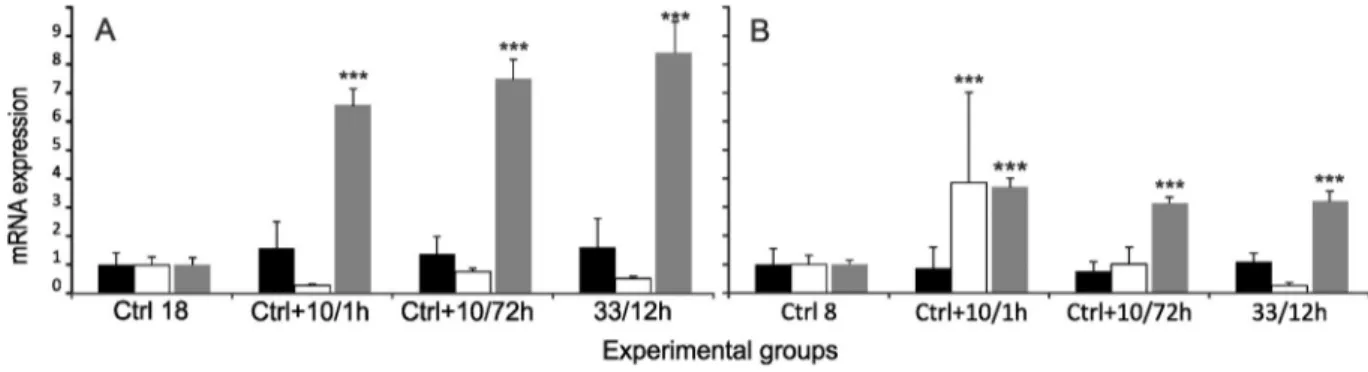
In the KIEL group exposed to elevated salinity, expression ofhsp70 level showed a slight tendency to increase (1.62 fold), but not statisti- cally significant (Fig. 1A). Cortisol level significantly increased (pb0.001) and remained at a high level in all experimental groups (Table 1,Fig. 1A). For better visualisation, the data for cortisol were nor- malised and shown together with gene expression levels on the same graphs. In the GDA group maintained in hypersaline water, expression ofhsp70was similar to the level observed in the control group. Expres- sion ofatp1asignificantly increased (3.8 fold, pb0.001) and fell after 72 h of exposure to 18 salinity (Fig. 1B). The cortisol level was signifi- cantly higher (pb0.001) in both experimental groups from Gdańsk Bay and Kiel Bight (3.71 fold and 8.51 fold, respectively). In the GDA group, plasma cortisol level slightly decreased after thefirst exposure to elevated salinity while in the KIEL group the level of this hormone increased continuously (Table 1,Fig. 1A, B).
Infish from KIEL exposed to reduced salinity, expression ofhsp70 was double to that of the control group (pb0.05) (Fig. 2A). Expression
Fig. 1.Elevated salinity. Relative expression ofhsp70(black bars) andatp1a(white bars) and concentration of cortisol (grey bars) in experimental groups of Baltic cod. The plasma cortisol concentrations and levels of gene expression were normalised to the control group. A—KIEL group, B—GDA group. Significant difference between salinities:⁎ —pb0.05,⁎⁎ —pb0.01,⁎⁎⁎
—pb0.001. Plasma cortisol concentration and expression of genes were calculated for each group in comparison to the control group. Data are expressed as mean ± SEM.
ofatp1awas visibly changed by salinity of 8 and its level of expression after 72 h of exposure was lower (0.25 fold) but not statistically signif- icant. Reduction of salinity to 3 caused a 1.72 fold increase ofatp1a expression but the difference was not statistically significant. Cortisol level elevated with reduced salinities. Only differences between the control group and those exposed to salinity of 8 after 72 h and exposed to 3 salinity after 12 h were statistically significant (Table 1). In the Gdańsk Bay group, expression ofhsp70was similar in all experimental groups. Expression ofatp1afirst increased after 1 h of exposition to re- duced salinity (3.09 fold, pb0.01) and then slightly decreased below the level of the control group (0.70 fold). Cortisol levels, compared to the control group, significantly increased (pb0.05) in the GDA group exposed to salinity of 3 for 72 h (Fig. 2B,Table 1).
The comparison between groups from the west and east Baltic Sea revealed statistically significant differences in both examined salinities.
The level ofatp1aexpression in the GDA group was up-regulated in the group with elevated salinity (18 after 1 h, pb0.001) and reduced salin- ity (3 after 12 h, pb0,01) (Fig. 2AB).
Fish from the Kiel Bight (KIEL) submitted to handling stress were characterized by extremely high concentration of plasma cortisol (Table 1) and 2.98 fold increase of expression of hsp70 gene (pb0.01). The expression ofatp1agene was 0.22 fold compared to the control group. Moreover, in KIELfish kept for 6 weeks in reduced salinity (7.5), plasma cortisol concentration was slightly elevated and
similar to that observed in the control group from the Gdańsk Bay (Table 1). The expression ofhsp70andatp1agenes was not changed compared to the control group.
Significant associations were observed between the expression of atp1aandhsp70and geographical origin of groups, and salinities. Asso- ciations betweenatp1aexpression and origin of cod were statistically significant in both groups (reduced salinity: pb0.01, elevated salinity:
pb0.001). Associations between expression ofatp1aand salinity were also strongly supported (reduced salinity: pb0.000, elevated salinity:
pb0.000). Statistically significant results forhsp70were observed for relationship between elevated salinity and the origin of the cod (pb0.05).
3.3. Osmolality
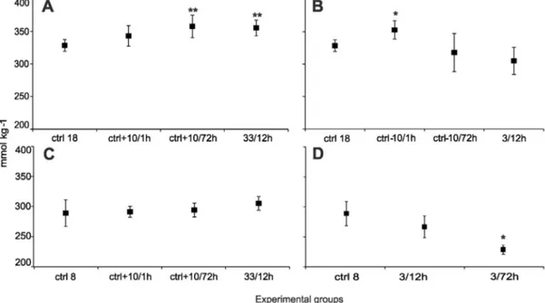
Plasma osmolality observed in the control groups from Gdańsk Bay and Kiel Bight was significantly different (pb0.01; GDA = 282 ± 11.11 mmol kg−1and KIEL = 328 ± 4.13 mmol kg−1). Osmolality observed in groups maintained in salinity of 33 was higher 8.2% in the KIEL and 5% in the GDA group than this in the control group. In extreme- ly low salinity 3/12 h, osmolality was reduced in comparison to the con- trol group: in the KIEL group 7.1% and in the GDA group 7.5% (Fig. 3B and D). In the KIEL group after 6 weeks of exposure to a reduced salinity of 7.5, the plasma osmolality was close to the level observed in the control group from the Gdańsk Bay (294.7 mmol kg−1).
4. Discussion
The results of the present study showed that in the KIEL group, the increase of plasma cortisol concentration was higher than in the GDA group under comparable conditions. The elevated level of cortisol can significantly suppress the level ofhsp70expression in gills as shown earlier byBasu et al. (2001)for tilapia (Oreochromis mossambicus) and rainbow trout (Oncorhynchus mykiss) and in the head kidney of sea bass (Dicentrarchus labrax) revealed byCeli et al. (2012).Boone and Vijayan (2002)have demonstrated that the administration of cortisol into the trout hepatocyte cell culture, significantly reducedhsp70 expression. A high concentration of cortisol and almost unchanged expression ofhsp70in the KIEL group can be explained by the cortisol effect on the hsp70 expression as mentioned above. Differences between plasma cortisol concentrations in experimental groups may provide evidence for the differences between mechanisms for coping with changes of salinity in cod. The different expressions ofatp1ain the KIEL and GDA groups coincide with the variable concentration of cortisol. In accordance withArmesto et al. (2014), the level ofatp1a expressed in gill ionocytes is regulated by salinity. Moreover, a different level of expression of gill ATP-ase is related to acclimatization to differ- ent salinities (Cutler et al., 1995; Deane and Woo, 2004). In east Baltic cod, the high expression ofatp1aandfluctuations of plasma cortisol level is a strong clue that suggests fast acclimatization to elevated
Fig. 2.Reduced salinity. Relative expression ofhsp70(black bars) andatp1a(white bars) and concentration of cortisol (grey bars) in experimental groups of Baltic cod. The plasma cortisol concentrations and levels of gene expression were normalised to the control group. A—KIEL group, B—GDA group. Significant difference between salinities:⁎ —pb0.05,⁎⁎⁎ —pb0.001.
Plasma cortisol concentration and expression of genes were calculated for each group in comparison to the control group. Data are expressed as mean ± SEM.
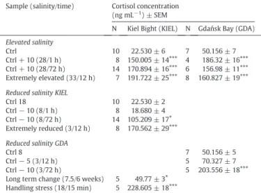
Table 1
Cortisol concentration in plasma samples of Atlantic cod under different salinit and stress conditions. Control (ctrl) indicates salinity of 18 in the Kiel Bight and salinity of 8 in the Gdańsk Bay. N–number of individuals in each group. Long term change sample is the sample exposed to lower salinity in extended time period. Handling stress sample is the sample exposed to acute stress in short time period. Asterisks (⁎: pb0.05;⁎⁎⁎: pb 0.001) indicate significantly different changes of plasma cortisol concentration.
Sample (salinity/time) Cortisol concentration (ng mL−1) ± SEM
N Kiel Bight (KIEL) N Gdańsk Bay (GDA) Elevated salinity
Ctrl 10 22.530 ± 6 7 50.156 ± 7
Ctrl + 10 (28/1 h) 8 150.005 ± 14⁎⁎⁎ 4 186.32 ± 16⁎⁎⁎
Ctrl + 10 (28/72 h) 14 170.894 ± 16⁎⁎⁎ 6 156.98 ± 11⁎⁎⁎
Extremely elevated (33/12 h) 7 191.722 ± 25⁎⁎⁎ 8 160.827 ± 19⁎⁎⁎
Reduced salinity KIEL
Ctrl 18 10 22.530 ± 2
Ctrl−10 (8/1 h) 8 18.680 ± 4
Ctrl−10 (8/72 h) 14 105.209 ± 17⁎ Extremely reduced (3/12 h) 8 170.562 ± 29⁎⁎⁎
Reduced salinity GDA
Ctrl 8 7 50.156 ± 5
Ctrl−5 (3/12 h) 5 70.327 ± 7
Ctrl−10 (3/72 h) 5 203.556 ± 18⁎⁎⁎
Long term change (7.5/6 weeks) 5 49.77 ± 3⁎ Handling stress (18/15 min) 5 228.605 ± 18⁎⁎⁎
salinity by the regulation of ion balance.Dang et al. (2000)noticed that cortisol treatment enhanced the amount of tubular membrane per cell inO. mossambicus. They suggested that,atp1expression was enhanced, because cortisol increased the density of immunoreactivity of this en- zyme in the tubular membrane area. This may explain why the observed cortisol concentrations in the GDA group increased and then revealed a tendency to decrease (Figs. 1 and 2).
In hypoosmotic water, the concentration of cortisol increased after exposure to reduced salinity in both groups. Whereas, in groups ex- posed to reduced salinity, a significantly higher concentration of cortisol was observed after 72 h. Significant changes inhsp70expression were observed in the KIEL group only. The reduced salinity in the GDA group changed only atp1a expression (Fig. 2B). McCormick et al.
(2008) have shown that in Atlantic salmon (Salmo salar), higher concentration of plasma cortisol enhances activity of Na+, K+-ATPase in elevated salinity. In turn,Arjona et al. (2007)revealed in Senegalese sole (Solea senegalensis) that, Na+, K+-ATPase activities in fish transferred to lower salinity were higher than those infish transferred to elevated salinity. In the GDA group exposed to reduced salinity, a 3.2 times higher and statistically significant change of expression of atp1awas observed. After 72 h, a rise in plasma cortisol was observed while the expression ofatp1adecreased and remained unchanged in comparison to the control group.
Significant differences in atp1a expression suggest a different mechanism of reaction of eastern cod to considerable changes in salinity during vertical and horizontal migrations. This may be an explanation of the slightly different results of previous experiments performed in At- lantic cod and Europeanflounder (Platichthysflesus) from the Danish Straits and western Baltic described byLarsen et al. (2008, 2012).
Lifelong residence of eastern cod in reduced salinity affects plasma osmolality. The results presented here, show significant differences in plasma osmolality between the two groups. A long-term exposure to re- duced salinity offish from the KIEL group revealed the osmolality was reduced to a level characteristic for eastern cod (GDA).Sampaio and Bianchini (2002)also noted that plasma osmolality was significantly changed in euryhalineflounder (Paralichthys orbignyanus) after long ac- climatization to reduced and elevated salinity (0 and 30, respectively).
From one side, different levels of the plasma cortisol and expression ofatp1ain both groups suggest that we observed subpopulations very
well acclimated to local environmental conditions. On the other hand, we found genetic differences between both groups, which strongly suggest a genetic background for salinity preferences.Poćwierz-Kotus et al. (2015) and Małachowicz et al. (2015) found significant differences, at the genomic and transcriptomic levels, of differentiation between the Baltic cod and western cod. Genetic differentiation between subpopulations of cod, mentioned previously by other authors (Nielsen et al., 2003; O'Leary et al., 2007; Kijewska et al., 2009, 2011;
Berg et al., 2015), strongly supports the hypothesis of a genetic background for acclimatization. Adaptations to reduced salinity, well described byNissling et al. (1994)for the eastern cod and byPetereit et al. (2014)for the western Baltic cod stock, are more arguments for the thesis that salinity is one of the major factors maintaining the genet- ic structure of the Baltic cod. Other authors likeLarsen et al. (2012)also mentioned that expression ofatp1agene demonstrates the population- specific patterns of gene regulation depending on the origin of the population.
There are significant differences between reaction to salinity chang- es in the eastern and western groups. This profile of response should be considered as an adaptation to variable salinity as supported by previ- ous results of genetic studies (Nielsen et al., 2003; O'Leary et al., 2007;
Poćwierz-Kotus et al., 2015). These adaptations protect eastern cod against the osmotic stress caused by vertical migrations and long- distance migration to spawning areas. Different profiles of reaction to changed salinity suggest that the border between brackish and marine waters is a barrier, which maintains the genetic and physiological separations betweenG. morhuastocks and affects the structure of each subpopulation in the Baltic Sea.
Acknowledgements
This study was partially funded by project: 2011/01/M/NZ9/07207 of the National Science Centre in Poland to RW and statutory topic IV.1 in the IO PAS. We would like to thank Dr. B. Więcaszek (West Pom- eranian University of Technology) for consultations. Special thanks go to Prof. E. Kulczykowska (IOPAS) for critically reading the manuscript and providing helpful advice. C.P. was supported from the BONUS project BIO-C3, funded jointly by the EU and the BMBF (grant no. 03F0682A) and by project funding‘Egg density project’by DTU-Aqua.[SS]
Fig. 3.Osmolality measured in plasma. A—Kiel Bight elevated salinity. B—Kiel Bight reduced salinity. C—Gdańsk Bay elevated salinity. D—Gdańsk Bay reduced salinity. Difference between salinity:⁎ —pb0.05,⁎⁎ —pb0.01. Data are expressed as mean ± SEM.
References
Antoszek, A., Antoszek, J., Więcaszek, B., Ogiński, T., Suszycki, S., 2011.Differences be- tween the Baltic codGadus morhua callariasL. and Atlantic codGadus morhua morhua L., by skeletal muscle protein polymorphism, analyzed with classical SDS-PAGE electrophoresis. EJPAU 14 (3) (#02).
Arjona, F.J., Vargas-Chacoff, L., Ruiz-Jarabo, I., Martín del Río, M.P., Mancera, J.M., 2007.Os- moregulatory response of Senegalese sole (Solea senegalensis, Kaup 1858) to changes in environmental salinity. Comp. Biochem. Physiol. A 148, 413–421.
Armesto, P., Campinho, M.A., Rodríguez-Rúa, A., Cousin, X., Power, D.M., Manchado, M., Infante, C., 2014.Molecular characterization and transcriptional regulation of the Na+/K+ATPaseαsubunit isoforms during development and salinity challenge in a teleostfish, the Senegalese sole (Solea senegalensis). Comp. Biochem. Physiol. B 175, 23–38.
Basu, N., Nakano, T., Grau, E.G., Iwama, G.K., 2001.The effects of cortisol on heat shock protein 70 levels in twofish species. Gen. Comp. Endocrinol. 124, 97–105.
Berg, P.R., Jentoft, S., Star, B., Ring, K.H., Knutsen, K., Lien, S., Jakobsen, K.S., André, C., 2015.
Adaptation to low salinity promotes genomic divergence in Atlantic cod (Gadus morhuaL.). Genome Biol. Evol. 20, 1644–1663.http://dx.doi.org/10.1093/gbe/evv093.
Boone, A.N., Vijayan, M.M., 2002. Glucocorticoid-mediated attenuation of thehsp70re- sponse in trout hepatocytes involves the proteasome. Am. J. Physiol. Regul. Integr.
Comp. Physiol. 283, R680–R687.http://dx.doi.org/10.1152/ajpregu.00125.2002.
Celi, M., Vazzana, M., Sanfratello, M.A., Parrinello, N., 2012.Elevated cortisol modulates Hsp70 and Hsp90 gene expression and protein in sea bass head kidney and isolated leukocytes. Gen. Comp. Endocrinol. 175, 424–431.
Cutler, Ch.P., Sanders, I.L., Hazon, N., Gordon Cramb, G., 1995. Primary sequence, tissue specificity and expression of the Na+,K+-ATPaseα1 subunit in the European eel (Anguilla anguilla). Comp. Biochem. Physiol. B Biochem. Mol. Biol. 111, 567–573.
http://dx.doi.org/10.1016/0305-0491(95)00037-9.
Dang, Z., Balm, P.H., Flik, G., Wendelaar Bonga, S.E., Lock, R.A., 2000.Cortisol increases Na(+)/K(+)-ATPase density in plasma membranes of gill chloride cells in the fresh- water tilapiaOreochromis mossambicus. J. Exp. Biol. 203, 2349–2355.
Deane, E.E., Woo, N.Y.S., 2004.Differential gene expression associated with euryhalinity in sea bream (Sparus sarba). Am. J. Physiol. Regul. Integr. Comp. Physiol. 287, R1054–R1063.
Deane, E.E., Kelly, S.P., Luk, J.C.Y., Woo, N.Y.S., 2002.Chronic salinity adaptation modulates hepatic heat shock protein and insulin-like growth factor I expression in black sea bream. Mar. Biotechnol. 4, 193–205.
Hüssy, K., Bastardie, F., Eero, M., Hemmer-Hansen, J., Mosegaard, E., Nielsen, J.R., 2013.
Improved management based on stock identification of eastern and western Baltic cod. DTU Aqua Report No. 265-2013, p. 17.
Kijewska, A., Burzyński, A., Wenne, R., 2009.Variation in the copy number of tandem re- peats of mitochondrial DNA in the North-East Atlantic cod populations. Mar. Biol. Res.
5, 186–192.
Kijewska, A., Więcaszek, B., Kijewski, T., 2011.Analysis of population and taxonomical structure of Atlantic cod,Gadus morhua(Actinopterygii: Gadiformes: Gadidae) from the Baltic sea with use of microsatellite DNA. Acta Ichthyol. Piscat. 41, 307–314.
Larsen, P.F., Nielsen, E.E., Meier, K., Olsvik, P.A., Hansen, M.M., Loeschcke, V., 2012. Differ- ences in salinity tolerance and gene expression between two populations of Atlantic cod (Gadus morhua) in response to salinity stress. Biochem. Genet. 50, 454–466.
http://dx.doi.org/10.1007/s10528-011-9490-0.
Larsen, P.F., Nielsen, E.E., Williams, T.D., Loeschcke, V., 2008. Intraspecific variation in expression of candidate genes for osmoregulation, heme biosynthesis and stress re- sistance suggests local adaptation in Europeanflounder (Platichthysflesus). Heredity 101, 247–259.http://dx.doi.org/10.1038/hdy.2008.54.
Livak, K.J., Schmittgen, T.D., 2001. Analysis of relative gene expression data using real- time quantitative PCR and the 2(−delta delta C(T)) method. Methods 25, 402–408.
http://dx.doi.org/10.1006/meth.2001.1262.
Madsen, S.S., Jensen, M.K., Nhr, J., Kristiansen, K., 1995.Expression of Na+, K+-ATPase in the brown trout,Salmo trutta:in vivomodulation by hormones and seawater. Am.
J. Physiol. 269, 1339–1345.
Małachowicz, M., Kijewska, A., Wenne, R., 2015. Transcriptome analysis of gill tissue of At- lantic codGadus morhuaL. from the Baltic Sea. Mar. Genomics 23, 37–40.http://dx.
doi.org/10.1016/j.margen.2015.04.005.
McCormick, S.D., Regish, A., O'Dea, M.F., Shrimpton, J.M., 2008. Are we missing a mineral- ocorticoid in teleostfish? Effects of cortisol, deoxycorticosterone and aldosterone on osmoregulation, gill Na+, K+-ATPase activity and isoform mRNA levels in Atlantic salmon. Gen. Comp. Endocrinol. 157, 35–40.http://dx.doi.org/10.1016/j.ygcen.2008.
03.024.
Mommsen, T.P., Vijayan, M.M., Moon, T.W., 1999. Cortisol in teleosts: dynamics, mecha- nisms of action, and metabolic regulation. Rev. Fish Biol. Fish. 9, 211–268.http://dx.
doi.org/10.1023/A:1008924418720.
Neuenfeldt, S., Andersen, K.H., Hinrichsen, H.-H., 2009. Some Atlantic codGadus morhua in the Baltic Sea visit hypoxic water briefly but often. J. Fish Biol. 75, 1095–8649.
http://dx.doi.org/10.1111/j.1095-8649.2009.02281.x.
Neuenfeldt, S., Hinrichsen, H.-H., Nielsen, A., Andersen, K.H., 2007. Reconstructing migra- tions of individual cod (Gadus morhuaL.) in the Baltic Sea by using electronic data storage tags. Fish. Oceanogr. 16, 1365–2419.http://dx.doi.org/10.1111/j.1365-2419.
2007.00458.x.
Nielsen, E.E., Hansen, M.M., Ruzzante, D.E., Meldrup, D., Grønkjær, P., 2003. Evidence of a hybrid-zone in Atlantic cod (Gadus morhua) in the Baltic and the Danish Belt Sea re- vealed by individual admixture analysis. Mol. Ecol. 12, 1497–1508.http://dx.doi.org/
10.1046/j.1365-294X.2003.01819.x.
Nissling, A., Westin, L., 1991.Egg mortality and hatching rate of Baltic cod (Gadus morhua) in different salinities. Mar. Biol. 111, 29–34.
Nissling, A., Westin, L., 1997.Salinity requirements for successful spawning of Baltic and Belt Sea cod and the potential for cod stock interaction in the Baltic Sea. Mar. Ecol.
Prog. Ser. 152, 261–271.
Nissling, A., Kryvi, H., Vallin, L., 1994.Variation in egg buoyancy of Baltic cod (Gadus morhua) and its implications for egg survival in prevailing conditions in the Baltic Sea. Mar. Ecol. Prog. Ser. 110, 67–74.
O'Leary, D.B., Coughlan, J., Dillane, E., McCarthy, T.V., Cross, T.F., 2007. Microsatellite vari- ation in codGadus morhuathroughout its geographic range. J. Fish Biol. 70, 1095–8649.http://dx.doi.org/10.1111/j.1095-8649.2007.01451.x.
Petereit, C., Hinrichsen, H.-H., Franke, A., Köster, F.W., 2014. Floating along buoyancy levels: dispersal and survival of western Balticfish eggs. Prog. Oceanogr. 122, 131–152.http://dx.doi.org/10.1016/j.pocean.2014.01.001.
Poćwierz-Kotus, A., Kijewska, A., Petereit, C., Bernaś, R., Więcaszek, B., Arnyasi, M., Lien, S., Kent, M.P., Wenne, R., 2015.Genetic differentiation of brackish water populations of codGadus morhuain the southern Baltic, inferred from genotyping using SNP-arrays.
Mar. Genomics 19, 17–22.
Reissmann, J.H., Burchard, H., Feistel, R., Hagen, E., Lass, H.U., Mohrholz, V., Nausch, G., Umlauf, L., Wieczorek, G., 2009.State-of-the-art review on vertical mixing in the Baltic Sea and consequences for eutrophication. Prog. Oceanogr. 82, 47–80.
Sampaio, L.A., Bianchini, A., 2002.Salinity effects on osmoregulation and growth of the euryhalineflounderParalichthys orbignyanus. J. Exp. Mar. Biol. Ecol. 269, 187–196.
Tomkiewicz, J., Lehmann, K.M., Stæhr, K.J., St John, M., 1998.Oceanographic influences on the distribution of Baltic cod,Gadus morhua, during spawning in the Bornholm Basin of the Baltic Sea. Fish. Oceanogr. 7, 48–62.
Vijayan, M.M., Pereira, C., Grau, E.G., Iwama, G.K., 1997. Metabolic responses associated with confinement stress in tilapia: the role of cortisol. Comp. Biochem. Physiol. C Pharmacol. Toxicol. Endocrinol. 116, 89–95. http://dx.doi.org/10.1016/S0742- 8413(96)00124-7.
Vizzini, A., Vazzana, M., Cammarata, M., Parrinello, N., 2007. Peritoneal cavity phagocytes from the teleost sea bass express a glucocorticoid receptor (cloned and sequenced) involved in genomic modulation of the in vitro chemiluminescence response to zymosan. Gen. Comp. Endocrinol. 150, 114–123.http://dx.doi.org/10.1016/j.ygcen.
2006.07.016.
Wendelaar Bonga, S.E., 1997.The stress response infish. Physiol. Rev. 77, 591–625.
Westin, L., Nissling, A., 1991.Effects of salinity on spermatozoa motility, percentage of fertilized eggs and egg development of Baltic cod (Gadus morhuaL.), and implications for cod stockfluctuations in the Baltic. Mar. Biol. 108, 5–9.