Nuclear Magnetic Resonance Investigations of Supramolecular Assemblies
D i s s e r t a t i o n
zur Erlangung des akademischen Grades d o c t o r r e r u m n a t u r a l i u m
(Dr. rer. nat.)
im Fach Biophysik eingereicht an der
M a t h e m a t i s c h - N a t u r w i s s e n s c h a f t l i c h e n F a k u l t ä t I d e r H u m b o l d t - U n i v e r s i t ä t z u B e r l i n
von
Andi Mainz, Diplom Biochemiker,
Präsident der Humboldt-Universität zu Berlin Prof. Dr. Jan-Hendrik Olbertz
Dekan der Mathematisch-Naturwissenschaftlichen Fakultät I Prof. Dr. Andreas Herrmann
Gutachter/in: 1. Prof. Dr. Bernd Reif
2. Prof. Dr. Harmut Oschkinat
Zusammenfassung
Eine Vielzahl zellulärer Prozesse wird durch große Proteinkomplexe reguliert. Strukturelle Untersuchungen dieser supramolekularen Maschinen auf atomarer Ebene sind essentiell um mechanistische Einblicke in biologische Vorgänge zu erlangen, sowie bei Fehlfunktion dieser Systeme therapeutische Ansätze zu finden. Die Kernspinresonanz- (NMR) Spektroskopie stellt eine vielseitige Methode dar um die Struktur und Funktion von Proteinen zu untersuchen. Die Strukturanalyse von großen Biomolekülen mittels Lösungs-NMR ist jedoch nur beschränkt möglich. Die Festkörper-NMR Spektroskopie mit Probenrotation im sogenannten magischen Winkel (MAS) stellt dagegen eine molekulargewichtsunabhängige Methode zur Charakterisierung biologischer Komplexe dar. Für die Anwendung der MAS NMR waren jedoch Kristallisationsverfahren für lösliche Proteine bisher unumgänglich.
Im Rahmen dieser Arbeit wurde eine neue Methode entwickelt, die die MAS NMR Spektroskopie an großen Biomolekülen in Lösung erlaubt. Proteinlösungen können demnach durch MAS und dessen Ultrazentrifugationseffekt homogene Proteinsedimente ausbilden, in denen die rotatorische Diffusion großer Proteinkomplexe überwiegend aufgehoben ist. Auf diese Weise können klassische Festkörper-NMR Methoden angewandt werden, ohne dass Präzipitations- oder Kristallisationsverfahren erforderlich sind. In Kombination mit Proteindeuterierung, Protonen-detektierten NMR Experimenten sowie paramagnetischer Relaxationsverstärkung ermöglichte diese neuartige Methode die Detektion und Zuordnung von Rückgrat-Amidresonanzen des 20S Proteasoms mit einem Molekulargewicht von 1.1 MDa. Die experimentellen Daten weisen außerdem daraufhin, dass die Sensitivität und die spektrale Auflösung mit zunehmender Molekülmasse des Zielproteins verbessert werden.
Die Anwendung von MAS NMR auf Proteinlösungen wurde des Weiteren genutzt um das kleine Hitzeschockprotein αB-Crystallin (αB) und dessen Cu(II)-Bindungseigenschaften zu untersuchen. Das Chaperon (600 kDa) spielt eine wesentliche Rolle in der zellulären Proteinhomeostase, ist jedoch aufgrund seiner Polydispersität strukturbiologisch nur schwer zugänglich. Die Anbindung von Cu(II) moduliert die oligomere Architektur und die
α-Crystallin-Domäne ein Cu(II)-Ion mit pikomolarer Affinität bindet. Die potentiellen Cu(II)- Liganden sind H83, H104, H111 und D109 in den β3-β4- und β5-β6+7-Schleifen nahe der Monomer-Monomer Interaktionsfläche. Darüber hinaus konnte eine Cu(II)-induzierte Konformationsänderung in der N-terminalen Domäne beobachtet werden. Letztere vermittelt nicht nur die intermolekulare Anordnung der Untereinheiten im αB-Oligomer, sondern auch die Wechselwirkung mit Substratproteinen. Das sich daraus ergebende Modell beinhaltet die Cu(II)-induzierte Freilegung von Substrat-Interaktionsflächen und Veränderungen in der dynamischen Quartärstruktur sowohl der dimeren Untereinheit als auch des oligomeren Komplexes von αB. Die hohe Bindungsaffinität von αB in Bezug auf redoxaktives Cu(II) hat Implikationen für die zelluläre Proteinhomeostase und die oxidative Stressresistenz.
Interessanterweise spielt oxidativer Stress eine wesentliche Rolle im Verlauf der Alzheimer Krankheit (AD). Elektronenmikroskopie und dynamische Lichtstreuungsexperimente deuten darauf hin, dass das Aggregationsverhalten des AD-relevanten Amyloid β-Peptids (Aβ) durch die Wirkungsweise von αB verändert wird. Über NMR-Studien konnte die Aβ-Bindungsstelle in der hydrophoben β4-β8-Furche der α-Crystallin-Domäne lokalisiert werden. Wie MAS Festkörper-NMR Experimente weiterhin zeigen konnten, führt die Inkubation von Aβ1-40 mit substöchiometrischen Mengen von αB zu strukturell definierten Aβ1-40-Aggregaten.
Die hier erbrachten Forschungsergebnisse leisten einen wesentlichen Beitrag zu dem mechanistischen Verständnis von kleinen Hitzeschockproteinen und ihrer Wechselwirkung mit leicht aggregierenden Substraten. Insbesondere die neuentwickelte MAS NMR Spektroskopie von sedimentierten Biomolekülen legt den Grundstein für zukünftige Struktur- und Dynamikuntersuchungen an großen molekularen Maschinen.
Summary
Various processes in the living cell are regulated by large machineries with molecular weights in the megadalton regime. Atomic-level investigations of these supramolecular assemblies are therefore fundamental to gain mechanistic insights into life and the ability to rationally intervene in case of malfunctioning of these systems. Nuclear magnetic resonance (NMR) spectroscopy is a versatile method to study the structure and function of proteins. However, structural investigations of large biomolecules by solution-state NMR are challenging in case the molecular weight of the complex exceeds 150 kDa. Magic-angle-spinning (MAS) solid- state NMR is a powerful tool for the characterization of biomolecular systems irrespective of their molecular weight. Crystallization, however, is required if this technique is applied to soluble proteins.
In the scope of this work, a novel approach was developed, which opens new perspectives in the investigation of supramolecular modules by MAS NMR. Using simply protein solutions, the ultracentrifugal forces during MAS yield fairly homogeneous sediments of high density, in which rotational diffusion of large protein complexes is impaired. Typical solid-state NMR techniques can thus be applied without the need of precipitation or crystallization procedures.
This novel approach in combination with protein deuteration, proton-detection and paramagnetic relaxation enhancement enabled the observation and the assignment of backbone amide resonances of a 20S proteasome assembly with a molecular weight of 1.1 MDa. The experiments show further that the sensitivity and the resolution improve with increasing molecular weight.
Similarly, this MAS NMR approach was used to characterize the small heat-shock protein αB- crystallin (αB) with respect to its Cu(II)-binding capability. The chaperone (600 kDa) plays an essential role in cellular protein homeostasis. Structural investigations at atomic resolution are hampered due to its polydispersity. Binding of Cu(II) modulates the oligomeric architecture and the chaperone activity of αB. The multimer assembly and its isolated dimeric core domain were studied by various NMR techniques and other biophysical methods to unravel the effects
β5-β6+7-loops at the dimer interface. Moreover, the metal ion triggers structural reorganization in the N-terminal domain, which is known to mediate the intermolecular arrangement in αB oligomers as well as the binding of client proteins. We therefore suggest that Cu(II)-binding unblocks potential client binding sites and alters quaternary dynamics of both the dimeric building block as well as the higher-order assemblies of αB. The high- affinity of αB towards redox-active Cu(II) has implications in protein homeostasis and oxidative stress resistance. Intriguingly, oxidative stress is also implicated in the progression of the Alzheimer’s disease (AD). Electron microscopy and dynamic light scattering experiments show that αB modulates the aggregation behavior of the AD-related amyloid β- peptide (Aβ). Our NMR data suggest that the Aβ-binding site of αB involves the hydrophobic β4-β8 groove of the α-crystallin domain. Moreover, co-incubation of Aβ1-40 and substoichiometric amounts of the chaperone yield well-defined Aβ1-40 aggregates, which are amenable for structural analysis by MAS solid-state NMR spectroscopy.
The findings of this work contribute to the mechanistic understanding of small heat-shock proteins and their interaction with aggregation-prone clients. In particular, we expect that MAS NMR employed to biomolecules in solution will be a novel tool to explore structural and dynamic properties of large biological machines in the future.
Table of Contents
Zusammenfassung ... I Summary ... III Table of Contents ... V Abbreviations ... IX
1 Introduction and Objectives ... 1
1.1 Protein Homeostasis ... 1
1.1.1 Protein Folding and Molecular Chaperones ... 2
1.1.2 Heat-Shock Proteins (HSPs) ... 3
1.1.3 Small Heat-Shock Proteins (sHSPs) ... 5
1.1.3.1 Structure and Function of αB-crystallin ... 8
1.1.3.2 Copper Biochemistry ... 11
1.1.4 Protein Degradation: The Proteasome ... 15
1.1.5 Amyloid Aggregation and the Alzheimer’s Disease ... 17
1.2 Nuclear Magnetic Resonance Spectroscopy ... 20
1.2.1 Nuclear Spins ... 20
1.2.2 Relaxation of Excited Spin States ... 23
1.2.3 NMR Investigations of Large Biomolecules ... 26
1.2.4 Solid-State NMR ... 27
1.2.4.1 Magic-Angle-Spinning (MAS) ... 27
1.2.4.2 Cross-Polarization ... 29
1.2.4.3 Perdeuteration and Proton-Detection ... 30
2 Materials and Methods ... 31
2.1 Solutions and Media ... 31
2.1.1 Buffer Solutions ... 31
2.1.2 Cell Growth Media ... 31
2.2 Microbiological and Molecular Biological Methods ... 32
2.2.1 Polymerase Chain-Reaction (PCR) ... 32
2.2.2 Agarose Gel-Electrophoresis ... 32
2.2.3 Determination of DNA Concentration ... 33
2.2.9 Colony-PCR ... 35
2.2.10 Amplification and Extraction of Plasmid-DNA ... 35
2.2.11 Recombinant Protein Expression ... 36
2.2.11.1 Unlabeled Protein ... 36
2.2.11.2 13C and 15N Enrichment in Proteins ... 36
2.2.11.3 13C/15N Enrichment and Perdeuteration ... 37
2.2.12 Cell Harvest and Storage ... 37
2.3 Biochemical Methods and Sample Preparation ... 38
2.3.1 Cell Lysis ... 38
2.3.2 Anion-Exchange Chromatography (AEC) ... 38
2.3.2.1 Weak AEC ... 38
2.3.2.2 Strong AEC ... 39
2.3.3 Size-Exclusion Chromatography (SEC) ... 39
2.3.4 Analytical SEC for Protein Characterization ... 40
2.3.5 SDS-PAGE ... 40
2.3.6 Dialysis ... 40
2.3.7 Concentration of Protein Samples by Ultrafiltration ... 41
2.3.8 Storage of Protein Samples ... 41
2.3.9 Preparation of Monomeric Aβ1-40 ... 41
2.3.10 Alignment of Protein Sequences ... 42
2.4 Biophysical Methods ... 42
2.4.1 Determination of Protein Concentration ... 42
2.4.2 Dynamic Light Scattering (DLS) ... 42
2.4.3 Inductively-Coupled-Plasma Mass Spectrometry (ICP-MS) ... 44
2.4.4 Isothermal Titration Calorimetry (ITC) ... 44
2.4.5 In-vitro Chaperone Activity Assay ... 45
2.4.6 Electron Microscopy (EM) ... 46
2.4.7 Circular Dichroism (CD) Spectroscopy ... 46
2.4.8 Fluorescence Spectroscopy ... 47
2.4.9 Fluorescence Correlation Spectroscopy (FCS) ... 48
2.4.10 Estimation of Rotational Correlation Times ... 49
2.4.11 X-ray Crystallography ... 50
2.4.12 Modeling of the Cu(II) Coordination Sphere ... 50
2.5 NMR Spectroscopy ... 51
2.5.1 Solution-State NMR Spectroscopy ... 51
2.5.1.1 Backbone Resonance Assignment of αB10m ... 51
2.5.1.2 Titration of αB10m with Divalent Metal Ions ... 52
2.5.1.3 13C Direct Detected Experiments ... 53
2.5.1.4 15NRelaxation Measurements ... 54
2.5.1.5 Estimation of Overall Rotational Correlation Times from T1/T2 ... 54
2.5.1.6 Resonance Assignment of Monomeric Aβ1-40 ... 55
2.5.1.7 Titration of αB10m with Monomeric Aβ1-40... 56
2.5.1.8 Perdeuterated αB in Solution ... 56
2.5.2 Nuclear Magnetic Relaxation Dispersion (NMRD) ... 56
2.5.3 Magic-Angle-Spinning (MAS) NMR Spectroscopy ... 58
2.5.3.1 Precipitation of αB for Conventional Solid-state NMR ... 58
2.5.3.2 Comparison of Soluble and Precipitated αB Multimers ... 59
2.5.3.3 Titration of αB Multimers with Divalent Metal-Ions ... 59
2.5.3.4 The 20S Proteasome Assemblies of Thermoplasma acidophilum ... 60
2.5.3.5 Aggregates of Aβ1-40 in the Presence of αB ... 60
2.5.3.6 1H-detected FROSTY MAS NMR ... 61
3 Results ... 63
3.1 Protein Preparation ... 63
3.1.1 Full-length αB ... 63
3.1.1.1 αB Constructs with N-terminal Affinity-tag ... 63
3.1.1.2 Cloning of Tag-free αB ... 64
3.1.1.3 Overexpression and Purification of αB ... 65
3.1.2 The Excised α-crystallin Domain: αB10m ... 66
3.1.2.1 Cloning of Construct ... 66
3.1.2.2 Overexpression and Purification of αB10m and its Mutants ... 67
3.2 αB-crystallin and Metal-Binding ... 68
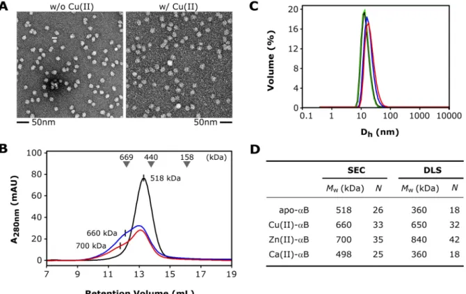
3.2.1 Effects of Metals on the Morphology of αB Multimers ... 68
3.2.2 Modulation of αB Chaperone Activity by Cu(II) ... 70
3.2.3 Stoichiometry of Cu(II)-binding: ICP-MS Experiments ... 72
3.2.4 Affinity and Stoichiometry of Cu(II)-binding: ITC Studies ... 72
3.2.5 Fluorescence Quenching: Cu(II)-Affinity of the ACD ... 73
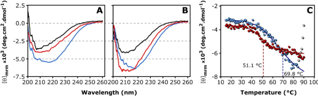
3.2.6 Secondary Structure and Thermal Stability ... 74
3.2.7 Effects of Cu(II) on the Quaternary Structure of the ACD ... 77
3.2.8 Crystallization of the ACD ... 79
3.2.9 NMR Investigations on Full-length αB Multimers ... 80
3.2.10 NMR Investigations on the Dimeric αB10m ... 81
3.2.10.1 Resonance Assignment of apo-αB10m ... 81
3.2.10.2 NMR Titration Studies ... 82
3.2.10.3 13C-detected NMR Experiments ... 86
3.2.10.4 The AP Interface and its Dynamics ... 89
3.2.10.5 The Cu(II) Coordination Sphere ... 95
3.3.4 Inhibition of Aβ1-40 Fibril Formation ... 100
3.3.5 Structural Investigations of Aβ1-40 Aggregates ... 102
3.4 MAS NMR on Protein Solutions ... 103
3.4.1 The Multimers of αB and the FROSTY Approach ... 104
3.4.2 The 20S Proteasome Assemblies of Thermoplasma acidophilum ... 107
3.4.3 Rotational Diffusion of Large Protein Complexes ... 110
3.4.3.1 Estimation of Rotational Correlation Times ... 110
3.4.3.2 Nuclear Magnetic Relaxation Dispersion (NMRD) ... 112
3.4.3.3 Fluorescence Correlation Spectroscopy (FCS) ... 113
3.4.4 1H-detected FROSTY MAS NMR ... 115
3.4.4.1 Perdeuterated αB Multimers in Solution ... 115
3.4.4.2 Perdeuterated 20S Proteasome Assemblies in Solution ... 119
4 Discussion and Conclusions ... 129
4.1 Protein Preparation ... 129
4.2 αB-crystallin and Metal-binding ... 129
4.3 The Interaction of αB-crystallin with Aβ1-40 ... 135
4.4 MAS NMR on Protein Solutions ... 138
5 Appendix ... 143
5.1 DNA and Protein Sequences ... 143
5.1.1 Vector DNA ... 143
5.1.2 PCR Primer Sequences ... 144
5.1.3 Primer Sequences for Site-directed Mutagenesis ... 144
5.1.4 Protein Sequences ... 145
5.1.5 Protein Parameters ... 146
5.2 NMR Data ... 147
5.2.1 PDSD Spectra of αB Oligomers in Solution ... 147
5.2.2 Chemical Shifts of apo-αB10m ... 148
5.2.3 Chemical Shifts of Cu(II)-αB10m ... 150
5.2.4 Chemical Shifts of Monomeric Aβ1-40 ... 151
5.2.5 Chemical Shifts of the α-subunit in α7β7β7α7-(11S)14... 152
5.2.6 Pulse Programs for 1H-detected Solid-state NMR Experiments ... 155
References ... 162
Acknowledgements ... 174
Publications ... 177
Conferences and Workshops ... 178
Eidesstattliche Erklärung ... 179
Abbreviations
2D/3D two dimensional, three dimensional
αB αB-crystallin
αB10m protein construct comprising residues 64-152 of αB and bearing the mutation N146D
Aβ amyloid β-peptide
ACD α-crystallin domain AD Alzheimer’s disease ADP adenosine diphosphate
AEC anion-exchange chromatography
AP anti-parallel
APP amyloid precursor protein ATP adenosine triphosphate BSA bovine serum albumin
C’ carbonyl atom
CCS copper chaperone for superoxide dismutase CD circular dichroism
CIP calf-intestinal phosphatase CP cross polarization
CS citrate synthase
CSA chemical shift anisotropy CSI chemical shift index CSP chemical shift pertubation
CV column volume
DLS dynamic light scattering DMSO dimethyl sulfoxide DNA desoxyribo-nuclein-acid E.coli Escherichia coli
EDTA ethylenediaminetetraacetic acid
Ek enterokinase
EM electron microscopy
EPR electron paramagnetic resonance FCS fluorescence correlation spectroscopy FID free induction decay
FROSTY freezing rotational diffusion of protein solutions at low temperature and high viscosity
fw forward
GSH glutathione
HMQC heteronuclear multiple quantum coherence HSP heat-shock protein
HSQC heteronuclear single quantum coherence ICP-MS inductively-coupled-plasma mass spectrometry IDP intrinsically disordered protein
INEPT insensitive nuclei enhanced by polarization transfer IPAP in-phase anti-phase
IPOD insoluble protein inclusion
IPTG isopropyl β-D-1-thiogalactopyranoside ITC isothermal titration calorimetry JUNQ juxtanuclear quality control
MPD 2-methyl 1,3-propanediol
MS mass spectrometry
NEF nucleotide-exchange factor NMR nuclear magnetic resonance
NMRD nuclear magnetic relaxation dispersion
NOE(SY) nuclear Overhauser enhancement (spectroscopy)
OD optical density
PAGE polyacrylamide gelelectrophoresis PBS phosphate buffered saline
PCR polymerase chain reaction
Pd polydispersity
PDB protein data bank
PDSD proton-driven spin diffusion PEG polyethylene glycol ppm parts per million
PRE paramagnetic relaxation enhancement
PrP prion protein
REDOR rotational echo double resonance RE restriction enzyme
rf radio-frequency
RFDR radio frequency driven recoupling ROS reactive oxygen species
rv reverse
SAXS small-angle X-ray scattering SDS sodium dodecylsulfate sHSPs small heat-shock protein SEC size-exclusion chromatography SOD superoxide dismutase
SSP secondary structure propensity TEV tobacco etch virus
TOCSY total correlation spectroscopy
TPPI time-proportional phase incrementation TROSY transverse relaxation-optimized spectroscopy UPS ubiquitin-proteasome system
UV ultraviolet
wt wildtype
Standard chemical and physical abbreviations are not listed. Nucleotides are abbreviated by the standard one-letter code. Amino acids are abbreviated by the standard one- or three letter code.
1 Introduction and Objectives
1.1 Protein Homeostasis
Proteins are the key players of life and their biological functions are multi-faceted.
Mammalian cells typically express more than 10,000 different types of proteins with diverse three-dimensional structures and cellular functions [1]. This multitude of protein species and its temporal flow must be stringently maintained and regulated in the living cell. Protein homeostasis (proteostasis) is therefore crucial for cellular viability. Several control machineries have evolved to fine-tune the balance between i) protein synthesis, ii) protein folding, and iii) protein clearance (Fig. 1A). Pathological conditions, environmental influences, metabolic stress, cancer and aging are known to disturb this sensitive balance [2,3]. Cellular strategies to cope with fluctuations of the proteome rely on the coordinated action of the quality control apparatus as well as the systems that regulate protein biogenesis, assembly, trafficking, and translocation [3]. All aspects of protein quality control involve the class of molecular chaperones, which accomplish de novo protein folding as well as the stabilization and the refolding of non-native proteins [4]. In futile cases, chaperones also guide misfolded proteins to the autophagy system and the degradation machinery, namely the ubiquitin-proteasome system (UPS) [1]. The sequestration of misfolded proteins in spatially distinct protein inclusions, known as quality control compartments, is a further mechanism to prevent the accumulation of toxic protein wastage [5]. Recently, two different compartments have been defined – the JUNQ (juxtanuclear quality control) and the IPOD (insoluble protein inclusion), which fulfill distinct functions within the proteostatic network [6]. Whilst JUNQ sequesters soluble non-native proteins and operates in conjunction with chaperones and proteasomes, the IPOD contains insoluble protein aggregates and is associated with the autophagy-related Atg8.
Hundreds of proteins participate in the regulation of cellular proteostasis [3]. In the following, selected members of molecular chaperones and the UPS are described in more detail. Protein
Fig. 1 Protein folding and the role of the chaperone machinery. (A) From the cradle to the grave: protein fates and the cellular proteome maintenance system. In mammalian cells, proteostasis is accomplished by a synchronized network of molecular chaperones, the UPS (ubiquitin-proteasome system) and autophagy system with approximately 180, 600 and 30 components, respectively. Together with the transcription and translation machinery, this network assures protein biogenesis, maintenance and clearance. This proteostatic balance is fundamental for cellular functionality and integrity. (B) Schematic illustration of the funnel-shaped free-energy landscape of protein folding, which might overlap with that of intermolecular aggregation. Polypeptide chains need to traverse free-energy barriers to reach downhill-paths towards the native fold (green). Folding intermediates and partially folded states accumulate and are imminent to form amorphous aggregates, pre-fibrillar oligomeric states or amyloid fibrils (red). Molecular chaperones inhibit protein aggregation by stabilizing partially folded states and assist in protein (re-)folding by lowering energetic barriers. Both figures are taken from [1].
1.1.1 Protein Folding and Molecular Chaperones
The amino acid sequence of a protein encodes the three-dimensional structure and consequently the biological function of the respective protein [7]. However, many proteins exhibit very slow folding rates or even fail to adopt their native state [4,8]. The huge conformational space of a polypeptide chain and the diverse combinations of many weak and non-covalent intramolecular interactions render the folding reaction a highly complex process.
It is the burial of hydrophobic residues within the protein interior that is one of the major driving forces, inducing an intramolecular chain collapse [9,10]. The conformational space becomes progressively restricted during the folding reaction, however, partially folded states can be transiently populated and kinetically trapped in local energy minima (Fig. 1B) [11]. In particular, larger and/or multi-domain proteins have an increased propensity to populate such misfolded states [12]. The incomplete folding reaction entails that hydrophobic residues and segments are partially exposed to the aqueous environment [13]. As in the case of
the formation of amorphous aggregates (Fig. 1B). This tendency of unstructured or partially folded proteins to associate in non-native fashions is especially pronounced in the crowded cellular environment with cytosolic protein concentrations in the range of 300–400 g/L [1]. In addition to the heterogeneous amorphous aggregates, amyloid fibrils with high structural order may also form [14]. Intriguingly, such fibrillar aggregates are often accompanied by soluble oligomeric states of rather heterogeneous appearance [14,15]. These less-ordered oligomeric structures might be considered as analogues of the kinetically trapped intermediates in protein folding. A recent model suggests, that aggregates of misfolded proteins provoke cell toxicity by diminishing the capacity of the protein folding machinery [16]. Accordingly, the chaperoning and proteolytic systems are sequestered and blocked by these toxic aggregates. This overstrain impairs the proteostatic capacity of the cell [16]. The up-regulation of protein quality control systems, namely chaperones, the UPS and autophagy, has been shown to reverse the deleterious effects of disease-related protein aggregates [17].
The formation of amorphous and fibrillar aggregates is restricted in vivo by the intervention of the cellular chaperone network [1,18]. Molecular chaperones are capable of stabilizing misfolded proteins prior to aggregation and to assist protein folding. Since all members of this protein class are up-regulated in response to cellular stress, e.g. elevated temperatures, they are also known as heat-shock proteins (HSPs) [1]. Commonly, chaperones are classified with respect to their molecular weight, namely HSP100, HSP90, HSP70, HSP60, HSP40 as well as the small HSPs (sHSPs) with molecular weights in the range of 12-43 kDa [19]. Chaperones can be further differentiated by their mode of action, i.e. the utilization of ATP, and are termed foldases (ATP-dependent) and holdases (ATP-independent), respectively [4]. These two types are outlined separately in the next chapters with a focus on the sHSP family.
1.1.2 Heat-Shock Proteins (HSPs)
ATP-dependent chaperones like HSP70s, HSP90s and the chaperonins (HSP60s) are capable of promoting de novo protein folding as well as refolding of stress-denatured proteins [19].
This assistance in structural maturation is achieved by repetitive substrate binding and release
release. The underlying principle is known as kinetic partitioning [20], which requires that the folding rate constant (kfold) is greater than the association constant (kon) for chaperone (re-)binding of partially folded substrates, and that kon exceeds the self-association rate constant kagg of higher-order aggregates (kfold > kon > kagg) (Fig. 2A). Under cellular stress conditions, kagg may become greater than kon resulting in protein aggregation, unless expression of the chaperone machinery is elevated via the stress-response pathway [21].
The HSP70 system and the chaperonins operate sequentially along the folding path of a protein [22]. Folding of newly synthesized and nascent polypeptide chains is assisted by the integrated action of the HSP40 and HSP70 chaperone systems (upstream chaperones) [23].
However, many proteins, such as actins and tubulins, encounter high energetic barriers en-route to their native and functionally active state [24]. In those cases, the association to the chaperone (kon) may become faster than the folding (kfold). These proteins are stabilized by the chaperone in a non-aggregated state, but require the intervention of the specialized chaperonins (downstream chaperones) [1]. An unique feature of the chaperonin family is the release of substrate proteins into a folding chamber, rather than the cytosolic environment.
Prominent members are the eukaryotic HSP60s and the bacterial GroEL, that form large double-ring complexes (800-900 kDa) [23]. The spacious interiors can accommodate substrate proteins up to molecular weights of ~60 kDa [1]. Non-native protein structures are sequestered via their hydrophobic surfaces and bound in the lumen of the chaperonin complex. The corresponding HSP10 proteins (eukaryotes) and GroES (bacteria) function as ATP-regulated lids of the folding cage, thereby completely encapsulating the substrate protein [25]. Closure of the folding chamber induces a highly hydrophilic (net negatively charged) surface of its inner wall, which promotes the folding reaction of the enclosed substrate [25].
The encapsulation of a single polypeptide chain impedes aggregation and the devastating interaction with other cellular components [26]. Furthermore, the steric confinement has been suggested to accelerate the folding rate by entropically destabilizing flexible folding intermediates and favoring more compact, native-like structures [27]. Chaperonins are thus enabled to diminish both enthalpic as well as entropic barriers in the free-energy landscape of protein folding. Mechanistic details of the chaperonin system GroEL-GroES are illustrated in Fig. 2B.
Fig. 2 Chaperone-mediated protein folding. (A) The chaperone cycle of the eukaryotic HSP70 system and the principles of kinetic partitioning (see text) are illustrated. Unfolded and misfolded proteins are sequestered by one of several HSP40 cofactors, that recruit the substrate to the ATP-bound HSP70 (low-affinity, open state). ATP hydrolysis induces closure of the peptide-binding domain (yellow). In this high-affinity (closed) state, the substrate is tightly bound to the chaperone. Several nucleotide-exchange factors (NEFs) catalyze the substitution of ADP by ATP, which finally promotes substrate release. Kinetic partitioning is achieved for kfold > kon > kagg, where kfold, kon, kagg denote the folding rate constant, the association constant of substrate binding to the chaperone and the aggregation rate constant of the substrate, respectively. (B) The bacterial chaperonin GroEL-GroES and its folding cage. Aberrantly folded proteins with slow kfold are guided to the chaperonin by the stabilizing HSP70 system. The apical domains of the seven-membered GroEL rings bind the substrate and are structurally rearranged upon ATP binding. Subsequently, the substrate is enclosed in the folding chamber of GroEL by the GroES lid (cis-complex), which causes the dissociation of ADP, GroES and substrate from the opposite side of the complex (trans-complex) (omitted for clarity). The GroES-induced hydrophilic nature of the inner wall allows the substrate to fold during the hydrolysis of seven ATP molecules (~10 s). The cycle is finished (substrate release) by binding of ATP and GroES to the trans complex. Both figures are taken from [1].
Certainly, there are several other chaperone and chaperonin systems known, such as the HSP90 and TRiC proteins, respectively, which are not discussed here [1,23]. The integrated action of all these multi-component molecular machines in cooperation with the sHSP family, the UPS and autophagy ensures the stability of the cellular proteome.
1.1.3 Small Heat-Shock Proteins (sHSPs)
Small heat-shock proteins (sHSPs) represent a diverse class of molecular chaperones that have been found in all six kingdoms of life except some pathogenic bacteria [28]. The number of sHSPs of an organism varies considerably, with the higher eukaryotes raising a broader
The sHSPs play a fundamental role during cellular stress by ‘rescuing’ misfolded or partially unfolded proteins from final proteolytic degradation [29,30]. Therefore, sHSPs constitute the
‘first line of defense’ in terms of cellular stress. The ATP-independent sequestration of client proteins prevents irreversible aggregation and enables subsequent intervention of refolding- competent chaperone systems like HSP40-HSP70 and HSP100 (Fig. 3) [31,32]. In vitro studies have shown that sequestered client proteins are folding competent, however, the trapped polypeptides need to be reactivated once cellular stress has been overcome [33].
Notably, sHSPs are not competent to spontaneously release bound clients, and thus cooperate with the ATP-dependent chaperones [28,32]. This cellular strategy to buffer aggregation is highly efficient, in particular considering the ATP-independent mechanism of sHSPs and the low-energy state of the cell under stress conditions. The possibility to rescue misfolded proteins from the sHSPs-stabilized reservoir of aggregation-prone polypeptides circumvents de novo synthesis of the respective proteins. These economic aspects may illustrate the important role of sHSPs in proteome maintenance.
Fig. 3 The anti-aggregation property of sHSPs. The multimeric assemblies of sHSPs are highly dynamic and exchange subunits rapidly (mostly dimeric substructures). Upon cellular stress, the activated state is capable of binding non-native proteins that otherwise undergo irreversible aggregation. The non-native protein is stabilized within the highly soluble client-sHSP-complex and is directed to the ATP-dependent refolding machineries, e.g.
HSP40/HSP70 and HSP100. The figure is adapted from [28].
Despite their diversity in sequence and size, sHSPs share common features such as small molecular weights (12-43 kDa), domain architecture, formation of large oligomeric assemblies with dynamic quaternary structure, induction in response to cellular stress and the prevention of protein aggregation (holdase function) [28]. This protective function of sHSPs remained elusive until bovine α-crystallin and murine HSP26 were reported to exhibit chaperone activity in vitro [30]. The ability to bind diverse non-native proteins at high client- sHSP stoichiometries (up to one substrate molecule per dimeric subunit) further highlights the efficiency of this cellular strategy to manage irreversible protein aggregation [34].
A characteristic trait of the sHSP family is the conserved α-crystallin domain, which is flanked by variable N- and C-terminal domains [35,36]. Dimerization of the α-crystallin core domain forms the basic building block of higher-order assemblies, which are established by intermolecular linkage via the terminal anchors (further structural details are given in Chapter 1.1.3.1). The sHSPs represent highly dynamic assemblies, which exchange subunits (primarily dimers) in a constant manner [37]. Due to this subunit exchange, hetero-oligomers may form in compartments with different types of sHSPs, as is the case for αA-crystallin (αA) and αB-crystallin (αB) in the mammalian eye lens [38]. This appears as a valid mechanism to modulate the characteristics and the substrate specificity of sHSPs.
Generally, molecular chaperones exist in states of low and high substrate affinity (Fig. 3) [28].
In ATP-dependent chaperones, the binding and hydrolysis of ATP triggers the transition between the two functional states [4]. By contrast, ATP has no direct role in the regulation of sHSPs chaperone activity [28]. Many members of the sHSP family are constitutively inactive, e.g. yeast HSP26, but become activated by typical stress factors such as elevated temperatures [39]. The dynamic behavior of sHSPs permits the potential client binding sites, which are hydrophobic in character and mostly buried in the oligomeric assembly, to become exposed upon subunit dissociation [40–42]. Indeed, several sHSPs become more hydrophobic under stress conditions [28,42,43]. Dynamic release and incorporation of subunits may thus reflect an inherent ‘sensing’ mechanism of sHSPs to recognize and sequester non-native proteins in the cellular environment as well as to stabilize the client proteins in the context of a soluble client-sHSP-complex. This hypothesis has been supported by studies on human HSP27, which
structural rearrangements within the multimeric assembly. It is tempting to note, that the mechanism of regulation may rely on modulation of quaternary dynamics and alteration of client binding site accessibility. The structural plasticity of thermally-activated pea HSP18.1 has been demonstrated to correlate with its ability to protect firefly luciferase from aggregation [37]. More than 300 different client-chaperone stoichiometries were observed in these studies. It appears that the ability to form polydisperse ensembles, constitutively or in response to stress, is an essential element for the chaperone mechanism of sHSPs.
1.1.3.1 Structure and Function of αB-crystallin
The sHSP αB-crystallin (αB) was originally discovered in the mammalian eye lens as the B- subunit of α-crystallin [30]. Its function is to maintain the transparency and high refractive index of the eye lens, thereby counteracting cataract formation and visual impairment [46].
Besides this specific lenticular function, the biological role of human αB is manifold.
Likewise it is localized in several other tissues such as the lung, kidney, brain, cardiac and skeletal muscle [47]. αB was shown to interact with a wide range of substrate proteins that either aggregate amorphously or form amyloid fibril structures [30,48–50]. As a consequence αB was found to be involved in multiple sclerosis, cancer, cardiomyopathy and various neurodegenerative diseases like Parkinson’s and Alzheimer’s disease [31,51,52].
The 20 kDa protein αB assembles into highly dynamic and polydisperse complexes of about 600 kDa (24-32 subunits) [53]. A general feature of sHSPs is the α-crystallin domain (ACD), which is a conserved core domain of about 90 residues adopting a β-sandwich structure (Fig.
4A,B) [28]. The ACD is flanked by variable N-terminal (~60 residues) and C-terminal domains (~25 residues) that accomplish the multimeric assembly. The formation of dimeric building blocks via shared β-sheets (strands β6 and β7’) is a conservative trait of sHSPs (Fig.
4C) [35,36]. The dynamic nature of sHSPs, and the αB oligomers in particular, hindered their structural characterization [54]. Crystal structures of sHSPs have been determined only for HSP16.9 from wheat and HSP16.5 from Methanococcus jannashii, revealing 12-mer and 24- mer assemblies, respectively (Fig. 4D,E) [35,36]. The C-terminal anchors comprise a conserved IXI-motif that packs into hydrophobic grooves (strands β4-β8) of neighboring molecules on the surface of the complexes. By contrast, the N-terminal domains are usually involved in intermolecular interactions within the central cavity of sHSPs assemblies.
Fig. 4 Subunit assembly of sHSPs. (A) The domain organization of sHSPs is illustrated. The α-crystallin domain (ACD) is flanked by N- and C-terminal sequences. Residue numbers are exemplarily shown for αB. (B) Crystal structure of wheat HSP16.9 illustrating the topology of the monomeric subunit [35]. The conserved ACD (red) adopts an immunoglobulin-like fold with two anti-parallel β-sheets forming a β-sandwich structure. The strands β6 and β7 that form the dimer interface are indicated. The ACD is flanked by variable N- (green) and C-terminal sequences (blue), which accomplish the higher-order assembly. (C) Dimerization of the α-crystallin domain via the strands β6 and β7’ forms the building block of oligomeric assemblies. The two monomers of wheat HSP16.9 are colored in cyan and grey, respectively [35]. (D) The 12-mer assembly of wheat HSP16.9 is shown with three dimeric building blocks colored in red, blue and yellow [35]. Two hexameric rings with three-fold symmetry arrange in a double-disk fashion.
(E) For comparison, HSP16.5 of Methanococcus jannashii is depicted [36]. Three dimeric building blocks are highlighted in red, blue and yellow, respectively. Four of such hexameric rings are arranged with tetrahedral geometry in the 24-mer. The C-terminal anchors interact with neighboring monomers and thereby stabilize the complex.
Only truncated variants of αB, containing the ACD without the terminal anchors, were amenable for X-ray crystallography and solution-state NMR [55–57]. Electron microscopy (EM) of negatively-stained αB revealed the envelope of a 24-mer with tetrahedral symmetry [58,59] (Fig. 5C). Recently, solid-state magic-angle-spinning (MAS) NMR enabled structural studies of full-length αB and provided insights into the oligomeric architecture of αB [57,60,61] (Fig. 5B,D). In all atomic-level structures of αB variants, the ACD folds into a β- sandwich structure comprising the two sheets β8-β9-β3-(β2) and β4-β5-β6+7 (Fig. 5A,B). The separated β6 and β7 strands, as observed for HSP16.5 and HSP16.9, have merged into one
[56]. A shared groove is located above the AP interface, which appears to be polymorphic, since three different alignment registers were observed [55,56]. The curvature of the dimeric building block varies significantly. The solid-state NMR structure of αB multimers reveals a highly bended ACD dimer [60]. This curvature is supposed to respond to changes of the pH [60]. An extensive network of ionic interactions spans the interface. This network involves charged residues of the strands β3, β4 as well as β5, β6+7 and the intervening loops. Notably, the conserved R120 of one monomer is involved in a bidentate ion pair with D109 of the neighboring molecule [55,56,60]. The missense mutation R120G causes an increase of the oligomeric size and a decrease in the in vitro chaperone efficiency [62]. Co-precipitates of desmin and αB-R120G are found in cardiac muscle tissue of patients suffering from desmin- related cardiomyopathy [52].
Fig. 5 Structural information for the αB assembly. (A) Crystal structure of the isolated ACD dimer (αB residues 67-157) [55]. The two monomers are colored cyan and grey, respectively. β-strands are labeled. Strands β6+7 form the anti-parallel (AP) interface and an extended bottom-sheet. The two top-sheets (β2-β3-β9-β8) form a shared groove above the AP-interface. The variable β2 strand is disordered in one of the two monomers. (B) Solid-state NMR structure of the ACD dimer as present in full-length αB multimers (residues 64-152 are shown) [60]. The two monomers are colored cyan and grey, respectively. β-strands are labeled. In comparison to the isolated ACD, the dimer is much more curved in the context of αB multimers. Hence, the shared groove becomes more accessible.
(C) EM density map of the αB 24-mer [58]. The view along one threefold axis shows the tetrahedral arrangement of four hexameric rings, each formed by three dimeric building blocks. (D) Structural model of the 24-mer of αB as obtained by combined data from solid-state NMR, EM and SAXS (small-angle X-ray scattering) [61]. Three dimeric building blocks are highlighted in red, blue and yellow, respectively. The flexible C-terminal extensions are omitted for clarity.
The dimeric substructure of αB represents the building block of its higher-order assemblies [60,61]. Due to the dynamic subunit-exchange, the resulting αB ensemble is consequently heterogeneous. The intrinsic subunit-exchange and domain dynamics have been shown to be essential for αB chaperone activity [54,63,64]. Intriguingly, divalent metal ions like Cu(II) and Zn(II) can act as modulators of this chaperone property [65–67]. It was shown recently that αB can coordinate Cu(II) with a picomolar affinity, whereas the interaction with other divalent metal ions is significantly weaker [68]. Cu(II) affects the secondary structure and oligomeric size of αB [68,69]. Furthermore, αB is able to reduce Cu(II)-mediated formation of reactive oxygen species (ROS), thereby conferring cytoprotection to cells under conditions of oxidative stress [68]. Moreover, it has been shown, that αB exhibits redox activity, which induces oxidation of residue M35 in the amyloid β-peptide Aβ1-40 [49].
To date, no structural information concerning the Cu(II)-binding property of αB has been obtained, and it is a total mystery, how the metal ion can affect the chaperone mechanism of αB, which itself is far from being understood. This work focuses to a great extent on the metalloprotein αB and its interaction with paramagnetic Cu(II). A comprehensive biophysical characterization was envisaged in order to locate the metal center in αB and thus to understand the effects on the chaperone structure and function. The following chapter outlines the biological role of copper as well as the structural details of copper-binding proteins.
1.1.3.2 Copper Biochemistry
Copper is an essential trace element in biological systems, in which it is mainly found in the cupric state (Cu(II)) [70]. The average diet of adult humans in western countries contains from 0.6 to 1.6 mg copper per day [71]. On entering the blood plasma and interstitial fluid from intestinal cells, copper becomes sequestered by albumin, transcuprein, small peptides and amino acids [70]. However, albumin and transcuprein appear to be the primary components of the exchangeable plasma copper pool [72,73]. Most of the copper is transported to the liver, where it is incorporated into ceruloplasmin during its biosynthesis [74]. The distribution of copper in the organism is mainly achieved by ceruloplasmin [75].
The major role of copper within the living cell is to serve as a cofactor for enzymes and
However, due to its redox activity, excessive levels of copper can result in adverse oxidative damage of proteins, DNA and lipids, whereby ROS are produced through a Fenton-type reaction (Fig. 6A) [76,77]. Therefore, copper homeostasis is carefully regulated through a system of copper transporters, such as Ctr1 (influx) and ATP7A/B (efflux) [78]. Genetic defects of these systems lead to copper deficiencies known as Menke’s and Wilson’s disease [79]. The intracellular transport of copper has been shown to be accomplished by several copper-binding proteins, that have been termed copper chaperones [80]. Those proteins escort the potentially toxic copper ion to its final destination within the cell (Fig. 6B). For instance, CCS (copper chaperone for superoxide dismutase) binds Cu(I) and transfers the metal to apo- SOD [81]. Copper is also escorted into mitochondria by the copper chaperone COX17, and thus plays a crucial role for copper delivery to the respiration-related cytochrome-c oxidase [82]. Other copper chaperones have been reported as well [83]. The majority of yet identified copper chaperones adopts an ‘open-faced β-sandwich’ fold with a conserved MXCXXC metal-binding motif, which coordinates Cu(I) in a trigonal complex via the three sulfur ligands [84]. Due to the network of copper chaperones and copper-binding proteins, there is basically no free copper within the living cell [85].
The most common endogenous copper ligands in proteins are nitrogen (His, backbone amide), sulfur (Cys, Met) and oxygen (Asp, Glu, Tyr) [86,87]. The metal centers in copper proteins can be classified into three different types according to their spectroscopic properties. Proteins which bind type I copper are known as cupredoxins or blue copper proteins due to their extraordinarily intense absorption near 600 nm (charge transfer in the copper-cysteine bond) [88]. The corresponding EPR spectra of the oxidized state (Cu(II)) are characterized by unusually small hyperfine coupling constants (A|| < 95 × 10-4 cm-1) [88–90]. Type I copper proteins usually bind the metal ion with distorted tetrahedral geometry using two histidines, one cysteine and one methionine [89]. However, different arrangements are possible and led to subgroups of type I copper centers. Azurin and plastocyanin are the most prominent members of this class, and are in involved in electron transfer processes [91]. The cupredoxin fold, being a Greek key β-barrel structure with a loop containing three copper ligands and an adjacent β-strand bearing the fourth ligand, is similar to that of the immunoglobulin domain (Fig. 6C) [89].
Fig. 6 Fates of copper in the living cell. (A) Production of reactive oxygen species through a copper-mediated Fenton-type reaction. Reductants (Yred), such as ascorbic acid, are able to reduce Cu(II) to Cu(I), which in turn induces the generation of the cell-toxic superoxide, hydrogen peroxide and hydroxide radicals. (B) Copper trafficking in an epithelial cell. The apical and the basolateral plasma membrane domains are separated by tight junctions (TJ).
The transporter Ctr1 controls the influx of Cu(I) (orange), which is either stored in a complex with metallothioneins (MT), most probably mediated by glutathione (GSH), or escorted by specific copper chaperones to its final cellular destination. Accordingly, the chaperone CCS transfers Cu(I) to Cu/Zn-SOD, COX17 conveys Cu(I) to the CuA and CuB sites of mitochondrial COX (via SCO1/2 and COX11), and Atox1 escorts the metal ion to the P-type ATPases ATP7A/B. The major pool of copper ions is incorporated into ceruloplasmin (CP) in the Golgi apparatus.
Ceruloplasmin, albumin (ALB) and transcuprein function to distribute copper within the organism. The figure is taken from [92]. (C) The cupredoxin fold. The X-ray structure of the blue copper protein plastocyanin is shown to illustrate the Greek key β-barrel structure, which is found in all cupredoxins and blue oxidases [93]. A similar arrangement is present in Cu/Zn-SOD. The copper coordination sphere of plastocyanin consists of 1×Met, 1×Cys and 2×His residues. The copper ion is illustrated as a green sphere.
Type II copper sites exhibit normal extinction coefficients and large hyperfine couplings constants (A|| > 140 × 10-4 cm-1) in EPR spectra [89,90]. The biological functions of type II copper proteins are diverse, but chemical reactivity is the common trait of these proteins.
Prominent examples include SOD, galactose oxidase, lysine oxidase and amine oxidases, such as dopamine β-monooxygenase [94]. The enzyme Cu/Zn-SOD catalyzes the dismutation of superoxide to oxygen and hydrogen peroxide, and is thus part of the oxidative stress response of the cell [95,96]. Cu/Zn-SOD coordinates copper in a tetrahedrally distorted square plane using four histidine residues [97,98]. One of these histidines bridges copper and zinc being
Type III copper centers are characterized by a strong absorption at 330 nm, by the absence of an EPR signal and a pair of copper atoms, which is antiferromagnetically coupled [89]. Such a type III pair of copper atoms can be found in hemocyanins – a class of multimeric proteins which function as oxygen transporters in arthropods and mollusks [99].
Furthermore, several copper proteins have been reported to feature multiple types of copper centers [100]. Such multinuclear copper sites are described for the blue oxidases ascorbate oxidase, nitrite reductase and ceruloplasmin. The copper enzyme ascorbate oxidase contains four copper ions per monomeric subunit and catalyzes the four-electron reduction of molecular oxygen to water [101]. Metal coordination is achieved via a type I copper site and a trinuclear copper cluster, which involves a type III copper pair and a type II copper [102].
Eight histidine residues are involved in the coordination of the trinuclear copper cluster [102].
The corresponding domains of ascorbate oxidase and the other blue oxidases adopt an eight- stranded Greek key β-barrel as in the case of the cupredoxins [103]. This conserved fold has led to the conclusion that the cupredoxins and the blue oxidases evolved from a common ancestor [89,104,105]. A Greek key β-barrel was originally discovered and described for SOD [98,106]. This structural motif, though distinct from that of cupredoxins, seems to be a common trait of most copper proteins.
The coordination spheres of selected copper proteins are illustrated in Fig. 7A-C. Besides these well-established copper proteins and enzymes, several other copper-binding proteins have been recently discovered. In particular, polypeptides which are associated with neurodegenerative disorders, such as Parkinson’s disease and Alzheimer’s disease, have been shown to specifically bind copper ions [107]. Prominent examples are the prion protein [108,109], α-synuclein [110,111], the amyloid precursor protein (APP) [112,113] and its proteolytic products, the amyloid β peptides (Aβ1-40/Aβ1-42) [114–116] (see Chapter 1.1.5).
Fig. 7 Coordination spheres of selected copper proteins. Ligating residues are labeled. Copper and zinc ions are depicted as green and orange spheres, respectively. (A) Type I copper site of the cupredoxin azurin (PDB 4AZU) [117]. The distorted tetrahedral geometry is accomplished by 1×Cys, 1×Met and 2×His residues. (B) Type II copper center of Cu/Zn-SOD (PDB 1SDA) [118]. The copper ion is coordinated by four histidines in a distorted square plane. Zinc is ligated by 1×Asp and 3×His residues, with H61 bridging the two metal ions. (C) Type II copper site of calgranulin C (PDB 1ODB) [119]. The tetrahedral coordination sphere comprises 1×Asp and 3×His residues.
1.1.4 Protein Degradation: The Proteasome
Proteins that fail to be reactivated by the chaperone network need to be cleared from the cell in order to prevent the toxic effects of those misfolded and aggregated protein states [16].
Proteolytic degradation is thus an essential element for protein homeostasis and cellular integrity [120]. The majority of soluble misfolded proteins is degraded via the ubiquitin- proteasome system (UPS) [121,122]. This proteolytic pathway involves the E1/E2/E3 ligase cascade, which stringently controls the polyubiquitination of target proteins [123]. This tagging of misfolded proteins assigns them for proteosomal degradation, which may require additional factors such as the p97/Cdc48-Ufd1-Npl4 complex [124,125]. The proteolytic 20S core particle (670 kDa) represents the enzymatic machinery of the 26S proteasome. Four heptameric rings assemble in an α7β7β7α7 fashion to form the barrel-shaped structure of the 20S proteasome complex (Fig. 8) [126–128]. The outer α7 rings control the uptake of substrate proteins into the lumen of the complex via its N-terminal gates [129–131].
Furthermore, the α7 rings interact with the polyubiquitin-recognizing 19S proteasome [132]
and the immune-response-related 11S activator (Fig. 8) [133]. The catalytic chamber is
machinery for most proteins in the cell, and it is thus an attractive target for anti-cancer drug development [136,137].
The α-subunit and the β-subunit of the 20S proteasome can be produced separately by recombinant expression, with each of the subunits spontaneously forming a complex of two heptameric rings [138]. The mixture of both components results in the native α7β7β7α7
assembly. Rational mutant design has also succeeded to generate α7 single-ring assemblies (180 kDa) as well as monomeric variants accessible for conventional solution-state NMR spectroscopy, focusing on the protein backbone [138]. The modular character is a prerequisite for selective isotopic labeling of subunits and enables NMR spectroscopic investigations with reduced signal overlap. The application of methyl-TROSY (transverse relaxation-optimized spectroscopy) techniques on 13C1HD2-labeled isoleucine-δ1, leucine and valine methyl groups in otherwise fully-deuterated proteins has shown that large protein complexes such as the 20S proteasome can be investigated by solution-state NMR [131,138,139] (see Chapter 1.2.3). In particular, information on internal dynamics can be obtained by this approach, leading to a better understanding of biological mechanisms in large molecular machines.
Fig. 8 The modular architecture of the proteasome. Different proteasome assemblies of Thermoplasma acidophilum and Trypanosoma brucei are illustrated with the molecular weight increasing from left to right (see bottom). The heptameric rings of the α-, β- and 11S-subunits are colored in green, red and white, respectively, whereas the single subunits are highlighted in orange, salmon and blue, respectively. The structures were visualized using PDB entries 1PMA [126] and 1FNT [133], respectively.
1.1.5 Amyloid Aggregation and the Alzheimer’s Disease
Alzheimer’s disease (AD) is the most common form of dementia resulting in the loss of memory and intellectual abilities [140]. The greatest risk factor for this neurodegenerative disorder is age [140]. Therefore, the incidence and prevalence of AD has increased dramatically in the last century due to the extended life span in modern countries [141].
Pathologically, AD is characterized by extracellular cortical amyloid deposits as well as by intraneuronal helical filaments, which are referred to as senile plaques and neurofibrillary tangles, respectively (Fig. 9A) [141]. The primary component of the amyloid plaques has been identified as a 39-42 residue polypeptide [142–144]. This so-called amyloid β peptide (Aβ) is a proteolytic fragment of the α-amyloid precursor protein (APP) [145,146] – an ubiquitously expressed 110-130 kDa transmembrane protein, whose biological function is still poorly understood [147–149]. However, there is growing evidence, that APP is involved in metal ion homeostasis of the cell [112,113,150]. According to that, APP possesses a cysteine- rich metal-binding domain within the extracellular N-terminal region (Fig. 9B) [150]. The copper binding site of APP, which is structurally similar to that of the copper chaperones Atx1 and CCS, exhibits a strong affinity for Cu(II) (Kd ≈ 10-8 M) and is capable of reducing the Cu(II) ion in vitro [112,113].
Proteolytic processing of APP is primarily accomplished by the endoproteases β- and γ- secretase, which generate the two major cleavage products Aβ1-40 and Aβ1-42, respectively (Fig. 9B) [151]. In a healthy individual, the shorter fragment Aβ1-40 is the most abundant species (> 85%) [145], whereas the production of the elongated peptide Aβ1-42 increases by a factor of 1.5-1.9 in patients with familial forms of AD [152]. The early-onset familial AD can be ascribed to mutations in the genes for APP (chromosome 21), presenilin-1 (chromosome 14) and presenilin-2 (chromosome 1) [153,154]. Those mutations result either in elevated levels of total Aβ peptides or an increase of Aβ1-42 only [155,156]. Moreover, patients with Down’s syndrome (trisomy 21) develop features of dementia due to increased AD-like brain depositions of amyloid plaques [157]. The extra copy of chromosome 21 in these individuals is suggested to elevate the intraneuronal accumulation of Aβ1-42 [157].
inherently devoid of any structural order in solution [158]. AD is thus referred to as a protein misfolding or conformational disease [159]. The hydrophobic nature of the Aβ peptide causes the self-association into soluble oligomers, protofibrils and finally mature filaments of 10- 20 nm diameter [160]. Aβ fibrils adopt a so-called cross-β structure, in which extended β- sheets run perpendicular, and their interstrand hydrogen bonds parallel, to the fibril axis (Fig.
9D) [161]. The cross-β structure is a conserved structural motif of amyloid fibrils and yields a characteristic 4.8 Å reflection in X-ray fiber diffraction [160]. However, amyloid fibrils of Aβ1-40 occur in a wide range of morphologies differing in fibril width and helical twist [160,162,163]. This morphological heterogeneity hampered structural characterization of the fibrillar assemblies. The most detailed information on Aβ fibril structure has been provided by solid-state NMR spectroscopy. Studies on both Aβ1-40 and Aβ1-42 revealed two stacked parallel in-register β-sheets as the structural basis of the fibrillar assemblies (Fig. 9D) [164,165].
Fig. 9 Production, self-assembly and deposition of Aβ. (A) Senile amyloid plaques in the cerebral cortex of AD patients. The extracellular spherical lesions are immunostained against Aβ [166]. (B) Metabolic processing of the transmembrane protein APP by the endoproteases α-, β- and γ-secretase. Proteolytic recognition sites are indicated as red arrows. Processing by the β- and γ-secretases yields the fragment Aβ (red), which is a 39-42 residue peptide. The sequence of Aβ is enlarged with the corresponding residue numbers shown on top. The sequence encoding for the hydrophobic transmembrane region of Aβ is highlighted in green. The metal-binding domain of APP is indicated in blue. (C) Mature amyloid fibrils of Aβ1-40 as seen by electron microscopy (negative stain). The 0.2 µm scale bar is shown on the lower right. (D) Structural model of Aβ1-42 fibrils illustrating the cross-β motif [165]. The peptide forms two β-strands that stack to each other in a ‘steric-zipper’ fashion. Intermolecular assembly of these β-strands yields two extended β-sheets. The orientation of the fibril axis (arrow) converges with that of the interstrand hydrogen bonds. The boxes highlight intermolecular salt-bridges between D23 and K28. The figure is adapted from [165].
Fibril formation is controlled by two kinetic parameters: the nucleation rate and the elongation rate [167,168]. The nucleation process is the rate-limiting step in amyloidogenesis [167–169].
Once the amyloid nucleus has formed, fibril elongation proceeds rapidly. Soluble oligomers may be considered as on-pathway aggregation states that drive the nucleation process [170,171]. However, these oligomeric assemblies may also be kinetically trapped similar to misfolded states in protein folding (see Chapter 1.1.1) [1]. It has been shown that oligomeric states of Aβ, likewise rich in β-strand structure, represent the actual cell-toxic species, rather than the insoluble amyloid deposits [171]. Soluble Aβ oligomers were found in AD brains, and their abundance correlates well with loss of synapses and cognitive perception [172,173].
The deleterious effects of oligomeric Aβ species have been potentially ascribed to pore formation in biological membranes, caspase activation and elevated oxidative stress [174–
176]. Indeed, Aβ has been identified as a metalloprotein specifically binding Cu(II) in the N- terminal region [177]. NMR and EPR studies implicated a square planar 3N1O configuration via three histidine residues (H6, H13, H14) and a yet not determined oxygen ligand [177,178].
Cu(II)-Aβ has been shown to generate ROS in vitro [179,180]. Increased levels of lipid peroxidation as well as DNA and protein oxidation products in AD brains point towards a significant role of oxidative stress in AD pathology [76,181]. Furthermore, the production of free radicals most likely provokes oxidation of Aβ residues M35 and Y10 [76,181].
Dityrosine-bridged Aβ oligomers were shown to be highly neurotoxic, whereas oligomers of the variant Aβ-Y10A were devoid of cell-toxic effects [182]. However, the causes of neuronal cell death in AD brains still remain unclear.
Interestingly, expression of the sHSP αB has been shown to be up-regulated in brains of patients suffering from neurodegenerative disorders such as AD [183–185]. Despite its protective role as a member of the protein quality control machinery, αB has been found to be co-aggregated in AD plaques [183,186]. The relation between αB and Aβ was further established by the observation of frequent equatorial supramolecular cataracts in lenses of AD patients [187]. Experimental data provided evidence, that αB interacts transiently with the hydrophobic sequence L17FFAV21 of monomeric Aβ1-40, and that αB is capable of oxidizing residue M35 [49]. Moreover, the chaperone was shown to attach to the fibrillar forms of
suggested that the chaperoning action of αB provokes the accumulation of non-fibrillar aggregation intermediates, which exhibit neurotoxic properties such as pore formation and/or redox reactivity [176,190,192]. However, other results have demonstrated that the αB- mediated inhibition of Aβ fibril formation is accompanied by a decline of the toxicity towards human brain pericytes [189]. Due to the conflicting results, it is far from being clear, how the elevated expression of αB in response to cellular stress and its co-localization in amyloid deposits affects the progression of AD. So far, atomic resolution details concerning the interaction between αB and Aβ are rare. Here, NMR spectroscopy was employed to further define the interaction sites of both proteins and to understand the effects of αB on the aggregation properties of Aβ1-40.
1.2 Nuclear Magnetic Resonance Spectroscopy
This chapter introduces selected concepts of modern nuclear magnetic resonance (NMR) spectroscopy, in particular with respect to relaxation properties and the investigation of biomolecules with high molecular weights. However, for a comprehensive understanding of the physical principles of NMR, the reader is referred to the literature [193–196].
1.2.1 Nuclear Spins
Nuclei may possess an intrinsic angular momentum I
, known as spin, depending on their spin quantum number I (with I = 0, 1/2, 1, 3/2, 2, …). Relevant nuclides for biological NMR spectroscopy are 1H, 13C, 15N, 19F, 31P (‘spin-½’ nuclei) and 2H, 14N (‘spin-1’ nuclei), respectively. The magnitude of the spin angular momentum is quantized according to
) 1 ( +
= I I I
, (1)
with ħ = h/2π and h being the Planck constant. The spin angular momentum of a spin-I nucleus has 2I+1 projections onto an arbitrarily chosen axis. Accordingly, the z-component of
I
, denoted Iz, is quantized:
m
Iz = . (2)
The magnetic quantum number m has values of I, I–1, I–2, …, –I. The spin angular momentum of a spin-½ nucleus (e.g. 1H, 13C) thus has two permitted orientations, that is