miRNAs in the kidney and their role in podocyte (dys)function
DISSERTATION ZUR ERLANGUNG DES
DOKTORGRADES DER NATURWISSENSCHAFTEN (DR. RER. NAT.) DER FAKULTÄT FÜR BIOLOGIE UND VORKLINISCHE MEDIZIN
DER UNIVERSITÄT REGENSBURG
vorgelegt von Susanne Baumgarten
aus Ingolstadt
im Jahr
2015
2 Das Promotionsgesuch wurde eingereicht am:
18.09.2015
Die Arbeit wurde angeleitet von:
Prof. Dr. Ralph Witzgall Unterschrift:
--- Susanne Baumgarten
3 I TABLE OF CONTENT
1 INTRODUCTION ... 10
1.1 Non-coding small RNAs ... 10
1.1.1 Biogenesis and function of miRNAs in mammalian cells ... 11
1.1.2 Classification of identified miRNAs ... 15
1.2 The mammalian kidney ... 17
1.2.1 The anatomy of the mammalian kidney... 17
1.2.2 The glomerulus and the renal filtration barrier ... 18
1.2.3 Models for working with podocytes ... 19
1.3 The role of miRNAs in podocyte integrity ... 21
1.3.1 miRNA expression profiles of the mammalian kidney ... 21
1.3.2 Podocyte specific knockout of miRNA processing enzymes ... 21
1.3.3 Profiles from murine podocytes ... 22
1.3.4 Known miRNA functions in the glomeruli and podocytes ... 22
1.4 Identification of miRNA targets in podocytes ... 26
1.4.1 Proteins important for podocyte structure and function... 27
1.4.2 Manipulation of miRNA levels in cell culture and animal models ... 33
1.4.3 Methods for miRNA-mRNA pair identification ... 38
2 AIMS OF THIS WORK ... 40
3 MATERIALS AND METHODS ... 41
3.1 Materials ... 41
3.1.1 Equipment and consumables... 41
3.1.2 Chemicals and reagents ... 43
3.1.3 Kits, enzymes and markers ... 45
3.1.5 Cells and plasmids ... 46
3.1.6 Software ... 47
3.1.7 Prepared solutions and buffers... 47
3.1.8 External services ... 49
3.2 Cell Culture Work ... 49
3.2.1 General handling of cells ... 49
3.2.2 Culturing of cells ... 49
3.2.3 Freezing and thawing of cells... 50
3.2.4 Harvesting of cells ... 51
4
3.2.5 Immunostaining of cells ... 51
3.3 Mouse Work ... 51
3.3.1 Used and generated mouse lines ... 51
3.3.2 Genotyping of animals ... 52
3.3.3 Induction of Lmx1b knockout in the mouse model ... 54
3.3.4 Isolation of glomeruli and tubules from mice ... 55
3.3.5 Isolation of podocytes and endothelial/mesangial cells from mice ... 55
3.3.6 FACS analysis of isolated podocytes ... 57
3.4 RNA Work ... 57
3.4.1 General handling of RNA samples ... 57
3.4.2 RNA isolation and quantification... 57
3.4.3 Gel electrophoresis and Northern Blotting... 59
3.4.4 Quantitative real-time PCR (qPCR) ... 61
3.5 In silico predictions of putative miRNA-mRNA pairs ... 65
3.6 Argonaute Immunoprecipitation (Ago-IP) ... 65
3.6.1 Western Blotting detection of Argonaute2 ... 66
3.6.2 Argonaute Immunoprecipitation using hPCLs and mPCLs ... 68
3.6.3 Argonaute Immunoprecipitation using murine podocytes ... 70
3.7 Luciferase Assay ... 72
3.7.1 Design and generation of Luciferase Assay constructs ... 72
3.7.2 pMir-Report constructs containing 3’-UTRs of putative target genes ... 75
3.7.3 pSuper constructs for miRNA overexpression ... 78
3.7.4 Constructs for sponge functionality Luciferase Assay ... 79
3.7.5 Luciferase Assay procedure ... 81
3.8 miRNA knockout techniques in vivo and in cell culture ... 83
3.8.1 miRNA level manipulation by Vivo-Morpholinos ... 83
3.8.2 miRNA knockout by TALEN constructs ... 83
4 RESULTS ... 85
4.1 Characterization of human podocyte cell line ... 85
4.1.1 Immunostaining for podocyte proteins in hPCLs ... 85
4.1.2 Northern Blotting of miRNAs expressed in human podocyte cell line ... 89
4.2 miRNA expression in murine glomeruli and tubules... 90
4.2.1 Deep sequencing profiles ... 90
4.2.2 miRNA library validation by Northern Blotting and qPCR ... 90
5
4.3 Differential miRNA expression in the different glomerular cell types ... 94
4.3.1 Confirmation of cell populations ... 95
4.3.2 Deep-sequencing profiles ... 95
4.3.3 Profile validation by qPCR... 98
4.4 miRNA target prediction by different in silico tools ... 100
4.5 Argonaute immunoprecipitation (Ago-IP) applied for target identification ... 112
4.5.1 Ago-IP using hPCLs and subsequent target detection by qPCR ... 112
4.5.2 Ago-IP using mPCLs and subsequent target identification by qPCR ... 113
4.5.3 Ago-IP using freshly isolated murine podocytes and analysis using microarray115 4.6 Luciferase assay for confirmation of predicted miRNA-mRNA pairs ... 116
4.7 Sponge constructs for miRNA knockdown ... 123
4.8 In vivo knockdown of miRNAs using Vivo-Morpholinos ... 125
4.9 TALE-Nucleases for miR-30a knockout in HEK293T cell line ... 126
4.10 Renal miRNA expression patterns in NPS disease models ... 128
4.10.1 Profiles of glomerular and tubular miRNAs in Lmx1b knockout mice ... 128
4.10.2 Validation of miRNA expression by qPCR ... 130
4.10.3 Profiles of podocyte and endothelial/mesangial miRNAs in Lmx1b knockout mice ... 132
5 DISCUSSION ... 135
5.1 Models for investigation of podocytes ... 135
5.1.1 Differences between animal model and cell culture ... 135
5.1.2 Immortalized human cell line as model for podocytes ... 136
5.1.3 Freshly isolated murine podocytes ... 138
5.2 miRNA expression in murine glomeruli and podocytes ... 139
5.2.1 Identification of glomerular and podocyte enriched miRNAs ... 139
5.2.2 miRNA expression validation ... 148
5.3 Identification of miRNA-mRNA pairs ... 151
5.3.1 In silico prediction of miRNA-mRNA pairs ... 151
5.3.2 Regulated mRNAs identified by Ago-IP and microarray ... 155
5.3.3 miRNA-mRNA pair validation by luciferase assays ... 156
5.4 miRNA level manipulation methods ... 162
5.4.1 miRNA knockdown in cell culture by sponge expression ... 162
5.4.2 TALEN mediated genomic miRNA knockout ... 163
5.4.3 Podocyte specific knockdown/knockout techniques in mice ... 165
6
5.5 miRNA expression in podocytes of NPS mouse model ... 166
6 SUMMARY ... 170
7 ZUSAMMENFASSUNG ... 173
8 LIST OF ABBREVIATIONS ... 176
9 REFERENCE LIST ... 181
10 APPENDIX ... 204
11 ACKNOWLEDGEMENTS ... 242
EIDESSTATTLICHE ERKLÄRUNG ... 244
7
II List of figures
Fig. 1.1: Pathways leading to functional RISCs Fig. 1.2: Non-canonical miRNA precursors
Fig. 1.3: Microscopic anatomy of the mammalian kidney Fig .1.4: Microscopic anatomy of the filtration barrier
Fig. 1.5: Immunofluorescence of immortalized human podocytes Fig. 1.6: The podocyte slit diaphragm and podocyte-matrix interactions
Fig. 1.7: Links between the actin cytoskeleton and the slit diaphragm proteins and the cell- matrix adhesions in podocytes
Fig. 1.8: Chemical structure of a Vivo-Morpholino Fig. 1.9: Principle of sponge functionality
Fig. 1.10: Overview of TALE array nucleases Fig. 1.11: Principle of Ago-IP experiment
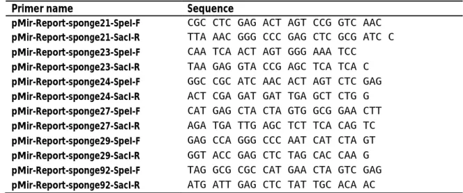
Fig. 1.12: Principle of miRNA binding Luciferase Assay Fig. 3.1: Sequences of sponge inserts
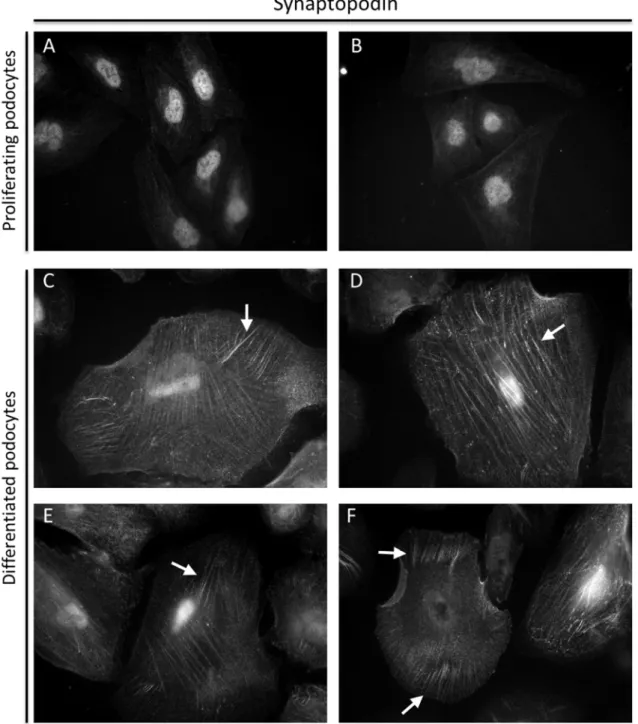
Fig. 4.1: Immunostaining of proliferating and differentiated hPCLs with -Nephrin antibody Fig. 4.2: Immunostaining of proliferating and differentiated hPCLs with -Synaptopodin
antibody
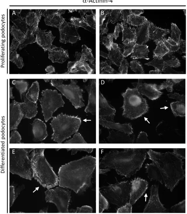
Fig. 4.3: Immunostaining of proliferating and differentiated hPCLs with --Actinin-4 antibody
Fig. 4.4: Northern Blotting of candidate miRNAs isolated from proliferating and differentiated cells
Fig. 4.5: Northern Blotting of mature miRNAs in total RNA isolated from murine glomeruli and tubules
Fig. 4.7: Cell populations in mammalian glomeruli
Fig. 4.8: Double fluorescent glomeruli from mTmG x P2.5Cre mice Fig. 4.9: Identification of cell types in glomerular cell population Fig. 4.10: miRNAs enriched in podocytes
Fig. 4.11: qPCR quantification of mature miRNAs enriched in podocytes
Fig. 4.12: qPCR quantification of mature miRNAs enriched in the red fluorescent cell population
Fig. 4.13: Alignment of the 3’UTRs of CD2AP transcripts from human (NM_012120) and mouse (NM_009847)
Fig. 4.14: 3’-end of human 3’-UTR of CD2AP transcript (NM_012120)
Fig. 4.15: Alignment of the 3’UTRs of longest Fyn transcripts in human (NM_002037) and mouse (NM_008054)
Fig. 4.16: Alignment of the 3’UTRs of longest Nck2 transcripts in human (NM_003581) and mouse (NM_010879)
Fig. 4.17: Alignment of the 3’-UTR of Neph1 transcripts in human (NM_018240) and mouse (NM_130867)
Fig. 4.18: Ago-IP from proliferating hPCLs
Fig. 4.19: -Ago2 Western Blot with lysates from mPCLs and N2A cells Fig. 4.20: Ago-IP from proliferating mPCL
8
Fig. 4.21: Transfection efficiency test using HEK293T cells
Fig. 4.22: Luciferase assay using 3’-end of human CD2AP-3’-UTR, screening for putative regulatory miRNAs
Fig. 4.23: Luciferase assay using 3’-end of human CD2AP-3’-UTR and miR-92a/b-3p Fig. 4.24: Luciferase assay using 3’-end of human CD2AP-3’-UTR and miR-30a-e-5p Fig. 4.25: Luciferase assay using complete human FYN-3’-UTR, screening for putative
regulatory miRNAs
Fig. 4.26: Luciferase assay using complete human FYN-3’-UTR, screening for putative regulatory miRNAs and putative cooperative regulation
Fig. 4.27: Luciferase assay using complete human NCK2-3’-UTR, screening for putative regulatory miRNAs
Fig. 4.28: Luciferase assay using human NEPH1-3’-UTR, investigating miR-29a-3p regulation Fig. 4.29: Luciferase assay using human NEPH1 3’-UTR, investigating miR-29a-3p regulation Fig. 4.30: Northern Blotting of sponge candidate miRNAs using proliferating hPCLs
Fig. 4.31: Sponge functionality analysis by luciferase assay using hPCLs Fig. 4.32: Urine gel of vivo-Morpholino treated mice
Fig. 4.33: Identification of miR-30a-5p knockout clones
Fig. 4.34: Genomic organization of miR-30a-5p knockout clones
Fig. 4.35: Genomic organization of the triple transgenic Lmx1b knockout mice Fig. 4.36: Coomassie stained urine gel of triple transgenic Lmx1b knockout mice Fig. 4.37: Characterization of Lmx1b knockout validation animals
Fig. 4.38: Relative expression levels of mature miRNAs in Lmx1b knockout animals and control animals
Fig. 4.39: Relative expression levels of mature miRNAs in Lmx1b knockout animals and control animals
Fig. 4.40: Genomic organization of the quadruple transgenic Lmx1b knockout reporter mice Fig. 4.41: Coomassie stained urine gel of quadruple transgenic Lmx1b knockout mice Fig. 4.42: Coomassie stained urine gel of quadruple transgenic control mice
Fig. 5.1: Relative enrichment of miRNAs identified in murine glomeruli and podocytes Fig. 5.2: Putative podocyte miRNA cluster
Fig. 5.3: Comparison of miRNA profile validation techniques
Fig. 5.4: Putative regulatory pathways of transcription factor LMX1B
III List of tables
Tab. 1.1: List of amino acid combinations in TALE array monomers that bind specifically to DNA bases
Tab. 1.2: Overview of methods for miRNA level manipulation Tab. 3.1: Antibodies used in immunofluorescence staining Tab. 3.2: PCR Primer for mouse genotyping
Tab. 3.3: Sequences of probes used for snRNA U6 and miRNA detection in Northern Blotting Tab. 3.4: Primers for miRNA detection by qPCR
9
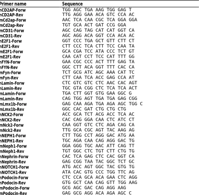
Tab. 3.5: Primer for detection of long transcripts in human (h) and murine (m) samples by qPCR
Tab. 3.6: Antibodies used in Ago immunoprecipitation
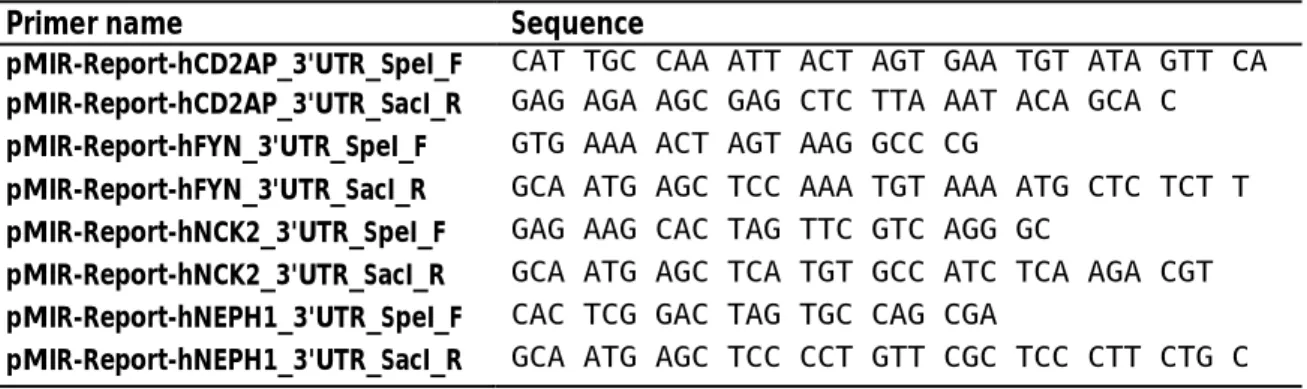
Tab. 3.7: Primers for PCR amplification of 3’-UTR fragments from genomic hPCL DNA Tab. 3.8: Primers for binding site mutations in 3’-UTRs
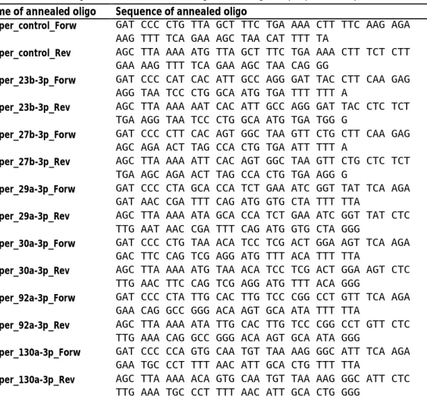
Tab. 3.9: Precursor oligonucleotides for annealing and cloning into pSuper overexpression vector
Tab. 3.10: Primer for PCR amplification of sponge sequences for cloning into pMir-Report vector
Tab. 3.11: Primer for amplification of the human genomic locus of mir-30a
Tab. 4.1: Relative expression of mature miRNAs in proliferating and differentiated hPCLs Tab. 4.2: Relative expression of mature miRNAs in murine glomeruli (G) and tubules (T) Tab. 4.3: Glomerular enrichment of miRNAs
Tab. 4.4: Podocyte enriched miRNAs
Tab. 4.5: Enrichment of mature miRNAs in podocytes
Tab. 4.6: Enrichment of mature miRNAs in red fluorescent cell population Tab. 4.7: Podocyte miRNAs target prediction using miRWalk
Tab. 4.8: miRWalk2 predictions of miRNAs targeting human CD2AP 3’-UTR Tab. 4.9: miRWalk2 predictions of miRNAs targeting murine Cd2ap 3’-UTR;
Tab. 4.10: miRWalk2 predictions of miRNAs targeting human FYN 3’-UTR Tab. 4.11: miRWalk2 predictions of miRNAs targeting murine Fyn 3’-UTR Tab. 4.12: miRWalk2 predictions of miRNAs targeting human NCK2 3’-UTR Tab. 4.13: miRWalk2 predictions of miRNAs targeting murine Nck2 3’-UTR Tab. 4.14: miRWalk2 predictions of miRNAs targeting human NEPH1/KIRREL Tab. 4.15: miRWalk2 predictions of miRNAs targeting murine Neph1/Kirrel Tab. 4.16: miRNA targets isolated in Ago-IP from murine podocytes
Tab. 5.1: Glomerular and tubular expression of podocyte enriched miRNAs Tab. 5.2: Genomic organization of murine podocyte miRNAs
Tab. 5.3: Genomic organization of podocyte miRNAs in human Tab. 5.4: Sequences of mature miR-30 family members
Tab. 5.5: Podocyte miRNAs and targets – prediction and known functions Tab. 5.6: Analyzed miRNA targets in podocytes
Tab. 10.1: miRNAs expressed in murine glomeruli and tubules
Tab. 10.2: miRNAs expressed in glomerular cells: podocytes and “red cell population”
Tab. 10.3: Alignment of RT-PCR products with mRNA sequences of potential human target genes
Tab. 10.4: Alignment of RT-PCR products with mRNA sequences of potential murine target genes
Tab. 10.5: miRNA expression in Cre- control mice and Cre+ Lmx1b knockout mice
Tab. 10.6: miRNA expression in Lmx1b knockout podocytes and red fluorescent cell population Tab. 10.7: Human genomic organization of murine podocyte miRNAs
10
1 INTRODUCTION
1.1 Non-coding small RNAs
In the last two decades, a vast amount of evidence was discovered that RNA is not only functional as a messenger between DNA and protein, but also plays a crucial role in regulation of genome organization and gene expression (reviewed in Morris & Mattick 2014). The ENCODE (Encyclopedia of DNA elements) project identified 80% of the human genome to serve a biochemical purpose (Pennisi 2012), e.g. providing a binding site for transcription factors. In total, 76% of the human genome is transcribed to RNA, while only 3% of the whole genome codes for a total of 20,687 proteins (Pennisi 2012). In addition, 11,224 pseudogenes, 8,800 small RNA molecules and 9,600 long RNA molecules were found to be encoded in the genome (Pennisi 2012). Many classes of non-protein coding RNAs have been identified to operate on several regulatory levels in eukaryotic as well as in prokaryotic cells, adding new levels of complexity to the regulatory network in living cells. The regulation of gene expression on a post-transcriptional level is the mechanism best described for miRNA (microRNA) dependent regulation (Carthew &
Sontheimer 2009). Non-coding RNAs are also able to mediate epigenetic regulation, as small RNAs as well as long non-coding RNAs can recruit histones, DNA methyltransferases and chromatin modifying complexes (reviewed by Holoch & Moazed, 2015).
This work focuses on one specific, highly conserved class of non-coding RNAs, the miRNAs, and their role in post-transcriptional regulation of protein coding transcripts in mammalian kidneys.
miRNAs have already been identified to be involved in many important signaling processes in mammalian cells, e.g. the regulation of somatic stem cell proliferation and differentiation (reviewed by Shenoy & Blelloch 2014) or the modulation of autophagy and apoptosis (reviewed by Su et al. 2015). Since miRNAs are important for the homeostasis of physiological conditions in cells, dysregulation of their levels was proposed to be involved in genesis and progression of a broad variety of diseases. They play an important role in the progression of different cancers (reviewed by e.g. Hata & Lieberman 2015, Melo & Esteller 2011, Jansson & Lund 2012, Volinia et al. 2010), but also in other diseases like Alzheimer’s disease (Codoceno et al. 2015, Feminella et al. 2015) or diabetes (reviewed by Guay et al. 2011) and diabetic nephropathy (reviewed by Kato
& Natarajan 2015). miRNAs have been proposed as potential activators of diseases, biomarkers (reviewed by Wang et al. 2015A) and therapy targets.
11
1.1.1 Biogenesis and function of miRNAs in mammalian cells
miRNAs are small, non-coding RNA molecules with a length of 19-25 nt (nucleotides) that are processed in mammalian cells from endogenously generated transcripts. They play an exclusively regulatory role in cells, functioning as guide molecules in post-transcriptional gene silencing by base pairing with their target, leading to its silencing or degradation (Kim 2005). It has been estimated that up to 60% of human protein coding genes are regulated by miRNAs (Friedman et al. 2009). For many miRNAs, the sequence of the mature miRNA molecules as well as the genomic organization of the precursor transcripts is highly conserved between different species, especially between mammalian species like human and mouse.
miRNA precursor molecules
The primary miRNA transcript, the pri-miRNA, consists of a hairpin with an imperfectly paired stem of ca. 33 nucleotides and flanking sequences that extend on both the 5’-end and the 3’-end from the hairpin structure (Carthew & Sontheimer 2009). Transcription of miRNAs is typically performed by RNA polymerase II, and the primary transcripts are capped and polyadenylated (Kim 2005). miRNA transcripts synthesized by RNA polymerase III have also been described (Dieci et al. 2007). miRNAs can be categorized according to their genomic locations: exonic miRNAs are processed from their own, non-coding primary transcript that contains one or more hairpin structures, while intronic miRNA hairpins are derived from the introns of a protein coding or non-protein coding transcript (Kim 2005). A transcript containing several miRNA hairpins is called a miRNA cluster (chapter 1.1.2).
In the first pri-miRNA processing step (Fig. 1.1), occurring in the nuclei of mammalian cells, the stem loop structure is cleaved by the enzyme Drosha, releasing the miRNA precursor, the pre- miRNA (Kim 2005). Drosha, a member of the RNAse III family, and its cofactor DGCR8 (DiGeorge syndrome critical region gene 8), which is important for miRNA maturation, are called the microprocessor complex (Denli et al. 2004, Nguyen et al. 2015).
An alternative miRNA maturation pathway uses splicing to form transcripts that mimic the structural features of pre-miRNAs, the so-called “mirtrons”, which also can enter the miRNA processing pathway after the Drosha processing step (Carthew & Sontheimer 2009).
12
Fig. 1.1: Pathways leading to functional RISCs; after transcription and processing by Drosha and Dicer, the mature miRNAs are bound by Ago proteins, forming the RISCs (Meister 2013, modified); Abbreviations:
DGCR8: DiGeorge syndrome critical region gene 8; NPC: nuclear pore complex; RISC: RNA induced silencing complex
miRNA maturation
After the pre-miRNA is exported out of the nucleus by XPO-5 (Exportin-5) and released from the transport complex in an energy dependent manner (Ha & Kim 2014), the second processing step is carried out by the conserved enzyme Dicer (Fig. 1.1), a member of the RNase III family (Kim 2005). It excises the terminal loop from the hairpin creating a mature miRNA duplex with a length of approximately 22 nucleotides (Carthew & Sontheimer 2009). Products of Dicer processing possess phosphates at the 5’-end and overhangs of two nucleotides at the 3’-end (Meister & Tuschl 2004) and can be loaded into the RISCs (RNA induced silencing complexes).
13 RISC complex formation
Mammalian cells possess four argonaute proteins, Ago1-4, which are all able to bind mature miRNAs to form RISCs. However, only Ago2 has a retained catalytic activity by a functional catalytic triad, while the other three Ago proteins might be partially redundant (Meister 2013). It has been shown that the different Ago proteins bind different subpopulations of miRNAs (Burroughs et al. 2011). The binding to the different Ago proteins seems to be influenced by the length of the miRNA. The Ago1 and Ago3 bound miRNAs often contain 23 to 24 nucleotides and may possess non-templated additions of mostly one nucleotide at their 3’-end (Dueck et al.
2012).
After Dicer processing, the mature miRNA is loaded into the Ago protein. The transfer of the double strand to the Ago protein is mediated by Hsp90 (Heat shock protein 90) in humans. By conformational changes of the Ago protein, its N domain is driven between the two duplex strands, leading to duplex opening and further unwinding (Meister 2013). How unwinding occurs in detail, and whether additional factors are needed, is not fully understood yet. After unwinding of the duplex, the strand with the less stably paired 5’-end is preferentially loaded into Ago proteins (Meister 2013). The passenger strand, often marked with a “*”, is removed and can be degraded, but may also be a functional miRNA itself. As an example, both mature miRNAs from the mir-9 precursor regulate the tumor suppressor CAMTA1 (Calmodulin binding transcription activator 1; Schraivogel et al. 2011).
Dicer is an enzyme with a broad range of possible substrates, e.g. it is able to generate small RNAs with a length of 22 nucleotides from a 70 nucleotide stem loop precursor (Kim 2005). Thus, also other double stranded precursor molecules can be processed by Dicer and loaded into Ago proteins, e.g. siRNAs (small interfering RNA).
Posttranscriptional regulation of gene expression by miRNAs
The miRNAs function as a guide for the Ago proteins to mRNAs, directing the RISC to binding sites which are often located in the 3’-UTR of the target genes (Meister 2013). miRNA binding in the coding region of genes also has been reported (reviewed by Brümmer & Hausser 2014;
Gebeshuber et al. 2013). Perfectly paired miRNA-target duplexes can be cleaved by the catalytically active Ago2 protein (Meister 2013). In mammals, miRNAs usually bind to partially complementary sites, while the “seed region”, the nucleotides on position 2 to 7 or 8 of the miRNA, is often fully paired and essential for interaction (Meister 2013).
14
The binding of a miRNA guided RISC to a target mRNA can lead to its translational silencing as well as its degradation, with the mRNA destabilization being the main contributor to regulation in animal cell cultures (Jonas & Izaurralde 2015). Mammals possess three GW (Glycine- tryptophan repeat containing) proteins, called TNRC6A-C (Trinucleotide repeat containing 6 A-C), that can bind to Ago proteins, PAN3 (Poly (A) specific ribonuclease subunit 3) and NOT9 (also:
RQCD1: Required for cell differentiation1 homolog) via their tryptophan residues (Jonas &
Izaurralde 2015). Via PAN3 and NOT9, the deadenylating complexes PAN2-PAN3 and CCR4-NOT are recruited to the regulated transcript, leading to its deadenylation, decapping and degradation via the cellular 5’-to-3’ mRNA decay pathway (reviewed by Jonas & Izaurralde 2015).
Non canonical miRNA processing
Two parallel studies identified a mature miRNA whose levels were not affected by Dicer loss of function, but reduced in mutant zebrafish and mice lacking enzymatic activity of Ago2 (Cifuentes et al. 2010, Cheloufi et al. 2010). Dicer requires a double strand of > 19 nucleotides for miRNA maturation from a canonical precursor (Fig. 1.2 A). A mature miRNA, miR-451-5p, is encoded in a conserved 42 nucleotide hairpin precursor with a stem of only 17 nucleotides (Fig. 1.2 B). This short hairpin can be directly bound by Ago2 and cleaved at the opposite strand (Fig. 1.2 C).
Fig. 1.2: Non-canonical miRNA precursors (A) Canonical hairpin precursor mir-144 coding for two mature miRNAs, miR-144-5p (blue) and miR-144-3p (red); (B) mir-451 hairpin precursor with shortened stem only coding for one mature miRNA, miR-451-5p (red); (C) mir-451 hairpin with predicted Ago2 cleavage site on the opposite strand (Cheloufi et al. 2010, modified)
The mir-451 pre-miRNA seems only to be bound by the only catalytically active argonaute protein, Ago2, and artificial precursors mimicking the pre-mir-451 structure will be bound and processed by Ago2 (Dueck et al. 2012). PARN (Poly (A) specific ribonuclease) could be identified as the enzyme responsible for 3’-5’ exonucleolytic trimming of Ago2 cleaved pre-mir-451 (Yoda et al. 2013).
15 Regulation miRNA expression and maturation
By guiding the RISCs to the 3’-UTRs of target genes, miRNAs function as gene expression regulators. Vice versa, since miRNAs are generated from primary transcripts produced by RNA polymerase II or III, their expression can be regulated by transcription factors. Many miRNAs have been shown to be regulated by transcription factors like p73 and p63 (Tumor proteins P73 and P63), who have been identified as activators of miR-200 family upregulation (Knouf et al.
2012). The transcription factor Lmx1b (LIM homeobox transcription factor 1 beta) has been found to form a regulatory circuit with miR-135a-2 in brain (Anderegg et al. 2013). A feedback loop between the miR-200 family and the transcription factors ZEB1 and ZEB2 (Zinc finger E-box binding homeobox 1 and 2), controlling EMT and MET (Epithelial–mesenchymal and Mesenchymal–epithelial transition), has also been discovered (Hill et al. 2013). It was reported that the miR-709 directly regulates miRNA-15a/16-1 biogenesis on a posttranscriptional level, hinting at a possible miRNA hierarchy system (Tang et al. 2012).
Additionally, processing of miRNAs can be regulated on every stage, i.e. nuclear procession, export from the nucleus, cytoplasmic procession and loading into the Ago complex as well as the structure of the miRNA itself, e.g. by tailing (reviewed by Ha & Kim 2014). miRNA binding to human Ago proteins can also be strongly reduced by Ago phosphorylation (Rüdel et al. 2011).
1.1.2 Classification of identified miRNAs
All miRNAs identified from different organisms are registered in the online database miRBase (http://www.mirbase.org; Kozomara & Griffiths-Jones 2014; Kozomara & Griffiths-Jones 2011;
Griffiths-Jones et al. 2008; Griffiths-Jones et al. 2006; Griffiths-Jones 2004). Until September 2015, 1881 miRNA precursors and 2588 mature miRNAs had been identified in human. In mouse, 1193 miRNA precursors and 1915 mature miRNAs had been identified. In the database TarBase v7.0 (Vlachos et al. 2014), more than 65,000 interactions of miRNAs and genes identified by high-throughput approaches or specific experiments had been indexed until September 2015. A database for miRNA involvement in diseases, miR2Disease (Jiang et al. 2009), listed 349 miRNAs to be involved in 163 diseases in September 2015.
Nomenclature of miRNAs
With the exception of the first two miRNAs discovered in C. elegans, lin-4 (Lee et al. 1993) and let-7 (Reinhart et al. 2000), miRNA precursors are numbered chronological according to their discovery. To indicate the precursor, the “r” in mir is not capitalized, e.g. mir-191. A prefix indicates the specimen the miRNA was found in (e.g. hsa for homo sapiens, mmu for mus
16
musculus, rno for rattus norvegicus). If the precursor is evolutionary conserved, the number will be the same in the different organisms, e.g. hsa-mir-22 and mmu-mir-22. To indicate that a precursor possesses two different loci in the genome, the precursor is numbered, e.g. hsa-mir- 24-1 and hsa-mir-24-2. The two mature miRNAs that originate from a common precursor are indicated with a suffix describing the arm of the precursor they are derived from, e.g. miR-9-5p and miR-9-3p. According to the old nomenclature, the miRNA with the lower expression level is sometimes marked with a star, e.g. miR-9*. When the sequence of two miRNAs is very similar and the sequence of the seed region, the positions 2 to 7 or 8, is identical, the miRNAs are grouped into one family, e.g. miR-23a-3p and miR-23b-3p. However, the division into families is not very consistent, as e.g. the miRNAs mir-141, mir-200a, mir-200b, mir-200c and mir-429 belong to the same family, while there is a single base mismatch in the seed regions of miR- 200a-3p and miR-200b-3p.
miRNA clusters
A miRNA cluster is a group of miRNA hairpin precursors that are processed from one common primary transcript yielding several mature miRNAs. One cluster that has been studied intensively is the miR-17~92 cluster, a genomic fragment of 786 base pairs containing 6 hairpin precursors (He et al. 2005, Hayashita et al. 2005). A common role of miRNAs organized in a cluster could be proven when overexpression of the miR-17~92 cluster enhanced tumor growth in a mouse B- cell lymphoma model (He et al. 2005) as well as lung cancer cell growth (Hayashita et al. 2005).
As a second example, the five members of the mir-200 family are organized in two genomic clusters, pointing to a common role of miRNAs that are encoded near to each other in the genome. The miR-200 family is involved in kidney fibrosis and diabetic nephropathy by regulation of the EMT and the EndMT (Endothelial mesenchymal transition) (Srivastava et al.
2013). Additionally, a feedback loop between the ZEB transcription factors and the miR-200 family was identified to be an important player in the progression of cancer (Brabletz & Brabletz 2010). The two mir-23~27~24 clusters, one harboring the precursors mir-23a, mir-27a and mir- 24-2 and the other containing mir-23b, mir-27b and mir-24-1, have been shown to be important for cardiovascular disorders, as they are involved in angiogenesis, endothelial apoptosis in cardiac ischemia and retinal vascular development (reviewed by Bang et al. 2011). A cooperative function of the encoded miRNAs was proposed, since the functional analysis revealed similar distributions of enriched genes (Liang et al. 2014). Additionally, the cooperative function of miRNAs organized in 20 different clusters in regulating several cell signaling pathways involved in cervical cancer has recently been reviewed (Servin-González et al. 2015).
17
1.2 The mammalian kidney
1.2.1 The anatomy of the mammalian kidney
The kidneys are an organ serving several important roles in the mammalian organism. Their main tasks are filtration of the blood to remove waste products from the organism, maintenance of homeostasis of electrolytes and thus, regulation of the blood pressure. The hilum, the place where the renal artery enters the kidney and the renal vein as well as the ureter leave the kidney, is located on the concave side of the bean-shaped organs. The two major segments of the kidney are the outer cortex and the inner medulla. Contrary to the murine kidney (unilobar kidney), the parenchyma of the human kidney is divided into several pyramids by projections of the renal cortex towards the center. The tip of each pyramid opens out into the calyxes that merge into the renal pelvis.
Fig. 1.3: Microscopic anatomy of the mammalian kidney (A) Nephrons spanning the cortex and medulla of a mammalian kidney: 1 glomerulus; 2 proximal convoluted tubule; 3 proximal straight tubule; 4 descending thin limb; 5 ascending thin limb; 6 thick ascending limb; 7 macula densa; 8 distal convoluted tubule; 9 connecting tubule; 10 cortical collecting duct; 11 outer medullary collecting duct; 12 inner medullary collecting duct (Mount 2014) (B) Schematic representation of a glomerulus with the podocytes covering the capillary convolute (Pollak et al. 2014, modified); (C) Scanning electron micrographs of the interdigitating processes of adjacent podocytes covering the glomerular capillaries. Micrographs by C.
Niemann & H. Schmidt, Institute for Molecular & Cellular Anatomy, University of Regensburg
18
The functional units of the kidney are the nephrons. They span the cortex and the medulla of the kidney and consist of a glomerulus, where the initial filtration of the blood takes place, and an adjacent tubular system in which the primary urine is concentrated (Fig. 1.3 A).
1.2.2 The glomerulus and the renal filtration barrier
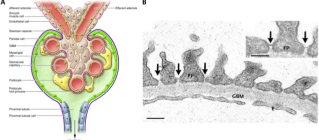
The mammalian glomerulus contains a capillary convolute, which is covered by highly specialized epithelial cells, the podocytes (Fig. 1.3 B, C). In the glomerulus, the primary urine is filtered from the blood into the Bowman’s space, from where it flows of into the tubular system. The inner part of the glomerulus consists of three main cell types: the endothelial cells lining the capillaries, the podocytes and the stabilizing mesangial cells, smooth muscle cells intercalated into the capillary convolute (Fig. 1.4 A). In addition, the Bowman’s space surrounding the capillary convolute is lined with the PECs (Parietal epithelial cells). In-between their interdigitating foot processes, the podocytes build up the slit diaphragm, a delicate protein structure crucial for blood filtration. The renal filtration barrier consists of three components:
The slit diaphragms, i.e. complex protein structures built by two neighboring podocytes in- between their interdigitating foot processes (Fig. 1.4 B), the glomerular basement membrane between the endothelial cells and the podocytes and the fenestrated endothelium of the capillaries.
Fig. 1.4: Microscopic anatomy of the filtration barrier (A) the glomerular capillaries are covered by the podocytes which form contacts to each other with their interdigitating foot processes; the capillary convolute is stabilized by mesangial cells; from the Bowman’s space bordered by the PECs, the primary urine is conducted towards the tubular system starting with the proximal tubule (Pozzi & Zent, 2012); (B) Electron micrograph of the glomerular filtration barrier: interdigitating processes of adjacent podocytes with linking slit diaphragms (arrows), the glomerular basement membrane and the fenestrated capillary endothelium (Tryggvason et al. 2006, modified); Abbreviations: FP: foot process; GBM: glomerular basement membrane; E: fenestrated endothelium; scale bar: 250 nm
19
In the last years, putative podocyte regeneration has been discussed. Different sources of progenitor cells, like parietal epithelial cells or cells of the renin lineage have been proposed (reviewed by Grahammer et al. 2013). PECs have been shown to play a role in podocyte regeneration in young mice from postnatal day ten (Appel et al. 2009) up to 28 days of age (Eng et al. 2015). No regeneration could be found in older mice from five weeks to 18 months of age (Berger et al. 2014), a finding that might be explained by the fact that progenitor cells are believed to be active in postnatal mice for one to two weeks (Rinkevich et al. 2014). Limited podocyte renewal from PECs was observed after diphtheria toxin induced podocyte injury, but not under physiological conditions (Wanner et al. 2014). Furthermore, miR-193a-5p was identified to play a role in modulating the phenotype and marker expression of cultured PECs towards a podocyte phenotype (Kietzmann et al. 2014).
1.2.3 Models for working with podocytes
To be able to investigate podocytes in cell culture, immortalized cell lines derived from isolated human and murine podocytes have been established previously (Saleem et al. 2002, Schiwek et al. 2004). The human as well as the murine cell line are proliferating at 33°C and can be differentiated by culturing at 37°C or 38°C, respectively.
Human podocyte cell line
Human podocytes were derived by biopsy from a three year old child (Saleem et al. 2002). By transfection with a retroviral construct coding for the SV40 large T antigen, primary podocytes were immortalized to form a cell culture line: the hPCL (Human podocyte cell line).
Fig. 1.5: Immunofluorescence of immortalized human podocytes; differential expression of Synaptopodin (top) and Nephrin (bottom) in proliferating (left) and differentiated (right) immortalized human podocytes (Saleem et al. 2002, modified)
When cultured at 33°C, the proliferating cells only show a low expression of synaptopodin and nephrin. After differentiation at 37°C for two weeks, expression of synaptopodin as well as
20
nephrin could be detected by immunofluorescence (Fig. 1.5). In the proliferating cells, PCNA (Proliferating cell nuclear antigen), a marker of cell proliferation, and CyclinA, a regulator of cell cycle progression, are expressed at high levels, but absent in the differentiated cells. Vice versa, p27 (Cyclin-Dependent Kinase Inhibitor 1B), a cell cycle inhibitor, and CyclinD1, a driver of G1/S phase (Gap 1 phase /Synthesis phase) transition, are absent in the proliferating cells but expressed in the differentiated cells.
Murine podocyte cell line
Murine podocytes were isolated from kidneys of the “Immorto-Mouse” (Charles River, St. Louis, MO, USA) by letting them grow out from isolated glomeruli (Schiwek et al. 2004). These mice carry a temperature sensitive mutant of the SV40 large T antigen under the control of the interferon-–inducible H-2Kb promoter. The mPCLs (Murine podocyte cell line) differentiate when cultured at 38°C for two weeks. Expression of the podocyte marker proteins nephrin and Cd2ap (CD2 associated protein) could be demonstrated by Northern Blotting, while expression of WT1 (Wilms tumor protein 1), Lmx1b, Neph1 (Kin of IRRE-like protein 1), Cd2ap, podocalyxin and other marker proteins was shown by RT-PCR (Reverse transcription polymerase chain reaction). By immunofluorescence microscopy, localization of Cd2ap and synaptopodin to the actin cytoskeleton at the cell-cell contacts was shown.
Cell culture of glomeruli isolated from mice
Glomeruli isolated from mice by magnetic bead perfusion (Takemoto et al. 2002) can be cultured in standard cell culture media for several days after isolation from mice (personal communication: M. Kubitza). From the seeded glomeruli, glomerular cells including podocytes grow out on the cell culture flask surface and can be used for experiments. However, the cells dedifferentiate and loose the typical podocyte architecture and marker protein expression within some days.
Freshly isolated murine podocytes
Using a double-fluorescent Cre reporter mouse (Muzumdar et al. 2007) crossed with a mouse with podocyte specific Cre expression (Möller et al. 2003), a mouse harboring podocytes marked by green fluorescence can be generated. By isolation of the glomeruli followed by additional digestion steps, a single cell suspension can be obtained from which the podocytes can be isolated by FACS (Fluorescent activated cell sorting) (Boerries et al. 2013). This method yields a pure podocyte sample. On the downside, the sorted podocytes do not adhere any more to cell culture substrates and thus cannot be cultured further (personal communication: T. Burghardt).
21
1.3 The role of miRNAs in podocyte integrity
1.3.1 miRNA expression profiles of the mammalian kidney
In several studies, miRNAs were isolated from different tissues in order to identify organ or cell type enriched miRNAs. In the first examination of nine different mouse tissues, highly enriched miRNAs for some of the tissues were identified (Lagos-Quintana et al. 2002). In order to obtain a mammalian miRNA expression atlas, more than 250 small RNA libraries from 26 different organ systems and cell types of human and rodent were sequenced, including total kidney from human as well as mouse (Landgraf et al. 2007). A study sequencing miRNAs isolated from kidney, ovaries and testis of adult mice as well as from the mesonephros, kidneys, ovaries and testis form the embryonic days E11, E12 and E13 revealed the miRNA profiles of these organs to vary during embryogenesis, hinting that miRNAs play an important role for development of murine urinary and reproductive systems (Aguilar et al. 2010).
1.3.2 Podocyte specific knockout of miRNA processing enzymes
In 2008, three parallel studies showed the importance of miRNAs for podocyte development and function (Harvey et al. 2008, Ho et al. 2008, Shi et al. 2008). Mice that expressed the Cre recombinase under the control of the podocyte specific NPHS2 promoter (Möller et al. 2003) were crossed with mice in which the exons 20 and 21, part of the second RNase III domain of the miRNA processing enzyme Dicer, are flanked by loxP (Locus of X-over P1) sites (Harfe et al.
2005), the Cre recombinase recognition sites. This lead to a conditional, podocyte specific Dicer knockout. The affected mice developed proteinuria two to five weeks after birth, leading to end- stage renal disease and death. During progression of disease, several glomerular abnormalities like foot process effacement, crescent formation, wrinkling and split areas of the glomerular basement membrane were observed. In the podocytes of the affected kidneys, the expression of synaptopodin, ezrin (Harvey et al. 2008), podocin (Shi et al. 2008) and nephrin (Ho et al. 2008) was reduced in the knockout podocytes, while expression of -actinin-4 (Harvey et al. 2008) and WT1 (Harvey et al. 2008, Ho et al. 2008) remained unchanged. It was shown by in situ hybridization directed against mature miRNAs, that miR-30a-5p expression in podocytes is lost in the Dicer knockout animals compared to the control animals, whereas the expression of the endothelial miR-126-3p and the mesangial miR-145-5p is not affected by the knockout (Harvey et al. 2008), thus identifying one mature miRNA highly enriched in podocytes. Vice versa, gene expression profiling of glomeruli isolated from Dicer knockout and control mice revealed an
22
enrichment of mRNAs with possible binding sites of the miR-30 family in the knockout glomeruli, pointing to a possible role of this miRNA family in podocytes (Shi et al. 2008).
In a different study, mice expressing the Cre recombinase under the control of the podocyte specific NPHS2 promoter (Möller et al. 2003) where crossed with mice possessing loxP sites on either side of exon 9 of the miRNA processing enzyme Drosha (Chong et al. 2008), leading to a conditional, podocyte specific Drosha knockout (Zhdanova et al. 2011). At about two to three weeks of age, mice affected by this knockout developed proteinuria that progressed to renal failure and death within four to eight weeks of age. The mice showed podocyte foot process effacement as a first pathologic sign, later leading to extensive collapsing glomerulopathy, pseudo crescent formation and sclerosis. This was accompanied by the loss of synaptopodin, WT1, nephrin and podocin expression, resembling the phenotype of the Dicer knockout mice, with the exception of WT1 expression which remained normal in the Dicer knockout mice. To determine if miRNAs are not only required for podocyte development, but also podocyte function, an inducible Tet-On (Tetracycline-controlled transcriptional activation) system was used in the same study to specifically delete Drosha at later time points. Mice showed proteinuria around two weeks after beginning of the doxycycline treatment. By histological examination, a phenotype very similar to the phenotype observed in the conditional Drosha knockout mice was revealed. Thus, this study showed that miRNAs are not only important for podocyte development, but also for maintenance of the podocyte filtration barrier in adult kidneys.
1.3.3 Profiles from murine podocytes
In 2013, expression profiles from murine podocytes were obtained (Boerries et al. 2013). The transcriptome as well as proteome of podocytes isolated freshly from double fluorescent Cre reporter mice was analyzed. Together with transcripts and proteins enriched in podocytes, a list of 35 miRNAs expressed in murine podocytes and non-podocyte glomerular cells was published that are in high congruence with the data of the present work (Tab. 5.5).
1.3.4 Known miRNA functions in the glomeruli and podocytes
Many studies tried to identify miRNAs important for kidney function. Profiling studies have been performed from patients, animal models and cultured podocytes cell lines under different conditions to identify miRNAs with altered expression levels (reviewed by Chandrasekaran et al.
2012, Kato et al. 2012). For some miRNAs expressed in mammalian kidney, not only expression data, but functional roles were identified. For instance, miR-26a-5p levels were found to be
23
lowered in swine and human post stenotic kidneys (Zhu et al. 2015) as well as in human patients with lupus nephritis or IgA nephropathy (Ichii et al. 2014). Recently, miR-26a-5p was identified to target CTGF (Connective tissues growth factor), connecting the downregulation of miR-26a-5p to diabetic nephropathy progression in mouse models and humans (Koga et al. 2015). In another study, enhanced nephrin acetylation with attenuated renal damage was reported in miR-155-5p double knockout mice suffering from hyperglycemia induced nephropathy (Lin et al. 2014).
Distinct targets have only been identified for some miRNAs in glomeruli and kidneys so far. It was reported that miR-135a-5p regulates TRPC1 (Transient receptor potential cation channel, subfamily C, member 1) during renal injury promoting renal fibrosis (He et al. 2014B). Under high glucose conditions, miR-93-5p regulates VEGF (Vascular endothelial growth factor) in vitro and in vivo (Long et al. 2010) and miR-195-5p promotes apoptosis in mouse podocytes by regulating BCL2 (B-cell lymphoma 2) activity (Chen et al. 2011). By downregulation of SOCS1 (Suppressor of cytokine signaling 1), miR-150-5p promotes renal fibrosis (Zhou et al. 2013). In immortalized cultured podocytes under mechanical stress and streptozotocin induced diabetic rats, miR-124- 3p was reported as a putative regulator of Itga3 (Integrin alpha 3) (Li et al. 2013A, Li et al. 2013B)
miR-17~92 cluster
The polycistronic miR-17~92 cluster contains 6 miRNA hairpin precursors and has firstly been identified to be a potential oncogene (He et al. 2005) which enhances cell proliferation and is highly expressed in lung cancers (Hayashita et al. 2005). The expression of this cluster is upregulated in mouse models for polycystic kidney disease, while inactivation of this cluster in these mouse models retards kidney cyst growth (Patel et al. 2013). Deletion of this cluster in nephron progenitor cells impairs cell proliferation and reduces the number of developing nephrons during kidney development (Marrone et al. 2014). Postnatally, the affected mice developed signs of kidney disease including albuminuria, podocyte foot process effacement and glomerulosclerosis, underlining the importance of the cluster for kidney development.
miR-21-5p
miR-21-5p has been shown to play an important role in the progression of fibrosis (for review see Patel & Noureddine 2012) as well as in the pathogenesis of acute kidney injury (for review see Li et al. 2013C). In a study using mir-21 knockout mice and mice treated with miR-21-5p antisense oligonucleotides, miR-21-5p was shown to promote fibrosis of the kidney by silencing metabolic pathways (Chau et al. 2012). In a different study using a mouse model for Alport nephropathy, miR-21-5p silencing resulted in a milder kidney disease (Gomez et al. 2015). On the other hand, it was shown that miR-21-5p can also ameliorate glomerular injury induced by
24
TGF-1 (Transforming growth factor beta 1) and hyperglycemia through repression of proapoptotic signals (Lai et al. 2015). These studies indicate that this miRNA may play different roles and that a tightly controlled physiological level is necessary for healthy kidney function.
miR-29 family
The miR-29 family, expressed in the glomeruli of mice, has been shown to play an important role in the regulation of kidney fibrosis. It was reported that miR-29b-3p regulates several collagens and related genes in Dahl salt sensitive rats (Liu et al. 2010A). By intravenous injection of LNA- antisense molecules into SS-13BN rats, expression of several collagen genes in the renal cortex as well as in the medulla was upregulated. Furthermore, down regulation of these genes by miR- 29b-3p was proven by luciferase assays.
miR-29c-3p was found to be a signature miRNA under high glucose conditions in diabetic db/db mice (mice with deficient leptin receptor activity, Long et al. 2011). It was proven by luciferase assays to target Spry1 (Sprouty homolog 1) which is an inhibitor of RhoA (Ras homolog family member A) and Rho kinase activation. Knockdown of miR-29c-3p by intraperitoneal injection of antisense oligonucleotides into db/db mice led to reduced matrix accumulation and reduced apoptosis in the murine glomeruli.
In another study, wild-type mice showed a reduced expression of miR-29 family and development of progressive renal fibrosis after induction of unilateral ureteral obstructive nephropathy, whereas Smad3 (Smad family member 3) knockout mice had an increased expression of the miR-29 family along with the absence of fibrosis (Qin et al. 2011). In vitro, overexpression of miR-29b-3p inhibited, but knockdown of miR-29 family enhanced TGF-1- induced expression of collagens I and III in renal tubular cells. Delivery of miR-29b-3p blocked progressive renal fibrosis, making it a downstream inhibitor of TGF-/Smad3-mediated fibrosis.
Furthermore, it was reported by a different group that expression of TGF-1 reduces the expression of the whole miR-29 family, thus enhancing the expression of proteins of the extracellular matrix (Wang et al. 2012). Ectopic expression of the miR-29 family repressed the expression of collagen I and collagen IV. Additionally, low miR-29 family levels in three different models of renal fibrosis were found.
Recently, it was shown that miR-29a-3p levels are lowered in glomeruli from mice with streptozotocin induced hyperglycemia (Lin et al. 2014). Transgenic overexpression of miR-29a-3p in these diabetic mice improved nephrin levels, podocyte viability and renal function while
25
reducing glomerular fibrosis and inflammation reaction compared to wild-type diabetic mice.
Overexpression of miR-29a-3p restored the acetylation of nephrin by modulation of HDAC4 (Histone deacetylase 4), thus ameliorating hyperglycemia induced podocyte dysfunction.
Taken together, maintenance of healthy miR-29 family levels seems to play an important role in the prevention of fibrosis and the maintenance of glomerular structure.
miR-30 family
miR-30a-5p was firstly identified to be an important podocyte miRNA in the podocyte specific Dicer knockout mice, when it was shown that targets with possible miR-30a-5p binding sites are upregulated after the knockout (Shi et al. 2008). Additionally, in situ hybridization showed the absence of this mature miRNA in glomeruli after the podocyte specific Dicer knockout (Harvey et al. 2008). It was shown that miR-30a-5p knockdown in the developing Xenopus pronephros phenocopies the defects caused by a pronephric Dicer knockdown, indicating that miR-30a-5p plays an important role in pronephros development (Agrawal et al. 2009). Additionally, a major transcriptional regulator during kidney development, Xlim1/Lhx1 (Lim homeobox 1), was shown to be targeted by miR-30a-5p. In contrast to wildtype mice, miR-30 family expression was lowered in albumin/TGF- transgenic mice who suffered from podocyte apoptosis and glomerulosclerosis (Shi et al. 2013). In vitro, TGF- is able to downregulate the miR-30 family in wildtype and Smad3 deficient, but not Smad2 (Smad family member 2) or Smad2/Smad3 deficient cultured podocytes. TGF- induced apoptosis could be hindered by lentiviral overexpression of miR-30d-5p. Taken together, these results hint that loss of miR-30 family signaling is a specific mechanism of TGF- signaling during glomerulosclerosis.
Later it was reported that downregulation of miRNA-30 facilitates podocyte injury (Wu et al.
2014). Podocyte cytoskeletal damage and apoptosis after treatment of cells with TGF- and PAN (Puromycin amino-glycoside) were ameliorated by exogenous miR-30 expression and aggravated by miR-30 knockdown. It was found that the miR-30 family exerts this protective role by direct inhibition of Notch1 (Notch homolog 1, translocation-associated) and the transcription factor p53 (Tumor protein P53). Similar results were shown in rats, when PAN treatment lead to a downregulation of podocyte miR-30s and podocyte damage, while exogenous overexpression of miR-30a-5p ameliorated this phenotype. Additionally, glucocorticoid treatment maintained the miR-30 expression levels in TGF- and PAN stressed cultured podocytes (Wu et al. 2014).
In another study, miR-30a-5p was identified to be upregulated in murine injured podocytes, while the inhibition of miR-30a-5p prevented PAN induced podocyte apoptosis (Xie et al. 2015).
26 miR-193a-5p
In 2013, it was shown that FSGS (Focal segmental glomerulosclerosis) can be induced by an upregulation of miR-193a-5p, a miRNA that can downregulate the podocyte transcription factor WT1 (Gebeshuber et al. 2013). A transgenic mouse with an inducible miR-193a-5p overexpression under the CMV (Cytomegalovirus) promoter showed a severe kidney phenotype with foot process effacement after two weeks and focal sclerosis after four weeks of induction.
WT1, a transcription factor regulating several genes important for maintenance of normal podocytes structure and function, was shown to be targeted by miR-193a-5p, as a predicted miR-193a-5p binding site at the end of the coding region of the WT1 transcript was confirmed as functional. miR-193a-5p upregulation in samples from patients suffering from focal segmental glomerulosclerosis was suggested to be a new pathogenic mechanism for genesis of this disease.
miR-193a-5p was also shown to play a crucial role in the transdifferention of PECs towards a podocyte phenotype (Kietzmann et al. 2014). PECs express miR-193a-5p in high levels. A stable knockdown of this miRNA in immortalized human PECs leads to a shift towards a podocyte-like morphology, expression of podocyte marker proteins like WT1, podocalyxin, synaptopodin, - actinin-4 and nephrin, and a simultaneous decrease of PEC markers. In a mouse model for rapid progressive glomerulosclerosis, the nephrotoxic nephritis, the formation of interglomerular crescent could be ameliorated by injection of miR-193a-5p-antisense oligonucleotides, again linking miR-193a-5p overexpression to progression of glomerulosclerosis.
1.4 Identification of miRNA targets in podocytes
The podocyte specific Dicer knockout mice as well as the inducible podocyte specific Drosha knockout mice displayed foot process effacement of the affected podocytes as well as loss of some of the podocyte specific marker proteins (Harvey et al. 2008, Ho et al. 2008, Shi et al. 2008, Zhdanova et al. 2011). Thus, miRNAs play an important role not only in the development, but also in the maintenance of podocyte ultrastructure. The structural proteins themselves as well as the signaling processes needed to maintain the slit diaphragms or the link of the slit diaphragm proteins to the actin cytoskeleton might by targets of miRNA dependent regulation.