Case report:
TRAUMATIC TRANSNASAL PENETRATING INJURY WITH CEREBRAL SPINAL FLUID LEAK
Tan Sui Teng1*, Noor Liza Ishak2, Sethu Thakachy Subha2, Saraiza Abu Bakar1
1 Department of Otorhinolaryngology, Head and Neck Surgery, Serdang Hospital, Jalan Puchong, Kajang, Selangor Darul Ehsan, 43000 Malaysia
2 Ear, Nose and Throat Unit, Department of Surgery, Faculty of Medicine and Health Sciences, Universiti Putra Malaysia
* Corresponding author: Tan Sui Teng, Tel: +60389475555, Fax : +60389475050, E-mail: suiteng1987@gmail.com
http://dx.doi.org/10.17179/excli2018-1971
This is an Open Access article distributed under the terms of the Creative Commons Attribution License (http://creativecommons.org/licenses/by/4.0/).
ABSTRACT
CSF leak in penetrating skull base injury is relatively rare compared to close head injury involving skull base fracture. We report a 5-year-old boy presented with epistaxis and impacted pencil into the left nostril. The child was hemodynamically stable without any neurological deficit. Intraoperatively, there was a nasal septal defect posteriorly with anterior skull base fracture associated with CSF leak. The pencil was removed from the left nos- tril and the CSF leak was repaired using harvested abdominal fat under the same setting. Computed Tomography (CT) of the brain showed right cribriform plate fracture with small pneumocranium. Postoperatively, a prophy- lactic antibiotic was given for seven days and he was discharged well. Subsequent clinic visits up to one-year postoperative period showed no recurrence of the CSF leak. History taking, physical examination and CT imag- ing give valuable diagnostic values in managing the penetrating skull base injury. Early intervention for removal of the foreign body and repair of the CSF leak is advocated to prevent catastrophic complication.
Keywords: pencil, penetrating trauma, skull base fracture, CSF leak
INTRODUCTION
Presence of fistulous tract in between in- tracranium and nasal cavity may result in cerebrospinal fluid (CSF) rhinorrhea. Crani- ofacial trauma contributed 80 % of the etiol- ogies of CSF rhinorrhea. Traumatic CSF rhi- norrhea is more commonly seen in skull base fractures. Majority of the skull base fractures (90 %) are caused by closed head injuries, whereas penetrating trauma constitutes the remaining 10 % of the etiology.
Most of the penetrating head injuries in- volved oro-cranial or transorbital intracranial penetration of the sharp object with a signifi-
cant external injury. Foreign bodies such as glass, air gun projectiles, metal splinters and plastic chair glides have been reported.
Traumatic transnasal penetrating injury to the skull base is rare in literature. Removal of the penetrating object followed by repair of the CSF leakage depicts more challenging management.
In this case report, we described an unu- sual case of penetrating injury of the anterior skull base by a pencil via the left nostril, without external, orbital and paranasal sinus- es involvement. The importance of radiolog- ical assessment prior to surgery was empha- sized in this report. We also discussed the
approaches of removal of the penetrating ob- ject and endoscopic CSF leakage repair in the same setting.
CASE REPORT
A 5-year-old boy presented to the emer- gency room with epistaxis following an im- pacted pencil over the left nostril. He admit- ted to inserting the blunted end into his left nostril and was pushed by his sibling from behind. He fell forward on his face causing impaction of the pencil into his left nostril.
His mother claimed that they have success- fully pulled out the pencil and the epistaxis
subsequently resolved. Upon review, he was hemodynamically stable, alert and conscious.
Anterior rhinoscopy showed part of the pen- cil shaft embedded in the left nasal cavity.
The child was however uncooperative for further assessment of the extent of the injury.
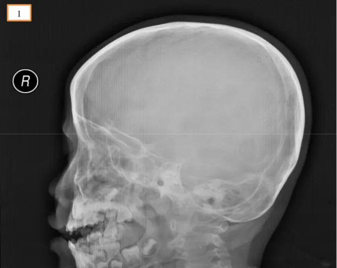
Otherwise, there was no external wound and deformity over the craniofacial region, bilat- eral eye examination and neurological exam- inations were unremarkable. Radiograph of the skull (Figure 1) showed faint pencil shadow over the left nasal cavity projecting towards the anterior skull base.
Figure 1: Radiograph of the skull showed faint pencil shadow over the left nasal cavity projecting to- wards the anterior skull base.
1
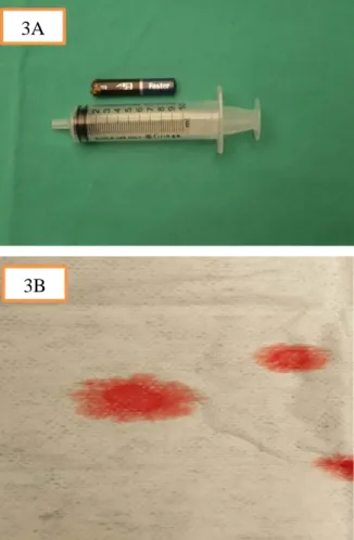
He was brought into the operating theatre for examination under anesthesia. In- traoperatively, the pencil was impacted firm- ly in the left nostril and nasal septum (Figure 2). The pencil was removed with ease using a Tilley’s forceps. There was a posterior na- sal septal perforation following the removal of the pencil. The embedded pencil meas- ured 8 cm in length and 1 cm in diameter (Figure 3A). An indentation mark with part of the lacerated septal cartilage was found covering the anterior skull base. Pulsatile clear fluid from the skull base was demon- strated immediately after removal of the pencil further which confirmed the skull base fracture with CSF leak (Figure 3B). We pos- tulated that the blunt end of the pencil served as the entry point via left nostril, penetrated the nasal septum with the extension of the in- jury to the anterior skull base.
Figure 2: Endoscopic view of the left nasal cavi- ty showed the pencil was impacted in the left nostril and nasal septum.
After removal of the foreign body, the CSF leakage was repaired with autologous graft by placing the septal cartilage and abdominal fat on to the defect until the CSF leakage ceased. The septal mucosal graft was reposi- tioned on top of the fat graft and covered with Gelfoam and tissue glue.
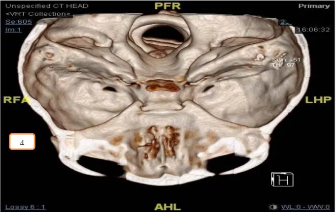
Post-operatively, he was ventilated for 24 hours and kept under bed rest with intrave- nous ceftriaxone administered for one week in view of overwhelming intraoperative find- ings. Computed Tomography (CT) scan of the brain, skull base and paranasal sinuses post-operatively showed right cribriform plate fracture with small pneumocranium (Figure 4). No evidence of intracranial hem- orrhage was seen. Post-extubation, his pupils were equal and reactive bilaterally with no neurological deficits. The neurosurgical team suggested for conservative management. He was discharged well one-week post-surgery.
Subsequent nasendoscopy during regular outpatient visits up to one year did not show any CSF leak.
Figure 3: (A) The embedded pencil measured 8 cm in length and 1 cm in diameter. (B) Pulsatile clear fluid from the skull base with positive ‘Halo’
sign on filter paper further confirmed the skull base fracture with CSF leak.
2
3B 3A
Figure 4: Three-dimensional (3D) images reconstructed from the fine cut CT scan of the brain, skull base and paranasal sinuses showed right cribriform plate fracture.
DISCUSSION
Nasal foreign bodies are common encounter in ENT setting. It ranges from simple foreign bodies to catastrophic scenarios with skull base fractures and intracranial complications.
Transnasal septal penetrating injury to the anterior skull base without an external wound and orbital involvement makes this a unique case. Furthermore, management of penetrating injury of the head and neck re- gion in a pediatric patient can be very chal- lenging as history may be inaccurate with limited cooperation during an examination.
Additionally, clinical examination showed no external wound over the face and orbital region with intact neurological function masquerade the extent of the injury made.
When the history is uncertain, the evalua- tion of suspected penetrating injury of the nose lies on the radiological assessment. A plain radiograph is easily available with low radiation, it offers an initial assessment of the bony framework of the facial and skull
bone to detect any fracture lines. The pano- ramic view of plain radiograph helps in lo- cating the foreign body. However, the visi- bility of the foreign body in plain radiograph depends on the types of object. Metallic for- eign body is radio-opaque on radiograph whereas organic foreign body like peanut or wood would be less radio-opaque hence easi- ly missed (Zilinskiene et al., 2014).
CT scan gives a good osseous delineation which is important in detecting subtle frac- tures. This fine thin cut of the CT scan with the image in axial, coronal and sagittal plane provides an accurate assessment of the loca- tion of the foreign body. With the three- dimensional (3D) images reconstructed from the fine cut CT scan, the extent of the injury can be accurately localized and essential in planning the surgical approach. For cases suspected with vasculature or intracranial in- jury, a CT angiography and Magnetic Reso- nance Imaging (MRI) brain would be war- ranted (Tsao et al, 2006).
4
Early removal of the penetrating foreign body is the definitive treatment in regards to avoid the unwanted sequelae (Brinson et al., 2004). The foreign body in the nasal cavity will obstruct the drainage and ventilation of the paranasal sinuses eventually causing si- nusitis. Penetration of the foreign body into the skull base with CSF leak predisposes the patient with intracranial infection by direct bacterial inoculation or ascending infection via the defects on the skull base (Cho et al., 2016). Early removal of foreign body and repair will reduce the risk of meningitis and cerebral abscess (Hettige et al., 2010). The patient was hemodynamically stable and surgical removal was carried out without fur- ther delay. He was given seven days of in- travenous ceftriaxone, which is the 3rd gen- eration of cephalosporin with the property of crossing blood-brain barrier for prophylaxis against intracranial infection.
There are two suggested methods in the removal of the penetrating foreign body into the maxillofacial region, namely the endo- nasal approach and external approach (Tsao et al., 2006). The shape and size of the object and the location of the entry point must be taken into consideration when planning for the removal. The penetrating object in this case report was a long and straight pencil with the entry point at the left nostril. This is best to be removed with endonasal approach via the natural orifice without leaving any additional scar on the face.
CSF leak was demonstrated immediately after removal of the foreign body. Most of the traumatic CSF leak will be resolved with conservative management or CSF diversion with lumbar drain. However, the incidence of ascending intracranial infection was rela- tively high in the group of CSF leak patient when conservative treatment was given.
Thus, repair of the CSF leak was advocated whereby it significantly reduces the risk of ascending intracranial infection (Bernal- Sprekelsen et al., 2005).
Presence of fistulous tract in between in- tracranial and nasal cavity predisposes the patient to CSF leaks, pneumocephalus and
intracranial infection such as ascending men- ingitis and abscess. The goals of skull base reconstruction include separation of the cra- nial cavity from the sinonasal tract, protec- tion of neurovascular structures, preservation of functions and avoidance of dead spaces.
As stated by Wormald and McDonough in 1997, CSF leaks have been traditionally managed by neurosurgeons via a frontal cra- niotomy. This traditional approach has a success rate of 60-80 % but has also been as- sociated with high morbidities such as frontal lobe retraction and anosmia. With the advancement of endoscopy technique, Oskar Hirsch from Austria was the first to use a strictly endonasal approach to close a CSF leak in sphenoid sinus using a mucosal peri- chondrial flap from nasal septum via a transseptal-transsphenoidal approach in 1952 (Hirsch, 1952).
Indications for endoscopic repair include post-traumatic CSF leak, iatrogenic and spontaneous CSF rhinorrhea associated with either malformation of the skull base, me- ningoencephaloceles, empty sella and in- creased intracranial pressure. With the im- provement of skills and instrumentation, amazingly high success rates with primary closure of the defect between 88 % and 94 % were achieved in the early 1990s (Lund et al., 2010).
The key to successful management of CSF leak is to precisely identify the site of the dural tear. Preoperative imaging such as high-resolution CT scan of the skull base and paranasal sinuses can be performed to locate the fracture site. Intrathecal application of sodium fluorescein in combination with the use of blue light and a blocking filter over the endoscope’s eyepiece will precisely guide the endoscopic surgeon to the site of the lesions. Laboratory techniques such as Beta-2-transferrin and beta-trace protein test- ing are the standard tests that help to differ- entiate non-CSF-related nasal secretion from the true CSF in an unclear situation (Le et al., 2016).
Multiple reports have validated that small CSF fistula can be reconstructed with a vari-
ety of free grafting techniques achieving success rate in more than 95 % of patients. In this case, the skull base defect was repaired via a transnasal endoscopic method by using autologous grafting. Examples of autologous grafts are fascia lata, fascia temporalis, carti- lage, bone and fat. The autologous graft is preferred as these avoid all the potential risks of heterogenous grafts like a prion- associated disease, HIV, hepatitis and other transmittable disease entities (Lund et al., 2010).
In this case, we harvested the fat graft from the periumbilical region as the autolo- gous graft for repair. The indentation mark over the anterior skull base was covered by the lacerated septal cartilage with septal mu- cosa. For defect closure, it is recommended that no mucosa should be placed under any graft or fat to avoid mucocele formation.
Hence the septal cartilage with mucosa was taken out temporary, the area of skull base defect was denuded of its mucosal layer. Ad- equate size of the harvested fat graft was gently placed over the skull base defect with pediatric Blakesley forcep. With the use of Freers elevator, the fat graft was gently placed in between the bone and surrounding mucosa, which is described as overlay tech- nique. Subsequently, the septal cartilage with mucosa was repositioned back on top of the fat graft. The grafted area was reinforced with Gelfoam and tissue glue. The end result of the repair was excellent without intracra- nial complication and further CSF leak up to date.
CONCLUSION
Management of transnasal penetrating in- jury of the anterior skull base can be very challenging in terms of diagnosis and treat- ment. When the history and physical exami- nation are in doubts, CT imaging is mandato- ry to rule out suspecting skull base injury.
Having said that accurate diagnosis and early intervention are important in order to prevent intracranial complications, continuous follow up is advocated to detect the delayed com-
Conflict of interest
The authors declare no conflict of inter- est.
REFERENCES
Bernal-Sprekelsen M, Alobid I, Mullol J, Trobat F, Tomás-Barberán M. Closure of cerebrospinal fluid leaks prevents ascending bacterial meningitis. Rhi- nology. 2005;43:277-81.
Brinson GM, Senior BA, Yarbrough WG. Endoscopic management of retained airgun projectiles in the pa- ranasal sinuses. Otolaryngol Head Neck Surg. 2004;
130:25-30.
Cho J, Kim JH, Hong SD. Complex anterior skullbase fracture caused by a bottle cap. A case report and re- view of literature. J Rhinol. 2016;23:49-54.
Hettige S, Kok K, Epaliyanage P, Thomas NWM.
Chopstick injury penetrating the skull base. Skull Base. 2010;20:219-22.
Hirsch O. Successful closure of cerebrospinal fluid rhinorrhea by endonasal surgery. AMA Arch Oto- laryngol. 1952;56(1):1-12.
Le C, Strong EB, Luu Q. Mangement of anterior skull base cerebrospinal fluid leaks. J Neurol Surg B. 2016;
77:404-11.
Lund V, Stammberger H, Nicolai P, Castelnuovo P, Beal T, Beham A, et al. European position paper on endoscopic management of tumours of the nose, pa- ranasal sinuses and skull base. Rhinol Suppl.
2010;22:1-143.
Tsao YH, Kao CH, Wang HW, Chin SC, Moe KS.
Transorbital penetrating injury of paranasal sinuses and anterior skull base by a plastic chair glide: Man- agement options of a foreign body in multiple ana- tomic compartments. Otolaryngol Head Neck Sur- gery. 2006;134:177-9.
Wormald PJ, McDonogh M. 'Bath-plug' technique for the endoscopic management of cerebrospinal fluid leaks. J Laryngol Otol. 1997;111:1042-6.
Zilinskiene L, Idle MR, Colley S. Emergency radiol- ogy: Maxillofacial and skull-base trauma. Trauma.
2014;16:243-55.