Eight-and-a-half syndrome: a rare potentially life-threatening disease
Abstract
Purpose:To report a case of eight-and-a-half syndrome presenting with a conjugate horizontal gaze palsy, an ipsilateral internuclear ophthalmo- plegia, and an ipsilateral lower motor neuron-like facial palsy.
Bryan Vincent Q.
Mesina
1George Michael N.
Sosuan
1Methods:A 56-year-old male who came in for limitation of extraocular muscle motion – wherein the only remaining movement is abduction
of the left eye – and right-sided facial weakness. Magnetic resonance
Karen B. Reyes
1imaging revealed a well-defined lesion resembling a cavernoma, at the level of the ponto-medullary junction.
1 Department of
Ophthalmology and Visual Results:Eight-and-a-half syndrome results from a lesion affecting the
paramedian pontine reticular formation, the median longitudinal fas-
Sciences, Sentro ciculus, and the facial nerve fascicle on one side. In this case, a cav-
Oftalmologico Jose Rizal, ernoma at the level of the ponto-medullary junction was compressing
the important pontine structures. University of the Philippines
– Philippine General Hospital, Manila, the Philippines Conclusion: Although eight-and-a-half syndrome is most commonly
caused by an infarction or demyelination, in rare instances, a space- occupying lesion at the level of the pons can be the etiology. It is of ut- most importance to recognize the features of this disease entity to be able to exhaust the proper diagnostic exams, localize the lesion and determine the proper treatment regimen catered to each patient.
Keywords:eight-and-a-half syndrome, cavernoma, neuro-ophthalmology
Introduction
Conjugate horizontal gazes are binocular movements that move the eyes synchronously toward one direction of gaze. These movements are supranuclear in origin, con- trol arising from the cerebrum and brainstem. The primary horizontal gaze control center is at the paramedian pontine reticular formation (PPRF) located at the pons, at the level of the facial colliculus. The PPRF sends signals towards its ipsilateral abducens nerve (VI) and contralat- eral medial longitudinal fasciculus. A lesion of this region would result to horizontal conjugate gaze palsy [1], [2].
Internuclear ophthalmoplegia (INO) is a disorder where there is defective adduction of the eye on the affected side and ataxic horizontal jerky nystagmus of the contralat- eral eye on abduction. Gaze towards the side of the lesion is, however, normal. INO arises from a lesion of the medial longitudinal fasciculus (MLF), a pair of crossed axonal tracts situated near the brainstem. It receives signals from the contralateral PPRF and in turn sends signals to its ipsilateral oculomotor nerve, coordinating conjugate eye movements. Lesions leading to INO include stroke, space-occupying lesions and demyelination [1], [2].
Lesions affecting both the PPRF and MLF on one side lead to the formation of the neuro-ophthalmologic disease entity one-and-a-half syndrome. First described by Fisher, one-and-a-half syndrome is a disorder comprised of ipsi- lateral conjugate horizontal gaze palsy, known as the
“one” and ipsilateral internuclear ophthalmoplegia (INO),
known as the “half”. The only remaining ocular movement is abduction of the contralateral eye [2]. In rare occasions, a lesion may affect the PPRF, MLF, and its ipsilateral fa- cial nerve fascicle around the area of the facial colliculus as it goes around the abducens nucleus. This lesion may give rise to a one-and-a-half syndrome with associated ipsilateral facial muscle weakness, also known as eight- and-a-half syndrome.
Eight-and-a-half syndrome is a rare pontine neuro-oph- thalmologic syndrome made up of a conjugate horizontal gaze palsy, ipsilateral internuclear ophthalmoplegia and ipsilateral lower motor neuron-like facial palsy [1], [3], [4], [5], [6], [7]. The facial palsy may involve forehead weakness, orbicularis weakness, facial asymmetry, and shallow nasolabial fold [1]. It is most often caused by a vascular etiology as in an infarction, as first described by Eggenberger in 1998 when magnetic resonance an- giography demonstrated eight-and-a-half syndrome caused by vertebral basilar disease [8]. Kakar et al., Sampath Kumar et al., Nandhagopal et al., and Bocos- Portillo et al. also demonstrated eight-and-a-half syn- drome as a sequelae of pontine infarction as seen in magnetic resonance imaging [3], [4], [5], [9].
Eight-and-a-half syndrome can also be caused by demy- elinating conditions at the level of the pons, such as multiple sclerosis, as demonstrated by Skaat et al. and Mortzos et al. [2], [10]. In even rarer instances, eight-and- a-half syndrome can be caused by a space-occupying le-
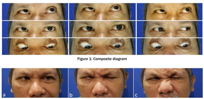
Figure 2: a) Shallow nasolabial fold, right, b) lagophthalmos, right, c) corrugator supercilli and procerus muscle weakness
sion at the level of the pons such as a tuberculoma, as demonstrated by Van Toom and colleagues [11].
Since a majority of the cases is secondary to infarction or ischemia at the pontine level, its onset is often sudden [3]. No sexual, race or age group predilection has been noted since the disease has first been documented. Hy- pertension, diabetes mellitus, and hypercholesterolemia are the co-morbidities noted commonly amongst patients with the disease [3], [5], [6], [9]. Prognosis of cases in- volving infarcts depends on the ability of the affected area to recover [4], [5], [9]. Several studies note improve- ment of the facial palsy before other signs and symptoms [4]. Neuro-rehabilitation is mostly warranted [3]. Prognosis of cases due to demyelination depends on the progres- sion of the primary disease entity [2], [10]. In cases dealing with space-occupying lesions, recovery depends on the removal of the lesion, as demonstrated by Van Toom and colleagues [11].
Case description
A fifty-six-year-old hypertensive male came in for consult due to an inability to abduct the right eye. History of the present illness started about thirty-four years prior when he first noted a limited abduction of the right eye. During the interim, he noted a gradually progressive limitation in all movements of the right eye, and a limitation in ad- duction of the left eye, associated with occasional dip- lopia. No consults were sought until a month prior when he noticed a sudden-onset right-sided facial weakness and dysarthria, with no hemiparesis or hemiplegia.
The patient came into the clinic with good visual acuity, good color vision, and no relative afferent pupillary defect, but with a right horizontal conjugate gaze palsy, a limita- tion of adduction of the right eye with associated nystag- mus of the left eye on abduction, and a right peripheral facial nerve palsy demonstrated by a four-millimeter
lagophthalmos, a shallow nasolabial fold, and an inability to wrinkle the forehead. The rest of the neurologic exam- ination was unremarkable. Slit lamp examination was also essentially unremarkable except for immature cataracts. Indirect ophthalmoscopy showed evidence of hypertensive retinopathy on both eyes.
Figure 1 shows the patient’s extraocular muscle move- ments in a composite diagram. There is a right horizontal gaze palsy and a limited adduction of the right eye. The sole extraocular muscle movement remaining is an ab- duction of the left eye.
Figure 2 highlights the peripheral facial nerve palsy. The patient has a shallow nasolabial fold on the right, a four- millimeter lagophthalmos on the right, and weakness of the occipitofrontalis, corrugator supercilli and procerus muscles.
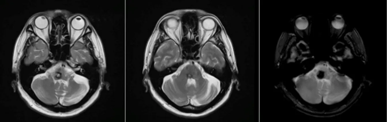
On magnetic resonance imaging, there is a hemorrhage with a complete hemosiderin ring at the level of the pons/ponto-medullary junction, associated with a well- defined lesion with a “popcorn ball” appearance resem- bling that of a cavernoma (Figure 3, Figure 4, and Figure 5).
The patient was co-managed with neurosurgery and neurology services for possible resection.
Discussion
Presented above is the case of a middle-aged male with a horizontal gaze palsy, an ipsilateral internuclear oph- thalmoplegia, and an ipsilateral peripheral facial nerve palsy, with no other associated findings. On imaging, a cavernoma with surrounding hemorrhage is noted at the level of the pons. Anatomically, the area in which the le- sion is situated in correlates well with the features seen in the patient’s case. Cavernomas, also known as cerebral cavernous malformations, are common vascular malform- ations in the brain, accounting to 10–25% of all vascular
Figure 3: T2-weighted images from the patient’s cranial magnetic resonance imaging
Figure 4: T1-weighted images from the patient’s cranial magnetic resonance imaging
Figure 5: Time of flight image from the patient’s cranial magnetic resonance angiogram
malformations [12]. The incidence in the general popula- tion is 0.4–0.8%, and the age group in which most are first diagnosed is within the twenty to forty year-olds [12].
Cavernomas are composed of clusters of abnormal, hya- linized capillaries surrounded by hemosiderin deposits and gliotic reaction [12]. They can be found in any location in the brain, but 70–80% are supratentorial in origin [12].
Supratentorial cavernomas present with new-onset seizures and headaches while infratentorial cavernomas present with progressive neurologic deficits. Due to their
slow-growing but dynamic nature, patients with caverno- mas may remain asymptomatic or may only have focal neurologic deficits. However, sudden-onset, neurologic deficits occur in cases of acute hemorrhage. If left un- treated, cavernomas may lead to intracerebral hemor- rhage, seizures, focal neurologic deficits, or headaches of varying severity [12]. The annual risk of hemorrhage is 0.7–1.1% per lesion in patients with no history of hemorrhage, but rises to 4.5% in patients with a previous episode of intracerebral hemorrhage [12]. Going by loca-
Cavernomas are angiographically occult lesions, which is why conventional T1- and T2-weighted magnetic reson- ance imaging (MRI) is still being used for its diagnosis [12]. The MRI is able to accurately detect cavernomas, which have a pathognomonic “popcorn-ball” appearance due to loculated hemorrhages, hemosiderin deposition around the primary lesion, and gliotic reaction [12]. Based on imaging, differentials may include hemorrhagic brain metastasis, gliomas, and hemorrhagic telangiectasias (Osler-Weber-Rendu disease) [12].
Microsurgical resection via the trans-sulcal approach, stereotactic radiosurgery (gamma knife surgery), and conservative management are the treatment options [12], which is why the patient was referred to the neurosurgery service. The choice of management de- pends on a myriad of factors including accessibility and location of the lesion, size of the lesion, severity of clinical presentation, and patient preference.
However, presented with the risks and benefits of the surgical procedure, the patient and his family opted to choose conservative management and have the disease run its course. In conservative management, alleviation of clinical symptoms is the primary goal. Pain relievers and anti-seizure medications may be prescribed and pa- tients are advised to refrain from strenuous exercises, anticoagulant use and pregnancy [12].
The area encompassed by the structures affected in eight- and-a-half syndrome spans a large portion of the pons.
Its presentation can mimic a cerebrovascular accident, but an affected area that large should irk an ophthalmo- logist to consider other etiologies, such as space-occupy- ing lesions. Hence, urgent and immediate localization of the lesion via radiologic examinations is of utmost import- ance.
Conclusions
Conjugate horizontal gaze palsy, ipsilateral internuclear ophthalmoplegia, and ipsilateral peripheral facial nerve palsy can occur together as a constellation of features seen in eight-and-half syndrome. Although it is most commonly caused by an infarction or demyelination, in rare instances, a space-occupying lesion, such as a cav- ernoma located at the level of the pons, can be the eti- ology. It is important to recognize the features of the dis- ease so as to be able to exhaust the proper diagnostic
Literature search
PubMed was searched for English-language articles on May 10, 2017, using the following terms: Eight-and-a- Half Syndrome, Cavernoma, Neuro-Ophthalmology.
Sources in retrieved articles were cross-referenced.
Acknowledgments
The authors thank the Neuro-Ophthalmology Service of the University of the Philippines-Philippine General Hos- pital Department of Ophthalmology and Visual Sciences for the support and guidance in making this paper.
Ethical consideration
The case report is a minimal risk study which was con- ducted in full compliance with the principles of the 7thit- eration of the Declaration of Helsinki and Good Clinical Practice of the WHO. All identifying patient information was kept confidential. Informed consent from the patient was obtained prior to inclusion in the study.
References
1. Kirkpatrick C. Eight-and-a-Half Syndrome. In: EyeRounds.org.
University of Iowa Health Care – Ophthalmology and Visual Sciences; 2015 [cited 2016 Jul 2]. Available from:
https://webeye.ophth.uiowa.edu/eyeforum/atlas/pages/eight- and-a-half-syndrome/index.htm
2. Skaat A, Huna-Baron R. Eight-and-a-half syndrome: a rare pontine neuro-ophthalmologic syndrome. Arch Neurol. 2012
Jul;69(7):934-5. DOI: 10.1001/archneurol.2011.2185 3. Kakar P, Brown Z, Banerjee S. Eight-and-a-half syndrome: an
unusual presentation of brainstem infarction. QJM. 2013 Mar;106(3):273-6. DOI: 10.1093/qjmed/hcr187
4. Bocos-Portillo J, Ruiz Ojeda J, Gomez-Beldarrain M, Vazquez- Picon R, Garcia-Monco JC. Eight-and-a-Half Syndrome. JAMA Neurol. 2015 Jul;72(7):830. DOI:
10.1001/jamaneurol.2015.0255
5. Sampath Kumar NS, Raju CG, Kiran PR, Kumar TA, Gopal BV, Khaseem DB. Eight-and-a-half syndrome: a rare presentation of pontine infarction. J Stroke Cerebrovasc Dis. 2014
Sep;23(8):e389-e391. DOI:
10.1016/j.jstrokecerebrovasdis.2014.01.002
6. Marquart C, Strauss C, Alfieri A. Eight-and-a-half syndrome combined with an ipsilateral vertical gaze palsy: a
pathophysiological explanation. Clin Neurol Neurosurg. 2013 Jun;115(6):767-9. DOI: 10.1016/j.clineuro.2012.06.039
7. Sarwal A, Garewal M, Sahota S, Sivaraman M. Eight-and-a-half syndrome. J Neuroimaging. 2009 Jul;19(3):288-90. DOI:
10.1111/j.1552-6569.2008.00261.x
8. Eggenberger E. Eight-and-a-half syndrome: one-and-a-half syndrome plus cranial nerve VII palsy. J Neuroophthalmol. 1998 Jun;18(2):114-6.
9. Nandhagopal R, Krishnamoorthy SG. Neurological picture. Eight- and-a-half syndrome. J Neurol Neurosurg Psychiatry. 2006 Apr;77(4):463. DOI: 10.1136/jnnp.2005.078915
10. Mortzos P, Nordling MM, Sørensen TL. Eight-and-a-half syndrome as presenting sign of childhood multiple sclerosis. J AAPOS. 2014 Oct;18(5):490-2. DOI: 10.1016/j.jaapos.2014.05.007 11. van Toorn R, Schoeman JF, Donald PR. Brainstem tuberculoma
presenting as eight-and-a-half syndrome. Eur J Paediatr Neurol.
2006 Jan;10(1):41-4. DOI: 10.1016/j.ejpn.2005.11.003 12. Mouchtouris N, Chalouhi N, Chitale A, Starke RM, Tjoumakaris
SI, Rosenwasser RH, Jabbour PM. Management of cerebral cavernous malformations: from diagnosis to treatment.
ScientificWorldJournal. 2015;2015:808314. DOI:
10.1155/2015/808314
Corresponding author:
George Michael N. Sosuan, MD
Department of Ophthalmology and Visual Sciences, Sentro Oftalmologico Jose Rizal, University of the Philippines – Philippine General Hospital, Taft Avenue Ermita, Brgy 670 Zone 72, 1000 Manila, the Philippines
gmsosuan@yahoo.com
Please cite as
Mesina BVQ, Sosuan GMN, Reyes KB. Eight-and-a-half syndrome: a rare potentially life-threatening disease. GMS Ophthalmol Cases.
2018;8:Doc04.
DOI: 10.3205/oc000086, URN: urn:nbn:de:0183-oc0000862
This article is freely available from
http://www.egms.de/en/journals/oc/2018-8/oc000086.shtml Published:2018-03-08
Copyright
©2018 Mesina et al. This is an Open Access article distributed under the terms of the Creative Commons Attribution 4.0 License. See license information at http://creativecommons.org/licenses/by/4.0/.