of Acanthocheilonema viteae tropomyosin.
Dissertation
zur Erlangerung des akademischen Grades doctor rerum naturalium
(Dr. rer. nat.) im Fach Biologie
eingereicht an der
Mathematisch-Naturwissenschaftlichen Fakultät I der Humboldt-Universität zu Berlin
von
Michał Janusz Sereda (M.Sc. Biology) geboren am 04.06.76 in Warschau, Polen
Präsident der Humboldt-Universität zu Berlin Prof. Dr. Christoph Markschies
Dekan der Mathematisch-Naturwissenschaftlichen Fakultät I Dr. Christian Limberg
Gutachter/innen: 1. Prof. Dr Richard Lucius 2. Prof. Dr Norbert Brattig
3. PD Dr Susanne Hartmann
Tag der mündlichen Prüfung: 14.02.08
I. Summary
II. Zusammenfassung
1. Introduction……….. 11
1.1. Filarial infections... 11
1.2. Filarial proxi models for research………... 13
1.3. The A. viteae proxi model for O. volvulus……… 13
1.4. Immunological aspects of Helminth infections……….. 15
1.5. Tropomyosin... 16
1.6. Structure and function of tropomyosin………. 17
1.7. Conserved structure of tropomyosin... 18
1.8. Tropomyosin isoforms in nematodes... 19
1.9. Allergenicity of tropomyosin and cross-reactivity among different species……. 19
1.10. Tropomyosin as a vaccine candidate……… 20
1.11. Aims and objectives of this study………. 22
2. Results………. 23
2.1. Molecular cloning of A. viteae and O. volvulus tropomyosins………... 23
2.2. Alignment of tropomyosin sequences of A. viteae and O. volvulus with other invertebrates and vertebrates reveals a great level of similarity………... 23
2.3. Purification of filarial tropomyosins... 25
2.3.1. Recombinant tropomyosin of A. viteae and O. volvulus……….. 25
2.3.2. Purification of tropomyosin by electroelution from SDS- PAGE gel……….. 26
2.3.3. Purification of native A. viteae tropomyosin by affinity chromatography with monoclonal antibody……….. 27
2.3.4. Both eAvTropo and rAvTropo retain the secondary structure of an D-helical protein………... 27
2.4. Transfected COS7 cells express A. viteae tropomyosin after 48 h………... 28
2.5. Use of tropomyosin based vaccine against A. viteae challenge infections……… 29
2.5.1. Recombinant protein vaccine efficacy depends on adjuvant used……….... 30
2.5.4. cDNA based vaccines effects... 33
2.5.5. Effect of vaccination on parasite length and development……….. 35
2.6. Analysis of antibody responses of immunized jirds and mice………... 35
2.6.1. rAvtropo vaccination elicits IgM and IgG that are not restimulated in the course of infection……… 35
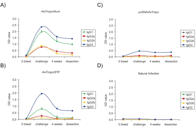
2.6.2. IgG subclasses... 37
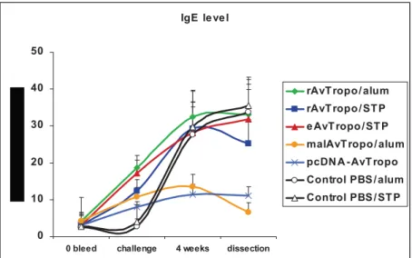
2.6.3. Immunization with A. viteae tropomyosin induces IgE responses………. 38
2.6.4. The role of specific anti-tropomyosin antibodies in protecton against L3 challenge………. 39
2.7. Analysis of antibody responses of immunized jirds and mice………... 40
2.7.1. Screening of peptide libraries revealed 13 IgG epitopes………. 40
2.7.2. A. viteae tropomyosin has 11 IgE epitopes……… 42
2.7.3. Key amino acid residues within A. viteae tropomyosin IgE epitopes are important for antibody binding……… 45
2.8. Allergenicity of tropomyosin... 50
2.8.1. A. viteae tropomyosin is a potent IgE inducer………... 50
2.8.2. Nematode tropomyosins are strongly cross-reactive……… 52
2.8.3. Determination of the cross-reactivity degree between tropomyosins by competition assay……….. 54
2.9. Raising monoclonal antibodies against A. viteae tropomyosin………. 55
2.9.1. NR1 but not R21 and N11 mAbs is specific to invertebrate tropomyosins………... 55
2.9.2. Epitopes of NR1 and R21 mAb are located in C-terminal part of A. viteae tropomyosin………. 57
2.9.3. N11 is presumably targeted against posttranslational modification on A. viteae tropomyosin………. 58
2.9.4. Tropomyosin is a surface component of A. viteae L3……….. 59
2.9.5. Tropomyosin specific mAb help to kill microfilariae in vitro by ADCC……… 60
2.10. T cell responses to tropomyosin……… 64
2.10.1. Both, the whole molecule and synthetic peptides based on A. viteae tropomyosin are capable to stimulate T cell proliferation……… 64
2.11. A. viteae tropomyosin influences expansion of various cell populations after immunization……… 66
2.11.1. Splenocytes from animals immunized with A. viteae tropomyosin produce IL-4 and IL-10 but no INF-J 66 2.11.2. Immunization with A. vitae tropomyosin expand cells Gr1+/CD11b+in spleen……. 67
2.11.4. The subpopulation of Gr1+/CD11b+ cells raises in peritoneal exudate after
immunization with rAvTropo………. 72
2.11.5. The constitution of PECs after immunization with A. viteae tropomyosin…………. 74
3. Discussion……… 76
3.1. Tropomyosin of A. viteae as a vaccine………. 76
3.2. Allergenicity of A. viteae tropomyosin……….. 84
3.3. mAb against A. viteae tropomyosin, their epitopes and specificity……….. 90
3.4. Cellular alteration and cytokine profile induced by A. viteae tropomyosin……… 92
3.5 Outlook………... 96
4. Methods………... 97
4.1. Parasitological methods……… 97
4.1.1. Laboratory animals………. 97
4.1.2. Maintenance of the life cycle of Acanthocheilonema viteae……… 97
4.1.3. Immunisation experiments……… 97
4.1.4. Antigen injection in Gr1+/CD11b+ experiments……….. 98
4.1.5. Quantification of microfilarial load in blood of jirds……… 98
4.1.6. Isolation of adult A. viteae from M. unguiculatus……….. 98
4.1.7. Isolation of L3 stages from the vector Ornithodoros moubata……… 99
4.2. Cell culture methods………... 99
4.2.1. Maintenance of mammalian cells……… 99
4.2.2. Preparation of stocks………. 99
4.2.3. Transfection of COS7 and HeLa cells………. 100
4.2.4. Raising the monoclonal antibody………. 100
4.2.5. Subcloning………... 101
4.2.6. Rat Basophil Leukemia (RBL) cell mediator release assay……… 101
4.2.7. T cells proliferation assay………. 101
4.3. Immunochemical and immunological methods………. 102
4.3.1. Bleeding of animals for production of sera……… 102
4.3.2. Western blot……… 102
4.3.3. Detection of specific antibody in sera of M. unguiculatus and BALB/c mice……… 103
4.3.4. Immunostaining of A. viteae larvae………. 103
4.3.5. Creation of the synthetic peptides libraries………... 103
4.3.8. IgE ELISA……… 105
4.3.9. Cytokine ELISA……….. 105
4.3.10. Flow cytometry (FACS)………. 105
4.3.11. Depletion of IgG antibodies from sera of immunized mice and jirds……….. 106
4.3.12. Deglycosylation of proteins on ELISA plate………... 106
4.4. Protein analytical methods………... 107
4.4.1. Preparation of soluble protein extracts………... 107
4.4.2. Determination of protein concentration………... 107
4.4.3. Sodium dodecyl sulfate polyacrylamide gel electrophoresis (SDS-PAGE)……….. 107
4.4.4. Maleylation of rAvTropomyosin……… 108
4.4.5. Preparation of gel-eluted antigen………. 108
4.4.6. Maleyation of recombinant A. vitae tropomyosin……….. 108
4.5. Molecular biology methods……….. 108
4.5.1. Preparation of DNA vaccine………. 108
4.5.2. Electrophoresis and detection of DNA on agarose gels……….. 109
4.5.3. Isolation of DNA from agarose gels………. 109
4.5.4. Extraction with Nucleospin kit……….. 109
4.5.5. Precipitation of DNA……….. 109
4.5.6. Isolation of plasmid DNA……….. 109
4.5.7. Isolation of total RNA………. 110
4.5.8. Electrophoresis of total RNA……… 110
4.5.9. Determination of the concentration of nucleic acids………. 110
4.5.10. Restriction analysis of isolated plasmids……… 111
4.5.11. Restriction digestion of DNA………. 111
4.5.12. Polymerase Chain Reaction (PCR)………. 111
4.5.13. Reverse Transcription PCR (RT-PCR)………... 112
4.5.14. Ligation………. 113
4.5.15. Cloning of A. viteae tropomyosin into pCDNA 3.1+ vector……….. 113
4.5.16. Cloning of O. volvulus tropomyosin into pET28b vector……….. 114
4.6. Microbiological methods……….. 114
4.6.1. Preparation of competent E. coli……….. 114
4.6.2. Transformation of competent E. coli……… 114
4.6.3. Colony PCR………. 115
4.6.4. Bacteria cultures and long term storage of stocks……… 115
4.6.5. Expression and purification of protein expressed in E. coli………. 115
5. Materials………. 117
5.1. Laboratory equipment……… 117
5.2. Primers………... 117
5.3. Vectors………... 118
5.4. E. coli strains……… 118
5.5. Consumables……… 118
5.6. Reagents……… 119
5.7. Commercial Kits……….. 120
5.8. Enzymes………. 120
5.9. Solutions, mediums and buffers………. 121
5.9.1. Agarose gel electrophoresis buffers……… 121
5.9.2. Bacterial culture medium……….. 121
5.9.3. Antibiotics……… 122
5.10. Protein and Immunochemistry……… 122
5.10.1. SDS-PAGE………. 122
5.10.2. Solutions for SDS-Polyacrylamide gel……… 123
5.10.3. Western Blot……… 123
5.10.4. Immunostaining……….. 123
5.10.5. Protease inhibitors………. 124
5.10.6. Ni-NTA-Affinity chromatography……….. 124
5.10.7. mAb-Affinity chromatography………... 124
5.10.8. ELISA………... 124
5.10.9. Antibodies……… 125
5.11. Immunization of M. unguiculatus……… 125
5.12. Software………. 125
6. Abbreviations... 126
7. References... 128
The study presented here describes the immunological properties of Acanthocheilonema viteae tropomyosin, a muscle associated protein reported to be a promising vaccine candidate. A. viteae is a filarial parasite of jirds that is used as a laboratory model for the important human parasite Onchocerca volvulus. Our experiments focus on unraveling the functional properties of A. viteae tropomyosin in the context of a natural infection, experimental immunization, or vaccination against parasite challenge. Additionally, the allergenic potential of tropomyosin was investigated along with the ability to induce high levels of specific IgE. A part of this study was also aimed at the development of anti-tropomyosin monoclonal antibodies (mAb) and their characterization.
This study revealed that tropomyosin can be a promising antigen for a vaccine against filarial nematodes, however, effective only in a Th1 biased environment.
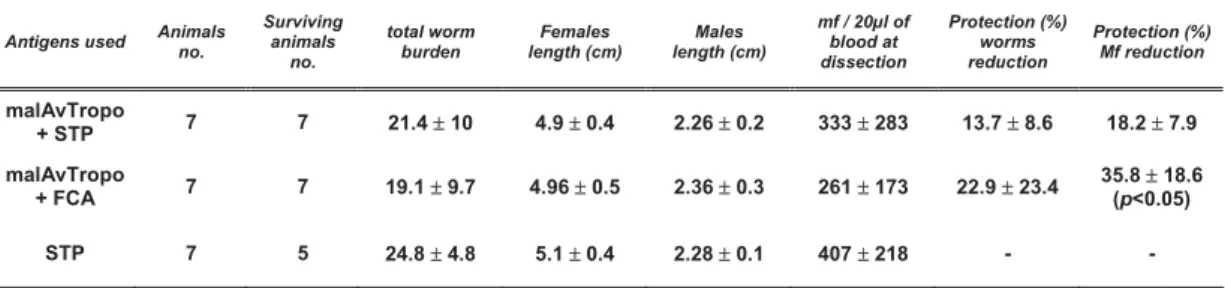
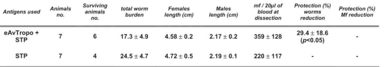
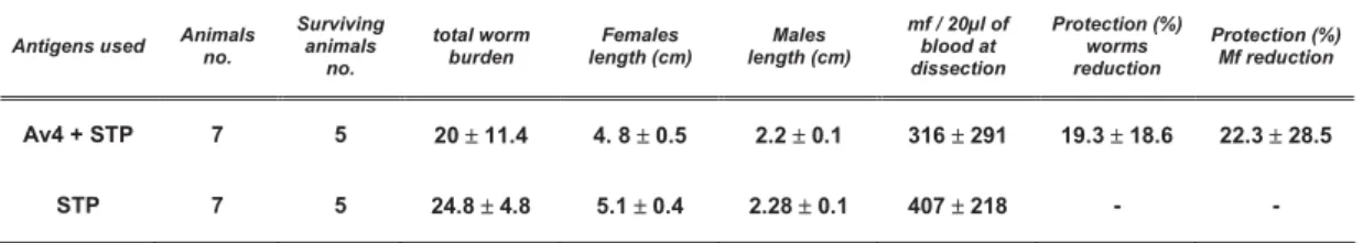
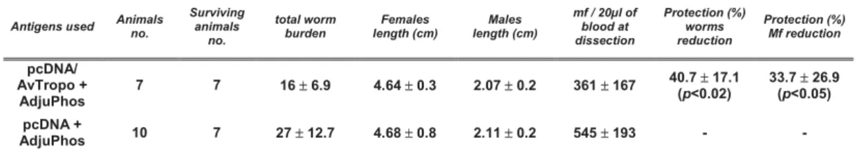
Vaccination with protein or DNA resulted in 30% and 45% protection, respectively, and protection against experimental challenge was not associated with specific IgG or IgE antibodies.
A. viteae tropomyosin is a functional allergen in the course of a natural infection and leads to the production of high levels of specific IgE. By screening of synthetic peptide libraries 13 IgG and 11 IgE co-located epitopes were characterized.
Functional assays showed that this protein cross-reacts with tropomyosins of other nematodes and shrimp, and that these proteins share IgE epitopes.
Three mAb were raised against recombinant and native A. viteae tropomyosin and for two of them (NR1 and R21) the regions of binding were identified. Specific mAb showed that tropomyosin is abundant on the cuticle of L3 and mf of the parasite.
Deglycosylation of the native protein showed that the epitope of the third antibody (N11) appeared to be formed by the posttranslational modifications.
Additionally, immunization of mice showed that A. viteae tropomyosin induced a similar pattern of cell activation and antibody production as aluminium hydroxide adjuvant, yet additionally it leads to the induction of IL-10 and the increase of a GR1+/CD11b+cell population. These cells are regarded as natural suppressors.
Taken together, these results show that A. viteae tropomyosin has immunomodulating properties and can in addition be considered as a component of an efficient vaccine.
Die vorliegende Arbeit beschreibt die immunologischen Eigenschaften von Acanthochilonema viteae Tropomyosin, einem Muskel-assoziierten Protein, das als vielversprechend für die Vakzinierung gegen Filariosen angesehen wird. A. viteae ist ein zu den Filarien gehörender Parasit von Gerbilen und wird als Labormodell für die humanpathogene Filarie Onchocercha volvulus genutzt. Diese Arbeit hatte die funktionelle Charakterisierung von A. viteae Tropomyosin im Kontext der natürlichen Infektion sowie der experimentellen Immunisierung und Vakzinierung zum Ziel.
Zusätzlich wurde das allergene Potential des Tropomyosins und die Produktion von spezifischen IgE-Antikörpern gegen das Protein überprüft. Ein weitere Teil dieser Arbeit beschäftigt sich mit der Entwicklung von Tropomyosin-spezifischen monoklonalen Antikörpern und deren Charakterisierung.
Es konnte gezeigt werden, dass Tropomyosin als vielversprechender Kandidat zur Vakzinentwicklung gegen Filariosen angesehen werden kann, wobei berücksichtigt werden muss, dass deutliche Effekte nur unter Th1-Bedingungen einer Immunantwort erzielt werden konnten. Die Vakzinierung mit Protein oder DNA führte zu um 30 bzw. 45% reduzierten Adultwurmzahlen und dieser Schutz ging nicht mit der Bildung von spezifischen IgG oder IgE Antikörpern gegen Tropomyosin einher.
A. viteae Tropomyosin fungiert im Zuge einer natürlichen Infektion als funktionelles Allergen und führt zur Produktion von hohen Mengen spezifischen IgEs. Durch ein Screening synthetischer Peptid-Bibliotheken konnten 13 IgG- und 11 IgE-Epitope charakterisiert werden. IgE Antikörper gegen A. viteae Tropomyosin zeigten in funktionellen Analysen eine Kreuzreaktivität mit aus anderen Nematoden sowie Garnelen gewonnenem Tropomyosin.
Es wurden drei monoklonale Antikörper gegen rekombinant exprimiertes und natives Tropomyosin generiert. Für zwei Klone (NR1 und R21) wurden die Bindungsregionen identifiziert. Mit Hilfe dieser Antikörper konnte gezeigt werden, dass Tropomyosin auf der Oberfläche der Kutikula der Larvenstadien L1 und L3 des Parasiten vorkommt.
Durch die Deglykosylierung des nativen Proteins wurde deutlich, dass ein durch den dritten generierten Antikörper (N11) erkanntes Epitop von posttranslationellen Modifikationen gebildet wird.
Schließlich führte die Immunisierung von Mäusen mit A. viteae Tropomyosin zu einem ähnlichen Profil der Zellaktivierung und Antikörperproduktion wie das Adjuvant
Oberflächenmoleküle Gr1 und CD11b charakterisierten Zellpopulation und zur IL-10 Produktion in der Milz immunisierter Tiere. Es konnte gezeigt werden, dass Gr1+CD11b+ Zellen an der Produktion dieses Zytokins Teil haben.
Zusammenfassend kann gesagt werden, dass A. viteae Tropomyosin immunmodulierende Eigenschaften aufweist und zusätzlich als Komponente eines zu entwickelnden Vakzins gegen Filariosen in Frage kommt.
1. Introduction
1.1. Filarial infections
One of the major problems of public health in tropical countries are infections with filarial nematodes witch affect about 120-150 million people (http://www.who.int/inf- fs/en/fact102.html). The forms of filariasis that have the most significant influence on global population health are: lymphatic filariasis caused by Wuchereria bancrofti and two species of genus Brugia: B. malayi and B. timori, onchocerciasis (called also “river blindness”) caused by Onchocerca volvulus, loiasis caused by Loa loa (Hoerauf et al., 2003). Filarial nematodes are classified as a part of a phylum Nematoda within a Filarioidea superfamily.
Onchocerciasis occurs in 34 African and Latin American countries. It is also reported from the Arabian Peninsula. The vast majority of an estimated 37 million population of people that are infected with O. volvulus live in sub-Saharan Africa. Parasite infections have caused blindness in about 270,000 and left another half million with severe visual impairment (Burnham et al., 1998). Onchocerciasis is not only a blinding disease, but in some cases also a chronic systemic illness that causes changes in the immune system.
Severe infections can cause symptoms like extensive and disfiguring skin changes, musculoskeletal complaints, weight loss and growth arrest. The disease is endemic in some of the world's poorest areas and its influence on public health has also a major impact on the economy of the regions (Evans et al., 1995).
The life cycle of O. volvulus starts when infective L3 larvae (L3) enter the human during the blood meal of an infected female black fly Simulium. Within 1-3 months the L3 larvae moult to the L4 and later to adults. Male and female worms dwell in connective tissue and eventually are encapsulated in callagenous nodules, mostly under the skin of a host.
Each female worm releases 1,300-1,900 microfilariae per day for 9-11 years (Schulz- Key and Karam, 1986). Microfilariae that migrate out of the nodules into the dermis of the skin are ingested by Simulium flies. In an intermediate host microfilariae penetrate the gut and settle in the thoracic flight muscles where they moult and develop into the L2 and subsequently into the infective L3 stage. The manifestation of onchocerciasis in humans is mainly due to the host inflammatory responses directed against dead or dying
microfilariae (they release their endosymbiotic Wolbachia sp., bacteria against which, the elements of immune system act, Pearlman and Gillette-Ferguson, 2007). These reactions are responsible for the development of the “river blindness” syndrome. When microfilariae from the skin enter the cornea and conjunctiva a punctuate keratitis develops around dead ones which can clear when inflammation settles. Sclerosing keratitis and iridocyclities are likely to develop after years of prolonged infection causing permanent visual impairment or blindness (Chan et al., 1987). One of the consequences of the disease is also chronic and acute papular onchodermatitis.
So far, strategies to eliminate onchocerciasis have been based on vector control and mass treatment with the anti-microfilaricidal drug Ivermectin (Molyneux et al., 2003) carried out by the Oncocerciasis Control Programme (OCP). The OCP sponsored by several organisations including World Bank and WHO aimed at eradication of onchocerciasis as an important public health disease and as an obstacle for the social and economic development of regions infested with O. volvulus. OCP used biodegradable insecticides to eradicate the simulium vector but despite high initial success it faced the rising problem of developing resistance to used chemicals and reinvasion of previously cleared areas by migrating black flies. Invermectin acts as an agonist of the parasite neurotransmitter J-aminobutyric acid (Soboslay et al., 1987) and induces an influx of Cl- through channels not regulated by J-aminobutyric acid.
Ivermecitin is an efficient microfilaricidal but unfortunately it has no real effect on adult worms. It affects only temporarily the embryonic development and release of microfilariae from the female (Schulz-Key et al., 1992). Moreover, there are several drawbacks to the chemotherapeutic treatment (Burnham et al., 1998). It is extremely difficult to achieve eradication of parasite infection unless a very costly treatment is sustained for a period of 12 to 15 years (estimated life span of the adult worm). Thus, a more efficient treatment and parasite control approach has been suggested. Concept is based on simultaneous use of a drug and a prophylactic vaccination. There are good reasons to believe that such a combined treatment that acts on both infectious stages of the parasite (mf with a drug and L3 with a vaccine) lead to eradication of onchocerciasis.
Therefore, it is important to characterise antigens playing critical roles in parasite development and transmission that could be targets for an efficient vaccine.
1.2. Filarial proxi models for research
Research in filariasis is dependent on the use of laboratory animal models owing to the inability to carry out experiments on human population. The filarial parasites are host- specific and this is one of the significant limitations that have hindered the functional characterisation of relevant target molecules of Onchocerca species. Thus, model systems, that involve the use of parasites in surrogate models, were developed. Study on Onchocerca spp. involves the implantation of Onchocerca spp. in subcutaneous chambers in CBA/J or DBA/2J mice (Townson and Bianco, 1982; Abraham et al., 1992).
A similar approach with Brugia spp. / BALB/c mouse system has been used as a chemotherapeutic (Devaney et al., 1985) and immunological (Carlow and Philipp 1987) model for the brugian filariasis. These systems have a major disadvantage because they rely on studies of parasites in non-natural hosts. It can be overcome by the use of natural life-cycle models of the filaria Brugia pahangi in cats (serves as a model for Brugia and Wuchereria in humans; Grenfell et al., 1991), bovine Onchocerca spp. or rodent filariae like Acanthocheilonema viteae and Litomosoides sigmodontis in their natural hosts (plays similar role as B. pahangi / cat model but for Onchocerciasis;
Abraham et al., 2002). Unfortunately, owing to the lack of adequate immunoreagents the underlying immunological mechanisms can not be investigated fully in these model systems. Recently, a promising model for Onchocerciasis based on full cycle of L.
sigmodontis in BALB/c mice was developed. Due to the full knowledge of the mouse genetic background, complete and correct development of filarial parasites in that host, it does not have the limitations of previously utilised models.
1.3. The A. viteae proxi model for O. volvulus
A. viteae in its natural host Meriones unguiculatus serves as a model for onchocerciasis, the disease caused by O. volvulus.A. viteae has a very similar life cycle (Fig. 1) and has some parallels to O. volvulus. Both reside in the subcutaneous tissue of their hosts and are therefore in the same immunological compartment. However, A. viteae parasites do
not form nodules and microfilariae that are present in blood cause no eye lesions like O.
volvulus. Furthermore, both parasites share an array of antigenic similarities as demonstrated by anti-O. volvulus monoclonal antibodies (Nogami et al., 1986), and a high homology in corresponding molecules that have so far been characterised from both parasites. In addition, cross-protection between species has been shown in filariasis (Storey and Al-Mukhtar, 1982; Geiger et al., 1996) so that vaccine candidates established in one system could be tested in others. The A. viteae / M. unguiculatus system allows the study of resistance to challenge infection following immunisation (Abraham et al., 2002). Interestingly, it has been shown that immunisation with irradiated A. viteae L3 led to 90% protection against challenge infection, while immunisation with excretory-secretory products (ESP) led to 70% protection (Lucius et al., 1991). Parallel results were also obtained using irradiated L3 in other filariasis models (Lange et al., 1993; Johnson et al., 1998; Trees et al., 2000) and it was shown in this model that immunisation with irradiated L3 could also lead to resistance against homologous challenge infection (Abraham et al., 2002). Therefore, the A. viteae / Meriones system was used in this study.
Figure 1. The life cycle of Acanthocheilonema viteae. The microfilariae are taken up by the arthropod host (tick – Ornithodoros moubata) during the blood meal and develop into the L2 and L3 stages. The infective L3 are transmitted to the rodent host where after 6 weeks of development and migration they settle usually under the skin and develop to adults. Source: Department of Molecular Parasitology.
1.4. Immunological aspects of Helminth infections
Infections with nematodes and other helminths are regularly associated with T-helper cell 2 (Th2) responses, eosinophilia and high levels of IgE, symptoms that are usually found in allergic patients. In the context of helminth infections, IgE and eosinophils can efficiently attack certain helminth stages, e.g. larval stages of schistosomes, by antibody dependent cellular cytotoxicity (Capron and Capron, 1994, see Fig. 2). However, certain immune effector mechanisms are modulated during infection, an effect probably owing to helminth immunomodulators acting on the host. Thus, although nematode infections polarize immune responses towards Th2, which would theoretically favor atopic reactions, they additionally trigger powerful anti-inflammatory mechanisms limiting the magnitude of the in vivo-response to allergens. The resulting immunological phenotype has been termed “anti-allergic phenotype”.
Figure 2. Induction of IgE-mediated anti-helminth effector reactions. Parasitic nematodes release allergens that are presented to naïve T cells by dendritic cells, macrophages or B cells. Activated T cells (effector T cells) release cytokines that activate other cells of the immune system and among others induce production of allergen-specific IgE by B cells. Allergens released by the nematodes can cross-link specific IgE on the mast cell surface and initiate their degranulation. Specific IgE bound to the surface of the parasites can be recognized by eosinophils and initiate their degranulation, eventually leading to parasite killing and tissue damage. CCR3, CC chemokine receptor 3; TCR, T cell receptor; MHC II, major histocompatiblity complex II; FcHR I, receptor I for Fc part of IgE;
CD80/86, CD28, CD40, CD40L: costimulatory molecules.
Nematode infections will continuously confront the host with a large number of different antigens. This is particularly evident in the case of filarial infections, where worms living within the host’s tissues reportedly reach life-spans of more than a decade. These worms constantly release large amounts of microfilariae that eventually die and degrade after having lived for several months. It was reported that patients heavily infected with O. volvulus might harbor about 100,000,000 microfilariae in addition to their adult worms (Schulz-Key and Karam, 1986). This number highlights the enormous amount of foreign material a patient’s immune system must deal with.
Out of the many nematode antigens a host is exposed to, some are allergens, meaning that they induce IgE antibodies, which can trigger allergic responses. Such allergic responses consist of the degranulation of basophils or mast cells, when their surface bound IgEs are cross-linked by allergen (Fig. 2). Release of histamine and other mediators, subsequent attraction and activation of effector cells lead to inflammatory responses that may kill the parasite, but can also result in damage of the host tissue.
1.5. Tropomyosin
Tropomyosin was characterized as one of important antigens in the filariae. Studies in A.
vitae model showed that antibodies derived from serum of animals immunized with irradiated L3 and resistant to the following challenge, when used in Western Blot on whole parasite extract, among others, bind strongly to the 41 kDa band on the whole worm extract. Protein in this band was characterized as the filarial tropomyosin (Hartmann et al., 1997) thus, A. viteae tropomyosin seemed to be an immunodominant antigen and a promising potential vaccine candidate. Moreover, invertebrate tropomyosins were described to account for allergic reactions against seafood, cockroaches and house dust mites (Reese et al., 1999). As reported tropomyosin also induces reactive IgE in nematode infections (Asturias et al., 2000; Hartmann at al., 2006). It was also observed that tropomyosin is present on the cuticle of O. volvulus larvae (Jenkins et al, 1998). All these arguments place tropomyosin as a major antigen in the context of filarial invasion.
So far vaccination with E. coli-expressed proteins of filariae has met with difficulties, because the level of protection observed was generally much lower and showed high
variability between experiments than with irradiated larvae (Abraham et al., 2002;
Lustigman et al., 2002). Nonetheless, proteins are still considered as a promising alternative, although it is difficult to find a suitable antigen. Due to its immunogenic properties tropomyosin is an interesting candidate for vaccination experiments.
1.6. Structure and function of tropomyosin
Tropomyosin is a microfilament-associated protein present in all eukaryotic cells. It is found in multiple isoforms that are characteristic for specific types of tissue or cells (Perry, 2001). The most common isoform of tropomyosin has a molecular weight of around 32.5 kDa and is abundant in muscle, where it forms a structure characteristic for all helical proteins, a dimer in the form of a coiled-coil rod with two polypeptides swirling one around the other, of approximately 20 Å in diameter and 400 Å in length (Stewart et al., 2001). In thin filaments, tropomyosin molecules associated in a head to tail manner form overlapping chains that span along actin filaments (Fig. 3A). They play a crucial role in the process of thin filament activation by blocking or freeing actin binding sites in the presence of calcium ions and myosin (Vibert et al., 1997). In this respect tropomyosin is essential in the process of muscle work, proper action of the movement apparatus and the basic functionality of filaments within the cytoskeleton. Activation of tropomyosin is calcium dependent and is controlled by a complex of three troponins, which have a great influence on tropomyosin and its flexibility (Solaro and Rarick, 1998).
The coiled-coil conformation of tropomyosin is a result of the architecture of the protein, which is built from heptapeptides (abcdefg). This motif is made up of a sequence of amino acids (aa) with a similar chemical nature, resulting in imperfect repeats of short aa sequences (Paulucci et al., 2002, see Fig. 3B). Both, the N- and C-terminus of tropomyosin have conserved aa residues, which are vital for the creation of “head-to-tail overlaps” allowing tropomyosin polymers to extend over the thin muscle filaments (Sousa and Farah, 2002).
A) B)
Figure 3. Structure of tropomyosin. Details see text. A) Tropomyosin in the context of a thin muscle filament. The tropomyosin multimer is arranged along an actin filament. In the resting stage, tropomyosin covers myosin-binding sites of actin. Ca2+ and the proximity of myosin heads lead to activation of troponins (TnI, TnC, TnT) that start a cyclic reaction resulting in a shift of tropomyosin by 35o. This reversible shift exposes the hitherto blocked myosin-binding sites and allows myosin heads to bind, a prerequisite for locomotion (modified after Gordon, 2000). B) Structure of the tropomyosin coiled-coil, viewed along the polymer (residues g1 to f7 create a single heptapeptide turn). The close association between two polypeptides results from binding of hydrophobic side chains of aa a and d of one molecule to aa a’ and d’ of the partner molecule. Salt bridges between aa e,g and g’,e’ stabilize the bond. Side chains of aa in positions b,c,f, are generally hydrophilic and face outside where they can react with a residues of adjacent proteins (modified after Stewart, 2001).
1.7. Conserved structure of tropomyosin
The complex interaction with other proteins involved in the process of movement or transport allows only a low level of freedom in aa composition of tropomyosin. Therefore, it is not surprising that the degree of homology is very high among tropomyosins even of phylogenetically distant species. Thus, tropomyosins represent a family of very conserved proteins (Perry, 2001). The homology of aa sequences of tropomyosins originating from invertebrate and vertebrate organisms reaches over 50%. The sequence identity between tropomyosin isoforms of closely related invertebrate species might reach 95% as e.g. between the orthologues of the human pathogenic filarial nematode O. volvulus and the rodent filarial A. viteae (Hartmann et al., 2006). Detailed studies of a muscle isoform of vertebrate tropomyosin implicated seven regions along the tropomyosin polypeptide that play a key role in interactions with actin. These actin- binding regions are very conserved (e.g. region 5 with 31% of the residues identical throughout the animal kingdom) and typically show a pattern of conserved aa in certain positions within a heptapeptide repeat (Hitchcock-DeGregori et al., 2002).
a’2
g’1 d’5 g1
d5 f 7
a2 e6
f’7 e’6
b3 c4
c’4 b’3 20 Å
a’2
g’1 d’5 g1
d5 f 7
a2 e6
f’7 e’6
b3 c4
c’4 b’3 a’2
g’1 d’5 g1
d5 f 7
a2 e6
f’7 e’6
b3 c4
c’4 b’3 20 Å
1.8. Tropomyosin isoforms in nematodes
The free-living nematode Caenorhabditis elegans has one tropomyosin gene (tmy-1/lev- 11) that is located on chromosome I, spans 14.5 kb and includes 14 exons. Alternative splicing of its transcript leads to creation of 4 different tissue-specific isoforms, CeTM I-IV (Kagawa et al., 1995; Anyanful et al., 2001). RNAi-mediated gene silencing in C.
elegans revealed that tropomyosin is essential for body morphology, coordinated movement and embryogenesis (Anyanful et al., 2001). Considering the high degree of conservation of tropomyosins, it is conceivable that these proteins in parasitic nematodes have similar functions and architecture. Tropomyosin of parasitic worms was found to be abundant in the muscle layer and was shown to occur also in the cuticle of O. volvulus (Jenkins et al., 1998) and Trichinella spiralis (Nakada et al., 2003).
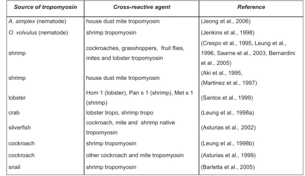
1.9. Allergenicity of tropomyosin and cross-reactivity among different species Tropomyosins of invertebrates have been reported to be potent allergens for humans, especially among food allergens (Lehrer et al., 2002). It was established that patients allergic to shrimp are not only allergic to other crustaceans, but also to more distantly related taxa of invertebrates. This cross-reactivity among tropomyosins of different origin was mostly evaluated with competition assays, where binding of serum IgE to its target epitopes was inhibited by addition of tropomyosin from a different source (Tab. 1). It was shown that the cross-reactivity is due to conserved IgE-recognized epitopes shared by closely or distantly related species. Several studies revealed that IgE against tropomyosin of shrimp cross-reacts with tropomyosin of non-edible arthropods such as house dust mites (Witteman et al., 1994; Aki et al., 1995) and insects like cockroaches, grasshoppers and fruit flies (Lehrer et al., 2002). Tropomyosin is therefore considered a cause for widespread cross-reactivity particularly among seafood and aeroallergens of invertebrate origin, suggesting that invertebrate tropomyosins are cross-reactive pan- allergens (Reese et al., 1999).
Table 1. Observed cross-reactivity of human serum IgE with tropomyosins from different edible and nonedible species of invertebrates
Source of tropomyosin Cross-reactive agent Reference
A. simplex (nematode) house dust mite tropomyosin (Jeong et al., 2006) O. volvulus (nematode) shrimp tropomyosin (Jenkins et al., 1998)
shrimp cockroaches, grasshoppers, fruit flies, mites and lobster tropomyosin
(Crespo et al., 1995, Leung et al., 1996, Saarne et al., 2003, Bernardini et al., 2005)
shrimp house dust mite tropomyosin (Aki et al., 1995, (Martinez et al., 1997) lobster Hom 1 (lobster), Pan s 1 (shrimp), Met s 1
(shrimp) (Santos et al., 1999)
crab lobster tropo, shrimp tropo (Leung et al., 1998a) silverfish cockroach, mite and shrimp native
tropomyosin (Asturias et al., 2002)
cockroach shrimp tropomyosin (Leung et al., 1998b)
cockroach other cockroach and mite tropomyosin (Asturias et al., 1999)
snail shrimp tropomyosin (Barletta et al., 2005)
Helminth allergens comprise several other nematode proteins like well characterized polyprotein allergens (NPA). These include ABA-I from Ascaris suum or TBA-I from Toxocara canis, and also from other intestinal nematodes and filariae (gp15/400 from B.
malayi or LL20 from Loa loa). NPA are lipid-binding proteins with a suspected immunomodulatory function (Kennedy, 2000). Allergens of parasitic nematodes include also the 31 kDa Tco-api-1 protein from Trichostrongylus colubriformis (Shaw et al., 2003), the major high molecular weight allergen (HMWtc) from Teladorsagia circumcincta (Huntley et al., 2001) and the NIE allergen from Strongyloides stercoralis (Ravi et al., 2005). All these proteins evoke strong IgE responses and most induce eosinophilia and Th2 biased cytokine responses as well. Interestingly, to the group of nematode allergens belongs also paramyosin from Anisakis simplex (Perez-Perez et al., 2000), a protein that shares many features with tropomyosin.
1.10. Tropomyosin as a vaccine candidate
Nematode tropomyosin is one of the few antigens described to induce significant protective immunity against parasite challenge in vaccination experiments. The existing
data on tropomyosin immunization were brought together in Tab. 2.
Table 2. Vaccine studies with tropomyosin of various parasites
Parasite Host Vaccine formulation Reduction in worm burden (%)
Comments / approach / reference Trichostrongylus
colubriformis
Guinea pigs 41 kDa fraction of L3 extract
43-51 (O'Donnell et al., 1989)
Onchocerca volvulus a)BALB/c mice
b) Jirds
C-terminus fragment+MBP (Ov14)
48-62
46
89 (blood mf)
a) Ov14 was used against O. lienalis challenge (Taylor et al., 1996)
b) Ov14 useds against A. viteae challenge (Taylor et al., 1996) Onchocerca lienalis BALB/c mice Aqueous extract of mf
immune serum transfer
51 (mf) 54 (mf)
(Folkard et al., 1996)
Onchocerca volvulus BALB/c mice Full lenght cDNA 20 i.m. and epidermal (Gene Gun) (Harrison and Bianco, 2000) Acanthocheilonema
viteae
Jirds a)Native protein 40-64 93 (blood mf)
(Hartmann et al., 1997)
Schistosoma japonicum
BALB/c mice Native protein 21
30 (liver eggs)
(Cao and Liu, 1998)
These studies describe the protective capacity of filarial tropomyosin as well as tropomyosin of the intestinal nematode Trichostrongylus colubriformis and a trematode Schistosoma japonicum. However, they did not address the effector mechanisms leading to the protective immunity. Generally, it is believed that filarial nematodes are attacked by mechanisms of antibody dependant cellular cytotoxicity (ADCC). Recent studies in mouse model systems have described IgE responses as pivotal for protective anti- helminth immune responses induced by irradiated larvae (Abraham et al., 2004). Thus, it appears attractive to re-evaluate a role of a surface associated, IgE inducing candidate antigen with respect to potential effector mechanisms in a natural host-parasite- association.
1.11. Aims and objectives of this study
Understanding the physiological functions of proteins required for parasitism could help to develop novel therapeutic and preventive strategies. With this principle in mind the objective of this study was to characterize the molecular and immunological properties of A. viteae tropomyosin.
Tropomyosin is an attractive intervention target suitable for vaccine studies. Therefore, it was an aim of this study to investigate its function during infection of A. viteae within a rodent and to evaluate the potential of tropomyosin as a protective antigen in an environment of a model that simulates the natural cycle of O. volvulus.
In addition, due to described allergenic potential of invertebrate tropomyosin it was important to study the allergenic properties of filarial tropomyosin and other characteristics and physiological functions that could influence the host immune system and the process of infection.
2. Results
2.1. Molecular cloning of A. viteae and O. volvulus tropomyosins
For this study tropomyosin from two filarial nematodes A. viteae and O. volvulus was used.
A. viteae tropomyosin gene was previously cloned into the pQE30b vector by the group of Dr. S. Hartmann (Hartmann et al., 2006, GeneBank accession number AF000607) within the Department of Molecular Parasitology. That construct was used in this study to transform competent bacteria and produce recombinant A. viteae tropomyosin (rAvTropo).
O. volvulus tropomyosin cDNA was isolated from a cDNA library created from female O.
volvulus RNA by PCR with specific primers. A clone of 855 bp in length was isolated, with a deduced amino acid sequence of 284 residues. The nucleotide sequence of the clone was very similar to that of A. viteae tropomyosin and contained an open reading frame extending from the start methionine to a stop codon at position 853-855. Both highly conserved regions, features of tropomyosin (N-terminal region aa 1-8: M-D-X-I-K- K-K-M and the C-terminal aa 240 – 249 L-K-X-A-E-X-R-A-E) were also present in the sequence of the obtained clone. The clone was named O. volvulus tropomyosin and inserted into the pET28b+ expression vector. The resulting construct was used to transform competent bacteria and to express and purify the recombinant protein. The theoretical molecular weight of O. volvulus tropomyosin (rOvTropo) was 33.2 kDa and the theoretical isoelectric point was pI 4.71. The recombinant protein had an apart molecular weight of 44 kDa on SDS-PAGE.
2.2. Alignment of tropomyosin sequences of A. viteae and O. volvulus with other invertebrates and vertebrates reveals a great level of similarity
Sequence alignment of A. viteae tropomyosin with tropomyosin genes of other nematodes showed a high degree of homology. On the amino acid level AvTropo shows 93% identity and (94% similarity) to O. volvulus tropomyosin and 87% (92%) identity to Caenorhabditis elegans tropomyosin. The homology to chicken cardiac tropomyosin is 52% (70%), to mouse muscle tropomyosin (TM1) 54% (70%) and to human tropomyosin
(TM1) 53% (73%). Homology of A. viteae tropomyosin to tropomyosins from other relevant species of invertebrates and vertebrates was summarized in Tab. 3. Amino acid sequences of both filarial tropomyosins used in this study are presented in figure 4 were they were aligned for comparison with sequences of tropomyosins from other species.
Figure 4. Alignment of tropomyosin genes sequences of Acanthocheilonema viteae and Onchocerca volvulus used in this study together with sequences of tropomyosin genes from several major species.
Table 3. Homology and similarity of tropomyosins from several relevant species. Homology shown by upper black number, while blue bottom numbers represent similarity.
A. vitae O. volvulus H. polygyrus C. elegans T. colubriformis T. spiralis T. pseudospiralis E. multiocularis S. mansoni H. americanus P. aztecus P. americana D. melanogaster X. laevis G. gallus M. musculus H. sapiens
A. vitae 100
O. volvulus 95
96 100
H. polygyrus 89
94 93 97 100
C. elegans 88
92 92 95 94
97 100 T. colubriformis 89
94 93 97 98
99 95 97 100
T. spiralis 82
90 87 94 86
93 85 92 86
93 100 T. pseudospiralis 83
91 87 94 87
94 85 92 87
94 99 99 100 E. multiocularis 53
73 56 76 56
76 56 75 56
76 59 78 59
78 100
S. mansoni 55
71 58 74 58
73 58 74 58
74 61 76 61
76 74 88 100
H. americanus 70
85 74 88 72
87 72 86 72
86 78 89 78
89 61 79 60
76 100
P. aztecus 68
83 71 85 71
84 70 85 71
85 76 87 76
87 60 80 60
76 92
95 100
P. americana 65
80 68 83 69
84 68 83 69
84 73 84 73
84 55 76 56
73 82 91 81
89 100
D. melanogaster 65 79
68 82
68 82
68 82
68 82
72 83
72 83
56 74
56 72
79 88
77 89
83
90 100
X. laevis 55
71 57 74 58
74 58 74 57
74 58 76 59
77 49 71 50
71 57 74 57
75 54 73 55
72 100
G. gallus 52
71 55 74 54
73 54 73 54
73 55 75 56
76 45 68 48
69 54 72 54
73 52 72 52
72 82
91 100
M. musculus 54
70 57 73 56
73 56 72 56
73 57 75 57
75 47 68 48
68 56 72 55
72 53 71 54
71 86
93 90
95 100
H. sapiens 56
73 58 75 57
74 58 75 57
74 58 76 59
77 49 71 50
71 57 74 55
74 54 74 55
73 94
97 82
91 91 95 100
2.3. Purification of filarial tropomyosins
2.3.1. Recombinant tropomyosin of A. viteae and O. volvulus
Expression of the A. viteae cDNA in E. coli as a polypeptide fused to a C-terminal sequence of six histidines yielded a protein with an apparent molecular mass of about 42 kDa (rAvTropo) and 44 kDa (rOvTropo) in SDS-PAGE. The recombinant proteins were soluble in aqueous buffers and suitable for purification by nickel chelate affinity chromatography (Fig. 5 and 6). It was possible to purify 3.4 mg of rAvTropo and 4.8 mg of rOvTropo out of a 500 ml bacteria culture, but due to the precipitation during the
process of dialysis the final yield of soluble protein dropped to about 200 μg. Proteins were freed of LPS contaminant by passage through EndoTrap columns and sterilized by filtering through 0.2 μm filters and stored at -20C until further use.
rAvTropo rAvTropo
Figure 5. Purification of Acanthocheilonema viteae tropomyosin, shown by SDS-PAGE. The single band at the level of 42 KDa in the elution fractions was the recombinant protein. M – Prestained Marker; 1 – Bacterial lysate before Ni-NTA matrix binding; 2 – Lysate flow through; 3-4 – Washing steps; 5-12 – Elution fractions.
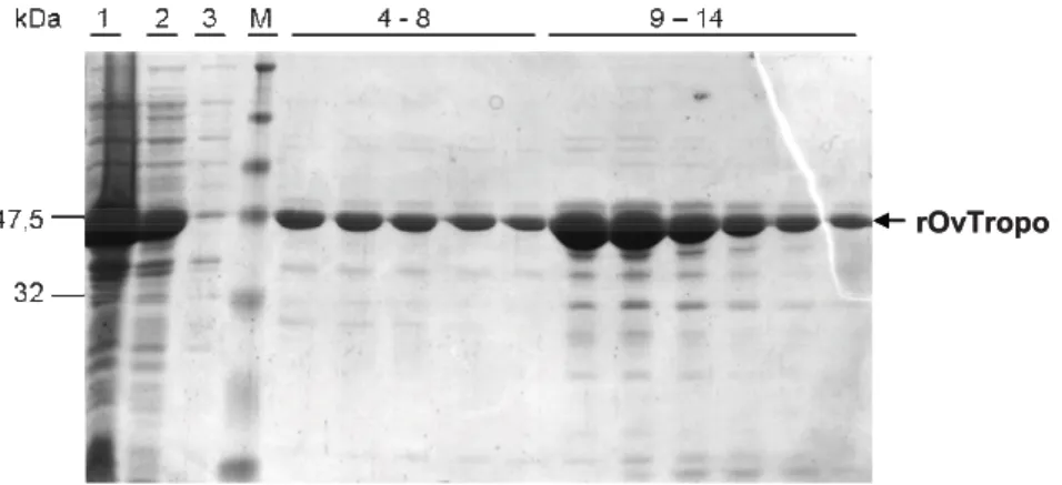
rOvTropo rOvTropo
Figure 6. Purification of Onchocerca volvulus tropomyosin, shown by SDS-PAGE. Single band at the level of 44 KDa in the elution fractions was the recombinant protein. 1 – Bacterial lysate after induction with IPTG; 2 – bacterial lysate befor binding to NiNTA matrix on Akta; 3 – bacterial lysate flow through; M – marker Prestained; 4-8 – Wash fractions; 9-14 – Elution fractions.
2.3.2. Purification of tropomyosin by electroelution from SDS-PAGE gel
One of the methods to obtain native A. viteae tropomyosin utilized in this work was electroelution of the protein band from the SDS-PAGE gel. PBS extract of female worms was first subjected to SDS-PAGE and a band corresponding to tropomyosin (as shown by mAb NR1 in Western-Blot) was cut out from the gel. Tropomyosin was electro-eluted from the gel (eAvTropo) and its quality and purity was checked on another SDS-gel where it appeared as a single band. It was later dialyzed in PBS. It was possible to
obtain about 400 μg of eAvTropo from an exctract of 150 worms. Protein was later filtered through the 0.2 μl mesh to sterilize and frozen at -20C until use.
2.3.3. Purification of native A. viteae tropomyosin by affinity chromatography with monoclonal antibody
Native tropomyosin from A. viteae (nAvTropo) was purified using monoclonal antibody NR1 bound to CNBr-activated Sepharose 4B. Tropomyosin was purified by the affinity chromatography from the female worms PBS extract. It was possible to purify about 200 g of pure nAvTropo out of 400 adult female worms (Fig. 7). Protein was purified on EndoTrap column to diminish LPS presence and dialyzed against PBS, sterilized through 0.2 μl filter and stored at -20C until further use.
Figure 7. Purification of Acanthocheilonema viteae native tropomyosin (nAvTropo), shown by SDS-PAGE. Single band at the level of 38 kDa in elution fractions corresponds to native protein. M – marker Prestained; 1 – mAb NR1 bound on sepharose; 2 – Female A.
viteae PBS extract; 3 – Wash fraction; 4 – Elution fractions.
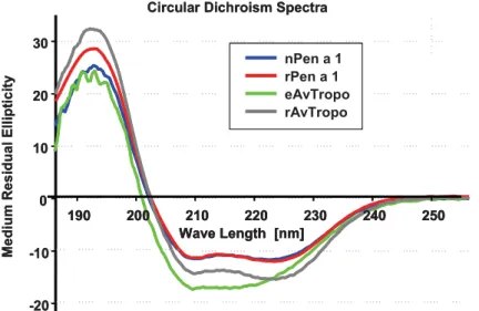
2.3.4. Both eAvTropo and rAvTropo retain the secondary structure of an D-helical protein
Purified eAvTropo and rAvTropo were a subject of a Circular Dichroism (CD) spectroscopy analysis. Both forms of A. viteae tropomyosin were compared with native and recombinant forms of well characterized shrimp tropomyosin. Analysis revealed that both eAvTropo and rAvTropo molecules have structures resembling D-helical proteins (Fig. 8). Spectra obtained for these proteins were similar to that obtained for proteins from shrimp (recombinant Penaeus aztecus tropomyosins - rPen a 1 and native P.
aztecus tropomyosin - nPen a 1). The shape of spectra for rAvTropo, but not for