Eur. J. Clin. Chem. Clin. Biochem.
Vol. 29, 1991,pp. 39-43
© 1991 Walter de Gruyter & Co.
Berlin · New York
Autoantibodies Against All the Phospholipids:
A Comparative Systematic Study
with Systemic Lupus Erythematosus and Healthy Sera
By Lilly Maneta-Peyret1, Christel Previsani1, Yvette Sultan2, J.-H. Bezian3 and C. Cassagne1
1 Centre National de la Recherche Scientifique, Institut de Biochimie Cellulaire et Neurochimie, Bordeaux, France
2 Höpital Cochin, Paris, France
3 Laboratoire d'Immunologie, Bordeaux, France
(Received July 11/October 22, 1990)
Summary: The sera of systemic lupus erythematosus patients were tested by ELISA for the presence of autoantibodies against all the phospholipids: cardiolipin, phosphatidic acid, phosphatidylcholine, phosphati- dylethanolamine, phosphatidylglycerol, phosphatidylinositol and phosphatidylserine. The quantity of phos- pholipid coated (in comparison with that initially deposited) on the microtitration plates was precisely evaluated in order to determine if the results (absorbance values) obtained for each phospholipid could be compared directly. The systemic lupus erythematosus sera tested gave positive results for all the phospholipids. The highest level of autoantibodies was observed with phosphatidic acid followed by phosphatidylserine, phos- phatidylinositol, cardiolipin, phosphatidylglycerol, phosphatidylcholine and phosphatidylethanolamine. The sera seemed to contain antibodies directed either against all the 8 phospholipids tested or more specifically against one or two phospholipids. The results were compared with those obtained with 17 healthy sera. Much lower values were obtained for the sera of healthy subjects, the majority of which showed a weak binding, similar for all the phospholipids. These results suggest that the anti-phospholipid autoantibodies present in systemic lupus erythematosus sera are significantly higher than those of healthy subjects. It is concluded that in the investigation of anti-phospholipid antibodies, tests should be carried out for all the phospholipids.
Introduction
Numerous papers have reported the presence of high levels of anti-phospholipid antibodies in human serum and their association with clinical disorders, especially systemic lupus erythematosus. However, these "anti- phospholipid" antibodies are detected either by the solid-phase Anti-Cardiplipin Test or the Standard Test for Syphilis; the antigen of both tests is cardiolipin.
This means that the term "anti-phospholipid" is most frequently employed in place of "anti-cardiolipin"
antibodies. It has nevertheless been reported that anti- phospholipid autoantibodies also react with other phospholipids, particularly the anionic ones. Harris et al. (1) showed in 1985 that 90% and 85% of the anti-cardiolipin activity can be inhibited with phos-
phatidic acid and phosphatidylglycerol respectively, whereas only 80% of Inhibition is obtained with the cardiolipin. In 1987, Gharaviet al. suggested that these antibodies are more specific for phosphatidylserine and phosphatidylinositol rather than for cardiolipin (2). Colaco & Male (3), Norberg et al. (4) and Costello
& Green (5) reported the presence of anti-phosphati- dylserine antibodies in systemic lupus erythematosus sera. Pengo et al. (6) found that affinity-purified anti- cardiolipin antibodies reacted with cardiolipin, phos- phatidylserine, phosphatidylinositol and phosphatidic acid. Brauch et al. (7) suggested that anticoagulant activity is always associated with the presence of anti- phosphatidylserine antibodies but also detected anti- bodies to cardiolipin, phosphatidylinositol, phospha-
Eur. J. Clin. Chem. Ciin. Biochem, / Vol. 29,1991 / No. l
iidylglycerol and phosphatidylethanolamine. McNeil et al. (8) compared the lupus anticoagulant and the anti-cardiolipin antibodies of two patient sera for their specificity towards all the phospholipids, and found reactivity towards the anionic phospholipids.
These comparatively rare reports deal with the pres- ence, in systemic lupus erythematosus sera, of auto- antibodies directed against phospholipids other than cardiolipin (for review, see I.e. (9)). Even though investigators agree that anionic rather than zwitter- ionic phospholipids (phosphatidylethanolamine, phosphatidylcholine) are involved, it is evident that the results reported are contradictory on the impor- tance of one or another phospholipid antigen in this affection. This question is complicated by the fact that most investigators have used the same test, i. e.
an ELISA in which the binding to antigen-coated wells is measured, to detect anti-phospholipid anti- bodies.
We suggest that the solution lies in the answers to the following questions:
1) are the phospholipids coated on the wells in a quantitative and reproducible manner?
2) are the different anti-phospholipid antibodies pres- ent in all systemic lupus erythematosus sera or are they only detected in particular cases?
To answer the first question, we titrated the coating of phospholipids on ELISA plates; to answer the second question, a systematic comparative study of systemic lupus erythematosus sera with all the phos- pholipids was undertaken.
Materials and Methods
The ELISA was developed for the study of anti-phosphatidyl- serine antibodies raised in rabbits (10). Phospholipids (from Sigma) were dissolved (100 mg/1) in hexane and 200 μΐ were deposited in each well. The hexane was evaporated under a gentle air stream at room temperature and the wells wefe saturated for 30 min at 37 °C with a solution of 10 g/l bovine serum albumin in 0.01 mol/1 phosphate buffer, pH 7.2, 9 g/l NaCl (PBS) and 0.5 g/l Tween 20. The plates were washed three times with PBS and incubated overnight at 4 °C with serum diluted (1/100) in PBS, 0.5 g/l Tween 20. After three further washes with PBS, the plates were incubated with anti-IgG- peroxidase conjugate (Inununotech) diluted in PBS, 0.5 g/l Tween 20 for l h at 37 °C. The plates were again washed three times with PBS and incubated with l g/l σ-phenylene diamine in 0.1 mol/1 citrate buffer pH 5.5 containing 0.25 ml/l H2O2 for 6 min in the dark. The reaction was stopped by the addition of 50 μΐ of 4 mol/1 H2S04 and the absorbance at 496 nm was measured by a Dynatech MR610 plate spectrophotometer.
Results and Discussion
For the coating of ELISA plates, phospholipids are generally dissolved in ethanol or in chloroform/meth-
anol (l + 4, by vol.) (9). Usually, cardiolipin is dis- solved in ethanol and the other phospholipids in chlo- roform/raethanol. The use of a single solvent should be more convenient and should give more comparable results. However, ethanol does not dissolve all the phospholipids and Chloroform is
fknown to attack plastic surfaces. We used hexane because it dissolves all the phospholipids, does not damage our plates (Nunc, Maxisorp) and can be evaporated rapidly (about 5 min for a plate containing 200 μΐ/well) under a gentle air stream.
The temperature of incubation during the ELISA is also of importance. In fact, the ELISA tests reported are carried out at 20 °C or 37 °C (especially the in- cubation of the plates with the serum to be tested).
However, when st dying the thermodynamics of the affmity of antigen-antibody iriteractiohs, it can be shown that the affmity (association constant K
ass) varies with temperature s foll ws:
dlnK
adT
ΔΗ°
RT
2'
where ΔΗ° is the Variation of the enthalpy of the reaction, R is the gas constant and T is the absolute temperature. This implies that for an interaction that is primarily exothermic (i.e. negative ΔΗ° by the formation of polar bonds), the affinity decreases with increasing temperature (11). Thus, many antigen-an- tibody interactions have a higher affmity at 4 °C than at 20 °C or 37 °C. In the case of phospholipids, there could be ionic interactions with the antibodies. C ri- sequently, the incubation with the serum should be carried out at 4 °C.
The absorbance measured at the end of the ELISA is proportional to the amount of antigen-antibody com- plexes formed. It is then important to know precisely the quantity of antigen (phospholipid) contained in the well after the coating procedure. Such a control has never been reported. The different kinds of ELISA plates s well s the different solvents used, could be responsible for the differences observed in the results reported.
We evaluated precisely the quantity of each phospho-
lipid coated on the plates. The supernatants resulting
from the washes of the wells were tested for their
phospholipid content by quantitative thin-layer chro-
matography (12). The exact quantity of coated phos-
pholipid was then defined s the quantity deposited
minus the loss during the washes. It was found that
for 20 μg of phospholipids deposited, 95.6% of car-
diolipin, 96.9% of phosphatidic acid, 98.1% of phos-
ph tidylcholine, 96.8% of phosphatidylethanolamine,
96.7% of phosphatidylglycerol, $7.8% ofphosphati-
dylinositol and 99.5% of phosphatidylserine were coated. These results show that the quantities of phos- pholipids coated (with hexane, in Nunc Maxisorp plates) are very close so that the absorbances obtained on the different phospholipids can be directly com- pared.
For each ELISA, a quadruple assay was carried out for every phospholipid. At least three experiments were performed (at different times) for each serum.
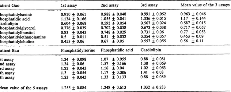
The incubation time for the reaction was 6 minutes and controlled carefully. Representative results with Standard deviations are given in table l, showing that the intra-assay äs well äs the inter-assay variations are low, thus permitting the direct comparison of the results without using Standards.
The totality of the results obtained with 38 systemic lupus erythematosus sera are presented in figure 1.
An important binding of auto-antibodies is observed for all the phospholipids and especially for phospha- tidic acid and phosphatidylserine. Much lower values were obtained for the healthy sera; furthermore, these values were very close for all healthy subjects, so that they are represented äs a mean value ± Standard deviation for each phospholipid (fig. 1).
The number of patients with a significantly higher level of autoantibodies than the healthy subjects were:
31 for phosphatidic acid (81.6%), 27 for phosphati- dylserine (71%), 14 for phosphatidylethanolamine (36.8%), 12 for phosphatidylglycerol (31.6%), 10 for cardiolipin (26.3%), 7 for phosphatidylinositol (18.4%) and 7 for phosphatidylcholine (18.4%).
It should be noted that 36.8% of the systemic lupus erythematösus patients have a significantly higher .level of autoantibodies to phosphatidylethanolamine
2.0
1.5
| 1.0
0.5
' (2.8)
•tt
.t:
* 4- $ S
PA PS CL Pl PG PC PE
Fig. l. ELISA on phosphatidic acid (PA), phosphatidylserine (PS), cardiolipin (CL), phosphatidylinositol (PI), phos- phatidylglycerol (PG), phosphatidylcholine (PC) and phosphatidylethanolamine (PE). Each point represents the mean value of absorbances obtained with one phos- pholipid for each systemic lupus erythematosus serura.
J represents the mean value and the Standard deviation of the absorbance obtained with one phospholipid for all healthy sera (n = 17).
compared with healthy subjects. The binding to zwit- terioriic phospholipids (phosphatidylethanolamine, phosphatidylcholine) is a subject of debate, äs it has often been reported that they are not recognized by the autoantibodies. Our results demonstrate that both phosphatidylcholine and phosphatidylethanolamine can bind specifically anti-phospholipid autoantibod- ies.
Tab. l. Mean values of absörbänces with the intra-assay Standard deviation (4 assays/phospholipid) and mean values of absorbances with the inter-assay Standard deviation (at least 3 experiments/serum).
Patient Gao Ist assay 2nd assay 3rd assay Mean value of the 3 assays
Phosphatidylserine 0.910 ± 0.061 0.988 ± 0,048 0.991 ± 0.052 0.963 ± 0.046 Phosphatidic acid 1.134 ± 0.166 1.055 ± 0.041 1.336 ± 0.015 1.17 ± 0.144 Cardiolipin 0.604 ± 0.068 0.593 ± 0.054 0.567 ± 0.024 0.587 ± 0.015 Phosphatidylglycerol 0.776 ± 0.039 0.702 ± 0.038 0.673 ± 0.038 0.717 ± 0.057 Phosphatidylinositol 0.83 ± 0.043 0.748 ± 0.029 0.731 ± 0.06 0.77 ± 0.053 Phosphatidylethanolamine 0.5 ± 0.011 0.51 ± 0.032 0354 ± 0.057 0.453 ± 0.09 Phosphatidylcholine 0.453 ± 0.06 0.67 ±0.05 0.557 ± 0.055 0.56 ±0.11 Patient Bau Phosphatidylserine Phosphatidic acid Cardiolipin
Ist assay 2nd assay 3rd assay 4th assay 5th assay
1.34 + 0.098 1.34 +0.06 1.22 ±0.043 1.3 +0.054 1.23 +0.043
1.07 ± 0.055 1.57 ±0.166 1.16 + 0.04 1.17 ±0.086 1.33 + 0.133
0.88 + 0.081 1.38 ±0.069 1.02 + 0.063 1.41 + 0.08 0.88 ± 0.089 Mean value of the 5 assays 1.255 ± 0.084 1.248 ± 0.613 1.032 ± 0.283
Eur. J. Clin. Chem. Clin. Biochem. / Vol. 29,1991 / No. l
l ε 1
O m
i
El· in
E
l 1
PS PA CL PG Pl PE PC PS PA CL PG Pl PE PC
iiliiil
PS PA CL PG Pl PE PCIQ «»l
|D IOBPS PA CL PG Pl PE PC a) Healthy subjects
2
PS PA CL PG Pl PE PC PS PA CL PG Pl PE PC
E
l 1
ID oo
nOCra
PS PA CL PG Pl PE PC PS PA CL PG Pl PE PC 0 *^" -
PS "PA CL PG PI' PE'PC"
α DUV
ππ
PS PA CL PG Pl PE PC
Ec
D DUB
PS PA CL PG Pl PE PC b) Patients
PS PA CL PG Pl PE PC PS PA CL PG PI PE PC
PS PA CL PG Pl PE PC PS PA CL PG Pl PE PC
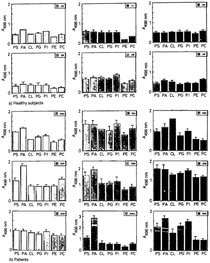
Fig. 2. ELISA on phosphatidic acid (PA), phosphatidylserine (PS), cardiolipin (CL), phosphatidylinositol (PI), phosphatidylgly- cerol (PG), phosphatidylcholine (PC) and phosphatidylethanolamine (PE).
Mean value and Standard deviation of absorbances obtained on each phospholipid for the serum studied.
a) healthy subjects (upper part)
b) systemic lupus erythematosus patients (lower part)
The results obtained with each phospholipid were than to other phospholipids; tfais explains the in- rather similar for the 17 healthy sera, s shown by creased Standard deviation of the mean value for the examples in figure 2a. For some healthy subjects, phosphatidylinositol (flg. 1).
the binding to phosphatidylinositol was slightly higher , t
For the systemic lupus erythematosus sera, different patterns were observed. Some sera had similar ab- sorbances for all phospholipids, other had increased binding to only some of them. Representative results are detailed in figure 2b. Of the phospholipids tested, the highest values were observed for phosphatidic acid in 28 patients (73.7%), for phosphatidylserine in 5 patients (13.1%), for phosphatidylinositol in 2 pa- tients, for cardiolipin in only 2 patients and for phos- phatidylcholine in one patient. In all the 5 sera con- taining a higher level of anti-phosphatidylserine an- tibodies, the levels of anti-phosphatidylserine and anti-phosphatidic acid antibodies were the same (fig.
2b, CHE or DUB). An exceptionally increased bind- ing of antibodies to phosphatidic acid was observed for the patient MALL (fig. 2b, MALL).
Conclusion
In this study, we measured the level of autoantibodies to each of the phospholipids (cardiolipin, phospha- tidic acid, phosphatidylcholine, phosphatidylethanol- amine, phosphatidylglycerol, phosphatidylinositol and phosphatidylserine) in the serum of systemic lu- pus erythematosus patients and compared their level with that of healthy subjects.
Our results demonstrate that
1) anti-phospholipid autoantibodies are present in healthy subjects;
2) their level is significantly increased in systemic lupus erythematosus patients and
3), they are directed against not only cardiolipin but also against phosphatidic acid, phosphatidylserine, phosphatidylinositol, phosphatidylglycerol, phospha- tidylethanolamine and phosphatidylcholine.
.The ELISA for anti-cardiolipin antibodies is more and more used äs a complementary clinical test. How- ever, it is generally accepted that this assay is less specific for predicting the clinical features than the lupus anticoagulant test (13). The development of an ELISA for all the phospholipids will give more Infor- mation about the role played by the anti-phospholipid antibodies. It has been reported that fluctuation of the level of anti-cardiolipin autoantibodies can be observed within weeks (14) or even years (5). This Problem should be further examined for all the phos- pholipids.
Our study is currently extended to the analysis of a larger number of sera äs well äs to the possible as- sociation of the different anti-phospholipid antibodies with the clinical features of the patients.
References
1. Harris, E. N., Gharavi, A. E., Loizou, S., Derue, G., Chan, J. K., Mackworth-Young, C. G., Bunn, C. C. & Hughes, G. R. V. (1985) Cross -reactivity of antiphospholipid anti- bodies. J. Clin. Lab. Immunol. 16, 1—6.
2. Gharavi, A. E., Harris, E. N., Asherson, R. A. & Hughes, G. R. V. (1987) Anticardjolipin antibodies: isotype distri- bution and phospholipid specificity. Ann. Rheum. Dis. 46, 3. Colaco, G. B. & Male, D. K. (1985) Antiphospholipid1-6.
antibodies in Syphilis and a throrabotic subset of systemic lupus erythematosus; distinct profiles of epitope specificity.
Clin. Exp. Immunol. 59, 449-456.
4. Norberg, R., Ernerudh, J., Hamsten, A., Unander, A. M.
& Arfors, L. (1986) Phospholipid antibodies in cardiovas- cular disease, Acta Med. Scand. Suppl. 775, 93—98.
5. Costello, R B. & Green, F. A. (1988) Binding affinity of serum immunoglobulin G to cardiolipin and other phos- pholipids in patients with systemic lupus erythematosus and Syphilis. Infect. Immun. 56, 1738-l742.
6. Pengo, V., Thiagarajan, R, Shapiro, S. S. & Heine, M. J.
(l 987) Immunological specificity and mechanism of action of IgG lupus anticoagulants. Blood 70. 69-76.
7. Branch, D. W., Rote, N;, Dostal, D. & Scott, J. (1987) Association of lupus anticoagulant with antibody against phosphatidylserine. Clin. Immunol. Immunopathol. 42,
63-75.
8. McNeil, R, Chesterman, C. & Krilis, S. (1989) Anticardio- lipin antibodies and lupus anticoagulants comprise separate antibody subgroups with different phospholipid binding characteristic. Br. J. Haematol. 73, 506-513.
9. Harris, E. N. (1990) Antiphospholipid antibodies. Br. J.
Haematol. 74, 1-9.
10. Maneta-Peyret, L., Bessoule, J. J., Geffard, M. & Cassagne, C. (1988) Demonstration of the high speciflcty of anti- phosphatidylserine antibodies. J. Immunol. Methods 108, 123-127.
11. Berzofsky, J. A., Epstein, S. & Berkower, I. J. (1989) An- tigen-antibody interactions. In: Fundamental Irmmmology, 2nd ed. (Paul, W. E., ed.) Raven Press Ltd. N. Y., pp. 315- 12. Heape, A., Juguelin, H., Fabre, M., Boiron, F. & Cassagne,326.
C. (1986) A quantitative developmental study of the pe- ripheral nerve lipid composition during myelinogenesis in normal and Trembler mice. Dev. Brain Res. 25, 181 — 189.
13. Mackworth-Young, C. (1990) Antiphospholipid antibodies:
more than just a disease marker? Immunol. Today I I , 60-65.
14. Cooper, R., Klemp, R, Stipp, C. J. & Brink, S. (1989) The relationship of anticardiolipin antibodies to disease activity in systemic lupus erythematosus. Br. J. Rheumatol. 28, 379-382.
Lilly Maneta-Peyret IBCN-CNRS
l rue Camille Saint-Saens F-33077 Bordeaux Eur. J. Clin. Chem. Clin. Biochem. / Vol. 29,1991 / No. l