Eur J Clin Chem Clin Biochem 1996; 34:343-347 © 1996 by Walter de Gruyter · Berlin · New York
Binding of Anti-Double Stranded (ds) DNA-Positive Sera to Denatured (d) DNA and Synthetic Poiy[dA-dT] x Poiy[dA-dT]
Double Stranded Copolymer in an ELISA Format
Drago Batinic1, Marijana Bozicevic1, Ana Krstulovic1, Dubravka Bosnia2, Mirna Sentic2, Jasenka Markeljevic2, Branko Malenica1, Nada Cikes2
and Matko Marusic
11
Division of Immunology, Clinical Department of Laboratory Diagnosis
2
Division of Clinical Immunology and Rheumatology University Hospital Center Zagreb and School of Medicine, Zagreb, Croatia
Summary: Using an ELISA assay anti-nuclear antibody-positive sera from 300 patients with various immune- related diseases and 64 anti-nuclear antibody-negative sera were analysed for binding to Sl-nuclease-treated double stranded (ds) DNA. In addition, the pattern of reactivity of 50 selected anti-dsDNA-positive sera was established using denatured (d) DNA and poly[dA-dT] X poly[dA-dT] double-stranded alternating copolymer (dAT) as addi- tional DNA antigens. None of the 64 anti-nuclear antibody-negative sera and 76 of the 300 anti-nuclear antibody- positive sera (25%) were anti-dsDNA-positive. Of the anti-nuclear antibody-positive and anti-dsDNA-positive sera, 48 (63%) were from systemic lupus erythematosus patients, and 7 (9%) from rheumatoid arthritis patients, whereas 21 patients (27.6%) suffered from various immune and non-immune related diseases. Anti-dsDNA-positive reactiv- ity was highly correlated with dDNA and dAT reactivity (r = 0.906, p < 0.0001 and r = 0.93, p < 0.0001, respec- tively). Although the majority of the 50 selected (37 systemic lupus erythematosus and 13 non-systemic lupus erythematosus) anti-dsDNA-positive sera concomitantly bound to both additional antigens, 7 of these (14%) did not bind to dAT, and 2 (4%) did not bind to dDNA. Anti-dsDNA-positive sera (n = 37) showed a similar pattern, in which 8.1% and 2.7% of sera did not bind to dAT and to dDNA, respectively. In contrast, anti-dsDNA-negative sera from various immune-related diseases bound either ssDNA (12.5%) or dDNA and dAT (12.5%). These data suggest that dsDNA and dAT-based assays detect similar but not identical specificities in the sera of patients suffering from systemic lupus erythematosus and in a proportion of non-systemic lupus erythematosus patients.
Introduction variety of diseases and have no diagnostic importance
A
.M ,- · .
Λt , .
Λj ,
A ^ ΤΛΧΤΑ f Λ (4). An intriguing hypothesis has been proposed thatAntibodies against double stranded (ds) DNA are found
v' ^ .
6. , . , , „ , in 50% to 75% of patients with systemic lupus erythe- »^-affimly anti-ssDNA') arise as a by-product of poly- matosus, and they represent one of the most helpful
clonal actlvatlon dunnS
imtial staS
es of systeirac luP
us,
f,. . . ! ., erythematosus, whereas anti-dsDNA antibodies emerge markers for diagnosing systemic lupus erythematosus , . ,. . . (reviewed in 1. c. (1)). In addition to their diagnostic po- *™°* ***?" P^
88'
0* ^
theP
rocess of antlS
en' tential, these antibodies are associated with the develop-
dnven selectlon of B'
cells (5)'
ment of renal complications in systemic lupus erythema- DNA is a rather flexible molecule that may adopt dif- tosus patients (2). Although anti-dsDNA
1) antibodies are ferent conformational forms (2, 6, 7). Even purified a central feature of systemic lupus erythematosus and dsDNA (which constitutes 85% of native DNA ex- are thought to play a role in its pathology, they have tracted from cells) can take up different conformations, also been found with lower frequency and in lower titre and further variations can be induced by temperature, in normal sera and in sera from patients with other auto- pH and ion concentration. Thus, these conditions may immune diseases (1—4). In contrast, antibodies to sin- influence the binding of antibodies to their target antigen gle-stranded (or denatured) DNA (dDNA
1)) occur in a (7). Early observations revealed that sera from systemic _ lupus erythematosus patients reacted with ssDNA,
1}
Abbreviations· dsDNA or with both forms, but the analysis of the fine dAT, poly[dA-dT] X po!y[dA-dT] double-stranded alternating co- specificity of DNA by monoclonal antibodies showed
polymer;
anextremely diverse fine specificity of DNA antibodies
dsDNA, double-stranded deoxyribonucleic acid; ,0. T. . n . . , ., . .. , ,-^ΧΤΑ *-u j·
dDNA, denatured DNA; (
8)·
ft 1S wel1 aPP
reciated thatanti-dsDNA antibodies
ssDNA, single stranded'DNA. have little specificity for base sequences (1,8) and bind
344
Batinic el al.: ANA"*" sera binding to DNA antigens in an ELISAto the epitopes centred on the phosphate backbone of the dsDNA. In contrast, the anti-DNA antibodies re- active with bases are found primarily among antibodies reactive with ssDNA or denatured DNA, where the bases are exposed (7).
ELISA is one of the most adequate techniques for the clinical measurement of the anti-dsDNA level (9). How- ever, since preparing and maintaining an antigenically pure dsDNA is not an easy task in the routine determina- tion of anti-sDNA, synthetic molecules, such as alternat- ing double stranded dAT
1) copolymer, were proposed as model molecules for an additional anti-native DNA as- say ((10) and reviewed in I.e. (11)). The rationale was the observed high correlation between native DNA and dAT-binding and the absence of contaminating mole- cules in the synthetic antigen (10). However, others have shown that systemic lupus erythematosus serum may have antibodies against different native DNA epitopes, i. e. those against the structure found in native DNA and those found in synthetic antigen (double helical configu- ration) (reviewed in I.e. (11)). In this study, we report a solid-phase assay for determination of antibodies against DNA and compare the results obtained using three dif- ferent antigen preparations.
Materials and Methods Control group
This group consisted of 64 adult (> 15 years of age) individuals of both sexes (c? : $ 1 : 2), whose sera gave a negative reaction for anti-nuclear antibodies in the immunofluorescent assay on rat liver sections. In general, these samples showed no abnormal findings during routine checking of immunological quantities, including to- tal haemolytic activity, C3, C4, Clq, circulating immunocom- plexes, rheumatoid-factor, anti-nuclear antibodies, anti-neutrophil cytoplasm antibodies and C-reactive protein.
Study group
These subjects were selected on the basis of their positive anti- nuclear antibody reaction (serum dilution > 1 : 16) and consisted of 300 adult subjects of both sexes with a male : female ratio of 1 : 4.8. Immunological testing was performed either as part of an out-patient diagnostic procedure or during hospitalisation of patients for treatment at the University Hospital Centre, Zagreb.
This group consisted of patients with various diagnoses of autoim- mune and non-autoimmune conditions, including degenerative, in- flammatory and malignant diseases. All patients with autoimmune diseases were classified and diagnosed according to standard cri- teria (American Rheumatology Association criteria) (12-14).
Patients with autoimmune diseases were either in active or inactive phase of disease. In order to avoid any bias toward a particular diagnosis, the sera were tested blindly, without prior knowledge of the patient's diagnosis. After the results of the ELISA were known, the diagnoses were obtained from responsible physicians, and sera from dsDNA"4" patients were checked in the DNA ELISA.
Sera
All samples were centriftiged immediately after receipt, heated (30 min at 56 °C) and stored at -20 °C (4-6 weeks) or at -70 °C (> 6 weeks).
Anti-nuclear antibody testing
The test was performed on acetone-fixed rat liver sections using FITC-labelled goat anti-human polyvalent Ig(A+G+M) (Imuno- loski Zavod, Zagreb, Croatia) as a second layer. The slides were analysed under a fluorescence microscope (Carl Zeiss, Jena, Ger- many) equipped with an epi-illuminator.
Immunoblot-assay for autoantigehs
In the majority of patients, a panel for the detection of autoantigens (ANA, dsDNA, SS/A, SS/B, Sm/RNP)2) was performed using an ImmunoDot assay (Gen Bio, San Diego, CA, USA).
Reagents and materials
Deoxyribonucleic acid from calf thymus (Sigma, Cat. #D3664), polydeoxyadenylic-thymidylic acid (poly[dA-dT] x poly[dA-dT]) (Cat. #Po883), methylated bovine serum albumin (Cat, #A-1009), nuclease-Sl from Aspergillus oryzae (Cat. #N7385), calf serum (Cat. #C6278), bovine serum albumin (Cat. #A2153), affmity- purified and alkaline phosphatase-conjugated anti-human polyva- lent immunoglobulins (α, γ and μ-chain specific) (Cat. #A5034), polyoxyethylenesorbitan (Tween-20 (Cat. #P2690)) and /7-nitro- phenyl phosphate disodium (Cat. #N2765) were purchased from Sigma, USA. All other chemicals - NaCl, sodium acetate (CH3CO2Na), NaOH, ZnCl2, ethanolamine (H2NCH2CH2OH) and NaH2PO4 X 2H2O - were purchased from Kemika, Zagreb, Croa- tia. The 96-well microtitre assay plates included Falcon PVC Microtest III (Cat. #3912) and PRO-BIND plates (Cat. #3915) (Becton Dickinson and Co., Oxnard, Canada) and 96-well flat bot- tomed plates from Behringwerke AG, Marburg, Germany. The ab- sorbance was read using a Behring ELISA Processor (Beh- ringwerke AG, Marburg, Germany).
Preparation of dsDNA
Double stranded DNA was prepared according to the original method of Rubin (15). Briefly, native DNA from calf thymus was dialysed against acetate buffer (0.03 mol/1 sodium acetate, 0.1 mol/1 NaCl, 5 mmol/1 ZnCl2, 0.1 g/1 bovine serum albumin, pH 4.4) and digested with Sl-nuclease (0.1 U^g DNA) for 3 h at 37 °C. After dialysis against phosphate-buffered saline (pH 7.2) at 4 °C, the concentration of DNA was determined (A260nm = 1 equ- als 50 mg/1) (15) and adjusted to 10 mg/1 in phosphate-buffered saline. Before coating with DNA, the wells of the microtitre plate were first coated overnight at 4 °C with methylated bovine serum albumin (10 mg/1 in phosphate-buffered saline 0.1 ml/well). After washing with phosphate-buffered saline, 0.1 ml of DNA solution (10 mg/1 in phosphate-buffered saline) was added to each well and the plate was further incubated overnight at 4 °C. The wells were then washed three times with phosphate-buffered saline, filled with 1 g/1 gelatin in phosphate-buffered saline and incubated overnight at 4 °C. After washing, DNA adhering to wells was redigested with 3 kU/1 of Sl-nuclease in phosphate-buffered saline (2 h, room tem- perature), followed by two washes with phosphate-buffered saline 0.5 g/1 Tween-20.
Preparation of denatured DNA
Denatured DNA was prepared by heating the DNA solution in a boiling water bath for 15 minutes, then cooling rapidly by immer- sion in an ice water bath (3). After coating the wells which had been pre-treated with methylated bovine serum albumin, the pro- cedure was continued as described for dsDNA.
Preparation of synthetic antigen
Poly([dA-dT] X poly[dA-dT]) was dissolved at 10 mg/1 in phos- phate-buffered saline and 0.1 ml was added to each well precoated
2) ANA, anti-nuclear antibody;
dsDNA, double stranded deoxyribonucleic acid;
SS, Sj rgen's syndrome;
Sm/RNP, small ribonucleoprotein.
Batinic et al.: ANA" sera binding to DNA antigens in an ELISA 345
with methylated bovine serum albumin. The procedure was then as described for dsDNA, including Sl-nuclease treatment in situ.
Enzyme-linked immunosorbent assay
Sera were diluted 1:400 in phosphate-buffered saline containing 5 g/1, 1 g/I gelatin and 0.5 g/1 Tween-20, then 0.1 ml of diluted serum was added per well (in triplicate for each serum). After incu- bation for 2 h at room temperature, the wells were washed three times with phosphate-buffered saline 0.5 g/1 Tween-20, then 0.1 ml of alkaline phosphatase-conjugated affinity-purified anti-human Ig (α, γ. and μ-chain specific) at 1 :5000 was added to each well.
After additional incubation for 2 h at room temperature, the wells were washed and 0.1 ml of p-nitrophenyl phosphate disodium (1 g/1 in ethanolamine buffer, pH 9.6) was added to each well. The reaction was stopped with 3 mol/l NaOH and the absorbance read at 405 nm in a Benring ELISA Processor. The results of the tripli- cate samples were reported according to the equation:
Fraction bound = mean A260nm (test serum)
mean (control)
Control sera were obtained from 10 healthy subjects of both sexes with normal laboratory findings, and were used throughout the study. Positive control of the test was an anti-nuclear antibody- positive serum (titre 1:1024) from a patient with an active sys- temic lupus erythematosus (M. LJ.) that strongly reacted with dsDNA in an ImmunoDot assay (Gen Bio, San Diego, CA, USA).
Statistics
Data were analysed using a computer statistical package NCSS, version 5.0, 10/87 (Dr. Jeiry L Hinlze, Kaysvile, UT, USA.). The statistics included parametric and non-parametric analysis for test- ing of the differences between the groups.
Results
Our preliminary experiments revealed significantly higher absorbance values for microtitre wells pre-coated with methylated-bovine serum albumin to support DNA binding (p < 0.05), and significantly weaker binding of sera to dsDNA redigested with Sl-nuclease immediately before running the test (p < 0.01) (data not shown). In the final dsDNA ELISA protocol (PVC Microtest III plates, methylated-bovine serum albumin precoating and Sl-nuclease redigestion of DNA in situ), the coefficient of variation between triplicate samples was 4.9%, with a high correlation of results obtained with the same sera run on two separate plates (r = 0.941, p < 0.0001).
None of the anti-nuclear antibody-negative subjects in this study had a detectable level of anti-dsDNA, whereas 76 of 300 (25.3%) anti-nuclear antibody-positive sub- jects had anti-dsDNA as judged by the sera reactivity of 3 S. D. above the mean of six control human sera run on each plate (tab. 1). Among these, systemic lupus erythe- matosus (both active and inactive) was the most frequent diagnosis (48/77 or 63.2%), including 3 patients in which a systemic lupus erythematosus was associated with rheumatoid arthritis and/or vasculitis ("overlapping syndrome") and four patients (5.2%) provisionally diag- nosed as suspected systemic lupus erythematosus. In the non-systemic lupus erythematosus group of patients, rheumatoid arthritis was the most frequent diagnosis
(7/77, 9.1%), followed by Sjogren's syndrome (2/77, 2.6%) and patients suffering from various autoimmune- related, malignant and inflammatory diseases. As seen from table 1, most of the systemic lupus erythematosus sera had higher median reactivity (188%) than rheuma- toid arthritis (151%) or those from other non-systemic lupus erythematosus patients (155%), but the difference was not statistically significant.
Using a cut-off level of 3 S.D. above the mean of nor- mal human sera, the specificity of the assay after testing 64 control anti-nuclear antibody-negative sera was 100%. Taking into account all non-systemic lupus ery- thematosus subjects with a positive anti-nuclear anti- body finding, the specificity of the assay was 82%, but this figure reached 91% after including anti-nuclear anti- body-negative sera. The sensitivity of the assay in the anti-nuclear antibody-positive systemic lupus erythema- tosus group with both active and inactive disease (in- cluding patients with overlapping syndromes, i.e. sys- temic lupus erythematosus with rheumatoid arthritis and/or vasculitis) was 66%.
By comparing the reactivities of 50 dsDNA-positive sera to different DNA preparations at the same coating concentrations, a high correlation was found between the binding of sera to dsDNA and dDNA (r = 0.906, p < 0.0001), dsDNA and dAT (r = 0.930, p < 0.0001) and between dDNA and dAT (r = 0.892, p < 0.0001)
Tab. 1 Anti-nuclear antibody-positive diseases with a positive anti-dsDNA resultDiagnosis n Reactivity with
dsDNA (% control)3
Median Range Systemic lupus erythematosus1* 4\
"Collagenosis"0 <
Rheumatoid arthritis
Sj rgen syndrome '.
M. Wegener Vasculitis CREST syndrome
Mixed connective tissue disease Polymyositis/scleroderma Polymyositis/vascul ids M. Befhet
Others:
Arthropathy Spondylarthritis M. Hodgkin Lymphadenopathy Nephrotic syndrome M. Crohn
Lost from the study ί
5 188 110-924
\ 152 117-405 7 151 121-352 1 150 131-169
(757) (136) (127) (104) (118) (137) (124) (110) (218) (292) (109) (157) (108)
1 172 118-225
a In relation to 3 S. D. above the mean of normal human sera.
b Including three patients with an overlapping syndrome, i. e. sys- temic lupus erythematosus in association with rheumatoid arthri- tis and/or vasculitis.
c Provisonal diagnosis (under verification).
346
Batinic et a!.: ANA+ sera binding to DNA antigens in an ELISA(tab. 2). However, by comparing the binding of indivi- dual sera to different DNA preparations, a significantly higher reactivity to dsDNA than to dDNA (mean, 203%
vs 179%, p < 0.005) and to dAT (mean, 203% vs 180%, p < 0.001) was found. At the same time, there was no difference in the reactivity level of individual sera to dDNA and to dAT (p = 0.7) (tab. 2).
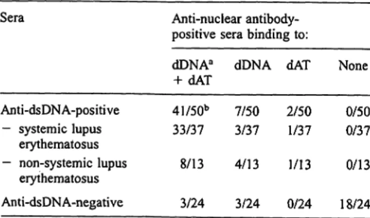
Although the majority of 50 selected anti-dsDNA-posi- tive sera (37 from systemic lupus erythematosus and 13 from non-systemic lupus erythematosus patients) bound to dDNA and dAT, 7 of 50 (14%) did not bind to dAT, whereas 2 of 50 (4%) did not bind to dDNA (tab. 3).
Anti-dsDNA-positive systemic lupus erythematosus sera (n = 37) showed a similar pattern with 8.1% and 2.7%
of sera lacking the reactivity towards dAT and to dDNA, respectively. In contrast, the majority (18/24 or 75%) of anti-dsDNA-negative sera from various diseases were negative for all three antigens: 3/24 (12.5% bound to dDNA only (1 systemic lupus erythematosus and 2 rheu- matoid arthritis patients), whereas 3/24 (12.5%) of non- systemic lupus erythematosus sera bound to dDNA and to dAT (tab. 3).
Tab. 2 Comparison of reactivity of 50 anti-dsDNA-positive sera to dDNA, dsDNA and dAT in an ELISA assay
Antigens Serum reactivity to DNA antigens3 Correlation
dDNA vs dAT dDNA vs dsDNA dsDNA vs dAT
r 0.89 0.91 0.93
P 0.001 0.001 0.001
Fraction bound0 (χ ± S. D.)
1.79 ±0.42 1.80 ±0.58 1.79 ±0.42 2.03 ± 0.55 2.03 ± 0.55 1.80 ±0.58
Difference0
P NS
< 0.005
< 0.001
a dsDNA, double stranded DNA; dDNA, denatured DNA; dAT, poly[dA-dT] double stranded alternating copolymer.
b In relation to 3 S. D. above the mean of normal human sera.
c Paired T-test.
Tab. 3 Pattern of anti-nuclear antibody-positive sera binding to DNA antigens
Sera Anti-nuclear antibody-
positive sera binding to:
Anti-dsDNA-positive - systemic lupus
erythematosus
— non-systemic lupus erythematosus Anti-dsDNA-negative
dDNAa
+ dAT 41/50b 33/37
8/13 3/24
dDNA 7/50 3/37 4/13 3/24
dAT
2/50 1/37 1/13 0/24
None 0/50 0/37 0/13 18/24
a dsDNA, double stranded DNA; dDNA, denatured DNA; dAT, poly[dA-dT] double stranded alternating copolymer.
b No. of positives/No, tested.
Discussion
By using an ELISA format for the anti-dsDNA anti- bodies based on Sl-nuclease-treated antigen (15), we were able to confirm the literature data on the reactiv- ity of systemic lupus erythematosus and non-systemic lupus erythematosus sera to dsDN/y (1-4). The speci- ficity of the assay in a group of anti-nuclear antibody- positive patients was 82%, and after including anti- nuclear antibody-negative control sera the overall spec- ificity was 91%. The assay was positive in 48 of 71 patients with both active and inactive systemic lupus erythematosus (including three patients with systemic lupus erythematosus complicated with rheumatoid ar- thritis and/or vasculitis), reaching a sensitivity of 66%, which is comparable to that reported in previous studies (1—4, 15). As observed previously (4, 15), a rather high proportion of non-systemic lupus erythe- matosus sera (27.6%) also gave a positive reaction to dsDNA. Although non-systemic lupus erythematosus showed weaker binding to dsDNA, the difference was not significant.
The question, however, arises as to whether this non-
systemic lupus erythematosus dsDNA binding repre-
sents a false result due to the presence of single strands
in our dsDNA preparation. Although we cannot rule out
this possibility, it is interesting to note that a positive
dsDNA finding in systemic lupus erythematosus and
non-systemic lupus erythematosus patients was paral-
leled by dAT binding, an antigen that is apparently free
of ssDNA strands (10). In addition, 3 of 24 anti-dsDNA-
negative sera from various non-systemic lupus erythe-
matosus immune-related diseases bound dDNA and
dAT, a finding that strongly argues against the presence
of single strands in our dsDNA preparation. Radio-
immunoassay showed a strong correlation between the
binding of sera to ss-nuclease-treated natural DNA and
to dAT (10). This correlation disappeared when ss-
nuclease-untreated natural DNA was used, indicating a
contamination of native, nuclease-untreated DNA with
ssDNA (10). It was also hypothesised that nuclease-
treated dsDNA and dAT displayed antigenic similarity
which resulted in high correlation of binding of anti-
dsDNA-positive sera to both antigens. On the basis of
these experiments it was concluded that synthetic anti-
gens such as dAT might offer practical advantages over
natural DNA preparations in detecting anti-dsDNA,
since it contained most of the native DNA specificity
(10). However, others have presented evidence that sys-
temic lupus erythematosus sera contain a mixture of an-
tibodies against different DNA epitopes, one population
binding solely to dsDNA and another to dAT (reviewed
in 1. c. (11)). Our results, obtained in an ELISA solid-
phase, assay, corroborate the finding of high correlation
between the binding of anti-dsDNA-positive sera to
dAT, but stress again that the binding was not 100%
Batinic et al.: ANA+ sera binding to DNA antigens in an ELISA
347 complementary. In support of this view and in contrast
to the findings of Steinman (10), we actually observed dAT binding in a proportion (16.7%) of non-systemic lupus erythematosus ssDNA-positive sera from patients with rheumatoid arthritis and "mixed collagenosis". This observation farther corroborates the heterogeneous pattern of individual anti-DNA specificity, as observed previously (10—11). It should be noted that the differ- ences observed might be associated with disease activ- ity, since dAT binding was mainly observed in patients with active, but not with inactive nephritis (10). Our
current work is aimed at the comparison of binding of isolated anti-dsDNA antibodies from patients with sys- temic lupus erythematosus and non-systemic lupus ery- thematosus patients to dAT in the context of disease ac- tivity.
Acknowledgements
We thank Dr. Zeljko Bosnjak from the Laboratory of Cellular Bio- logy, Medical College of Wisconsin, USA, for his continuous sup- port in the form of chemicals and literature, and Mrs. Nives Radio for excellent technical assistance.
References
1. Stollar BD. Immunochemistry of DNA. Int Rev Immunol 1989; 5:1-22.
2. Stollar BD. Anti-DNA antibodies. Clin Immunol Allergy 1981;
1:243-60.
3. Eaton BR, Schneider G, Schur PH. Enzyme immunoassay for antibodies to native DNA. Specificity and quality of antibod- ies. Arthritis Rheum 1983; 26:52-61.
4. Tan EM. Antinuclear antibodies: diagnostic markers for auto- immune diseases and probes for cell biology. Adv Immunol
1989; 44:93-151.
5. Steinberg AD, Krieg AM, Gourley MF, Klinman DM. Theoret- ical and experimental approaches to generalized autoimmunity.
Immunol Rev 1990; 118:129-63.
6. Buskila D, Shoenfeld Y. Anti-DNA antibodies. In: Lahita RG, editor. Systemic lupus erythematosus. New York: Churchill Livingstone 1992:205-26.
7. Staines NA. Autoantibodies against DNA. In: Panayi GS, edi- tor. Immunology of connective tissue diseases. Dordrecht:
Kluwer Academic Publishers 1994:257-78.
8. Morgan A, Buchanan RRC, Lew AM, Olsen I, Staines NA.
Five groups of antigenic determinants on DNA identified by monoclonal antibodies from (NZB X NZW)Ft and MRL/lpr/
/prmice. Immunology 1985;55:75-83.
9. Smeenk R, Hylkema M. Detection of antibodies to DNA: a technical assessment. Mol Biol Rep 1992; 17:71-9.
10. Steinman RC, Deesomchok U, Spiera H. Detection of anti- DNA antibody using synthetic antigens. Characterization and
clinical significance of binding of poly(deoxyadenylate-deoxy- thymidylate) by serum. J Clin Invest 1976; 57:1330-41.
11. Tan EM. Autoantibodies to nuclear antigens (ANA): their im- munobiology and medicine. Adv Immunol 1982; 33:167-240.
12. Tan EM, Cohen AS, Fries JF, Masi AT, McShane DJ, Rothfield NF, et al. The 1982. revised criteria for the classification of systemic lupus erythematosus. Arthritis Rheum 1982;
25:1271-7.
13. Arnett FC, Edworthy SM, Bloch DA, McShane DJ, Fries JF, Cooper NS, et al. The American Rheumatism Association 1987: Revised criteria for the classification of rheumatoid ar- thritis. Arthritis Rheum 1987; 31:315-24.
14. Fox RI, Robinson CA, Curd JG, Kozin F, Howell FV. Sjögren's syndrome: proposed criteria for classification. Arthritis Rheum 1986; 29:577-85.
15. Rubin RL. Enzyme-linked immunosorbent assay for anti-DNA and anti-histone antibodies. In: Rose NR, Friedman H, Fahey JL, editors. Manual of clinical laboratory immunology. 3rd ed.
Washington D. C.: American Society for Microbiology;
1986:744-9.
Received January 23'/December 6, 1995
Corresponding author: Dr. Drago Batinic, Division of Immunology, Clinical Department of Laboratory Diagnosis, Clinical Hospital Center Zagreb, Kispaticeva 12,
HR-10000 Zagreb, Croatia