INTRODUCTION
A functional photosynthetic apparatus and intact DNA are essential for the growth of plants. Although phototrophs depend on light, the 2 very important processes photosynthesis and DNA expression are sensitive to damage by sunlight (Peak & Peak 1983, Teramura & Sullivan 1994, Jansen & Bornman 2012, Vass 2012). Photodamage to DNA is mainly generated by ultraviolet-B (UVB, 280−315 nm) radiation (Cadet et al. 2012), creating covalent bonds between 2 neigh- boring cytosine or thymine bases. The majority of re- sulting dimers are cyclobutane-pyrimidine dimers (CPDs) (Setlow & Carrier 1966, Mitchell & Nairn 1989,
Douki et al. 2000). A single dimer may completely stop the progression of DNA and RNA polymerases and thereby inhibit replication and gene expression (Sauerbier et al. 1970). However, photoreactivation can efficiently repair such UVB-induced DNA lesions.
This photocatalytic splitting of the dimers is empow- ered by blue light and UVA radiation carried out by a specific enzyme, the photolyase (Britt 1996, Sancar 2003). This means that UVB-induced damage and UVA-driven repair occur simultaneously in sunlight, demonstrating that it is very important to evaluate the influence of solar radiation on the DNA integrity of an organism under the polychromatic composition of sunlight (Bray & West 2005, Caldwell et al. 2007).
© Inter-Research 2016 · www.int-res.com
*Corresponding author: fpescheck@bot.uni-kiel.de
Relative sensitivity of DNA and photosystem II in Ulva intestinalis (Chlorophyta) under natural
solar irradiation
Frauke Pescheck*, Hanna Campen, Lars Nichelmann, Wolfgang Bilger
Botanical Institute, Christian-Albrechts-University Kiel, Olshausenstr. 40, 24098 Kiel, Germany
ABSTRACT: High intensities of sunlight can result in DNA and photosystem II (PSII) damage.
However, the relative sensitivity of both these targets under natural sunlight and especially over a long period has not been studied in algae so far. Although DNA damage is highly specifically induced by ultraviolet-B radiation (UVB, 280−315 nm), PSII is inactivated by a broad spectrum.
The green macroalga Ulva intestinalis is an appropriate and interesting study organism with which to investigate the relative importance of the 2 different targets of sunlight because this alga contains no UV-screening protective pigments, although it is exposed to strong solar irradiation in its natural habitat. This entails a high risk of DNA damage. Therefore, diel time courses and long-term development of DNA damage and the optimal quantum yield of PSII (Fv/Fm) were studied in situ. Fv/Fm was extremely reduced at noon, but a fast recovery was observed in the afternoon. As dark-adapted basal fluorescence (Fo) of PSII was substantially decreased during the day, non-photochemical quenching is suggested to be a key photoprotective strategy in U. intesti- nalis. In contrast, even in samples with strongly reduced Fv/Fm, only very low DNA damage was found, irrespective of the accumulated UVB dose. We propose that efficient photoreactivation driven by natural sunlight balances the induction of dimers. This leads to a higher UVB tolerance of DNA than that observed in algae under experimental UVB irradition. In this field study, U.
intestinalissuffered more from photoinhibition than from DNA damage.
KEY WORDS: Diel cycle · DNA damage · Intertidal zone · Long-term data · Photosynthetic efficiency · Ultraviolet radiation · Sunlight · Ulva intestinalis
Resale or republication not permitted without written consent of the publisher
The action spectrum for inhibition of photosynthesis peaks in the UV region but extends far into the visible range (Takahashi et al. 2010). Destruction of the water splitting apparatus of photosystem II (PSII) can be one of the starting points of photoinactivation (Vass et al. 1996, Hakala et al. 2005). Damage to this protein complex can be explained by the direct ab - sorption of UVB and UVA radiation (Ohnishi et al.
2005). In the absence of water splitting, absorption of photosynthetically active radiation (400−700 nm, PAR) by photosynthetic pigments leads to the accu- mulation of positively charged P680+radicals and the subsequent oxidation of components of PSII. In addi- tion, the high photon flux densities (PFD) of sunlight can also lead to excessive and therefore potentially harmful irradiation as soon as an imbalance of light and dark reactions of photosynthesis set in. In this sit- uation, an uncontrolled transfer of excited electrons to oxygen might occur. It has been shown conclu - sively that UVB and UVA radiation as well as PAR can induce the formation of reactive oxygen species (ROS) in chloroplasts (Snyry chova et al. 2007). One of the central proteins of PSII, the D1 protein, and espe- cially its turnover are the major targets of these radi- cals (Jegerschöld et al. 1990, Nishiyama et al. 2001).
Thus, although sunlight-induced inactivation of PSII cannot easily be ascribed to a specific proportion of the solar spectrum, it may be quite a sensitive indica- tor of the presence of light stress.
UV and high PAR tolerance of PSII is mainly attrib- uted to an active repair cycle of PSII (Nishiyama et al.
2001, Allakhverdiev & Murata 2004). The function of this repair cycle can be protected by antioxidative substances and enzymes in the chloroplast against an inhibition by ROS (Nishiyama et al. 2001, Hakala- Yatkin et al. 2011). Similarly, non-photochemical quenching (NPQ) lowers the production of ROS by quenching of absorbed energy via efficient heat dis- sipation (Müller et al. 2001, Jahns & Holzwarth 2012).
This way the quantum yield of PSII is reduced as in photoinactivation, but in a controlled and regulated photoprotective manner (Osmond et al. 1993).
Besides the acute effects of high irradiances and UVB radiation, on longer time scales, other aspects, such as acclimation and accumulation of damage as a result of the balance between induction and repair, have to be considered (Jansen et al. 1998, Allakh - verdiev & Murata 2004). Long-term monitoring under natural solar irradiation is necessary to reveal the ecological relevance of light-induced damage. Up to now, only a few long-term studies of photosynthetic efficiency in plants or algae have been conducted in the field (Valladares & Pearcy 1997, Altamirano et al.
2000, Bischof et al. 2002, Michler et al. 2002). As re- gards DNA damage, long-term field observations are much rarer still, as well as sometimes being contra- dictory. Whereas a positive correlation of increased DNA damage with increased UVB irradiances due to ozone variations was ob served in a herbaceous plant, Gunnera magellanica, in south Argentina, and in 3 Antarctic mosses (Rousseaux et al. 1999, Turnbull &
Robinson 2009), no long-term accumulation of CPDs was found in Arctic tundra plants even under supple- mental UVB conditions (Rozema et al. 2006). In pi- coplankton from mid-latitudes, an increase in DNA damage after noon was reported (Buma et al. 2001).
However, in that study, DNA damage was not fol- lowed over complete diel courses or longer periods.
These results demonstrate the lack of knowledge about long-term effects of UVB on DNA in photosyn- thetic organisms and especially in macroalgae.
The organism of the present study, Ulva intestinalis L., is one of the most dominant green macroalgal spe- cies in the intertidal zone (Sousa 1979, Nelson et al.
2003, Wang et al. 2012) and the genus Ulva has gained a lot of interest as a model organism (Wichard et al. 2015). Ulva intestinalisestablishes dense popu- lations in the upper eulittoral zone, where emer- gence and concomitant exposure to unattenuated solar radiation regularly occur. In addition, be cause of its tubular morphology (Koeman & van den Hoek 1982), the gas-filled thalli often float on the water sur- face so that times of emergence can be extended. In this habitat, resistance to strong solar irradiance is obviously a prerequisite for survival. From short- term stress experiments, Ulva is well known to be highly tolerant to large variations in salinity (Reed &
Russell 1979) and temperature (Henley et al. 1992).
Whereas in these short-term experiments Ulva spe- cies also proved to be very tolerant towards light stress (Franklin et al. 1992, Henley et al. 1992, Figueroa et al. 2003, Gómez et al. 2004), in another study U. aff. rotundata showed strong UVB- enhanced bleaching after exposure to full sunlight for 3 d (Bischof et al. 2002). Furthermore, investiga- tions of UVB-induced DNA damage in macroalgae are scarce and most of these results have been gained from short-term laboratory studies (van de Poll et al. 2001, Roleda et al. 2006, Pescheck et al.
2014). The analysis of DNA damage in U. intestinalis is even more interesting as this species lacks UVB protective pigments, which are a major defense against UVB-induced damage in higher plants (Bornman et al. 1997). Hence, it seems important to investigate the high light tolerance of Ulva under long-term solar irradiation and to search for sensitive
structures. Therefore, we studied the effect of natural sunlight on CPD formation in the DNA, on short-term as well as on long-term scales in U. intestinalis in comparison to photochemical efficiency of PSII. We hypothesized that U. intestinalis accumulates CPD depending on the solar UVB dose because of the lack of UVB protective pigments. We further hypothe- sized that the proportion of DNA and PSII damage under solar irradiation would be different from that observed in a laboratory study (Pescheck et al. 2014).
The additional spectral components in sunlight may either mitigate or exacerbate the effects of UVB on both photosensitive structures.
MATERIALS AND METHODS Field experiments
From May 2009 to September 2013, 6 short-term field studies with Ulva intestinalis were conducted (Table 1). The optimal quantum yield of PSII (Fv/Fm) and the concentration of CPDs in the DNA were ana- lyzed over diel time courses. Additionally, UVB screening was determined for a subsample of the analyzed populations. The short-term investigations took place at different locations at the shore of the Baltic Sea around Kiel, Germany (Table 1). On the beaches of Meierwik, Dänisch-Nienhof (abbreviated as D.Nienhof) and Surendorf, dense populations of U. intestinalisgrow on small boulders from April to November. A long-term study site was established close to one of the locks on the Kiel Canal in Kiel Holtenau (hereafter referred to as the Lock, where by
Locks I, II, III refer to 3 different measuring days). An experimental setup with open boxes was installed in which the algae could grow and be observed for sev- eral weeks under natural light, temperature and salinity con ditions. For this experiment, stones (diam- eter ca. 15 cm) densely overgrown with thalli of U.
intestinalis were collected in Surendorf and trans- ferred to the Lock in buckets filled with seawater.
There, the stones with the algae were placed in float- ing plastic boxes (SN 106601, 600 × 400 × 400 mm, Schoeller Allibert) with a 1 × 1 cm grid of holes on all sides to allow water exchange with the fjord. Inside the boxes, the stones were kept in position by holes that were cut into a custom-made PVC floor. The height of this floor was adjusted so that most of the algae reached the water surface and floated horizon- tally as a result of the gas-filled thalli segments. The boxes were kept buoyant with the help of 2 con- nected air-filled plastic pipes (1 m × 15 cm). Four boxes were tied to the north side of 1 fender in the entrance to 1 of the 4 lock chambers on the fjord side.
This experiment was repeated with freshly collected algae (Expt A from 28 June to 24 July 2013, Expt B from 25 July to 16 August 2013). During both runs, the boxes were repeatedly cleaned of epibionts (e. g.
barnacles, diatoms, green microalgae) with a scrub- bing brush. Sometimes epiphytes accumulated on U.
intestinalisand were removed by carefully washing the algae in the open water at night. The boxes floated in a water area that was enclosed by fenders such that the water surface was very calm. The resulting stable exposure of the upper layer of thalli rarely occurs under natural conditions, but was also observed during the measurements in
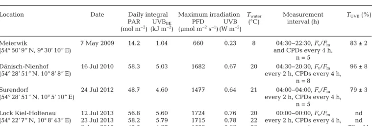
Location Date Daily integral Maximum irradiation Twater Measurement TUVB(%)
PAR UVBBE PFD UVB (°C) interval (h)
(mol m−2) (kJ m−2) (µmol m−2s−1) (W m−2)
Meierwik 7 May 2009 14.2 1.04 660 0.23 8 04:30−22:30, Fv/Fm 83 ± 2
(54° 50’ 9” N, 9° 30’ 10” E) and CPDs every 4 h,
n = 5
Dänisch-Nienhof 16 Jul 2010 58.3 5.03 1682 0.67 20 04:30−20:30, Fv/Fm 96 ± 8
(54° 28’ 51” N, 10° 8’ 8” E) every 2 h, CPDs every 4 h,
n = 8
Surendorf 24 Jul 2012 48.7 4.60 1477 0.64 21 04:00−04:00, Fv/Fm 79 ± 3
(54° 28’ 51” N, 10° 5’ 10” E) every 2 h, CPDs every 4 h,
n = 5
Lock Kiel-Holtenau 12 Jul 2013 56.8 5.60 1724 0.76 20 00:00−00:00, Fv/Fm nd (54° 22’ 7” N, 10° 8’ 43” E) 23 Jul 2013 58.2 5.79 1715 0.78 22 every 2 h, CPDs every 4 h, nd
6 Aug 2013 42.5 4.37 1592 0.68 20 n = 4 79 ± 11
Table 1. Overview of the daily courses with location, date, daily accumulated photosynthetically active radiation (PAR) and biologi- cally effective UVBBE(280−315 nm) dose (weighted after Ghetti et al. 1999), maximal photon flux density (PFD) and UVB irradiance, mean water temperature (Twater), measurement interval and number of replicates for optimal quantum yield (Fv/Fm) and DNA damage
(cyclobutane-pyrimidine dimers, CPDs), and UVB transmittance (TUVB, ± SD) of macrothalli. nd: no data
Surendorf when exceptionally calm wea ther con - ditions prevailed.
Environmental data
In Meierwik and at the Lock in Holtenau, a data log- ger (CR 1000, Campbell Scientific) was installed that recorded several environmental parameters. The wa- ter temperature was measured with a resistance ther- mometer PT100 in Meierwik or with a temperature probe (107-L, Campbell Scientific) 3 cm be low the water surface at the Lock. Approximately 2 m above the water surface, UVB and visible radiation were re - gistered with a UVB-sensitive photodiode (in Meier- wik: GUVB-T10GD, Roithner Lasertechnik; at the Lock: Tocon E + diffusor, sglux SolGel Technologies) and a photodiode (G1118, Hamamatsu Photo nics), respectively. The diodes were incorporated into custom-made housings constructed for cosine-cor- rected measurements. During the field measurements in D.Nienhof and Surendorf, the UVB radiation was measured with a hand-held custom-made UVB sensor (Veit et al. 1996) connected to a voltmeter approxi- mately 1.5 m above the water surface. Photon flux density (PFD, 400−700 nm) was measured using a LI- COR quantum sensor (LI-190), and water temperature with a standard thermometer. All radiation sensors were calibrated against a double monochromator spectroradiometer (DM 150, Bent ham Instruments) and the UVB signals were converted into W m−2UVB irradiance. To calculate the biologically effective UVB (UVBBE), the UVB irradiance was further weighted with the function of Ghetti et al. (1999) using a corre- lation factor for unweighted to weighted irradiance from a solar spectrum recorded in April 2009 in Kiel with the same spectroradiometer.
Measurements of chlorophyll fluorescence For all measurements, the upper 4 to 6 cm of green thalli floating on the water were collected. The up per 2 cm of these pieces were immediately placed in 2 ml reaction tubes and frozen in liquid nitrogen for sub- sequent DNA extraction. The remains of the same thalli were used for the determination of the optimal quantum yield of PSII so that it was possible to pair the Fm/Fmand CPD data from each individual thallus.
The thallus pieces were mounted in leaf clips (Walz) and kept in a water bath at ambient water tempera- ture for dark adaptation for 20 min (in Surendorf 15 min). Fo and Fm levels were then determined
using a Mini-PAM fluorometer (Walz) in Meierwik, D. Nienhof and Surendorf or a PAM-2100 fluorome- ter (Walz) at the Lock. In Surendorf and at the Lock after the first saturating pulse, the thalli were kept in darkness in the leaf clips and the measurement of Fo and Fmwas repeated after 1 h dark adaptation. After- wards, the fluorescence signal was recorded for each leaf clip with a blue fluorescence standard (Walz).
This allowed us to correct the ab solute fluorescence values for optical differences among the leaf clips and the temperature dependence of the instruments.
DNA damage detection
The frozen samples of the upper 2 cm of the thalli were transported to the laboratory in liquid nitrogen and stored at −85°C until analysis. All procedures for the detection of CPDs per megabase (Mb–1) by immuno-dot blotting followed the method described in Pescheck et al. (2014). The reagent for chemilumi- nescence was Pierce ECL (Thermo Fisher Scientific).
Experimentally-induced CPD formation and reduction of PSII efficiency
The intrinsic sensitivity of DNA and PSII in U.
intestinalis towards UVB radiation was determined by laboratory dose-dependency experiments. Field- collected algae were cleaned and cultured in Petri dishes with 40 ml nutrient-enriched half-marine sterile filtered seawater. The medium was changed every 2−3 d. The algae were kept in a temperature- controlled climate cabinet (Rumed, Rubarth Appa- rate) at 9°C, and illumination was from 3 fluorescence tubes (General Electrics) delivering approximately 10−12 µmol photons m−2s−1PAR for 16 h followed by 8 h darkness. This pre-experimental culture was maintained for 10−19 d after sampling to ascertain that no UVB-induced damages originating from the field were present in the algae anymore. Five UVB doses were applied by exposure for 1 h to different fluence rates (0−5 W m−2) of UVB irradiance emitted by UVB fluorescence tubes (TL40/12RS, Philips).
During the exposure, 2 open Petri dishes per UVB intensity were covered by WG295 glass filters (Schott) to exclude wavelengths under 295 nm. Two Petri dishes with control samples were kept under a UV-blocking plexiglass filter (GS 321, Röhm). From each dish, 4 subsamples were frozen for DNA dam- age detection immediately after UVB exposure.
Afterwards, the dishes were dark incubated for
20 min and Fv/Fm was determined with an Imaging PAM chlorophyll fluorometer (Walz).
UVB screening
From the analyzed populations, some thalli were brought to the laboratory directly after the last meas- urement in the field and used for screening measure- ments within a maximum of 3 h after sampling. The thalli were transported in 50 ml tubes filled with water from the sampling site. Basal fluorescence excited with blue-green light (420−550 nm) and UVB radiation (λmax = 314 nm, 18 nm half bandwidth) was deter- mined with a Xenon PAM fluoro meter (Walz) as de- scribed in Pescheck et al. (2010). The excitation beam had a pulse rate of 4 Hz. Measuring was applied for 30 s for each excitation wavelength. Apparent UVB transmittance was calculated as defined in Pescheck et al. (2010).
Data analysis
Statistics were calculated using SigmaPlot version 11.0 (Systat Software) or GraphPad 5 (GraphPad Software).
The diel cycles of Fv/Fmand CPDs Mb−1 were analyzed by one-way repeated measures ANOVA, and if a significant difference was detected the groups were separated with a Tukey post hoc comparison. The data for PSII efficiency from Surendorf and Lock III were not normally distributed and thus were ana- lyzed using Friedman repeated meas- ures ANOVA on ranks. In the long-term study, linear regression analysis was
performed of Fv/Fm and ac cumulated CPDs Mb−1. Additionally, linear regressions were calculated against the daily dose of PFD and UVBBE(both inte- grated until the time point of measurement: 13:00 h).
From the light measurements recorded with the data logger, mean values of every 15 min were calculated and integrated over 24 h for the accumulated doses.
RESULTS Environmental data
Environmental conditions at all sites were gener- ally similar except for 7 May 2009 in Meierwik and
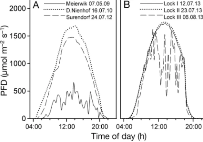
6 August 2013 at the Lock (Table 1). Although on 7 May 2009 and 6 August 2013 the sky was overcast, the rest of the days were clear and the irradiance courses resembled sinusoidal curves (Fig. 1). The maximal irradiances of 660−1700 µmol photons m−2 s−1were observed be tween 13:15 and 14:00 h (CEST) (Table 1).
Short-term observations
On all days, characteristic diel variations of the optimal quantum yield of PSII following irradiance were found (Fig. 2). The highest values of Fv/Fmwere always at night (Meierwik), pre-dawn (D.Nien hof) or Fig. 1. Diel courses of horizontal photon flux density (PFD) at (A) the 3 beach locations and (B) the Lock in Kiel-Holtenau on 3 measuring days (Lock I, Lock II and Lock III). PFD was measured automatically as 15 min means in Meierwik and at the Lock. In D.Nienhof and Surendorf, PFD was measured
approximately every 30 min
Fig. 2. Diel courses of optimal quantum yield of photosystem II (Fv/Fm). (A) In situmeasurements on the 3 beaches; (B) in boxes at the Lock. Data are means ± 1 SD, n ≥4. Notice the different scaling of the time of day in
(A) and (B)
in the early morning (Surendorf, Locks I−III, re - peated measures ANOVA p < 0.05). During the after- noon, concomitant with the peak of irradiance, a sig- nificant reduction of Fv/Fm occurred between 12:00 and 16:00 h except for 7 May 2009 in Meierwik and 6 August 2013 at the Lock. Maximal reduction of Fv/Fm
was 55−77% (Table 2).
In D.Nienhof, PSII recovery started with a signifi- cant increase from 16:00 to 18:00 h. From 20:00 h onwards, the Fv/Fm values were identical to the morning values. Likewise, recovery was completed at 22:00 h in Surendorf and at Locks I and II. At that time, no statistically significant difference to the maximum Fv/Fmvalues of the early morning could be found. According to the low irradiance observed in
Meierwik (Fig. 1), Fv/Fm was not significantly re - duced throughout the day at this site, except for the late-afternoon value of the previous day (repeated measures ANOVA, p < 0.05; Fig. 2A). On Day 3 at the Lock, the weather was cloudier, which is mirrored in the time course in a more variable Fv/Fm (Fig. 2B).
Fv/Fm and the instantaneous photon flux density had a relatively constant relationship during the 6 diel courses (Fig. 3). One can observe a distinct ab - sence of a hysteresis, indicating that the reduction of Fv/Fmduring the day may not have been caused by long-lasting damage but by fast reversible downreg- ulation of PSII.
Location Date Max. reduction Max. CPD of Fv/Fm(%) Mb−1 Meierwik 7 May 2009 8 (13:00)ns 44 ± 24 (04:00)ns D.Nienhof 16 Jul 2010 63 (13:00) 44 ± 38 (13:00)ns Surendorf 24 Jul 2012 56 (16:00) 6 ± 2 (12:00)ns Lock I 12 Jul 2013 66 (12:00) 40 ± 35 (04:00)ns Lock II 23 Jul 2013 77 (12:00) 49 ± 14 (14:00)ns Lock III 6 Aug 2013 55 (10:00) nd
Table 2. Maximum reduction of optimal quantum yield (Fv/Fm) and maximum concentration (means ± SD) of cyclo - butane-pyri midine dimers per megabase (CPD Mb−1) during diel courses. The time of day of the respective observation is given in pa ren theses (h). ns: not significantly different from all other day times (repeated measures ANOVA);
nd: not determined
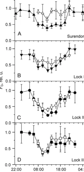
Fig. 4. Normalized basal fluorescence (Fo) val- ues after 15 or 20 min (open symbols) and 60 min (closed symbols) darkness during the diel courses in (A) Surendorf and (B–D) at the Lock in Kiel-Holtenau. Relative units of Fowere calculated from the raw data of Fodivided by the signal of a blue fluorescence standard, and were normalized to the maximal value during each diel course. Data are means ± SD, n ≥4 Fig. 3. Relationship of optimal quantum yield of photosystem II (Fv/Fm)
measured after 15 or 20 min dark incubation and incident photon flux density (PFD) during the daily courses from (A) the in situmeasurements on the 3 beaches and (B) the boxes at the Lock. Solid lines connect values from increasing PFDs; dotted lines connect values from decreasing PFDs. Data are from Figs. 1 and 2. Fv/Fmdata are means ± 1 SD, n ≥4
In Surendorf and at the Lock, the diel variation of basal fluorescence (Fo) was determined after 15 or 20 min and 60 min of dark incubation (Fig. 4). Despite a considerable scatter of the data, a strong decrease of up to 76% of the normalized Fosignal was apparent during the 4 d. A substantial quenching of Fowas also observed after 1 h dark incubation at the Lock, but not at Surendorf, indicating a faster reversibility at the lat- ter location. One explanation for these results may be that before the in situmeasurements at Surendorf, the
algae had been high light stressed for only 1 d, whereas inside the boxes at the Lock they had been exposed for 2 wk.
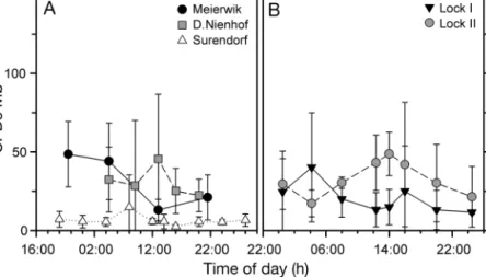
The concentration of DNA damage did not show a distinct diel pattern in any of the experiments (Fig. 5). No statistically significant differences were detected be - tween times of day (1-way re peated measures ANOVA, p > 0.05; Table 2).
There was also no relationship of CPDs Mb−1with accumulated UVB dose in the field (p = 0.61; Fig. 6). For comparison, CPDs in duced in Ulva in testinalis under experimental exposure conditions are shown (Fig. 6). The y-intercept at 36 CPDs Mb−1of the re gres sion line of these data was higher than the mean of all field- measured CPD concentrations (28 CPDs Mb−1). In the laboratory, the same dose response was observed when the dura- tion, rather than the intensity, of the UVB exposure was varied instead of the intensity (data not shown).
This re vea led time−dose reciprocity in CPD forma- tion under experimental UVB exposure.
Relative sensitivity of DNA and PSII
In situ CPD formation did not increase with de - creasing PSII quantum yield determined with paral- Fig. 5. Concentrations of cyclobutane-pyrimidine dimers per megabase
(CPDs Mb−1) during the diel courses in U. intestinalisfrom (A) the in situ measurements at the 3 beach locations and (B) the boxes at the Lock. Data are means ± SD, n ≥4. Notice the different scaling of the time of day
in (A) and (B)
Fig. 6. Concentration of cyclobutane-pyrimidine dimers per megabase (CPDs Mb−1) in Ulva intestinalisin relation to ac- cumulated (Acc.) UVB dose. Field CPD data are from Fig. 5.
For comparison, the induction of CPDs Mb−1in U. intesti- nalis exposed to experimental (Exp.) UVB is given (dashed line, r2= 0.83). Laboratory data are mean values of n = 8 from
6 independent experiments
Fig. 7. Relationship between cyclobutane-pyrimidine di mers per megabase (CPDs Mb−1) and optimal quantum yield (Fv/Fm) determined in parallel samples in Ulva intes tinalis.
Data points are from diel courses shown in Figs. 2 and 5 or from experimental (Exp.) exposure to UVB irradiation alone in the laboratory. The regression coefficient of laboratory
results is r2= 0.82
lel samples (Fig. 7). It is clearly demonstrated that the 2 targets do not respond similarly, Fv/Fm showing a much higher sensitivity towards sunlight than DNA integrity. In contrast, under experimental UVB expo- sure, a tight correlation of DNA lesions and optimal quantum yield was found in U. intestinalis (Fig. 7, open circles).
Long-term study
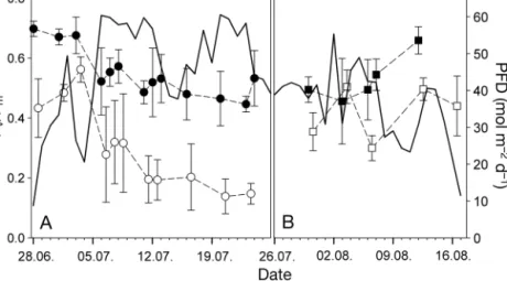
At the Lock, the boxes were exposed for 4 wk in July and the experiment was repeated with fresh al- gae for 3 wk in August. The optimal quantum yield during these periods varied strongly (Fig. 8). In the first week of July, daily sums of PFD remained around 30 to 40 mol m−2 and Fv/Fm at midnight and noon stayed high (Fig. 8A). On 6 July, the daily PFD doses suddenly increased due to fine weather. During this period of high irradiance, the night and day maximum quantum yields clearly dropped. No trend of Fv/Fmat night was found over the time span of low or high irra- diance (linear regression, p = 0.4165 and p = 0.1668, data not shown). However, the absolute levels of Fv/Fmwere significantly lower during the high light period, as evidenced by different y-intercepts of the regression lines. The overall minimum Fv/Fmof 0.14 was observed on 20 July at 12:00 h, coincident with the highest daily PFD dose. During the second run of the experiment, the variation of Fv/Fmwith the daily PFD doses was similar (Fig. 8B). Here, linear regres- sion analysis identified a significant dependence of the Fv/Fm noon level on the accumulated PFD dose (integrating PFD values before 13:00 h), with an ad-
justed R2of 0.611 for the first run of the long-term experiment (regression not shown). In the second run, no significant re gression was de tectable, probably due to fewer observations. However, in a combined data set of both runs, all val- ues showed the same dependency and the regression was highly significant (R2adj= 0.685, not shown).
CPD concentrations were not corre- lated with the concomitant daily UVBBE doses during the first run of the long- term study (p = 0.071, R2adj= 0.243, re- gression not shown; Fig. 9). In strong contrast to PSII activity, no differences of DNA damage between day and night were observed. During the first week the concentrations of CPDs were barely above the detection limit. Even on 6 July, when the daily UVB dose doubled, no strong in- crease in CPDs Mb−1occurred. DNA damage was not determined during the second run of the experiment.
DISCUSSION
Accumulation of CPDs under strong sunlight was lacking
For the first time, the concentration of DNA dam- age and the course of the optimal quantum yield of PSII in an intertidal macroalga were simultaneously investigated in the field over a long period and with a partially high temporal resolution. Three diel courses Fig. 8. Long-term observation of daily sums of photon flux density (PFD; solid
line) and optimal quantum yield of photosystem II (Fv/Fm) in U. intestinalisat the Lock in 2013. (A) First run; (B) second run. Data are means ± SD, n = 4.
Closed symbols: midnight values; open symbols: noon values
Fig. 9. Long-term observation of daily sums of biologically effective UVB irradiance (UVBBE, solid line) and cyclobu- tane-pyrimidine dimers per megabase (CPDs Mb−1) in Ulva intestinalisfrom the first run at the Lock. Data are means ± SD, n = 4. Closed symbols: midnight values; open symbols:
noon values
were measured in situat the natural growth site of Ulva intestinalis. In a long-term study, we were able to keep the algae in open boxes in nearly undistur - bed natural conditions with respect to the main abio - tic parameters light, temperature, nutrient supply and salinity. The conditions in the boxes deviated from that naturally experienced by the algae only in one aspect. Inside the boxes, the water movement was strongly reduced, which inhibited mixing of the thalli, normally leading to shifting irradiation of a single thallus. However, U. intestinaliscan also natu- rally be found steadily floating on the water surface, as was the case during the measuring period at Surendorf. On that day the weather conditions were such that the water surface was very calm and the thalli were not mixed by waves over the whole day.
Thus, the light absorption of the thalli was extremely high on this day, as was the case during the entire long-term study.
Most of the time during the investigation period the sky was clear and daily sums of PAR and UVB ra - diation reached values well above 50 mol m−2d−1and 5 kJ m−2d−1, respectively. Unexpectedly, in spite of this strong irradiation, probably the maximum achievable in this habitat, we observed very low con- centrations of CPDs in U. intestinalis. There was no correlation of DNA damage with UVB radiation, nei- ther on diel nor on long-term scales (Figs. 5 & 9, Table 2). This indicates that U. intestinalis did not accumulate relevant concentrations of CPDs over our study period of more than 1 mo and points to a minor influence of UVB radiation on long-term DNA in - tegrity of U. intestinalisunder field conditions. Simi- larly low CPD concentrations were described in the green macrophyte Prasiola stipitata in Antarctica (Lud et al. 2001). Likewise, there was only a very weak correlation of CPDs and UVB dose under ambi- ent UVB conditions, which was stronger under artifi- cial UVB enhancement. However, and in contrast to U. intestinalis, the genus Prasiolais known to contain UV-absorbing mycosporine-like amino acids (Kars - ten et al. 2005). The UVB resistance of the DNA in U.
intestinalisis also quite remarkable if compared with that of higher plant leaves. For example, in maize, rice and Gunnera magellanica, significant levels of CPDs were induced depending on time of day or cumulative UVB dose, respectively, (Stapleton et al.
1997, Kang et al. 1998, Rousseaux et al. 1999). We also expected to occur in U. intestinalis, particularly because higher plants are usually well protected by screening compounds.
The obvious discrepancy between the lack of UVB- screening pigments and the absence of DNA damage
(Figs. 5 & 6) in such a high-light habitat raises the question of photoprotective mechanisms alternative to screening pigments. Such mechanisms might be especially efficient in terms of photoreactivation of DNA. However, photoreactivation is poorly investi- gated in green macroalgae and, so far, has been re - ported to be relatively inefficient compared with other macrophytes or higher plants (Takeuchi et al.
1996, van de Poll et al. 2002, Pescheck et al. 2014).
Nevertheless, there are 2 important considerations that let us assume that photoreactivation may have been very efficient in U. intestinalis under natural sunlight conditions. Firstly, photoreactivation rates are always higher in plants acclimated to natural sunlight compared with culture-grown individuals (Takayanagi et al. 1994, Kang et al. 1998). It was shown that the expression of the photolyase is upreg- ulated with a strong light dependence in Arabidopsis thaliana(Li et al. 2015). A similar acclimation poten- tial of photolyase expression might exist in U. intesti- nalisas a 10 times increased photoreactivation rate was observed in freshly collected compared with low-light acclimated thalli (F. Pescheck unpubl. re - sults). To our knowledge, investigations of the accli- mation of photolyase activity are lacking for macro- algae. Secondly, irradiation with sufficient UVA radiation or blue light concomitant with the UVB exposure is obligatory for the activity of photolyase in higher plants (Takeuchi et al. 1996, Dany et al. 2001).
In our experiments under artificial UVB exposure, only very low UVA radiation was present, whereas in sunlight the natural ratio of UVA:UVB existed. So far, regarding macroalgae, only for Palmaria palmatahas a reduced accumulation of CPDs under simultaneous photoreactivation light been shown (Pakker et al.
2000). Therefore, we assume that one reason for the non-significant levels of CPDs we measured under strong solar irradiation may have been photo - reactivation, which was highly efficient due to both induction of high photolyase activity and the pres- ence of sufficient radiation at the appropriate wave- lengths.
An additional mechanism contributing to the lack of DNA damage may be the shielding of the DNA by the chloroplasts. The concept of ‘chloroplast shield- ing’ was first described for arctic diatoms (Karentz et al. 1991). In each Ulva cell, one large cup-shaped chloroplast is located at the periphery of the cyto- plasm, which during illumination orientates towards the outer surface of the thallus and thus might shade the nucleus efficiently from radiation (Britz et al.
1976). In U. clathrata this effect is suggested to in - crease the UVB resistance of the DNA by 50% in
comparison to Rhizoclonium riparium, a multinucle- ated siphonocladal species with a net-like chloro- plast structure (Pescheck et al. 2014). One can spec- ulate that the maintenance of gene expression and DNA replication is of major importance in a growing organism or in reproductive tissues, making the sac- rifice of chloroplasts a reasonable strategy. In pho- tolyase-deficient mutants of Synechocystis, the loss of PSII activity was significantly increased and the recovery significantly delayed after UVB exposure (Vass et al. 2013). This means that prevention of nuclear DNA damage would also be beneficial for repair processes in photosynthesis.
Fast reversible variations of PSII efficiency indicate high potential for photoprotection
In contrast to the observed integrity of the DNA, a strong reduction of optimal quantum yield of PSII was found in situ(Figs. 2 & 8). However, it is not clear to what extent the observed changes in Fv/Fmwere due to photodamage or photoprotection. Both pro- cesses are a ‘function of PFD ..., duration of exposure, and spectral quality’ (Henley 1993) and occur simul- taneously during the day in Ulva rotundata(Henley et al. 1992). During the diel cycles (Fig. 2) and the long-term study (Fig. 8, open symbols), Fv/Fm was considerably reduced around noon, but a large por- tion of this reduction was reversible within a few hours (Figs. 2 & 3). This recovery had already begun while irradiances were still oversaturating (Fig. 2), which was also observed in the field study of Henley et al. (1992). The apparent absence of a pronounced hysteresis (Fig. 3) suggests either the presence of protective downregulation or a very rapid repair cycle (Allakhverdiev & Murata 2004). A fast repair capacity of PSII was found in a laboratory study of the closely related species U. clathrata, reaching 50% re - pair within 3 h of a strong UVB challenge (Pescheck et al. 2014). Therefore, it is possible that repair rates in U. intestinalis were able to compensate for the induction of protein damage.
However, recovery during the evenings was not fully complete until darkness set in (Figs. 2 & 8). In some diel time courses, a further increase in Fv/Fm was observed during the early morning hours when the sun rose and irradiation potentially enabled addi- tional recovery (Allakhverdiev & Murata 2004). The divergence of the optimal quantum yield at night from the empirical maximum (close to 0.8 in U. rotun- data[Henley et al. 1992], and 0.75 as the overall aver- age of U. intestinalisin the present study) may be
considered as chronic damage to PSII. In the long- term course of the midnight values a sudden de - crease in Fv/Fmwas observed upon the change from cloudy to bright weather conditions, but during the high light period no further significant drop was detected by a regression line analysis (Fig. 8, closed symbols). It appears that during the bright weather period a new, more or less stable balance between damage and repair was established. Because this balance was already achieved in the first days of high light stress, apparently no further acclimation took place. In another Ulva species, Ulva rigida, it took more than 7 d to reach constant growth under full sunlight after transfer of the thalli from their nat- ural site (Altamirano et al. 2000). Reduced growth in that study was accompanied by an increase in heat dissipation of PSII (NPQ), which rose over the study period of 20 d. Other species of Ulva, such as U. rotundata, also increase their dissipation capac- ity when acclimated to strong light (Osmond et al.
1993). Thus, the constantly reduced quantum yield we measured under long-term high-light conditions might have been caused by the development of a longer lasting NPQ, i.e. a sustained downregulation of PSII quantum efficiency.
Support for the presence of a high downregulation of PSII comes from the observation of a strong de - crease of the basal fluorescence (Fo) during the diel courses (Fig. 4). As described above, in Ulva, the chloroplasts do not move to the sides of the cells in high light but orient themselves parallel to the outer thallus surface. Hence, the lowering of Fois probably not due to chloroplast movements but must be ascribed to the development of a substantial NPQ of Fo(Franklin et al. 1992). The in situ Fo-quench during the exposure to sunlight may have been even stronger than we detected, as a significant dark period (of 15 or 20 min) was applied before fluores- cence was measured (Fig. 4, open symbols). At noon, Fowas decreased compared with the morning level even after 60 min dark adaptation (Fig. 4, closed symbols). Interestingly, the reversibility of the Fo- quench was much slower at the Lock than in Suren- dorf. While the algae in Surendorf received relatively little sunlight during the preceding week, the algae were strongly exposed for at least 1 wk at the Lock.
Hence, the different kinetics of Foquenching could be an indication of a long-term acclimation to high irradiance. Similarly, Henley et al. (1992) observed a clear Fo-quench in U. rotundata acclimated to full sunlight in contrast to algae acclimated to only 9% of sunlight. The quenching in U. intestinalis, which was much stronger than that usually observed in higher
plants, indicated an intense NPQ and might have been associated with zeaxanthin formation. It is known that in higher plants under light stress condi- tions, a higher amount of zeaxanthin is retained overnight in the chloroplasts to facilitate NPQ (Barker et al. 2002). Conclusively, photoprotective downregulation of PSII quantum yield seems as power ful in U. intestinalisas in U. rotundata(Henley et al. 1992). Notably, irrespective of the underlying mechanism that caused the quenching, the chloro- phyll fluorescence data in total show that a strong high light stress was acting on the sampled thalli.
Relative sensitivity of DNA integrity and PSII The parallel analysis of 2 major photosensitive com- ponents in the present study enabled us to investigate their relative sensitivity in situ (Fig. 7). Moreover, comparing these in situdata with laboratory results helps to evaluate the relative sensitivities of both tar- gets. In laboratory experiments, the same UVB dose generated from fluorescence tubes caused signifi- cantly more CPDs than in situ(Fig. 6). Our data sug- gest that, due to the mitigating effect of UVA ra - diation in sunlight, the balance of CPD induction and photo reactivation is shifted towards the latter under natural sunlight compared with the experimental UVB exposure. Clearly, exposure to natural sunlight is necessary to be able to judge the ecological impact of UVB on DNA integrity and PSII function. In contrast to DNA integrity, it seems that PSII function is not similarly favored by solar irradiation, especially in the long-term comparison where PSII function was lowered under high solar irradiances, whereas DNA integrity was unaffected. This means that both targets displayed different long-term responses.
Avoidance of long-term accumulation of DNA dam- age might be an important ecological advantage of U.
intestinalisto gain dominance in the intertidal. Cellu- lar tolerance processes as the repair cycle of PSII rely on the continuous availability of intact trans cription.
It was shown that unrepaired DNA damage can re- duce the UVB tolerance of a cyanobacterium (Vass et al. 2013). The observed different sensitivity of PSII and the DNA in Ulva is in agreement with results from dinoflagellates, where photoinhibition domi- nated over DNA damage under natural sunlight (Hel- bling et al. 2008). However, the observed reduction of PSII function may not be problematic for U. intesti- nalisin its natural high-light habitat. Under oversatu- rating irradiances, as were observed at noon, this reduction does not necessarily lower the maximal
photosynthesis rate and therefore would not inhibit growth (Henley 1993). To conclude, in contrast to ex- pectations based on laboratory studies, U. intestinalis proved to be a very high-light- tolerant species.
Acknowledgements. During the fieldwork, Nele Hein, Cayssa Staggen, Christine Bönniger, Hanna Jähmlich, Paul Wieners and Jens Hermann lent many helping hands. Dr.
Florian Weinberger gave useful advice for the conduct of the long-term experiment. Many thanks to Susanne Wolf for help with DNA extraction and blotting. Prof. H. Brendel- berger generously loaned us several life jackets. Prof. K.
Krupinska is thanked for the use of a photo laboratory. Sin- cere thanks are due to Mr. H. Koch of the Waterways and Shipping office in Kiel Holtenau for support and the gener- ous permission to access the lock area in Kiel Holtenau. The improvement of the manuscript by the comments of 2 ano - nymous reviewers is gratefully acknowledged.
LITERATURE CITED
Allakhverdiev SI, Murata N (2004) Environmental stress inhibits the synthesis de novoof proteins involved in the photodamage-repair cycle of photosystem II in Synecho - cystis sp. PCC 6803. Biochim Biophys Acta Bioenerg 1657: 23−32
Altamirano M, Flores-Moya A, Figueroa FL (2000) Long- term effects of natural sunlight under various ultraviolet radiation conditions on growth and photosynthesis of intertidal Ulva rigida(Chlorophyceae) cultivated in situ.
Bot Mar 43: 119−126
Barker DH, Adams WW, Demmig-Adams B, Logan BA, Ver- hoeven AS, Smith SD (2002) Nocturnally retained zea - xanthin does not remain engaged in a state primed for energy dissipation during the summer in two yucca spe- cies growing in the Mojave Desert. Plant Cell Environ 25:
95−103
Bischof K, Hanelt D, Aguilera J, Karsten U, Vogele B, Sawall T, Wiencke C (2002) Seasonal variation in ecophysio - logical patterns in macroalgae from an Arctic fjord. I.
Sensitivity of photosynthesis to ultraviolet radiation. Mar Biol 140: 1097−1106
Bornman JF, Reuber S, Cen YP, Weissenböck G (1997) Ultraviolet radiation as a stress factor and the role of pro- tective pigments. In: Lumsden P (ed) Plants and UV-B:
responses to environmental change. Cambridge Univer- sity Press, Cambridge, p 157−168
Bray CM, West CE (2005) DNA repair mechanisms in plants:
crucial sensors and effectors for the maintenance of genome integrity. New Phytol 168: 511−528
Britt AB (1996) DNA damage and repair in plants. Annu Rev Plant Physiol Plant Mol Biol 47: 75−100
Britz SJ, Pfau J, Nultsch W, Briggs WR (1976) Automatic monitoring of a circadian rhythm of change in light trans- mittance in Ulva.Plant Physiol 58: 17−21
Buma AGJ, Helbling EW, de Boer MK, Villafañe VE (2001) Patterns of DNA damage and photoinhibition in temper- ate South-Atlantic picophytoplankton exposed to solar ultraviolet radiation. J Photochem Photobiol B 62: 9−18 Cadet J, Mouret S, Ravanat JL, Douki T (2012) Photo -
induced damage to cellular DNA: direct and photosensi- tized reactions. Photochem Photobiol 88: 1048−1065
➤
➤
➤
➤
➤
➤
➤
➤
➤
➤
➤
➤
➤
➤
➤
➤
➤
➤
Caldwell MM, Bornman JF, Ballare CL, Flint SD, Kulan - daivelu G (2007) Terrestrial ecosystems, increased solar ultraviolet radiation, and interactions with other climate change factors. Photochem Photobiol Sci 6: 252−266 Dany AL, Douki T, Triantaphylides C, Cadet J (2001) Repair
of the main UV-induced thymine dimeric lesions within Arabidopsis thaliana DNA: evidence for the major in - volvement of photoreactivation pathways. J Photochem Photobiol B 65: 127−135
Douki T, Court M, Sauvaigo S, Odin F, Cadet J (2000) For- mation of the main UV-induced thymine dimeric lesions within isolated and cellular DNA as measured by high performance liquid chromatography-tandem mass spec- trometry. J Biol Chem 275: 11678−11685
Figueroa FL, Nygård C, Ekelund N, Gómez I (2003) Photobi- ological characteristics and photosynthetic UV responses in two Ulvaspecies (Chlorophyta) from southern Spain.
J Photochem Photobiol B 72: 35−44
Franklin LA, Levavasseur G, Osmond CB, Henle WJ, Ramus J (1992) Two components of onset and recovery during photoinhibition of Ulva rotundata. Planta 186: 399−408 Ghetti F, Herrmann H, Häder DP, Seidlitz HK (1999) Spec-
tral dependence of the inhibition of photosynthesis under simulated global radiation in the unicellular green alga Dunaliella salina. J Photochem Photobiol B 48: 166−173 Gómez I, Lopez-Figueroa F, Ulloa N, Morales V, Loven-
green C, Huovinen P, Hess S (2004) Patterns of photo- synthesis in 18 species of intertidal macroalgae from southern Chile. Mar Ecol Prog Ser 270: 103−116
Hakala M, Tuominen I, Keranen M, Tyystjärvi T, Tyystjärvi E (2005) Evidence for the role of the oxygen-evolving manganese complex in photoinhibition of photosystem II. Biochim Biophys Acta Bioenerg 1706: 68−80
Hakala-Yatkin M, Sarvikas P, Paturi P, Mäntysaari M and others (2011) Magnetic field protects plants against high light by slowing down production of singlet oxygen.
Physiol Plant 142: 26−34
Helbling EW, Buma AGJ, van de Poll W, Fernández Zenoff MV, Villafañe VE (2008) UVR-induced photosynthetic inhibition dominates over DNA damage in marine dino- flagellates exposed to fluctuating solar radiation regimes. J Exp Mar Biol Ecol 365: 96−102
Henley W (1993) Measurement and interpretation of photo- synthetic light-response curves in algae in the context of photoinhibtion and diel changes. J Phycol 29: 729−739 Henley WJ, Lindley ST, Levavasseur G, Osmond CB, Ramus
J (1992) Photosynthetic response of Ulva rotundata to light and temperature during emersion on an intertidal sand flat. Oecologia 89: 516−523
Jahns P, Holzwarth AR (2012) The role of the xanthophyll cycle and of lutein in photoprotection of photosystem II.
Biochim Biophys Acta Bioenerg 1817: 182−193
Jansen MAK, Bornman JF (2012) UV-B radiation: from generic stressor to specific regulator. Physiol Plant 145: 501−504 Jansen MAK, Gaba V, Greenberg BM (1998) Higher plants
and UV-B radiation: balancing damage, repair and accli- mation. Trends Plant Sci 3: 131−135
Jegerschöld C, Virgin I, Styring S (1990) Light-dependent degradation of the D1 protein in photosystem II is accel- erated after inhibition of the water splitting reaction. Bio- chemistry 29: 6179−6186
Kang HS, Hidema J, Kumagai T (1998) Effects of light envi- ronment during culture on UV-induced cyclobutyl pyri - midine dimers and their photorepair in rice (Oryza sativa L.). Photochem Photobiol 68: 71−77
Karentz D, Cleaver JE, Mitchell DL (1991) Cell-survival characteristics and molecular responses of Antarctic phytoplankton to ultraviolet-B radiation. J Phycol 27:
326−341
Karsten U, Friedl T, Schumann R, Hoyer K, Lembcke S (2005) Mycosporine-like amino acids and phylogenies in green algae: Prasiola and its relatives from the Tre - bouxio phyceae (Chlorophyta). J Phycol 41: 557−566 Koeman RPT, van den Hoek C (1982) The taxonomy of
Enteromorpha Link, 1820, (Chlorophyceae) in the Nether lands. I. The section Enteromorpha. Arch Hydro- biol Suppl Algol Stud 32: 279−330
Li N, Teranishi M, Yamaguchi H, Matsushita T and others (2015) UV-B-induced CPD photolyase gene expression is regulated by UVR8-dependent and -independent pathways in Arabidopsis. Plant Cell Physiol 56:
2014−2023
Lud D, Buma AGJ, van de Poll WH, Moerdijk TC, Huiskes AHL (2001) DNA damage and photosynthetic perform- ance in the Antarctic terrestrial alga Prasiola crispa ssp.
antarctica(Chlorophyta) under manipulated UV-B radia- tion. J Phycol 37: 459−467
Michler T, Aguilera J, Hanelt D, Bischof K, Wiencke C (2002) Long-term effects of ultraviolet radiation on growth and photosynthetic performance of polar and cold-temperate macroalgae. Mar Biol 140: 1117−1127 Mitchell DL, Nairn RS (1989) The biology of the (6-4) photo-
product. Photochem Photobiol 49: 805−819
Müller P, Li XP, Niyogi KK (2001) Non-photochemical quenching. A response to excess light energy. Plant Physiol 125: 1558−1566
Nelson TA, Nelson AV, Tjoelker M (2003) Seasonal and spa- tial patterns of ‘green tides’ (ulvoid algal blooms) and related water quality parameters in the coastal waters of Washington State, USA. Bot Mar 46: 263−275
Nishiyama Y, Yamamoto H, Allakhverdiev SI, Inaba M, Yokota A, Murata N (2001) Oxidative stress inhibits the repair of photodamage to the photosynthetic machinery.
EMBO J 20: 5587−5594
Ohnishi N, Allakhverdiev SI, Takahashi S, Higashi S, Watanabe M, Nishiyama Y, Murata N (2005) Two-step mechanism of photodamage to photosystem II: step 1 occurs at the oxygen-evolving complex and step 2 occurs at the photochemical reaction center. Biochemistry 44:
8494−8499
Osmond CB, Ramus J, Levavasseur G, Franklin LA, Henley WJ (1993) Fluorescence quenching during photosynthe- sis and photoinhibition of Ulva rotundata Blid. Planta 190: 97−106
Pakker H, Martins RST, Boelen P, Breeman AGJ (2000) Effects of temperature on the photoreactivation of ultra- violet-B-induced DNA damage in Palmaria palmata (Rhodophyta). J Phycol 36: 334−341
Peak M, Peak J (1983) Use of action spectra for identifying molecular targets and mechanisms. Physiol Plant 58:
367−372
Pescheck F, Bischof K, Bilger W (2010) Screening of ultra - violet-A and ultraviolet-B radiation in marine green macroalgae (Chlorophyta). J Phycol 46: 444−455 Pescheck F, Lohbeck KT, Roleda MY, Bilger W (2014) UVB-
induced DNA and photosystem II damage in two inter- tidal green macroalgae: distinct survival strategies in UV-screening and non-screening Chlorophyta. J Photo - chem Photobiol B 132: 85−93
Reed R, Russell G (1979) Adaptation to salinity stress in pop-
➤
➤
➤
➤
➤
➤
➤
➤
➤
➤
➤
➤
➤
➤
➤
➤
➤
➤
➤
➤
➤
➤
➤
➤
➤
➤
➤
➤
➤
➤
➤
➤
➤
➤
➤
➤
➤
➤
➤
➤
➤
➤
➤
➤
➤
➤
➤
➤
➤
➤
➤
➤
➤
➤
➤
➤
➤
➤
➤
➤
➤
➤
ulations of Enteromorpha intestinalis (L) Link. Estuar Coast Mar Sci 8: 251−258
Roleda MY, Wiencke C, Hanelt D (2006) Thallus morpho - logy and optical characteristics affect growth and DNA damage by UV radiation in juvenile Arctic Laminaria sporophytes. Planta 223: 407−417
Rousseaux MC, Ballaré CL, Giordano CV, Scopel AL and others (1999) Ozone depletion and UVB radiation:
impact on plant DNA damage in southern South Ame - rica. Proc Natl Acad Sci USA 96: 15310−15315
Rozema J, Boelen P, Solheim B, Zielke M, and others (2006) Stratospheric ozone depletion: high arctic tundra plant growth on Svalbard is not affected by enhanced UV-B after 7 years of UV-B supplementation in the field. Plant Ecol 182: 121−135
Sancar A (2003) Structure and function of DNA photolyase and cryptochrome blue-light photoreceptors. Chem Rev 103: 2203−2237
Sauerbier W, Millette RL, Hackett PB Jr (1970) The effects of ultraviolet irradiation on the transcription of T4 DNA.
Biochim Biophys Acta Nucleic Acids Protein Synth 209:
368−386
Setlow R, Carrier W (1966) Pyrimidine dimers in ultraviolet- irradiated DNAs. J Mol Biol 17: 237−254
Snyrychova I, Kos PB, Hideg É (2007) Hydroxyl radicals are not the protagonists of UV-B-induced damage in isolated thylakoid membranes. Funct Plant Biol 34:
1112−1121
Sousa W (1979) Experimental investigations of disturbance and ecological succession in a rocky inter-tidal algal community. Ecol Monogr 49: 227−254
Stapleton AE, Thornber CS, Walbot V (1997) UV-B compo- nent of sunlight causes measurable damage in field- grown maize (Zea maysL): developmental and cellular heterogeneity of damage and repair. Plant Cell Environ 20: 279−290
Takahashi S, Milward SE, Yamori W, Evans JR, Hillier W, Badger MR (2010) The solar action spectrum of photo - system II damage. Plant Physiol 153: 988−993
Takayanagi S, Trunk J, Sutherland J, Sutherland B (1994) Alfalfa seedlings grown outdoors are more resistant to UV-induced DNA damage than plants grown in a UV- free environmental chamber. Photochem Photobiol 60:
363−367
Takeuchi Y, Murakami M, Nakajima N, Kondo N, Nikaido O (1996) Induction and repair of damage to DNA in
cucumber cotyledons irradiated with UV-B. Plant Cell Physiol 37: 181−187
Teramura AH, Sullivan JH (1994) Effects of UV-B radiation on photosynthesis and growth of terrestrial plants. Photo- synth Res 39: 463−473
Turnbull JD, Robinson SA (2009) Accumulation of DNA damage in Antarctic mosses: correlations with ultra - violet-B radiation, temperature and turf water content vary among species. Glob Change Biol 15: 319−329 Valladares F, Pearcy RW (1997) Interactions between water
stress, sun-shade acclimation, heat tolerance and photo - inhibition in the sclerophyll Heteromeles arbutifolia.
Plant Cell Environ 20: 25−36
van de Poll WH, Eggert A, Buma AGJ, Breeman AM (2001) Effects of UV-B-induced DNA damage and photoinhibi- tion on growth of temperate marine red macrophytes:
Habitat-related differences in UV-B tolerance. J Phycol 37: 30−37
van de Poll WH, Hanelt D, Hoyer K, Buma AGJ, Breeman AM (2002) Ultraviolet-B-induced cyclobutane-pyrimi- dine dimer formation and repair in Arctic marine macro- phytes. Photochem Photobiol 76: 493−500
Vass I (2012) Molecular mechanisms of photodamage in the photosystem II complex. Biochim Biophys Acta Bioenerg 1817: 209−217
Vass I, Sass L, Spetea C, Bakou A, Ghanotakis DF, Petrouleas V (1996) UV-B-induced inhibition of photo- system II electron transport studied by EPR and chloro- phyll fluorescence. Impairment of donor and acceptor side components. Biochemistry 35: 8964−8973
Vass IZ, Kos PB, Sass L, Nagy CI, Vass I (2013) The ability of cyanobacterial cells to restore UV-B radiation induced damage to photosystem II is influenced by photolyase dependent DNA repair. Photochem Photobiol 89: 384−390
Veit M, Bilger W, Mühlbauer T, Brummet W, Winter K (1996) Diurnal changes in flavonoids. J Plant Physiol 148:
478−482
Wang Y, Wang Y, Zhu L, Zhou B, Tang X (2012) Compara- tive studies on the ecophysiological differences of two green tide macroalgae under controlled laboratory con- ditions. PLOS ONE 7: e38245
Wichard T, Charrier B, Mineur F, Bothwell JH, De Clerck O, Coates JC (2015) The green seaweed Ulva:
a model system to study morphogenesis. Front Plant Sci 6: 72
Editorial responsibility: Morten Pedersen, Roskilde, Denmark
Submitted: December 8, 2015; Accepted: June 13, 2016 Proofs received from author(s): August 1, 2016
➤
➤
➤
➤
➤
➤
➤
➤
➤
➤
➤
➤
➤
➤
➤
➤
➤
➤
➤
➤
➤
➤
➤
➤
➤
➤
➤
➤
➤
➤
➤
➤
➤
➤
➤
➤
➤
➤
➤
➤
➤
➤