Review
New Dimensions in Microbial Ecology—Functional Genes in Studies to Unravel the Biodiversity and Role of Functional Microbial Groups in the Environment
Johannes F. Imhoff
GEOMAR Helmholtz-Zentrum für Ozeanforschung, Düsternbrooker Weg 20, D-24105 Kiel, Germany Academic Editor: Senjie Lin
Received: 8 March 2016; Accepted: 20 May 2016; Published: 24 May 2016
Abstract: During the past decades, tremendous advances have been made in the possibilities to study the diversity of microbial communities in the environment. The development of methods to study these communities on the basis of 16S rRNA gene sequences analysis was a first step into the molecular analysis of environmental communities and the study of biodiversity in natural habitats.
A new dimension in this field was reached with the introduction of functional genes of ecological importance and the establishment of genetic tools to study the diversity of functional microbial groups and their responses to environmental factors. Functional gene approaches are excellent tools to study the diversity of a particular function and to demonstrate changes in the composition of prokaryote communities contributing to this function. The phylogeny of many functional genes largely correlates with that of the 16S rRNA gene, and microbial species may be identified on the basis of functional gene sequences. Functional genes are perfectly suited to link culture-based microbiological work with environmental molecular genetic studies. In this review, the development of functional gene studies in environmental microbiology is highlighted with examples of genes relevant for important ecophysiological functions. Examples are presented for bacterial photosynthesis and two types of anoxygenic phototrophic bacteria, with genes of the Fenna-Matthews-Olson-protein (fmoA) as target for the green sulfur bacteria and of two reaction center proteins (pufLM) for the phototrophic purple bacteria, with genes of adenosine-51phosphosulfate (APS) reductase (aprA), sulfate thioesterase (soxB) and dissimilatory sulfite reductase (dsrAB) for sulfur oxidizing and sulfate reducing bacteria, with genes of ammonia monooxygenase (amoA) for nitrifying/ammonia-oxidizing bacteria, with genes of particulate nitrate reductase and nitrite reductases (narH/G, nirS, nirK) for denitrifying bacteria and with genes of methane monooxygenase (pmoA) for methane oxidizing bacteria.
Keywords:marine habitats; hypersaline lakes; hydrothermal vents; soda lakes; phototrophic bacteria;
ammonia oxidation; denitrification; sulfate reduction; sulfur oxidation; methane oxidation
1. Introduction
The structure and functions of microbial communities are highly complex, and environmental factors as well as biological interactions are significant factors in dynamically shaping these structures.
Prior to the application of molecular methods, it was almost impossible to even roughly estimate the diversity and structure of these communities. It was the application of methods based on 16S rRNA gene sequences that for the first time enabled studies on microbial diversity in environmental samples and opened our eyes to the tremendous diversity and complexity of environmental communities.
Over the past decades, dramatic progress has been made in the knowledge and understanding on microbial diversity and function in the environment. It is fortunate to be a contemporary witness of the developments over this time, which started with first considerations on prokaryote phylogeny based on oligonucleotide catalogues of the 16S rRNA [1,2]. Soon thereafter, with the establishment of sequencing
Microorganisms2016,4, 19; doi:10.3390/microorganisms4020019 www.mdpi.com/journal/microorganisms
Microorganisms2016,4, 19 2 of 41
techniques in the late 1970s, complete sequences of the 16S rRNA gene became available and large data bases were established which made these sequences the primary reference for phylogenetic and systematic studies (e.g., the Ribosomal Database Project, http://rdp.cme.msu.edu/index.jsp) [3].
The sequences of the 16S rRNA gene rapidly became indispensable information for the identification of species and the phylogeny of microorganisms. Consequently, the 16S rRNA gene was also the first gene successfully applied in systematic studies on the microbial diversity of environmental communities, which might be considered as the first metagenetic studies. Despite the great enthusiasm regarding the possibility to approach microbial diversity of environmental communities, researchers soon realized that the possibilities of application of 16S rRNA gene sequences for studies of functional groups in the environment was rather limited and that functional genes were much better suited and therefore the clearly preferred choice for such studies.
Today, the analysis of functional bacterial groups using genes of metabolic key proteins as targets has become a central topic of microbial ecology and brings a new dimension to environmental studies of microbial communities. An increasing number of investigations are concerned with diversity analysis using functional genes and receives support from the exponentially growing number of genome and metagenome sequences emerging along with the tremendously advancing sequencing technologies. These sequence data form a strong backbone for the application of functional genes in environmental studies.
This review will focus on the application of functional genes for the analysis of environmental microbial communities and gives examples of selected functions of major ecological significance. It will be the author’s personal view on the developments during the past two decades. The diversity of selected important functional groups based on genes involved in these functions is discussed with special emphasis on marine and hypersaline habitats. Also, a few key studies related to deep sea hot vent communities are highlighted.
2. The Start: 16S rRNA Genes for Studies in the Environment
Dramatic advances in methodology and research strategies for the analysis of prokaryote (bacterial/archaeal) phylogeny and their environmental communities have been made over the past two decades. Culture-dependent studies, the only way to approach environmental microbial communities until the 1980s, were inadequate to assess the diversity of bacterial communities.
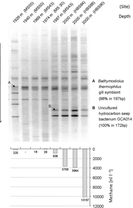
The availability of PCR techniques and extended phylogenetic trees based on 16S rRNA gene sequences from pure cultures not only enabled detailed considerations on microbial phylogeny, but also formed an initial backbone for biodiversity studies in the environment. Together with improved methods of DNA separation and purification using gel electrophoresis and cloning techniques, for the first time studies of mixed bacterial communities were possible by analysis of environmental DNA [4,5]. The favorite methods in the initial phase of these studies included separation of the mixed PCR products by gradient gels either with salt/solute gradients (denaturing gradient gel electrophoresis, DGGE) or with temperature gradients (temperature gradient gel electrophoresis, TGGE) [4,5]. These methods made possible the direct comparison of the community composition, e.g., of different samples alongside natural physicochemical gradients, from different geographic regions and from series of temporal or experimentally induced changes on a single gel. One example of such a study from the authors lab demonstrated the dependence of the community composition in the vicinity of warm deep sea vents of the Fiji Basin, in which a clear correlation was found between the presence of methane in the water samples and the bacterial community composition as revealed by 16S rRNA gene analysis using DGGE separation (Figure1) [6].
However, these methods allowed identification of the bacterial species based on the 16S rRNA gene sequences only to a limited extent. In addition to biases of DNA extraction and amplification protocols, to a great part this was due to the insufficient separation of complex mixtures in the gradient gels and to the limited sequence information obtained from the short DNA stretches that yielded the best separation within the gels (approx. <500 nt). The lack of phylogenetic resolution
due to the consideration of only small sequence stretches was alleviated by selection of primers that cover the complete sequence of the 16S rRNA gene, separation of the PCR products by cloning techniques and sequencing from the clone libraries [7]. These methods represented a tremendous progress in the methodology to approach environmental microbial communities at that time and caused a complete shift in research approaches and strategies. They were and still are intensively used in many laboratories worldwide.
yielded the best separation within the gels (approx. <500 nt). The lack of phylogenetic resolution due to the consideration of only small sequence stretches was alleviated by selection of primers that cover the complete sequence of the 16S rRNA gene, separation of the PCR products by cloning techniques and sequencing from the clone libraries [7]. These methods represented a tremendous progress in the methodology to approach environmental microbial communities at that time and caused a complete shift in research approaches and strategies. They were and still are intensively used in many laboratories worldwide.
Figure 1. A gradient gel of amplified 16S rRNA gene sequences from deep ocean water in the vicinity of hydrothermal vents of the Fiji-Basin (top) and concentration of methane in the same samples (bottom) are shown. The band pattern clearly indicates that the presence of various strains is strongly correlated with the presence of methane in the water (from [6]).
A major drawback of the molecular approaches in microbial ecology for many years was the failure to identify microorganisms in environmental samples which was related to the discrepancies of results from culture-independent molecular analyses and culture-dependent studies. It was a general tendency that both approaches gave non-congruent views on the environmental communities
Figure 1.A gradient gel of amplified 16S rRNA gene sequences from deep ocean water in the vicinity of hydrothermal vents of the Fiji-Basin (top) and concentration of methane in the same samples (bottom) are shown. The band pattern clearly indicates that the presence of various strains is strongly correlated with the presence of methane in the water (from [6]).
A major drawback of the molecular approaches in microbial ecology for many years was the failure to identify microorganisms in environmental samples which was related to the discrepancies of results from culture-independent molecular analyses and culture-dependent studies. It was a general
Microorganisms2016,4, 19 4 of 41
tendency that both approaches gave non-congruent views on the environmental communities (e.g., [8]).
The strong selectivity of culture-based approaches on the one hand together with the really small number of microorganisms being cultured at all and the limited resolution of molecular approaches on the other hand have to be considered as major reasons for the striking discrepancies between these two approaches. Ongoing efforts to increase the cultivation success, in particular of microorganisms from so far not cultured groups, together with an improved coverage of the microbial metagenome by new sequencing techniques, is expected to alleviate this discrepancy.
Another major drawback of studies using 16S rRNA gene sequences is the failure to convincingly depict the diversity of specific functional groups in environmental communities. This is a basic problem of the 16S rRNA gene approach, as the 16S rRNA molecule is not related to metabolic propertiesper se, but only through phylogenetic observations and assumptions. Therefore, more specific approaches have to be applied in order to convincingly unravel the environmental diversity of functional groups having a unifying physiological property. This is achieved by the analysis of suitable functional genes.
3. Progress and Limitations of Sequence Studies of 16S rRNA Genes and Metagenomes
In view of the great enthusiasm due to the tremendous advances in environmental microbiology coming along with the use of DNA sequences for microbial biodiversity studies in the 1990s, which caused complete shifts in research approaches and initiated the field of molecular microbial ecology, the limitations of these molecular approaches appeared of minor importance. With the rigorous advances in molecular techniques over the past decade, molecular microbial ecology has grown up and analyses of the complex metagenome of environmental samples became possible. Although a steady increase in sensitivity and resolution in sequencing has been obtained, it should not be overlooked that a basic limitation in studies on the environmental metagenome has remained. This limitation is connected to the generally low number of analyzed sequences of the metagenome from a given environmental sample, with the effect that not the complete community is depicted. (The same holds for metagenetic studies that have been performed with the 16S rRNA gene as target.) If one considers that an individual sample of a marine sediment may contain more than 1010bacterial cells with >10.000 different microbial species in a cm3and surface waters of the oceans have an average of 1 million bacteria, a number of 100 or 1000 clone sequences of a sample (as handled in most biodiversity studies using 16S rRNA gene sequences) could cover only a minor part of the community. Only the most abundant and the most easily amplified sequences could be retrieved from the tremendous diversity of the environmental communities. Although this fraction of the more abundant organisms could be regarded to include the most important players (at least under the conditions and the time of sampling) by no means it represents the potential of a selected sample and habitat under variation of the environmental conditions. In fact, changes of the environmental conditions are expected to provoke significant and complex changes of the community composition with the effect that minor and not detected members of the community become major or even dominant parts of it [9]. Probably one of the most extreme realizations of this effect is seen in enrichment cultures used by generations of microbiologists to specifically manipulate the natural community. The design of media recipes and the definition of the culture conditions specifically select microorganisms with a desired function such as sulfate reduction, photosynthesis or methane oxidation and a desired property such as halophilic, alkaliphilic or thermophilic responses [10].
Overall, the speed of new methodological developments in sequencing technologies and the progress of knowledge in molecular microbial ecology over the past decades was unbelievable.
Since the invention of PCR and sequencing techniques of the Sanger method [11,12], the most significant advances in molecular microbial ecology are realized by the high throughput sequencing technologies also called next generation sequencing technologies [13], which still continue to further improve the efficiency and performance. The first single capillary sequencer (ABI Prism 310) using the Sanger sequencing technology was on the market in 1996 and only two years later commercial 96 capillary sequencers were available [14]. This is not the place to go into details of the fascinating
story of the genomic and metagenomic achievements and methodological development over the past decades. A nice overview on the development of sequencing technologies and the different platforms of high-throughput sequencing can be found in the article by Kircher and Kelso [14]. Advances in the development of microbial metagenomics, which is strongly correlated to the progress in sequencing technologies, are nicely summarized by Gilbert and Dupont [15].
Sequencing technologies made possible the sequence analysis of the first bacterial genome in 1995 (Haemophilus influencae, 1.8 Mb) [16]. At that time, this was a great effort made possible by use of the Sanger method, today this amount of sequence information can be obtained within hours from the new high throughput sequencing platforms [14]. For comparison, the sequencing throughput with the well-established Sanger method now is at approx. 6 Mb/day (three-times the genome size of Haemophilus influencae), while the throughput of the Illumina technology and similar other technologies is at 5000 Mb/day (almost 2800 times the genome size ofHaemophilus influencae, more than 1000 times the genome size ofEscherichia coliand approx. 1.5 times the human genome size) [14]. These are impressive numbers and it is interesting to compare them with the sequencing efforts of metagenomic studies and the size of environmental metagenomes. In a great effort published in 2004, microbial metagenomes in the Sargasso Sea were studied with the Sanger technology resulting in the production of 1 billion non-redundant base pairs [15,17]. This number compares to 1 million bacteria present within 1 mL sea water and an average content of 5 million base pairs per bacterium,i.e., an information content of 5ˆ1012(5 trillion) base pairs in 1 mL, 1015base pairs per Liter and 1018base pairs per 1000 L, with the implication that metagenomic studies before 2010 have used sample volumes from 1–1000 L sea water [15]. With a further thousand-fold increase of the sequencing effort of the high-throughput technology on a daily basis from 5000 Mb/day and considering a sampling size of only 1 L (equivalent to 1015 base pairs of the metagenome), the coverage would be 0.1%. These numbers reflect both, the incredible advances in sequencing technologies on the one hand and the immense complexity of microbial metagenomes on the other hand. At this point it is necessary to recall the breathtaking improvements in the handling of data by computing facilities and software programs over the past 2 decades that all of us have witnessed in the daily life. Without special efforts and specific software programs, the handling of sequence information of a complete metagenome containing 1 billion single prokaryote genomes would not be possible. The evaluation of metagenomic data by bioinformatics tools is another great challenge to metagenome analysis [15].
4. Functional Gene Studies
Because much of the motivation to study bacterial and microbial biodiversity is coming from the desire to understand the functions of the environmental communities and the interactions within these communities, it was a logic step to include functional genes into biodiversity studies and to study functionally defined fractions of the whole microbial community. Functional gene approaches to a great part overcome limitations of resolution and in particular of lacking specificity in microbial community analyses by the 16S rRNA gene approach. They have a number of advantages for investigations of environmental communities. First of all, these approaches focus their view on a selected functional aspect of the environment and on organisms involved in this specific function.
Second and most important, these approaches achieve a better resolution regarding the community composition, because the number of genes of a particular function in a given sample is much smaller compared to the number of 16S rRNA genes (and whole genomes) and (assumed that the used primer system not only is specific to the target gene but also covers the diversity of this gene in the widest possible range) less abundant strains may be much easier detected and ideally the whole functional community can be resolved. In addition, many functional genes show faster rates of evolution relative to the 16S rRNA gene, which specifically has been selected to trace prokaryote phylogeny because of its phylogenetic conservative properties [1,2]. In consequence, their sequence comparison results in a higher phylogenetic resolution, supposed that specific and informative sequence stretches are selected for the analyses. In particular, closely related microorganisms within a defined functional
Microorganisms2016,4, 19 6 of 41
group often can be more easily distinguished by functional genes compared to the 16S rRNA gene. Therefore, functional gene approaches are excellent tools to study the diversity of a particular functional group and to demonstrate changes in the composition of prokaryote communities contributing to the selected function upon natural or experimentally induced changes of the environmental conditions [9,18].
The main challenges at the initiation of functional gene approaches in the late 1990s included the selection of suitable functional genes, the design of appropriate primer sets to amplify sequence stretches of sufficient length and information as well as the establishment of databases with sequences of these genes in order to allow phylogenetic assignment and eventually identification of the players in the environment. Today, it is hardly imaginable, that the lack of appropriate gene sequences in the databases was a major limitation at the initial stages of such investigations and even made it difficult to design proper primer sequences to specifically approach the target gene [8].
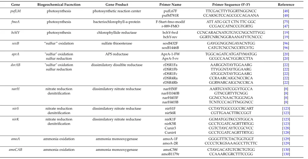
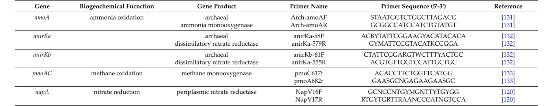
The selected genes and the established primers on one hand need to be specific for the selected metabolic pathway on the other hand the primers/selected sequence stretches should enable a complete consideration of the biodiversity of the selected pathway and function. In addition, the selected sequences should contain sufficient phylogenetic information and ideally contain more than 1000 nucleotides to enable good phylogenetic resolution. For a number of functional genes, appropriate primer systems have been described (see Table1) and databases of functional gene sequences rapidly grow up, in particular due to the growing number of complete genome sequences and the increased potential in performing metagenetic analyses with functional genes.
Although lateral gene transfer may be relevant for most if not all functional genes and potentially may blur the phylogenetic signal as compared to that of the 16S rRNA gene, experience with quite a number of functional genes and the comparison of their phylogeny with that of the 16S rRNA makes clear that events of lateral gene transfer were not frequent events [19–24]. They can be localized and do not limit the application of functional genes in diversity studies and in the identification of species in the environment. In consequence, advanced phylogenetic trees of functional genes can establish a functional phylogeny, independent from 16S rRNA phylogeny, and functional gene sequences enable species identification in the environmental metagenome with increasing confidence, as outlined below with examples of the anoxygenic phototrophic bacteria.
In general, most eco-physiological key functions are found in various phylogenetic lineages and in addition multiple pathways may exist for the same biogeochemical function, as for instance for the oxidation of sulfur compounds, ammonia oxidation, methane oxidation and autotrophic carbon dioxide fixation. The different pathways of the same biochemical function also present different target genes for the molecular analysis. This necessitates the design of different primer systems for each of the target genes of a particular function considered for community analysis, but at the same time it provides the advantage to distinguish the different groups of the studied function. In addition, the same genes may be involved in different functional roles, as exemplified by adenosine-51phosphosulfate (APS) reductase (apr) and dissimilatory sulfite reductase (dsr) playing roles in sulfate reduction as well as in sulfur oxidation [19,20,25]. Furthermore, different functional reactions may have common evolutionary roots and show strong sequence homologies of their genes, as known for methane monooxygenase (pmo) in methane-oxidizing bacteria and ammonia monooxygenases (amo) in ammonia-oxidizing bacteria [26].
The presence of a particular target gene and the sequences thereof yield valuable information on the organisms involved in this function in a particular habitat, on their phylogenetic relationships and eventually on their identity. If a comprehensive database is available on functional target gene sequences from identified type cultures, even species identification of the participating microorganisms may be possible [9]. With these implications, functional gene analysis is perfectly suited to link culture-based microbiological work with environmental molecular genetic studies. Major goals on the future perspective for such studies are to achieve not only the function-specific diversity but enable species identification in environmental communities, relate specific functions to microbial species or
phylotypes, determine their habitat preference and environmental adaptation as well as their biological activity based on functional transcriptome analyses.
To approach the functional diversity in environmental systems today two strategies are available, (i) the analysis of whole metagenomes and (ii) the selective analysis of functional genes. Even if the coverage of the whole metagenome is small, a tremendous amount of information is becoming available from metagenomic studies, which includes information on functional genes. Thus metagenome studies will continue to provide valuable information for functional aspects in the environment. However, for the specific functional analysis of a particular microbial community, the efforts of metagenomic analysis are not required and the coverage of the functional gene sequences is expected to be much better using up-to-date sequencing techniques for amplicons of specific functional genes. The major advantages of functional metagenetic approaches are the high specificity and the use of a comparable small data volume of sequence information to be analyzed and evaluated.
In order to specifically study communities of functional microbial groups and their responses to environmental factors, genetic tools were established for various important eco-physiological functions. Examples are presented with the following target genes/proteins: for two types of anoxygenic phototrophic bacteria, the Fenna-Matthews-Olson-protein (fmoA) as target for the green sulfur bacteria and two reaction center proteins (pufLM) for the phototrophic purple bacteria, adenosine-51phosphosulfate (APS) reductase(aprA), sulfate thioesterase (soxB) and dissimilatory sulfite reductase (dsrAB) for sulfur oxidizing and sulfate reducing bacteria, ammonia monooxygenase (amoA) for nitrifying/ammonia-oxidizing bacteria, nitrate and nitrite reductases (narH/G, napA, nirS, nirK) for denitrifying bacteria and methane monooxygenase (pmoA) for methane oxidizing bacteria (see Table1for recommended primers). In addition, a few studies are highlighted in which multiple functional genes have been used to characterize the potential of deep sea hot vent communities.
5. Photosynthesis and Anoxygenic Phototrophic Bacteria
Anoxygenic phototrophic bacteria are major players in a number of ecological niches, which primarily are strictly anoxic but extend to microoxic and even oxic environments [27–31].
We distinguish several major phylogenetic lines as well as different physiological groups which occupy defined and different niches in freshwater, marine and hypersaline environments [29,30,32].
These lines are represented by members of different bacterial phyla, by the green sulfur bacteria or Chlorobiaceae (Chlorobi) [33], the Heliobacteriaceae (Firmicutes) [34], the green nonsulfur bacteria or Chloroflexaceae [35], the Chloroacidobacteria [36,37], and the phototrophic purple bacteria (purple sulfur and purple nonsulfur bacteria), which are Alpha-, Beta- or Gammaproteobacteria [38–41].
The so-called aerobic phototrophic purple bacteria, at least most of those known to date, are close relatives of the purple nonsulfur bacteria, in particular of the Alphaproteobacteria (though representatives of Beta- and Gammaproteobacteria are known as well) close toRhodobacter and Rhodovulum species, have a primarily chemoheterotrophic metabolism and are aerobic respiring bacteria containing bacteriochlorophyll [31,42,43]. In metagenetic studies using shortpufMsequences, they have been recognized as important players in the open ocean [44,45] and also in brackish water lagoons [46]. In the following, we will focus on the phototrophic green and purple sulfur bacteria, because systematic and diversity studies of these groups are most advanced.
Pitfalls of applying 16S-rRNA-based approaches to the analysis of communities of anoxygenic phototrophic bacteria, in particular the phototrophic Proteobacteria (the purple sulfur and nonsulfur bacteria), were due to the close phylogenetic relationship between phototrophic and nonphototrophic bacteria within the Proteobacteria. In consequence, specific sequence stretches of 16S rRNA genes that would clearly allow the design of specific primers and the identification of phototrophic representatives in complex mixtures and environmental samples could not be identified. This necessitated the use of functional genes, e.g., those related to photosynthesis to study the environmental diversity of these bacteria.
Microorganisms2016,4, 19 8 of 41
In order to specifically analyze communities of anoxygenic phototrophic bacteria and their adaptation to different environmental conditions and their geographic distribution, primer systems have been developed that specifically target these bacteria: thefmoA gene encoding for a bacteriochlorophyll-a protein specific for the green sulfur bacteria and Chloroacidobacteria [47] and thepufLMgenes encoding for reaction center proteins of the bacterial photosystem II which is specific for all phototrophic purple Proteobacteria and Chloroflexaceae [9,48].
Important steps for the possible identification of species in environmental DNA sequences were the establishment of a phylogenetic-based taxonomy supported by 16S rRNA gene sequences and the demonstration of a general congruence of the phylogenies of 16S rRNA genes with those offmoA andpufLMgenes [49–54]. The formation of a comprehensive database offmoAgenes of green sulfur bacteria [47] and ofpufLMgenes of purple sulfur bacteria [48] from most cultured reference and type strains enabled detailed studies of environmental communities of these bacteria and the recognition of genera and species in the natural habitat.
5.1. The Phylogeny of the fmoA Gene in Green Sulfur Bacteria
The FMO protein is a bacteriochlorophyll-a protein that mediates energy transfer between the chlorosomes and the reaction center in the cytoplasmic membrane of green sulfur bacteria [55] and the recently described phototrophic thermoacidophilicChloroacidobacterium thermophilum,which occupies special ecological niches in hydrothermal springs and belongs to the Acidobacteria phylum [36,37].
FMO is absent in another major phylogenetic line of phototrophic green bacteria containing chlorosomes, the Chloroflexi [56]. Therefore, fmoAis an appropriate target to specifically analyze environmental communities of the green sulfur bacteria [57] and phototrophic Chloroacidobacteria.
A comprehensive phylogeny of the green sulfur bacteria, based on phylogenies of both 16S rRNA and fmoAgene sequences was established including available type strains of the established species [47].
Remarkably, the phylogenies of the two independent genes were largely congruent and species and strains can be easily well identified by either of the two genes. The available information on sequences from bothfmoAand 16S rRNA genes was used in the taxonomy of these bacteria to rearrange the strains and species, to redefine some species and in a few cases establish new species and a new genus [54]. With this background, environmental communities of green sulfur bacteria can be resolved on the species level byfmoAsequence analyses [58].
5.2. The Phylogeny of the pufLM Genes in Purple Sulfur Bacteria
In order to selectively approach the phylogeny of the phototrophic purple bacteria and to develop tools for the analysis of natural communities of these bacteria, thepufLMgenes were selected, which code for the light (L) and medium (M) subunit of the photosynthetic reaction center type II structural proteins of all phototrophic Proteobacteria (purple sulfur bacteria, purple nonsulfur bacteria as well as the aerobic phototrophic purple bacteria producing bacteriochlorophyll and forming a photosynthetic apparatus) and in addition of the phototrophic members of the Chloroflexi [48]. An improved primer system was designed covering a large part of the combinedpufLandpufMgenes and in addition, a comprehensive database ofpufLMgene sequences of most of the recognized type strains of the purple sulfur bacteria was established [48]. The phylogenetic relationship demonstrated bypufLM gene sequences of the purple sulfur bacteria (Gammaproteobacteria) was in good correlation to that of 16S rRNA gene sequences [48]. A phylogenetically based taxonomy of the purple sulfur bacteria facilitated the sequence-based species recognition in environmental samples [52,53,59].
Thus, the pufLM genes qualified as a valuable tool for studies of environmental communities of the phototrophic purple bacteria allowing species recognition of these bacteria even in complex mixtures of environmental communities.
5.3. Molecular Ecology and Species Recognition of Phototrophic Sulfur Bacteria in the Environment
Sequence information is predestined to link bacterial systematics and functional ecological studies because (i) sequence information is now well established as a property in bacterial systematics and forms a backbone for bacterial phylogeny and systematic treatment; and (ii) sequence information becomes easily available from environmental communities, from individual clones as well as from complete metagenomes and can contain important information about the physiological potential at the level of individual strains and species. Environmental gene sequences (or rather the bacteria associated with the sequences) can be arranged in phylotypes, which comprise sequences above a defined similarity. If a distinction of phylotypes is made at a sequence level that compares to the level of distinction between species with pure cultures, phylotypes can be used to approach the species diversity of environmental communities. If environmental clone sequences are sufficiently similar to known species, represented by their type strains, it is quite likely that they are representatives of this species or close relatives thereof. If considerations concerning sequence similarities as a rough guide for species differentiation of pure cultures are transferred to sequences from the environment, species recognition and an estimate of the species diversity in environmental samples can be achieved with phylotypes as an equivalent to the taxonomic defined species. Similar considerations as have been proposed as guidelines for the use of 16S rRNA gene sequences in taxonomy can be made for functional genes [60]. With the consideration of evolutionary rates in comparison to the 16S rRNA gene, borderlines of 86% and 95% sequence similarity of thepufLMgenes have been proposed for the distinction of genera and species, of purple sulfur bacteria, respectively [9,48].
5.4. Environmental Communities of Green Sulfur Bacteria
Phototrophic green sulfur bacteria are common in coastal lagoons and marine sediments, where they often form massive colored mass developments, while in freshwater lakes their development is mostly hidden in the depth at the chemoclines. They are dominant primary producers in the Black Sea [61] and develop in other stratified ocean basins [62]. They have the most advanced antenna system to harvest light and accordingly are adapted to life at minute amounts of light [63].
With the comprehensive study offmoAand 16S rRNA gene phylogenies of pure cultures of green sulfur bacteria as a solid basis, a first detailed molecular genetic study on the species composition of green sulfur bacteria communities was made with samples from marine and saline habitats of different geographical locations, of the Baltic Sea, the Mediterranean Sea, Sippewissett salt marsh (MA, USA) and Bad Water (Death Valley, CA, USA) using the established primers [57]. Quite interestingly, all of the clone sequences (more than 370 16S rRNA gene sequences and more than 130fmoAsequences) were associated with salt-dependent phylogenetic lines of green sulfur bacteria which had been previously established with pure cultures [47]. The clear dominance of representatives of the true marine green sulfur bacteria, in particular of the genusProsthecochloris[47,54] was in support of the experience from culture-dependent approaches, which regularly yieldedProsthecochloris aestuariiin enrichments and as isolated cultures from many marine habitats (Figure2) [32].
Though culture-based studies always had left doubt whether the specific cultivation conditions had selected just strains ofProsthecochlorisfrom a more complex environmental community, the genetic analyses conclusively demonstrated that members ofProsthecochlorisare the dominant green sulfur bacteria in suitable marine and saline habitats. Apparently, the phylogenetic diversity of marine green sulfur bacteria belonging toProsthecochlorisis significantly higher than was known from pure cultures. Available sequence information allowed the recognition of at least four different groups within this genus which are likely to represent different species [57], including the established species Prosthecochloris aestuarii,Prosthecochloris vibrioformeandProsthecochloris indica[54,64] and additional phylogenetic lines represented by environmental sequences [57,65].
Microorganisms2016,4, 19 10 of 41
Microorganisms 2016, 4, 19 10 of 39
Figure 2. The phylogenetic relationship of marine green sulfur bacteria (connected with red lines) according to fmoA sequences (according to [57]) is shown. Reference strains and type strains of known species are indicated and environmental sequences are highlighted and originate from the Croatian Lake Malo Jezero (“Adria”), a Baltic Sea lagoon (“Ostsee”) and Sippewissett salt marsh USA (“Atlantik”). It highlights the phylogenetic relation of green sulfur bacteria of the Prosthecochloris group from different geographic locations. Strain 2K represents the branch including Prosthecochloris indica [64].
5.5. Environmental Communities of Phototrophic Purple Bacteria
Phototrophic purple sulfur bacteria are Gammaproteobacteria and are forming massive colored blooms in coastal sediments and lagoons and regularly are accompanied by green sulfur bacteria and purple nonsulfur bacteria (Alphaproteobacteria and Betaproteobacteria) [32,62]. In the coastal zone the phototrophic bacteria are exposed to considerable changes in salinity, temperature and other parameters (in particular concentrations of oxygen and sulfide forming countercurrent gradients), which are expected to significantly shape and modify the composition of these communities. Adaptation to these changing conditions is prerequisite for the successful competition of the species and differential adaptation determines the dominance of different species alongside the natural gradients.
First molecular genetic studies to characterize the communities of phototrophic purple bacteria were based on sequences of the pufM gene and revealed a remarkable high diversity in different habitats [66–68]. Most sequences retrieved from a frozen Antarctic lake represented a diverse array of sequences related to the Betaproteobacteria Rhodoferax and Roseateles (a bacteriochlorophyll-containing aerobic bacterium) and to the aerobic bacteriochlorophyll-containing Alphaproteobacteria Acidiphilium and Bradyrhizobium [68]. The high diversity and abundance of phototrophic bacteria related to the aerobic anoxygenic phototrophic Roseobacter and to the facultative aerobic anaerobic phototrophic Rhodobacter/Rhodovulum clades in the open oceans was subject of a number of metagenomic studies and supported the high abundance and great diversity of this group of phototrophic bacteria in ocean waters [44,69]. Their abundance and wide distribution was substantiated in the Red Sea and the Mediterranean Sea [70,71], in shrimp ponds in
Figure 2. The phylogenetic relationship of marine green sulfur bacteria (connected with red lines) according tofmoAsequences (according to [57]) is shown. Reference strains and type strains of known species are indicated and environmental sequences are highlighted and originate from the Croatian Lake Malo Jezero (“Adria”), a Baltic Sea lagoon (“Ostsee”) and Sippewissett salt marsh USA (“Atlantik”).
It highlights the phylogenetic relation of green sulfur bacteria of theProsthecochlorisgroup from different geographic locations. Strain 2K represents the branch includingProsthecochloris indica[64].
5.5. Environmental Communities of Phototrophic Purple Bacteria
Phototrophic purple sulfur bacteria are Gammaproteobacteria and are forming massive colored blooms in coastal sediments and lagoons and regularly are accompanied by green sulfur bacteria and purple nonsulfur bacteria (Alphaproteobacteria and Betaproteobacteria) [32,62]. In the coastal zone the phototrophic bacteria are exposed to considerable changes in salinity, temperature and other parameters (in particular concentrations of oxygen and sulfide forming countercurrent gradients), which are expected to significantly shape and modify the composition of these communities.
Adaptation to these changing conditions is prerequisite for the successful competition of the species and differential adaptation determines the dominance of different species alongside the natural gradients.
First molecular genetic studies to characterize the communities of phototrophic purple bacteria were based on sequences of the pufMgene and revealed a remarkable high diversity in different habitats [66–68]. Most sequences retrieved from a frozen Antarctic lake represented a diverse array of sequences related to the BetaproteobacteriaRhodoferaxandRoseateles(a bacteriochlorophyll-containing aerobic bacterium) and to the aerobic bacteriochlorophyll-containing AlphaproteobacteriaAcidiphilium andBradyrhizobium[68]. The high diversity and abundance of phototrophic bacteria related to the aerobic anoxygenic phototrophicRoseobacterand to the facultative aerobic anaerobic phototrophic Rhodobacter/Rhodovulumclades in the open oceans was subject of a number of metagenomic studies and supported the high abundance and great diversity of this group of phototrophic bacteria in ocean waters [44,69]. Their abundance and wide distribution was substantiated in the Red Sea and the
Mediterranean Sea [70,71], in shrimp ponds in Thailand [72] and in Delaware estuary [73]. Most of these studies used separation of environmental amplicons on DGGE gels and sequencing of the separated bands to demonstrate the diversity of thepufMgene. However, due to the limitations of separation of bands on the gels and the short sequence fragments (<228 nt) considered, these studies lacked sufficient phylogenetic resolution to identify species or even genera. Nonetheless, they have demonstrated the presence of an immense biodiversity of aerobic phototrophic bacteria in marine habitats, which is to a great part not represented by cultured bacteria and in part not even significantly related to known bacterial species [45,71].
Remarkable are the studies on Soap Lake (Washington), which is a small soda lake with a distinct chemocline (at 20 m depth), a maximum depth of 24 m and high concentrations of sulfide reaching up to 175 mM near the sediment [74]. Both culture-based and culture-independent approaches were used to study the community of phototrophic bacteria in this lake. With the exception of a strain related to the purple nonsulfur AlphaproteobacteriumRhodobaca bogoriensis,several purple sulfur bacteria (representatives ofEctothiorhodospira,a strain related toThiorhodopsira sibiricaandThiocapsa imhoffii) were isolated [74]. Though the same primers as in a previous study on an Antarctic lake [68] were used, no aerobic phototrophic bacteria, but primarily purple sulfur bacteria (Gammaproteobacteria) were found inpufMamplicons from environmental DNA. This is in accord with the expected presence of bacteria specifically adapted to the highly sulfidic and also alkaline environment. Sequences from a total of 12 bands (136 nt long) ofpufMamplicons could be obtained after reamplification of bands from DGGE [74]. Only two of these were identical to one of the isolates from Soap Lake, toThiocapsa imhoffii, which apparently is the dominant purple sulfur bacterium at the chemocline, while others showed only distant relations toRhodoferax,RhodocyclusandEctothiorhodospira[74]. In conclusion, only a small part of the diversity of phototrophic purple bacteria of this extreme habitat was brought into culture and a better resolution of the phylogenetic diversity will probably be obtained with primer systems targeting the complete sequence ofpufLMgenes.
Systematic studies with higher phylogenetic resolution and the species-specific analysis of complex environmental communities of phototrophic purple bacteria were enabled by the application of a primer pair that amplified sequences of bothpufLandpufMgenes (approx. 1400 nt) of most purple bacteria. With these primers, a comprehensive database using the almost complete sequences of most cultured purple sulfur bacteria was established and their phylogenetic relationship was verified [48]. The established phylogeny of thepufLMgenes allowed confident identification of species and phylotypes in environmental samples. Two case studies of salt lakes in the Chilean highland and of a coastal lagoon of the Baltic Sea (Germany) may highlight the possibilities of this approach for community studies [9,75].
5.6. Baltic Sea Coastal Lagoon
The phototrophic bacterial community of a brackish water Baltic Sea coastal lagoon was characterized on the basis of almost completepufLMgene sequences and the impact of changes in temperature and salinity were determined under controlled conditions in the laboratory by using RFLP, cloning and sequencing [9,48]. This lagoon represents a habitat typical of many studies on phototrophic bacteria and quite a number of available cultures of phototrophic bacteria have been isolated from similar habitats [32]. Accordingly, major pufLM phylotypes of the community of purple sulfur bacterial of this brackish water lagoon affiliated to genera and species of phototrophic purple sulfur bacteria typically isolated from such habitats includingMarichromatium,Thiocystis,Thiorhodococcus, Allochromatium,Thiocapsa,Thiorhodovibrio; but some sequences were related to moderately halophilic HalochromatiumandThiohalocapsaspecies [32,76–78]. Quite important, at least 5 out of 20 identified phylotypes of purple sulfur bacteria could be clearly assigned to a known species, 10 additional phylotypes to a genus and only 5 phylotypes had sequence similarities (83.4%–85.6%) slightly below the proposed limit of 86%pufLMsequence similarity to the closest known type strain and therefore
Microorganisms2016,4, 19 12 of 41
might be regarded as representing new genera [9]. Thus, the purple sulfur bacteria in the lagoon more or less are known at the genus level but novelty of these bacteria is high at the species level.
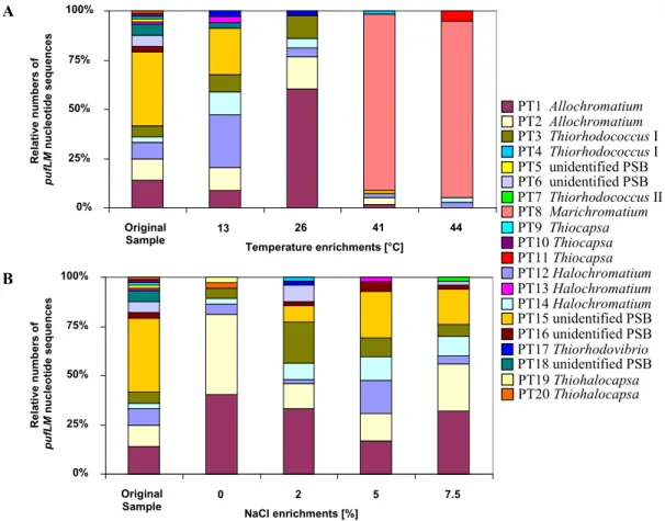
Both temperature and salinity had a significant influence on the community structure (Figure3).
In experimentally set up gradients of temperature (13–44 ˝C) and salinity (0%–7.5% NaCl) in identical culture media suited for purple sulfur bacteria, the community of purple sulfur bacteria was characterized by sequence analysis ofpufLMclone libraries and compared to that from the natural habitat. The highest diversity of identified phylotypes was observed in the natural sample (23.5˝C, 2% salinity). Most phylotypes in the habitat were tolerating the complete range of salt concentrations and developed up to 7.5% NaCl. While the community at 0% NaCl was clearly dominated by only twoAllochromatiumphylotypes, the community was more divers at all other conditions [9]. With the exception of three phylotypes found as single clones in the environmental sample, all were retrieved at least from one of the enrichments. In addition, six phylotypes of purple sulfur bacteria were retrieved only after enrichments, but not found in the environmental sample. Among these were phylotypes most similar to the type strains ofThiorhodococcus mannitoliphagus(99.8%),Thiorhodococcus kakinadensis (98.2%), andMarichromatium gracile(100%). This is quite remarkable and indicates an even higher diversity in the environmental sample than resolved by its metagenetic analysis and also demonstrates that media and culture conditions were quite appropriate for almost all purple sulfur bacteria recognized in the environmental sample by the genetic approach.
Microorganisms 2016, 4, 19 12 of 39
bacteria in the lagoon more or less are known at the genus level but novelty of these bacteria is high at the species level.
Both temperature and salinity had a significant influence on the community structure (Figure 3). In experimentally set up gradients of temperature (13–44 °C) and salinity (0%–7.5% NaCl) in identical culture media suited for purple sulfur bacteria, the community of purple sulfur bacteria was characterized by sequence analysis of pufLM clone libraries and compared to that from the natural habitat. The highest diversity of identified phylotypes was observed in the natural sample (23.5 °C, 2% salinity). Most phylotypes in the habitat were tolerating the complete range of salt concentrations and developed up to 7.5% NaCl. While the community at 0% NaCl was clearly dominated by only two Allochromatium phylotypes, the community was more divers at all other conditions [9]. With the exception of three phylotypes found as single clones in the environmental sample, all were retrieved at least from one of the enrichments. In addition, six phylotypes of purple sulfur bacteria were retrieved only after enrichments, but not found in the environmental sample.
Among these were phylotypes most similar to the type strains of Thiorhodococcus mannitoliphagus (99.8%), Thiorhodococcus kakinadensis (98.2%), and Marichromatium gracile (100%). This is quite remarkable and indicates an even higher diversity in the environmental sample than resolved by its metagenetic analysis and also demonstrates that media and culture conditions were quite appropriate for almost all purple sulfur bacteria recognized in the environmental sample by the genetic approach.
PT19 Thiohalocapsa PT11 Thiocapsa PT7 Thiorhodococcus II PT4 Thiorhodococcus I
PT17 Thiorhodovibrio PT8 Marichromatium
PT20 Thiohalocapsa PT10 Thiocapsa PT9 Thiocapsa PT5 unidentified PSB
PT13 Halochromatium
PT18 unidentified PSB PT6 unidentified PSB
PT16 unidentified PSB PT15 unidentified PSB PT3 Thiorhodococcus I
PT14 Halochromatium PT12 Halochromatium PT2 Allochromatium PT1 Allochromatium
0%
25%
50%
75%
100%
Original
Sample 0 2 5 7.5
NaCl enrichments [%]
Relative numbers of pufLM nucleotide sequences 0%
25%
50%
75%
100%
Original
Sample 13 26 41 44
Temperature enrichments [°C]
Relative numbers of pufLM nucleotide sequences
A
B
Figure 3. The contribution of different phylotypes to the community of purple sulfur bacteria (PSB) in a Baltic Sea lagoon is shown. Twenty phylotypes were identified in original sample and under different enrichment conditions on the basis of pufLM gene sequences. The figure depicts the composition in the sample and under the influence of different temperatures and salt concentrations (from [9]).
Figure 3.The contribution of different phylotypes to the community of purple sulfur bacteria (PSB) in a Baltic Sea lagoon is shown. Twenty phylotypes were identified in original sample and under different enrichment conditions on the basis ofpufLMgene sequences. The figure depicts the composition in the sample and under the influence of different temperatures and salt concentrations (from [9]).
An interesting property ofMarichromatium gracile, which has been repeatedly isolated from marine coastal habitats [32,62], was demonstrated by these experiments. A dramatic shift in the
community composition occurred at elevated temperatures of 41 and 44˝C when a single phylotype ofMarichromatium gracilebecame most prominent, which was not detected at lower temperatures and not in the habitat sample. The clear preference ofMarichromatium gracilefor elevated temperatures points to its obvious competitive advantage in the shallow-water habitats if heated during daytime by the sun and supports findings of Serranoet al.who characterized a slightly thermophilic strain of this species [79].
5.7. Salt Lakes of the Salar de Atacama
The salt lakes of Chilean Salares represent an extraordinary and extreme habitat with special conditions regarding salt concentration and composition, irradiation and drastic diurnal changes in temperature [80,81]. Anoxygenic phototrophic purple bacteria, including a diverse group of aerobic phototrophic bacteria of the so-calledRoseobacter/Rhodobacterclade are common to these lakes [82].
Like other hypersaline environments, some shallow lakes of the Salar de Atacama (Laguna Chaxa and Laguna Tebenquiche) exhibit extended purple-red colored microbial mats in and on the sediment surface. A comprehensive study of environmental communities of anoxygenic phototrophic purple bacteria of these salt lakes using the advancedpufLMprimer system of [48] with focus on purple sulfur bacteria demonstrated highly diverse and variable communities of these bacteria [75]. The great majority of purple sulfur bacterial phylotypes of this habitat could be related to known purple sulfur bacteria, but was supposed to be new at the genus level or even at higher taxonomic rank [75].
The communities of purple sulfur bacteria from both salt lakes were characterized by the presence of representatives related to the type strains of moderately and extremely halophilic Chromatiaceaesuch asHalochromatium salexigens,Halochromatium glycolicum,Thiohalocapsa halophila, Ectothiorhodospira mobilis, Ectothiorhodospira variabilis and Halorhodospira halophila as closest relatives [76–78,83]. Evidence was also obtained for the presence of several phylotypes of BChl b-containing anoxygenic phototrophic bacteria distant to (<80% sequence similarity) the genera Thiococcus,ThioflavicoccusandThioalkalicoccus, that form a separate phylogenetic branch among the purple sulfur bacteria [48,52,84]. These bacteria are known as inhabitants of marine sediments and have a particular advantage in sediment habitats due to their special pigment content withbchl-b[85–87].
Therefore, their presence in these salt lake sediments is not surprising, though they have been rarely isolated from salt and soda lakes [84].
Only two out of 24 phylotypes identified as phototrophic Gammaproteobacteria could be clearly identified on the species level (Ectothiorhodospira mobilisandThiohalocapsa halophila), whereas the great majority of phylotypes had apufLMsequence of such a low similarity (<80%) to known purple sulfur bacteria that quite likely they might represent new genera. Most remarkable was the dominance and diversity (11 phylotypes) of a novel, so far unknown lineage ofpufLMcontaining Gammaproteobacteria, which was highly diverse and prevalent in different lakes of the Salar de Atacama [75].
Phototrophic Betaproteobacteria are rare in the salares of the Altiplano, but occasionally single clones of a Betaproteobacterium only distantly related to Rubrivivax [75] and others related to Rhodoferax antarcticus[82] were identified.
In conclusion and in contrast to the situation in the coastal Baltic Sea lagoon, most of the purple sulfur bacteria recognized bypufLMsequences in these salt lakes represent new bacteria at the genus level or higher taxonomic rank. This depicts quite well the extraordinary situation of the habitats in the Chilean highlands with extreme climatic and environmental conditions and the great geographic distance to all so far investigated habitats of phototrophic bacteria and points out the uniqueness of their bacterial communities.
5.8. Biodiversity of the bchY Gene
Another promising approach to analyze the diversity of phototrophic bacteria used the sequence of thebchYgene, which is present in phototrophic bacteria containing either a type I or a type II photosystem and was demonstrated to provide amplification products from various phototrophic
Microorganisms2016,4, 19 14 of 41
purple bacteria, green sulfur bacteria and also green nonsulfur bacteria [88]. ThebchYgene encodes the Y subunit of chlorophyllide reductase, which is at a branch point in the biosynthesis of chlorophyll and bacteriochlorophyll [89]. It is the only enzyme of this pathway present in all known anoxygenic phototrophic bacteria, but absent in oxygenic phototrophs. Therefore, genes coding for this enzyme should be good candidates for targeting the bacteriochlorophyll-containing anoxygenic phototrophic bacteria. A degenerate primer set for this gene was elaborated and primer specificity and coverage were evaluated using bothin silicoandin vitrotechniques. The primer set was applied tobchYsequences present in databases and to pure cultures and environmental populations of anoxygenic phototrophic bacteria [88]. It was suggested that these primers have a broad applicability to study environmental communities of the phototrophic bacteria [88], though the sequence information retrieved is much lower compared to the more specific target genesfmoAandpufLM. Expected length of PCR products of thebchYgene are 480 bp for green sulfur bacteria, green non-sulfur bacteria, andHeliobacteria and 510 bp for phototrophic purple bacteria [88]. With this primer set the diversity of phototrophic bacterial communities was analyzed in Lake Kinneret and in the Mediterranean Sea [88]. Although this approach appears promising to cover all anoxygenic phototrophic bacteria with a single primer set, extended studies on environmental samples and in particular a comprehensive database with sequences from all cultured anoxygenic phototrophic bacteria still is lacking. When the phylogeny of this gene will be established with the available type strains of phototrophic bacteria, thebchYgene may prove to be an additional tool to systematically analyze natural communities of phototrophic bacteria.
6. Oxidation and Reduction of Sulfur Compounds
Sulfur compounds play important roles in energy generation of microorganisms, either as final electron acceptor in respiratory processes (e.g., sulfate and sulfur respiration) or as electron donors for energy generation in chemo- and photolithotrophic ways of life. The cycling of sulfur compounds by oxidation and reduction processes is governed by a number of prokaryotes from different phylogenetic lineages that use different biochemical pathways. In marine sediments sulfate reduction is a major process in organic matter degradation [90,91] and produces sulfide, which in turn is an important electron donor for sulfur-oxidizing bacteria. Though sulfate is the most abundant electron acceptor for anaerobic bacterial respiration in the ocean, the primary niche of sulfate reduction is the anoxic marine environment and therefore sulfate reduction is largely restricted to the anoxic zone. The primary ecological niche of chemotrophic sulfur oxidizers is the oxic/anoxic interface of stratified environments where oxygen and sulfide co-occur, though nitrate-reducing sulfur oxidizers and phototrophic sulfur bacteria specifically thrive in the anoxic environment.
Different biochemical pathways are involved in the sulfur cycling. One of the pathways for oxidation of sulfur compounds is the well-known reaction sequence including the formation and oxidation of adenosine-51-phosphosulfate (APS) as intermediate with the involvement of disulfite reductase and APS reductase as key enzymes. First important insight into the evolution of these pathways was obtained from the comparable analysis of enzymes involved in the dissimilatory reduction of sulfate and in the oxidation of reduced sulfur compounds.
Key enzymes of sulfate-reducing bacteria are APS reductase and disulfite reductase and already Peck [25] concluded from properties of the involved proteins that these enzymes share common evolutionary precursors in sufur-oxidizing and sulfate-reducing bacteria. Highly significant sequence similarities of APS reductase (AprA) and dissimilatory disulfite reductase (DsrAB) from the phototrophic sulfur-oxidizing gammaproteobacteriumAllochromatium vinosum, the sulfate-reducing deltaproteobacteriumDesulfovibrio vulgarisand the sulfate-reducing archaeonArchaeoglobus fulgidus strongly supported the hypothesis that these systems are true homologues and common ancestral genes for dissimilatory sirohaem sulfite reductases as well as APS reductases exist [92]. Today we know thatArchaeoglobushas gained a true bacterialdsrgene by lateral gene transfer presumably from a bacterial ancestor [19]. Suitable marker genes for profiling the communities of both sulfate-reducing
bacteria as well as sulfur-oxidizing bacteria using this pathway are provided by the genes of APS reductase (aprA) and of the dissimilatory sulfite reductase (dsrAB) [20,92,93].
A second important and widely distributed pathway of sulfur oxidation is the so-called Sox-pathway, which includes complex reaction sequences [94,95]. ThesoxB gene encoding the sulfate thioesterase was established as a suitable marker gene for this pathway [21,96–98].
6.1. Adenosine-51-Phosphosulfate Reductase (APS Reductase), aprA
The adenosine-51-phosphosulfate reductase (APS reductase) is a key enzyme of microbial sulfate reduction and also of sulfur oxidation processes and is common to sulfate-reducing bacteria and archaea and also to an important group of sulfur-oxidizing bacteria. A modified primer pair targeting theaprAgene was applied to a large number of reference strains from sulfate-reducing and sulfur-oxidizing prokaryotes [20]. This study further demonstrated that the reductive- and oxidative-type APS reductases are highly conserved among sulfate-reducing bacteria (SRP) and sulfur-oxidizing bacteria (SOB) and that the applied primer system allowed the concomitant detection of both sulfate-reducing and sulfur-oxidizing prokaryotes, which form separate lineages [20].
These authors explicitly, state that “although affected by several lateral gene transfer (LGT) events involving representatives of SRP and SOB lineages, theaprAgenes have in general been transmitted vertically during evolution, which supports their usage as functional gene markers in microbial diversity surveys”. By using thisaprA gene-based approach, a high diversity of sulfur-oxidizers was detected and clear differences noted in communities of the studied habitats with putative chemolithoautotrophic sulfur oxidizers of Beta- and Gammaproteobacteria being dominant in the hydrothermal sphere while sulfur-oxidizing Alphaproteobacteria were the major component in non-hydrothermal samples [20]. The same authors demonstrated the presence of phylotypes related to five sulfur-oxidizing alphaproteobacterial and gammaproteobacterial species and one sulfate-reducing archaeon in the microbial community associated with the Caribbean deep-water spongePolymastia corticataby comparingdsr,aprand 16S rRNA gene sequences [99].
Also deep-sea subseafloor communities of sulfur-oxidizing and sulfur-reducing microorganisms from sediments of three sites of the Pacific margin off Japan revealed a markedly diverse community of aprA sequences [100]. 135 distinct phylotypes were recognized from a total of 692aprA gene clones. Though composition and diversity varied largely between the sites and with the sediment depths, Desulfobacteraceae in general represented the major group of sulfate-reducers in most samples, followed by Desulfobulbaceae in several andDesulfotomaculum-related phylotypes present only in a few samples [100]. At least eight clusters unrelated to known cultivated sulfate-reducers were identified in different abundance in the samples. Among two major lineages with 45 phylotypes of sulfur-oxidizing bacteria, Gammaproteobacteria were dominant and a larger number of them could not clearly be assigned to known taxa even at the family level, because of the lack of phylogenetically closeaprAreference gene sequences [100].
Novel groups of sulfur-oxidizing Gamma- and Alphaproteobacteria were recognized in studies of marine intertidal sediments from the German Wadden Sea by analysis of the diversity ofaprAanddsrAB gene libraries [101]. The phylogenetic relations ofdsrABsequences from this study revealed a large hidden diversity of so far unrecognized sulfur-oxidizers both of Alpha- and of Gammaproteobacteria, part of which was supposed to perform a chemoautotrophic way of life [101].
The analysis of sediments of the Black Sea and the deep biosphere of the Peru margin revealed that the sequences ofaprAanddsrApresent in the samples affiliated to corresponding phylogenetic clusters [102]. The authors concluded that both of the functional genes represent sulfate reducers of equal phylogenetic positions. This was particularly significant foraprAanddsrAclone libraries from the Black Sea sediment where almost allaprAand alldsrAsequences affiliated either toDesulfobulbus elongatusor toDesulfotalea psychrophila[102].
Microorganisms2016,4, 19 16 of 41
Altogether, these few investigations have demonstrated thataprgene sequences are valuable tools to analyze the environmental communities of sulfate-reducing as well as sulfur-oxidizing bacteria.
In both groups in addition toaprAthe sequences ofdsrABgive additional significance to the analyses.
6.2. Dissimilatory Sulfite Reductase, dsrAB
The dissimilatory siroheme sulfite reductase is a key enzyme in sulfate reduction and catalyzes the six-electron reduction of sulfite to sulfide. It is operating in the reversed reaction in sulfur oxidation as well and was first characterized from the sulfur-oxidizingThiobacillus denitrificans[103]. The enzyme is considered to be highly conserved and therefore well suited to specifically approach the diversity analysis of sulfate reducing and sulfur-oxidizing bacteria in the environment. A 1.9-kb fragment encoding most of the alpha- and beta-subunits of the enzyme could be amplified from all lineages of SRB with a single primer set and the phylogenies inferred from thedsrABgene of analyzed reference strains were consistent with those from the 16S rRNA gene, including the archaeal sulfate reducer Archaeoglobus fulgidus[104]. This view was extended to the sulfur-oxidizing bacteria, also showing largely congruent tree topologies ofdsrABand 16S rRNA gene sequences [105]. Though the enzymes from both groups can be approached with identical primer sets, the enzyme of sulfur-oxidizing bacteria is phylogenetically clearly distinct from that operating in sulfate reducers and both form separate phylogenetic groups [19,22].
In a study of a microbial mat of the hypersaline Solar Lake using dsr sequence analysis to evaluate the diversity of SRB, a highly diverse population of sulfate-reducing bacteria was found [105].
Special attention was paid in this study to the relationship of the sulfate-reducing community to oxygen within a microbial/cyanobacterial mat. The most striking observation was that the highest diversity of sulfate-reducers was found in the permanently oxic part of the mat and most of these sequences were closely related to members of theDesulfonemaandDesulfococcusgroup. Though several SRB were known to be oxygen tolerant and some may have limited capacity to respire with oxygen [106], the high diversity and even dominance ofDesulfonema-like SRB in the oxic part gave evidence for a possibly specific adaptation of these bacteria to life at the oxic/anoxic interface or even under microoxic conditions [105].
The analysis ofdsrABgenes was also applied to study the diversity of sulfate-reducing bacteria in the Guaymas Basin [107], in which members of the Desulfobacteriales were identified, a larger number of which were related toDesulfobacterspecies. In addition, a new group of sulfate-reducers forming a cluster separate from all known sulfate-reducers was found that could not be identified in clone libraries of the 16S rRNA gene [107].
The sulfate reducing bacteria represent a good example to demonstrate the advances in our understanding of the biology and ecology of a microbial group made over the past decades. Until the 1970s these bacteria were regarded as a highly specialized group, strictly dependent on anoxic conditions and highly restricted to the metabolism of a few organic molecules only. The pioneering work of Fritz Widdel had initiated a new vision on the biology of these bacteria [108]. He cultivated and characterized a large number of new species and groups of sulfate-reducing bacteria and added new metabolic capabilities to them, such as autotrophic growth and complete oxidation of substrates and significantly extended the knowledge on the substrates used by these bacteria [108]. Our recent advances on their ecology primarily come from metagenetic and metagenomic analyses, which significantly increased our knowledge on the ecology of the sulfate-reducing bacteria. In particular their high diversity and the occupation of special ecological niches was demonstrated and specifically well resolved by the application ofaprAanddsrABas functional genes.
The advances achieved by functional gene studies over the past decade are well represented in the extensive work ondsrABby Mülleret al.[19]. In order to establish a comprehensive reference of dsrABsequences, these authors constructed a consensus tree including more than 1200 sequences with approx. 1.9 kb length. Four major branches ofdsrABfamilies were recognized: the reductive bacterial type, the oxidative bacterial type, the reductive archaeal type and a branch represented so far only by
![Figure 2. The phylogenetic relationship of marine green sulfur bacteria (connected with red lines) according to fmoA sequences (according to [57]) is shown](https://thumb-eu.123doks.com/thumbv2/1library_info/5360048.1683462/10.892.142.762.143.595/figure-phylogenetic-relationship-bacteria-connected-according-sequences-according.webp)