Molecular Sciences
Review
Impact of Drosophila Models in the Study and Treatment of Friedreich’s Ataxia
Véronique Monnier
1,*
,†, Jose Vicente Llorens
2,*
,† IDand Juan Antonio Navarro
3,*
,† ID1
Unité de Biologie Fonctionnelle et Adaptative (BFA), Sorbonne Paris Cité, Université Paris Diderot, UMR8251 CNRS, 75013 Paris, France
2
Department of Genetics, University of Valencia, Campus of Burjassot, 96100 Valencia, Spain
3
Lehrstuhl für Entwicklungsbiologie, Universität Regensburg, 93040 Regensburg, Germany
* Correspondence: veronique.monnier@univ-paris-diderot.fr (V.M.); J.Vicente.Llorens@uv.es (J.V.L.);
juan.navarro@ur.de (J.A.N.); Tel.: +49-941-943-3083 (J.A.N.)
† These authors contributed equally to this work.
Received: 28 May 2018; Accepted: 3 July 2018; Published: 7 July 2018
Abstract: Drosophila melanogaster has been for over a century the model of choice of several neurobiologists to decipher the formation and development of the nervous system as well as to mirror the pathophysiological conditions of many human neurodegenerative diseases. The rare disease Friedreich’s ataxia (FRDA) is not an exception. Since the isolation of the responsible gene more than two decades ago, the analysis of the fly orthologue has proven to be an excellent avenue to understand the development and progression of the disease, to unravel pivotal mechanisms underpinning the pathology and to identify genes and molecules that might well be either disease biomarkers or promising targets for therapeutic interventions. In this review, we aim to summarize the collection of findings provided by the Drosophila models but also to go one step beyond and propose the implications of these discoveries for the study and cure of this disorder. We will present the physiological, cellular and molecular phenotypes described in the fly, highlighting those that have given insight into the pathology and we will show how the ability of Drosophila to perform genetic and pharmacological screens has provided valuable information that is not easily within reach of other cellular or mammalian models.
Keywords: Drosophila melanogaster; Friedreich’s ataxia; frataxin; iron; oxidative stress;
metal homeostasis; lipid metabolism; endoplasmic reticulum; genetic screens; drug screens
1. Molecular and Cellular Aspects of Friedreich’s Ataxia
1.1. Pathophysiology of the Disease
Friedreich’s ataxia (FRDA) is an autosomal recessive degenerative disease present only in Indo-European and Afro-Asiatic populations [1]. It is the most frequent autosomal recessive ataxia in the Caucasian population. The major clinical features of FRDA include age of onset around puberty, progressive ataxia, muscle weakness, sensory loss and non-neurological features such as skeletal defects and cardiomyopathy. FRDA neuropathology starts with the degeneration of the large sensory neurons of the dorsal root ganglia (DRG), followed by atrophy of the dorsal columns that produces loss of proprioception and vibration sense. Degeneration of the spinocerebellar tracts of the spinal cord results in upper motor weakness. Atrophy of the dentate nuclei is also observed and accounts for the cerebellar component of ataxia. The neuronal degeneration is also accompanied with demyelination of sural nerves and DRG fibres. Most FRDA patients develop hypertrophic cardiomyopathy with thickened septum walls and iron deposits in the myocardium [2,3]. Other clinical signs are diabetes
Int. J. Mol. Sci.2018,19, 1989; doi:10.3390/ijms19071989 www.mdpi.com/journal/ijms
mellitus and carbohydrate intolerance [3] and in agreement, postmortem analysis of pancreas from FRDA patients revealed loss of β cells in islets of Langerhans [4]. Although the expression of frataxin is ubiquitous [5,6], the selective tissue vulnerability and cell death in FRDA is still far from understood.
Reduced expression of the nuclear-encoded protein frataxin is the molecular cause of the disease.
Such a reduced expression originates from the presence of a guanine-adenine-adenine (GAA) expansion in the first intron of the gene [5]. However, around 2–4% of FRDA patients are compound heterozygotes with a GAA expansion on one allele and a small mutation on the second allele [7]. The GAA has been shown to alter chromatin structure and to induce epigenetic modifications that lead to a reduced transcription of the gene [8], whereas the point mutations induce a loss-of-function [7]. Human frataxin (FXN) is translated in the cytoplasm as a precursor of 210 amino acids that is imported into the mitochondria [6]. It is then proteolytically cleaved by the mitochondrial processing peptidase (MPP) in a two-step process that leads to the successive generation of an intermediate form of 19 kDa and of the mature form of 14 kDa [9,10]. Frataxin is a protein highly conserved among eukaryotes and some prokaryotes. Sequence alignment of the frataxin family shows two distinct regions, an N-terminal block of 70–90 amino acids that is absent in prokaryotes and poorly conserved among eukaryotes, and a highly conserved block of 100–120 residues in the C-terminus of the protein [11,12].
The three-dimensional structure of the human frataxin, the Escherichia coli homolog (CyaY) and the Yeast frataxin homolog 1 (Yfh1), have been fully characterized [13–15]. All three proteins share conserved C-terminal regions that consist of an antiparallel β -sheet flanked by two α -helices.
The N-terminal tail present in eukaryotes appears to be intrinsically unfolded. The presence of acidic residues within the first α-helix and the edge of the first β strand forms a negatively charged surface that is involved in iron binding, whereas a neutral flat area on the β -sheet probably allows protein-protein interactions [12,16]. A crucial difference among frataxin orthologs is their ability to undergo iron-dependent oligomerization. Moreover, the functional relevance of such oligomers is still controversial. In the presence of excess of iron, Yfh1 has been shown to assemble into trimmers, hexamers and to larger 12-mers, 24-mers and 48-mers [17,18]. However, some authors suggest that such oligomers are dispensable since a yeast frataxin defective for oligomerization was able to perfectly replace the endogenous protein [19]. Unlike CyaY and Yfh1, the mature form of the human frataxin did not seem to form oligomers [20], although recent experiments suggest the opposite [21].
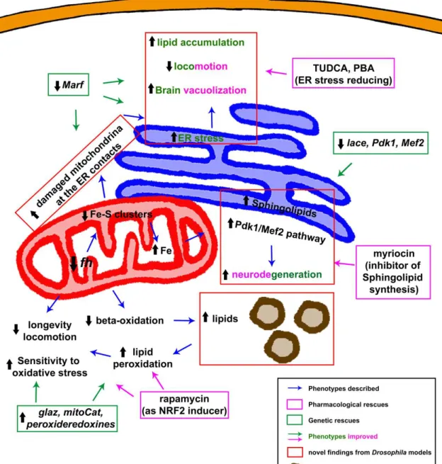
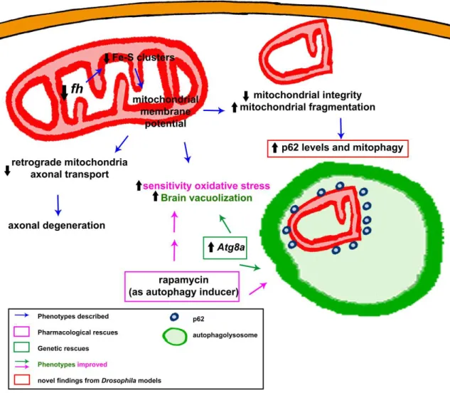
The function of frataxin has been linked to different mitochondrial pathways, but still remains partially unclear. The most accepted hypothesis based on all the available data supports the participation of frataxin in iron-sulfur (Fe–S) cluster biosynthesis in the mitochondrial matrix [22].
Fe–S clusters constitute one of the most ancient and ubiquitous of the biological prosthetic groups.
More than 200 types of proteins, exhibiting a remarkable functional and structural diversity, contain Fe–S centres. Fe–S clusters are composed by two or more iron atoms bridged by sulfide centres, most frequently in a [2Fe–2S] or a [4Fe–4S] conformation [23]. In higher eukaryotes, Fe–S biogenesis takes place in the mitochondria by means of homologous components of the bacterial Fe–S system that were transferred from the bacterial endosymbiotic ancestor of this organelle. Mitochondrial de novo Fe–S cluster biogenesis occurs in two steps. The first step involves the assembly of inorganic iron and sulfur on a scaffold protein, IscU in bacteria and Isu1 in yeast. It is known that this reaction needs a cysteine desulphurase as sulfur donor, IscS in bacteria and Nfs-1-Isd11 in yeast, whereas the origin of the iron still needs to be fully elucidated. In the second step, the clusters are transferred from the scaffold to recipient apoproteins for incorporation within specific amino acid residues [23].
The involvement of frataxin in this biosynthetic process was first suggested by the deficient
activity of proteins containing Fe–S clusters in FRDA patients and in mouse models [24,25]. Additional
data supporting this hypothesis was provided by studies in Saccharomyces cerevisiae [26]. Analysis
using mammalian recombinant proteins further characterized the interaction of human frataxin with a
preformed complex composed of NFS1, ISCU and ISD11 [20,27]. However, the exact role of frataxin in
the Fe–S cluster assembly process is still a matter of debate. On one hand, the iron-binding properties
of the human protein indicated that frataxin acts as the iron donor in the first step of the assembly
reaction [28]. On the other hand, other groups showed that frataxin functions as an allosteric regulator that facilitates the transfer of sulfur from NFS1 for the assembly and that iron is not required for the interaction between human frataxin and the NFS1/ISCU/ISD11complex [20,27,29]. Surprisingly, CyaY seems to inhibit the Fe–S cluster biogenesis [30]. In addition to its primary role in Fe–S cluster biogenesis, frataxin has been proposed to act directly as a chaperone that provides iron to aconitase [31]
and ferrochelatase [32] or electrons to ubiquinone/mitochondrial respiratory chain complex II [33] via protein-protein interaction.
1.2. Current and Prospective Treatments
Several clinical trials have been or are currently conducted to evaluate pharmacological compounds in FRDA patients. These trials are based on various strategies, such as increasing frataxin expression, lowering oxidative stress, improving mitochondrial function, reducing iron-mediated toxicity or modulating frataxin-controlled metabolic pathways [34]. However, to date, there is no pharmacological treatment with demonstrated efficacy to cure or even to stop the progression of the disease. It is therefore crucial to identify new candidate molecules for pharmacological approaches, and we believe that Drosophila models have a major role to play in this process.
An alternative, complementary and promising future approach is the possibility of a gene therapy. Two preclinical studies, performed on cardiac and sensory mouse models of FRDA using adeno-associated virus (AAV) vectors to express frataxin, showed that intravenous injection of an AAV vector expressing FXN not only prevented the development of cardiac and neurological features, but also improved cellular functions when injections were made in symptomatic mice [35,36].
The major challenges in the development of such gene therapy approaches will undoubtedly be to target efficiently the affected cells and tissues in FRDA patients and to succeed in inducing frataxin expression at levels that remain physiological. Indeed, studies in Drosophila showed that overexpression of endogenous or human frataxin in the nervous system decreased longevity, affected locomotor activity, and induced neurodegeneration [37,38], revealing the importance of a finely controlled level of expression of frataxin in the development of gene therapy approaches. Very recently, a study using human cells confirmed the hypothesis from these fly reports [39].
2. Analysis of Frataxin Function in Drosophila melanogaster
Frataxin homolog (fh) is the Drosophila homolog of FXN. The gene fh, of 965 bp, is in the region 8C/D of the X chromosome. It is composed of one intron flanked by two exons, encoding for the 190 amino acid Drosophila frataxin protein (Fh). Frataxin is a highly conserved protein, and the Drosophila protein shares conservation in both sequence and structure. Preliminary in silico analysis predicted the presence of a signal peptide for mitochondrial import [40]. The mitochondrial localization of fly frataxin was corroborated by a co-localization experiments in cell culture [41]. The biophysical and iron binding properties of the Drosophila frataxin are consistent with those from orthologs. Interestingly, Fh is stable as an iron-repleted monomer, not very prone to form oligomers and delivers iron to the scaffold on Fe–S cluster assembly [42]. This might be an advantage to study frataxin function. Furthermore, there has been some controversy regarding the existence and the function of an extramitochondrial frataxin pool that seemed sufficient to enhance survival [43] or to revert iron deregulation in FRDA cells [44].
Drosophila offers a nice experimental scenario to assess this issue in vivo. In agreement with evidence from Trypanosoma brucei [45], expression of a fly frataxin lacking the mitochondrial signal peptide failed to rescue the defects induced by the mutation present in the fh
1allele compared to the full length frataxin [46].
2.1. Methodologies Applied to Generate FRDA Models in Flies
The fruit fly is a versatile model organism used in biomedical research to study a broad range of
phenomena. The use of disease models is essential to understand the pathophysiological mechanisms
of human disorders. For over a century, different approaches have been developed to obtain Drosophila
models that recapitulate the hallmarks of human pathologies. Many of them take advantage of the GAL4/UAS system. This bipartite system, adapted from yeast, involves two transgenes and allows, up to some degree, the spatial and temporal control of the expression of the transgene of interest [47].
On the one hand, one that carries the GAL4 transcription factor under the control of a given promoter and on the other hand, the construct of interest downstream of an upstream activating sequence (UAS).
Using this approach, the fly has successfully mimicked human conditions by overexpressing mutant forms of human genes [48,49] or by promoting posttranscriptional silencing by means of the RNA interference (RNAi) strategy [50], of your favorite gene [51].
Using the RNAi strategy, John P. Phillip’s lab developed the first Drosophila model of FRDA [52].
This work was the first showing that loss of frataxin in flies reproduced the main molecular and biochemical features of the human disorder. Therefore, it was pivotal to stablish Drosophila as an excellent platform to reveal factors that can modulate the phenotypes and mechanisms involved in the disease. This first RNAi construct consisted of two inverted repeated copies of the first 391 nucleotides of fh cloned into the pUAST vector. Authors generated three transgenic lines—UDIR1, UDIR2 and UDIR3 (also named as fhRNAi, DfhIR, fhRNAi-1 in the literature)—able to almost completely suppress the expression of FH protein [52,53]. These lines have been also combined with the gene switch system [54,55] at the lab of Hervé Tricoire [56]. In this system, a modified GAL4 protein is fused to a progesterone steroid receptor, allowing the regulation of its GAL4 activity via the presence or absence of the synthetic progesterone analogue mifepristone (RU486). Using the same methodology and a different construct, Maria Dolores Moltó’s group developed a second RNAi model [41] named UAS-fhIR (also known as fhRNAi-2). In this model, two copies of the fh cDNA were cloned in the pUAST vector in an opposite direction and separated with a fragment of the GFP sequence as a linker to facilitate the formation of the loop [57]. Although based in a similar idea, there is a critical difference between both models. As stated above, the UDIR lines strongly abolish frataxin expression whereas, the UAS-fhIR line triggers a moderate and mild reduction of frataxin levels (around 70%
compared to controls) down to levels that better resemble the frataxin expression found in FRDA patients [58]. Importantly, this line has the advantage to allow working with adult flies upon ubiquitous downregulation of frataxin expression [41,59–61], whereas the UDIR lines are extremely useful to unveil pathological events and mechanisms upon tissue-specific silencing [56,61–64]. In this review, we decided to keep the original names—UDIR and UAS-fhIR—throughout the entire manuscript.
The last model was recently developed in Hugo Bellen’s lab. In an outstanding effort to unravel genes of the X chromosome involved in neuronal function and likely in neurodegeneration [65], authors performed an ethyl methanesulfonate (EMS) based mosaic genetic screen of lethal mutations.
Using this mutagenic alkylating agent, they identified a missense mutant allele of fh (S136R, named fh
1) [46]. The mutation is located in a highly conserved region of the protein used for the binding of frataxin to the ISC machinery [66]. Such a change triggers a strong loss of function leading to developmental arrest in larval stages. Remarkably, the fly stock used in the EMS screen also allowed the authors to carry out a mosaic mutant analysis by generating mitotic clones of adult photoreceptor neurons using the eyeless flippase and flippase recognition target (FLP/FRT) system [67]. This way, it is possible to generate a fly mutant for frataxin in a non-vital organ/tissue, while the rest of the fly remains wild-type like.
These are the models of FRDA developed so far in Drosophila melanogaster. All phenotypes induced by these genetic tools/models were recently described in detail [68]. Therefore, in the next chapters, we will focus on the detailed analysis of how all these models have contributed to the understanding of the pathophysiological mechanisms of the disease and the implications of such discoveries.
2.2. Fly Models Recapitulate FRDA Features
RNAi-mediated frataxin ubiquitous inactivation, using the daughterless (da-GAL4) driver, leads to
developmental lethality. Third instar-larvae (L3) fail to pupariate or reach the pupal stage much later
than controls and are not capable of becoming viable adults [52]. Similarly, fh
1hemizygote mutants
are lethal at the L3 or pupal stages, with prolonged larval stages. Removal of the maternal source of wild-type frataxin in these mutants leads to an earlier lethality, occurring at the embryonic stage [46].
This shows that frataxin is an essential protein during early embryogenesis in Drosophila, similarly to observations on mouse frataxin knock-out mutants [69] and in line with the lack of FRDA patients containing point mutations in both frataxin alleles [70].
To study the effects of partial frataxin inactivation on adults, and thus more closely mimic the situation of human FRDA patients, several strategies were used to bypass the preadult lethality. First, taking advantage of the fact that the activity of the UAS-GAL4 system is temperature dependent, some da-GAL4>UDIR adults were obtained by switching flies from 25 to 18
◦C at the beginning of the pupal stage. These adults are mostly short-lived, with a peak of mortality between 3 and 6 days of adult life. Interestingly, the cohort of flies still alive after this initial high-mortality phase could live up to 40 days of age, suggesting that frataxin is particularly needed in the early days of adulthood [52].
Another study used the actin-GAL4 ubiquitous driver to express the UAS-fhIR construct that allowed experiments on viable adults due a moderate but significant reduction of frataxin expression, as indicated above. Such flies also exhibited decreased lifespan and defective climbing activities [41].
In addition to these systemic approaches, a major advantage of RNAi-based Drosophila models lies in the ability to target specific tissues and therefore to evaluate which tissue or organs are particularly sensitive to frataxin depletion and to study tissue-specific physiopathological mechanisms.
Several studies targeted frataxin inactivation in neurons. Surprisingly, frataxin inactivation using various neuronal drivers resulted in viable progeny. Noticeably, flies in which RNAi-mediated frataxin inactivation is induced in the larval brain (c698a-GAL4) or in specific subtypes of neurons such as motorneurons (D42-GAL4 driver) or dopaminergic neurons (Ddc-GAL4) are viable without any obvious phenotypes [41,52]. Reduced adult lifespan and climbing activity were observed using the neur-GAL4 and C96-GAL4 drivers, allowing frataxin inactivation in the peripheral nervous system, suggesting that these neurons might be more sensitive to frataxin deficiency [41]. In agreement, in our unpublished observations, we did not observe any significant effect on fly fitness when frataxin was downregulated in all neurons by means of Elav-GAL4 and a moderate effect when using the RU486 inducible Elav-GS driver (as described in [71]). Silencing of frataxin in glial cells, using the pan-glial Repo-GAL4 driver, leads to partial developmental lethality, reduced adult lifespan and impaired locomotor activity [63]. Following this initial discovery in Drosophila, studies on human and mouse astrocytes confirmed a detrimental effect of frataxin silencing in astrocytes with non-cell autonomous effects on neurons, showing that glial cells are highly susceptible to contribute to the FRDA disease [72,73].
Heart-specific developmental inactivation of frataxin in flies, using the RU486 inducible Hand-GS driver, leads to cardiac dilatation and impaired systolic function [56]. Remarkably, those alterations are very similar to cardiac dysfunctions observed in patients and mouse models of FRDA [25,74,75].
Importantly, these phenotypes are fully rescued by complementation with human frataxin, showing conserved cardiac functions of frataxin between Drosophila and mammals. Interestingly, adult-specific frataxin inactivation did not lead to cardiac phenotypes, showing that the fly heart is particularly sensitive to frataxin depletion during developmental stages before adulthood. In human FRDA patients, the cardiomyopathy can already be observed in children, and patients with an earlier onset of disease generally also showed more severe cardiac involvement, suggesting a similar specific requirement of cardiac frataxin in young humans before adulthood [75]. It would be interesting to know whether such a developmental component is crucial exclusively in the heart or whether it is also a key element in other tissues/organs of the fly. Analyzing this aspect is of high interest in light of the new inducible and reversible mouse model [76] in which reactivation of frataxin expression after 12 weeks is sufficient to stop the progression of the disease and to recover morphological features in the affected heart and DRG.
Frataxin downregulation in muscles using the Mef2-GAL4 driver leads to reduced longevity and
locomotion, associated with expected mitochondrial dysfunctions [62]. Although often considered as
secondary in the disease, pathological manifestations are also observed in muscles of FRDA patients.
Those defects include loss of muscle strength, particularly affecting the lower limbs and prolonged recovery of calf muscle following exercise [77,78]. Moreover, magnetic resonance spectroscopy of the calf muscles in FRDA patients has also demonstrated impairment of ATP synthesis and inadequate oxygen utilization [78–80]. Therefore, Drosophila can be considered as an attractive and pertinent organism to study muscular dysfunctions, besides heart dysfunctions described above, induced by frataxin deficiency.
Finally, inactivation of frataxin in the steroidogenic prothoracic gland (the gland that produces ecdysteroids involved in larval molts, pupariation and metamorphosis) was sufficient to lead to developmental lethality. Although this appears at first glance to be specific to insect physiology, it could eventually reveal cellular and metabolic dysfunctions induced by frataxin deficiency relevant in the context of the FRDA disease [81].
As summarized in Table 1, ubiquitous and tissue-specific inactivation of frataxin allowed
recapitulation of clinical features characteristic of the FRDA disease, such as reduced lifespan, impaired
locomotor activity, cellular degeneration and cardiac dysfunction, providing relevant models to study
the physiopathological mechanisms involved in the disease at various scales, to screen for genetic and
pharmacological modifiers and ultimately propose therapeutic strategies.
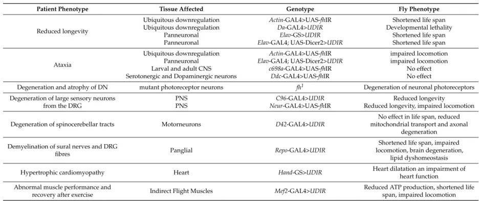
Table 1. Comparison of physiological hallmarks of FRDA in patients and phenotypes observed in FRDA fly models.
Patient Phenotype Tissue Affected Genotype Fly Phenotype
Reduced longevity
Ubiquitous downregulation Ubiquitous downregulation
Panneuronal Panneuronal
Actin-GAL4>UAS-fhIR Da-GAL4>UDIR
Elav-GS>UDIR Elav-GAL4; UAS-Dicer2>UDIR
Shortened life span Developmental lethality
Shortened life span Shortened life span
Ataxia
Ubiquitous downregulation Panneuronal Larval and adult CNS Serotonergic and Dopaminergic neurons
Actin-GAL4>UAS-fhIR Elav-GAL4; UAS-Dicer2>UDIR
c698a-GAL4>UAS-fhIR Ddc-GAL4>UAS-fhIR
impaired locomotion impaired locomotion
No effect No effect
Degeneration and atrophy of DN mutant photoreceptor neurons fh1 Degeneration of neuronal photoreceptors Degeneration of large sensory neurons
from the DRG
PNS PNS
C96-GAL4>UDIR Neur-GAL4>UAS-fhIR
Reduced longevity
Reduced longevity, impaired locomotion
Degeneration of spinocerebellar tracts Motorneurons D42-GAL4>UDIR
No effect in life span, reduced mitochondrial transport and axonal
degeneration Demyelination of sural nerves and DRG
fibres Panglial Repo-GAL4>UDIR
Shortened life span, impaired locomotion, brain degeneration,
lipid dyshomeostasis
Hypertrophic cardiomyopathy Heart Hand-GS>UDIR Heart dilatation an impairment of
heart function Abnormal muscle performance and
recovery after exercise Indirect Flight Muscles Mef2-GAL4>UDIR Reduced ATP production, shortened life span, impaired locomotion CNS: Central Nervous System; DN: Dentatte nucleus; DRG: Dorsal root ganglia.