NMR Spectroscopic Investigations on Flavin Catalyzed Photooxidations
Dissertation
zur Erlangung des Doktorgrades der Naturwissenschaften (Dr. rer. nat.) an der Fakultät für Chemie und Pharmazie
der Universität Regensburg
vorgelegt von Christian Feldmeier aus Wörth a. d. Donau
2014
Leitung von Prof. Dr. Ruth M. Gschwind durchgeführt wurden.
Promotionsgesuch eingereicht am 09.04.2014
Prüfungsausschuss: Prof. Dr. Alkwin Slenczka Vorsitzender Prof. Dr. Ruth M. Gschwind 1. Gutachter Prof. Dr. Werner Kremer 2. Gutachter Prof. Dr. Ingo Morgenstern 3. Prüfer
anderen Quellen direkt oder indirekt übernommenen Daten und Konzepte sind unter Angabe des Literaturzitats gekennzeichnet.
Diese Arbeit wurde bisher weder im In- noch im Ausland in gleicher oder ähnlicher Form einer anderen Prüfungsbehörde vorgelegt.
Regensburg, April 2014
NMR Spectroscopic Investigations on Flavin
Catalyzed Photooxidations
Contents
1 Introduction and Outline ... 1
2 Aggregation Effects in Visible-Light Flavin Photocatalysts: Synthesis, Structure, and Catalytic Activity of 10-Arylflavins ... 3
2.1 Abstract ... 4
2.2 Introduction ... 5
2.3 Results and Discussion ... 8
2.3.1 Synthesis ... 8
2.3.2 Crystal Structures ... 9
2.3.3 Aggregation Properties Determined by 1H-DOSY NMR Spectroscopy ... 11
2.3.4 Spectral and Electrochemical Properties ... 13
2.3.5 Photooxidation of 4-Methoxybenzyl Alcohol ... 15
2.4 Conclusion ... 18
2.5 Experimental Details ... 19
2.6 Supporting Information ... 25
2.6.1 Experimental Data for Aggregation Numbers ... 25
2.6.2 1H and 13C Assignments ... 27
2.7 Additional Experimental Findings ... 28
2.7.1 Self-Aggregation of Flavins ... 28
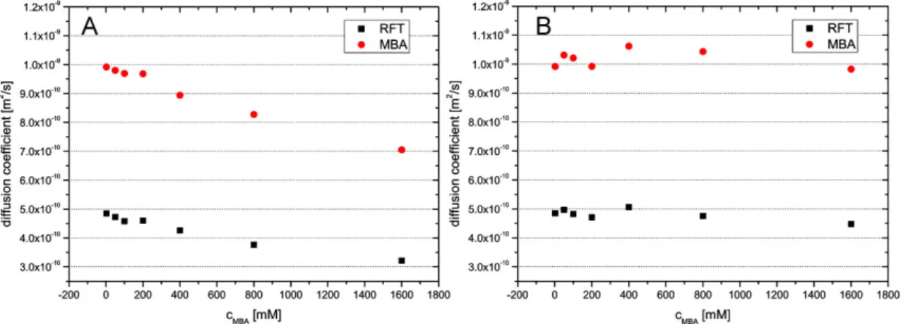
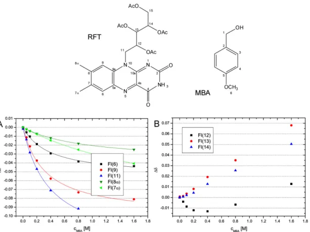
2.7.2 Aggregation between RFT and MBA ... 32
2.7.3 Experimental Details ... 39
2.8 References ... 40
3 LED based NMR Illumination Device for Mechanistic Studies on Photochemical Reactions - Versatile and Simple, yet Surprisingly Powerful ... 47
3.1 Abstract ... 48
3.2 Introduction ... 49
3.3 Materials and Methods ... 51
3.3.1 Circuit – Light Source ... 51
3.3.2 Light Source – Optical Fiber ... 52
3.3.3 Optical Fiber – Sample ... 53
3.3.4 Sample Preparation – Measurement ... 54
3.4 Results and Discussion ... 56
3.5 Conclusion ... 58
3.6 Additional Experimental Findings ... 60
3.6.1 Different Types of LEDs ... 60
3.6.2 LED Performance at Different Light Pulse Lengths ... 61
3.6.3 Photo-CIDNP Pulse Sequences ... 62
3.6.4 Modification of the Pulse Programs ... 64
3.6.5 Photo-CIDNP Build Up and Relaxation ... 64
3.6.6 Photo-CIDNP Cross Polarization ... 66
3.6.7 Experimental Details ... 67
3.6.8 1H and 13C Chemical Shift Assignment... 68
3.7 References ... 69
4 Photo-CIDNP Patterns of Free Flavins in Solution: Solvent, Structural and Heavy Atom Effects ... 73
4.1 Abstract ... 74
4.2 Introduction ... 75
4.3 Results and Discussion ... 77
4.3.1 Separation of Singlet and Triplet Pathway ... 77
4.3.2 Photo-CIDNP Polarization – Singlet vs. Triplet Pathway ... 79
4.3.3 Solvent Dependent Photo-CIDNP Cancelation ... 83
4.3.4 Photo-CIDNP Effects of Heavy Atom Flavins ... 88
4.3.5 Calculations ... 91
4.3.6 Photo-CIDNP Polarizations - Kaptein’s Sign Rules ... 98
4.4 Conclusion and Outlook ... 105
4.5 Additional Experimental Findings ... 107
4.5.1 Heavy Atom Flavins - Quantum Yields and Chemical Conversions ... 107
4.5.2 Temperature Dependent Photo-CIDNP Polarizations ... 108
4.6 Experimental Details ... 111
4.7 References ... 113
5 Solvent Stabilized Radical Intermediates in Flavin Catalyzed Reactions: One- vs. Two-Electron Oxidations ... 117
5.1 Abstract ... 118
5.2 Introduction ... 119
5.3 Results and Discussion ... 120
5.3.1 Model System ... 120
5.3.2 Reduced Flavin Species - Characterization and Stabilization ... 121
5.3.3 Kinetics ... 129
5.3.4 Mechanistic Proposal ... 131
5.3.5 Reoxidation of Flavin ... 132
5.4 Conclusion and Outlook ... 136
5.5 Supporting Information ... 139
5.5.1 1H and 13C Chemical Shift Assignment ... 139
5.6 Additional Experimental Findings ... 141
5.6.1 EPR Spectra of the Semiquinone Radical ... 141
5.6.2 Spin Density Dependent Line Broadening ... 142
5.6.3 Semiquinone Radical Stability ... 143
5.6.4 Oxidative Side of the Catalytic Cycle - Oxygen Concentration ... 145
5.6.5 H2O2 as the Second Reaction Product ... 149
5.6.6 Protonation of the Flavin Species ... 151
5.6.7 Amount of Semiquinone Radical ... 156
5.7 Experimental Details ... 159
5.8 References ... 162
6 Summary ... 167
7 Zusammenfassung ... 171
1
1 Introduction and Outline
The use of sunlight as an energy source for chemical conversions is a principle well known from nature where numerous processes in organisms are initiated and catalyzed by the absorption of photons in photoreceptors. In recent years the use of light as a driving force for chemical conversions has gained much attention in synthetic chemistry and the area of photocatalysis has seen a rapid development. However, detailed studies of the underlying reaction mechanisms lack behind these fast advances of visible light photocatalysis in synthetic applications despite the huge importance of mechanistic information for exploiting the full potential of photocatalysis and designing photocatalytic systems rationally.
The method of choice for mechanistic investigations on photocatalytic reactions is UV/Vis spectroscopy and in particular transient absorption spectroscopy as it provides access to the transient excited intermediates of the absorbing chromophore. Despite the manifold possibilities and techniques NMR provides for mechanistic studies, far too little attention has been paid to the application of NMR spectroscopy to investigations on photocatalytic reactions. Valuable information accessible directly by NMR is therefore often omitted in mechanistic studies. The objective of this thesis is therefore the application of modern NMR techniques to mechanistic studies of photocatalytic reactions. To enable the investigation of photochemical systems by means of NMR a device for in situ illumination of NMR samples was developed. With the help of this illumination device various NMR techniques were applied for the investigation of flavin catalyzed photooxidations as a central model system to get insights in the mechanism of the particular reaction, to test the possibilities of the NMR approach for the study of photochemical reactions and to reveal general underlying principles in photocatalysis. For a comprehensive approach considering also species invisible for NMR spectroscopy the NMR techniques were supplemented by Photo-CIDNP spectroscopy, UV/Vis spectroscopy and EPR spectroscopy.
The importance of flavin self-aggregation for its reactivity is shown in chapter 2. By means of 1H-DOSY solution NMR and crystal structures solvent dependent catalyst aggregates are revealed and the role of hydrogen bonds and π-π interactions are discussed. It is described how the catalyst aggregation, its photophysical properties and reactivity can be tuned by synthetically modifying the steric demand of the flavin side chain. Additionally, pre-associated flavin-substrate complexes are investigated by NMR-titration and 1H-DOSY NMR techniques.
2
In chapter 3, a new illumination device for NMR spectrometers is presented. Based on LEDs switched directly by the NMR spectrometer and an optical fiber with a roughened tip the light is guided directly into the NMR sample to illuminate it enabling the application of well-established NMR techniques to mechanistic investigations of all kinds of photochemical reactions. Pulsed operation of the LEDs at high powers in low duty cycles enables exceptional high optical powers and makes the setup suitable even for applications in Photo-CIDNP spectroscopy.
A comprehensive study of Photo-CIDNP effects of flavins is presented in chapter 4. The effect of the solvent on Photo-CIDNP polarization of flavins is discussed. Detailed investigations of the Photo-CIDNP effect in different flavin derivatives are conducted on a small molecule model system using acetonitrile as a solvent to enable the detection of Photo-CIDNP effects in flavins. The implications of different substituents at the isoalloxazine moiety on the Photo-CIDNP spectroscopic properties and the radical pair mechanism are addressed in terms of Kaptein’s sign rules and supplemented by DFT calculations of electron spin densities.
Finally, chapter 5 assesses solvent effects on the stabilization of the flavin semiquinone radical intermediate in flavin catalyzed photooxidations of benzyl alcohols. It is shown by a combination of NMR, Photo-CIDNP and UV/Vis spectroscopy that the semiquinone radical intermediate can be stabilized by the solvent water and that the stabilization makes flavin act as a one-electron oxidation agent for the photooxidation. The influence of solvent variation on the radical stabilization and the implications for the reaction mechanism and the efficiency of the photoreactions are discussed.
3
2 Aggregation Effects in Visible-Light Flavin Photocatalysts:
Synthesis, Structure, and Catalytic Activity of 10-Arylflavins
Jitka Dad’ová and Susanne Kümmel did the synthesis and performed the photocatalytic reactions. Jana Cibulková did the crystallizations. Christian Feldmeier did the NMR spectroscopic aggregation studies.
J. Daďová, S. Kümmel, C. Feldmeier, J. Cibulková, R. Pažout, J. εaixner, R. ε.
Gschwind, B. König, R. Cibulka, Chem. – A Eur. J. 2013, 19, 1066–1075.
Reproduced with permission of John Wiley and Sons
4
2.1 Abstract
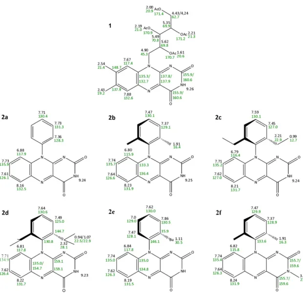
A series of 10-arylflavins (10-phenyl-, 10-(2’,6’-dimethylphenyl)-, 10-(2’,6’-diethylphenyl)-, 10-(2’,6’-diisopropylphenyl)-, 10-(2’-tert-butylphenyl)-, and 10-(2’,6’-dimethylphenyl)-3- methyl-isoalloxazine (2a–f)) was prepared as potentially nonaggregating flavin photocatalysts. The investigation of their structures in the crystalline phase combined with 1H-DOSY NMR spectroscopic experiments in CD3CN, CD3CN/D2O (1:1), and D2O confirm the decreased ability of 10-arylflavins 2 to form aggregates relative to tetra-O-acetyl riboflavin (1). 10-Arylflavins 2a–d do not interact by π–π interactions, which are restricted by the 10-phenyl ring oriented perpendicularly to the isoalloxazine skeleton. On the other hand, N3-H···O hydrogen bonds were detected in their crystal structures. In the structure of 10-aryl-3-methylflavin (2f) with a substituted N3 position, weak C-H···O bonds and weak π-π interactions were found. 10-arylflavins 2 were tested as photoredox catalysts for the aerial oxidation of 4-methoxybenzyl alcohol to the corresponding aldehyde (model reaction), thus showing higher efficiency relative to 1.
The quantum yields of 4-methoxybenzyl alcohol oxidation reactions mediated by arylflavins 2 were higher by almost one order of magnitude relative to values in the presence of 1.
5
2.2 Introduction
Flavins (isoalloxazines) are biologically active compounds that are responsible for redox processes in many types of enzymes, mostly in the form of flavin mononucleotide or flavin adenine dinucleotide cofactors.[1–4] Besides, synthetic flavin analogues are the subject of intensive research as organocatalysts of oxidation and reduction reactions.[5–25]
The redox activity of flavin derivatives is dramatically enhanced by absorption of visible light; the longest wavelength absorption maximum is at around max = 450 nm.[26] Thus, the photoexcitation of flavins enables the oxidation of substrates that cannot be oxidized thermally.[27–47] Until now, flavins have been applied to the photooxidation of benzyl alcohols,[27–37] benzyl amines,[38] and methylbenzenes[39] to benzaldehydes; the photooxidation of benzyl methyl ethers to methyl benzoates;[39] the photooxidation of dopamine,[40] amino acids,[41] indols,[42] unsaturated lipids and fatty acids,[43,44] glucose,[45]
and phenols;[46] and the selective photocatalytic removal of benzylic protecting groups.[47]
The photooxidation reactions mentioned are usually performed in the presence of air, thus allowing the regeneration of the flavin catalyst Fl from its dihydro form Fl-H2, which is formed from flavin in the excited state Fl* in the presence of a substrate (quencher) by a subsequent two-electron reduction and protonation process. Therefore, only a catalytic amount of flavin is required (Figure 2-1). Flavins are also known to sensitize singlet oxygen production.[48,49] Until now, flavin-mediated sulfoxidation reactions[50] and oxidation reactions of unsaturated lipids[36,51] that are proceeded by a singlet oxygen mechanism have been reported.
In almost all studies, the photooxidation of benzyl alcohols to benzaldehydes in acetonitrile was studied as a typical procedure to elucidate the efficiency of the flavin photocatalyst. The activity of simple flavins, for example, tetra-O-acetyl riboflavin (1) and lumiflavin (see Figure 2-2 for the structures), in the oxidation of 4-methoxybenzyl alcohol in acetonitrile is very low, with quantum yields of up to 0.03 %.[27,29] Several attempts to improve the efficiency of flavins have recently been reported. Substantially higher quantum yields of the oxidation of benzyl alcohol were achieved if the flavin photocatalyst was protonated or coordinated to rare-earth metal ions, with the highest value of 17 % in the case of a scandium(III) complex.[13–15,19] Also, thiourea accelerated the photooxidation of benzyl alcohol mediated by flavins and reached a high turnover number (TON) of up to 580.[29] A remarkable improvement in the catalytic efficiency of the flavin moiety was achieved by its covalent attachment to ZnII–cyclen or a β-cyclodextrin substrate binding site.[30,32] The reaction medium enhances the photooxidation if performed in sodium dodecyl sulfate (SDS) micelles.[31] A positive effect
6
of water on the rate of photooxidation reactions mediated by flavins has also been described.[27,30,52] Immobilization of flavins on fluorinated silica gel stabilizes the chromophore.[28]
Figure 2-1 Catalytic cycle for the aerobic photooxidation of a substrate S mediated by flavin Fl.
Figure 2-2 Structures of the flavin compounds typically used in photocatalysis.
Besides hydrogen bonding, flavins are known to interact with several molecules by π-π stacking,[53–60] donor–π interactions,[61] and cation or anion–π interactions.[62,63]
These interactions are essential not only for the binding of flavin cofactors in proteins, but also to modulate their redox properties and, consequently, the reactivity of flavin moieties in biological systems.[58,62,63] The effect of noncovalent interactions on the properties of flavins in artificial systems is also well documented.[53–61] There is evidence for flavin dimer formation, even in dilute solutions,[64] and such intermolecular aggregation may decrease the photocatalytic efficiency of flavins by quenching excited states or alter their redox properties.[27] With the aim of minimizing the ability of flavins to aggregate, we prepared a series of derivatives 2b–e, with an ortho-substituted phenyl ring in position 10 (Figure 2-3). The aryl ring should be oriented perpendicular to the flavin skeleton due to ortho substitution, thus making π–π interactions between flavins less possible. Compound 2a, without substitution on the phenyl ring, and the 3-methyl derivative 2f were prepared for comparison. The photochemical, electrochemical, and aggregation properties, crystal structures and ability to mediate the photooxidation of
7 4-methoxybenzyl alcohol (model reaction) of arylflavins 2 were studied and compared with those of 1.
Figure 2-3 Structure of 10-arylflavins synthesized and investigated as photocatalysts.
8
2.3 Results and Discussion
2.3.1 Synthesis
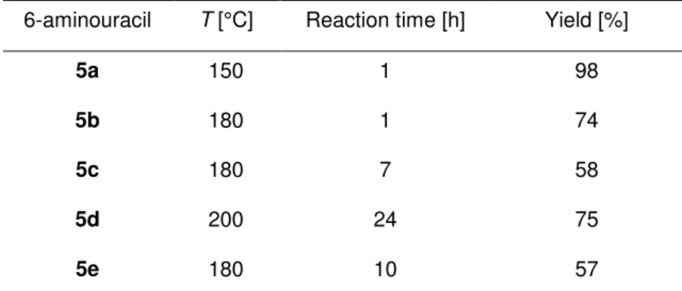
The synthesis of 10-arylisoalloxazines 2 (Figure 2-4) was started by converting commercially available substituted anilines 3a–e with 6-chlorouracil (4a) into 6-arylaminouracils 5a–e. It is evident from the reaction conditions and yields (Table 2-1) that the substitution becomes more difficult with increasing steric hindrance of the substituents on C2 and C6 of the phenyl ring. Although the nonsubstituted phenyl derivative 5a was obtained almost quantitatively, sterically hindered aminouracils were isolated only in moderate yields (i.e., 5c and 5e) or after a substantially longer reaction time (i.e. 5d).
Table 2-1 Reaction conditions and yields for the preparation of 6-aminouracils 5 by the reaction of 6-chlorouracil (4a) with substituted anilines 3.[a]
6-aminouracil T [°C] Reaction time [h] Yield [%]
5a 150 1 98
5b 180 1 74
5c 180 7 58
5d 200 24 75
5e 180 10 57
[a] A mixture of 4a and 3 was heated in a nitrogen atmosphere (see supporting information for further details).
The prepared aminouracils 5a–e were converted into the target flavins 2a–e by reaction with nitrosobenzene in acetic acid/acetic anhydride (1:1). Although this synthetic approach was effective for the synthesis of other sterically hindered flavins,[9,65]
derivatives 2 were obtained in relatively low yields (13–25 %). Unfortunately, the yield did not increase even when acetic acid and acetic anhydride in other ratios were used as solvent. The 3-methyl derivative 2f was prepared in an analogous process by using 6-chloro-3-methylaminouracil (4b; Figure 2-4). Interestingly, the conversion of 6-arylamino-3-methyluracil (5f) into 3-methylflavin (2f) proceeded in a substantially higher yield relative to the formation of 2b (44 versus 23 %, respectively), which possesses a nonsubstituted N3 position.
9
Figure 2-4 Synthesis of 10-arylflavins 2.
2.3.2 Crystal Structures
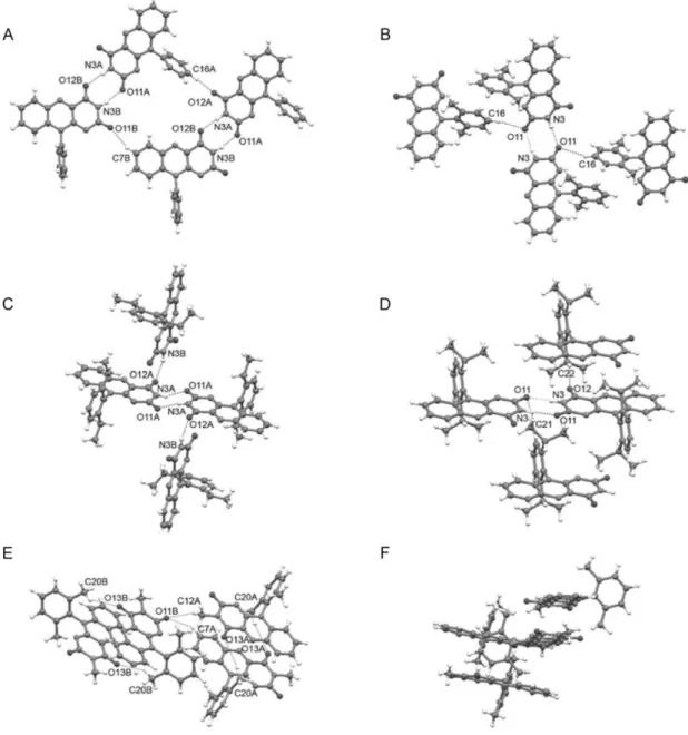
The interaction of flavin molecules in the crystal can provide information about the aggregation behavior in solution. For this purpose, crystals for single-crystal analysis were prepared of 2a–d and 2f. Interestingly, of the five structures only two 2b and 2d exhibited one molecule in the asymmetric unit, as could be expected. Three structures 2a, 2c, and 2f possessed two different molecules A and B in the asymmetric unit. A close inspection of the structures with A and B molecules showed that a significant difference between the two molecules was displayed only by 2c, in which one ethyl group of the ortho-substituted phenyl ring of the molecule B was rotated around the C(phenyl)-CH2 bond by 83.9(1)° (Figure 2-5 C). In the structure of 2f, molecules A and B differed only by a slightly different rotation of the phenyl ring (Table 2-2). In the case of the structure 2a, no marked difference between molecules A and B was observed.
Table 2-2 Dihedral angles between the aryl and isoalloxazine planes in the crystal structures.
Flavin Angle [°]
2a 79.73(5),[a] 78.43(4)[b]
2b 83.25(4)
2c 86.48(4),[a] 83.48(4)[b]
2d 85.69(5)
2f 79.49(5),[a] 82.02(5)[b]
[a] Molecule A. [b] Molecule B.
Structures of several simple flavin derivatives have already been investigated by X-ray diffraction.[66–71] In most cases, π-stacking interactions between the isoalloxazine moieties were recognized, which results in the packing of flavin molecules with distances
10
of between 3.3 and 3.6 Å. In such stacked systems, flavin molecules adopt an alternating orientation, and the benzene ring of one flavin moiety overlaps with the pyrimidine ring of the adjacent one (and vice versa). Tetra-O-acetyl riboflavin (1),[69] 3-methyl-tetra-O-acetyl riboflavin,[66] 3-benzyllumiflavin,[67] and 10-methylisoalloxazine[70] are examples of such stacked structures in the crystal phase. As expected, no π–π interactions between flavin moieties were found in the structures of 10-arylflavins 2b–d, even in the case of 2a with the unsubstituted phenyl ring, in which a coplanar orientation of the phenyl and isoalloxazine subunits was still allowed. In the structures of flavins 2a–d, the aryl ring is almost perpendicular to the mean plane of the isoalloxazine fragment, with a dihedral angle that ranges from 78.4 to 86.5°, thus preventing the stacking of flavins (see Table 2-2, Figure 2-5, and supporting information). A similar value of the dihedral angle of 79.7° was reported for 10-(2-hydroxyphenyl)-3-methylisoalloxazine.[72] The analysis of the X-ray crystallographic data showed pairs of symmetrical hydrogen N-H···O bonds between the pyrimidine rings of two adjacent molecules of flavins 2a–d (Figure 2-5 A–D).
Additionally, a relatively short N3B-H···O12A hydrogen bond in the structure of 2c and weak C-H···O interactions in 2a–d contribute to the aggregation. Hydrogen bonds C16-H···O11 in 2b (Figure 2-5 B) and C22-H···O12 in 2d (Figure 2-5 D), with participation of the hydrogen atoms on the (alkyl)phenyl ring on one hand, and hydrogen bond C7B-H···O11B in 2a (Figure 2-5 A), with participation of the hydrogen atoms on the isoalloxazine skeleton on the other hand, can be given as examples (see supporting information for all the hydrogen-bonding data). In contrast to flavins with a free N3-H bond (2a–d), compound 2f cannot form N-H···O bonds, and thus relatively weak C-H···O interactions dominate in the crystal structure of 2f (Figure 2-5 E). Methyl groups on both the N3 atom and the aryl ring participate in these C-H···O bonds. However, despite the presence of the ortho,ortho-disubstituted phenyl ring with perpendicular orientation toward the isoalloxazine skeleton (Table 2-2), little overlap of the flavin subunits that results in a weak π–π interaction was found in the structure of 2f (Figure 2-5 F). The distance between the neighboring planes in the stack is about 3.5 Å.
The investigation of the structure in the crystalline phase confirms that 10-arylflavins 2 have no structural prerequisites to interact by mean of strong π–π interactions and to form stacks in a similar manner to simple flavin molecules.[66–71] One could speculate about the situation in solution due to the conformational flexibility of the molecules. Flavin 2a may show rotation of the phenyl ring; however, this behavior is strongly limited by the ortho substituents in 2b–f. Therefore, only partial overlap of the isoalloxazine skeletons (e.g., by one ring) that results in a weak π–π interaction could be expected in solution.
As was shown in flavoenzyme models, the binding constants based on the overlap of
11 one or three rings of the flavin skeleton with an aromatic compound can differ by a factor of 30.[59]
Figure 2-5 Hydrogen bonding in the crystal structures of 10-arylflavins A) 2a, B) 2b, C) 2c, D) 2d, and E) 2f and F) a fragment that shows π stacking of the molecules 2f. The hydrogen bonds are shown as dashed lines, and the non-hydrogen atoms that participate in the hydrogen bonds are labeled. See supporting information for hydrogen-bonding data and more images.
2.3.3 Aggregation Properties Determined by 1H-DOSY NMR Spectroscopy
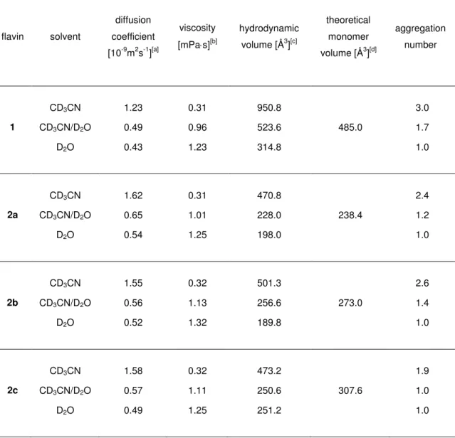
1H-DOSY NMR experiments[73] were used to measure the diffusion coefficients of 1 and arylflavins 2 in CD3CN, D2O, and CD3CN/D2O (1:1). The resulting aggregation numbers calculated from the experimental diffusion coefficients (see experimental section and supporting information) are presented in Table 2-3.
12
Table 2-3 Aggregation numbers of 1 and 10-arylflavins 2 in different solvents.[a]
Flavin aggregation number
CD3CN CD3CN / D2O (1:1) D2O
1 3.0 1.7 1.0
2a 2.4 1.2 1.0
2b 2.6 1.4 1.0
2c 1.9 1.0 1.0
2d 2.1 1.2 1.0
2e 2.2 1.0 1.0
2f 2.4 1.3 1.0
[a] Conditions: T=300 K, c=5x10⁻³ molL-1 of flavins 1 and 2 in solution with CD3CN and CD3CN/D2O (1:1) or a saturated solution in D2O.
A significant aggregation for 1 was detected in CD3CN, with an average aggregation number of 3.0, which decreased upon the addition of water down to monomers in pure D2O. Next, the aggregation trends for arylflavins 2a–f were investigated. For all the compounds, a significantly decreased aggregation number was found in CD3CN relative to 1. Again, the addition of water led to disaggregation for arylflavins 2a–f. These data show that the basic idea to decrease π-π interactions in the aggregates by the introduction of an aryl ring with sterically demanding substituents works. However, there was no direct correlation between the steric demand of the substituents in 2a–f and the aggregation number detected experimentally. For example, 2c shows decreased aggregation relative to 2b, as expected for ethyl groups relative to methyl groups as substituents; however, a further increase in the steric demand in 2d and 2e did not lead to decreased aggregation numbers. This finding suggests that not only π–π interactions between the isoalloxazine moieties contribute to the aggregation, but also additional stabilizing dispersion forces between the bulkier substituents[74] and other noncovalent interactions play an important role. Interestingly, analysis of the crystal structures of 2a–d revealed N-H···O hydrogen bonding as the dominant noncovalent interaction for these arylflavins and not π–π interactions, as found for 1. In the 3-methyl derivate 2f, hydrogen bonding through the N3-H bond is blocked. However, the diffusion measurements showed only a slightly decreased aggregation number relative to 2b. This outcome is in accordance with the crystal structure of 2f, which shows π–π interactions again besides weaker C-H···O interactions. The solvent-dependent disaggregation of arylflavins 2a–e going from CD3CN to CD3CN/D2O (1:1) and D2O correlates with the relative hydrogen- bond acceptor properties of these solvents in terms of better solute/solvent interactions
13 toward pure D2O.[75,76] Interestingly, the aggregates driven by π–π-interactions show a similar solvent dependence, thus demonstrating that the solvent dependence in flavins cannot be used as an indicator of the intermolecular interaction mode. Thus, the combination of aggregation numbers and crystal-structure analysis reveals that both π-π interactions and hydrogen bonding play a decisive role in the aggregation of the flavins, and their relative contribution can be tuned by the structure of the synthesized flavins.
2.3.4 Spectral and Electrochemical Properties
The spectral and electrochemical properties of the newly prepared 10-arylflavins 2 in acetonitrile were studied and compared to those of 1 (see Table 2-4 and supporting information).
Table 2-4 Spectroscopic data for flavins 1 and 2 in acetonitrile.
Flavin λ2 [nm]
( [mol-1dm3cm-1]) [a]
λ1 [nm]
( [mol-1dm3cm-1]) [a] λF [nm][b] ΦF[c]
1 343 (8500) 440 (12000) 505 0.499
2a 335 (6200) 436 (8900) 517 0.244
2b 330 (7000) 437 (10000) 498 0.447
2c 331 (7000) 434 (10000) 500 0.537
2d 330 (6200) 436 (8900) 501 0.434
2e 332 (7000) 437 (9900) 502 0.328
2f 321 (5500) 427 (6500) 498 0.282
[a] λ1and λ2 are the positions of the two lowest-energy bands in the absorption spectra;[b] The maximum of the fluorescence emission spectrum, λex = λ1; [c] The fluorescence quantum yield determined using quinidine sulphate as a standard.
The aryl substituent in position 10 of the isoalloxazine causes a small blue shift of the absorption maxima and a decrease in the absorption intensity in the UV/Vis spectra.
Substitution at the N3 position of the 10-arylisoalloxazine ring effects the position of the absorption maxima more significantly (cf. flavins 2b and 2f), than it was observed in the case of lumiflavin and 10-methylisoalloxazine.[77,78] All flavins 2 show intensive fluorescence with a maximum at around max = 500 nm. A small effect of the aryl substitution on the fluorescence maxima was only observed in the case of 2a, which bears a nonsubstituted phenyl ring. However, the fluorescence quantum yield of 2a is significantly decreased by half relative to 1 and 2b–d. Similarly, substitution at N3 decreases the fluorescence quantum yield of arylflavins, which corresponds to the
14
observed effect of N3 substitution in 1[79] and 10-methylisoalloxazine.[78] On the other hand, the fluorescence quantum yields reported for lumiflavin and 3-methyllumiflavin are almost the same.[77]
The reduction potentials of the synthesized flavin derivatives in acetonitrile that correspond to one-electron reduction (Fl → Fl.-)[79] were determined by cyclic voltammetry (CV) relative to ferrocene/ferrocenium. Moreover, the change in the Gibbs free energy ΔGET of the electron transfer from the substrate 4-methoxybenzyl alcohol to the excited flavins in the singlet state (Table 2-5) were calculated from the observed reduction potentials by using the Rehm-Weller equation (1):[80,81]
0 0 2
2 / 1 2 /
1
) /
( 4 .
96
G
ETE
oxE
rede a E
where Eox1/2 and Ered1/2 are the oxidation potentials of the substrate (Eox1/2=+1.19 V for 4-methoxybenzyl alcohol)[30] and the reduction potential of the flavin (Table 2-5); e2/ a is the Coulomb term (5.4 kJmol-1);[79] E0–0 is the flavin excitation energy (given in kJmol-1), which was estimated as the average of absorption (hc/ 1) and emission (hc/ F) energies with 1 and F values derived from the flavin absorption and fluorescence spectra (Table 2-4); h is the Planck constant (6.63 x 10⁻³⁴ m2kgs-1); and c is the velocity of light (2.99 x 10⁸ ms-1). The redox potential of arylflavins 2 shifts to more positive values, but by only 60 mV relative to 1, which did not seem to be enough to influence the oxidation power of the flavin significantly. According to Gibbs free energy changes, electron transfer between 4-methoxybenzyl alcohol and flavins 1 and 2 in their singlet excited state is exergonic, and thus favorable (ΔGET<0). The ΔGET values are less negative for 1 and 10-phenylisoalloxazine (2a) by about 10 kJmol-1 relative to 2b–f.
Fluorescence quenching for the newly synthesized derivatives 2 with 4-methoxybenzyl alcohol was studied in acetonitrile. Stern–Volmer plots constructed from the results are linear in all the cases (see supporting information). The values of Stern–Volmer constants KS (KS = kQτF, where kQ is the apparent rate constant and τF is the fluorescence lifetime) were calculated as the slope of Stern–Volmer dependence (I0/I = 1 + KS[Q]), that is, as the slope of the ratio of the fluorescence intensities I0/I in the absence and presence of 4-methoxybenzyl alcohol (quencher Q) plotted against concentration [Q].
Interestingly, for almost all the newly prepared flavins bearing substituted phenyl rings 2b–f, higher quenching KS constants were measured relative to 1. Only the value for 10-phenylisoalloxazine (2a) is equal to that of 1. The observed decreased values of Stern-Volmer KS constants for 1 and 2a probably result from the decreased rate of
15 electron transfer kQ, which corresponds to the decreased Gibbs free energy changes ΔGET (Table 2-5).
Table 2-5 Redox potentials of flavins 1 and 2, estimated free energy changes ΔGET and Stern-Volmer constants KS for the electron transfer from 4-methoxybenzyl alcohol to flavins 1 and 2 in acetonitrile.
Flavin Ered [V][a] ΔGET [kJ mol-1] [b] KS [L mol-1]
1 -1.18 -24 26
2a -1.12 -24 25
2b -1.11 -34 42
2c -1.10 -34 44
2d -1.11 -33 35
2e -1.10 -33 36
2f -1.11 -34 33
[a] Values obtained in acetonitrile at a scan rate of 50 mVs-1 in 0.001 molL-1 solutions of the flavins with 0.01 molL-1 Bu4NPF6 at 20 °C versus ferrocene / ferrocenium. [b] Free energy changes calculated from equation (1) using E1/2ox (4-methoxybenzyl alcohol) = 1.19 V versus ferrocene / ferrocenium. [30]
2.3.5 Photooxidation of 4-Methoxybenzyl Alcohol
The ability of the prepared flavins 2 to mediate the photooxidation of 4-methoxybenzyl alcohol with oxygen to the corresponding aldehyde was investigated under standard conditions, namely, 10 mol% of photocatalyst in deuterated acetonitrile at 25 °C under the atmospheric pressure of air (Figure 2-6). A high power light-emitting diode was used for the irradiation of the reaction mixture. A comparison of the efficiencies of flavins in the photooxidation reactions was made by determining 1) conversion after 90 minutes determined by 1H NMR spectroscopic analysis of the reaction mixture and 2) quantum yield of the photooxidation reactions determined independently. Importantly, the oxidation reaction does not proceed in the absence of flavin or light.
Figure 2-6 Model photooxidation reaction.
With 1 as a photocatalyst, only 5 % conversion was achieved after 90 minutes of irradiation (Table 2-6, entry 1). The use of 2a without substitution on the phenyl ring led to only a small improvement of the conversion (Table 2-6, entry 2). On the other hand, the introduction of an phenyl ring with substituents in ortho positions resulted in a
16
substantial increase in flavin efficiency to mediate photooxidation, thus reaching conversions of up to 37 % after 90 minutes of irradiation in the presence of 2b (Table 2-6, entry 3). The character of the alkyl substituents on the aryl ring seems to be important for the efficiency of the flavin photocatalysts. The diethyl derivative 2c showed nearly the same activity as 2b (compare entries 3 and 4 in Table 2-6), whereas the activity of 2d and 2e with branched isopropyl and tert-butyl substituents is slightly decreased (Table 2-6, entries 5 and 6). Interestingly, the alkylation of the N3 atom also decreases the efficiency of the flavin chromophore in the photooxidation reactions (Table 2-6, entry 7).
The conversion of the photooxidation reactions in the presence of 2b–f are relatively high after 1.5 hours of irradiation, but they are not remarkably increased during the next irradiation period. This behavior is caused by degradation of the flavin photocatalysts during photooxidation reactions, which was evidenced from bleaching of the reaction mixtures (see Table 2-6 and supporting Information). Nevertheless, the photo stability is not the most important factor that influences the activity of flavin photocatalysts. The least stable flavin 2c showed relatively high efficiency. Interestingly, all the synthesized catalysts 2 are less photostable than flavin 1.
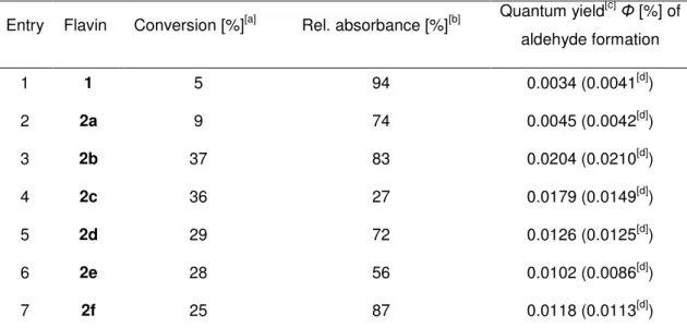
Table 2-6 Photooxidation of 4-methoxybenzyl alcohol to 4-methoxybenzaldehyde in CD3CN catalyzed by 1 and 10-arylflavins 2a–f.
Entry Flavin Conversion [%][a] Rel. absorbance [%][b] Quantum yield[c]Φ [%] of aldehyde formation
1 1 5 94 0.0034 (0.0041[d])
2 2a 9 74 0.0045 (0.0042[d])
3 2b 37 83 0.0204 (0.0210[d])
4 2c 36 27 0.0179 (0.0149[d])
5 2d 29 72 0.0126 (0.0125[d])
6 2e 28 56 0.0102 (0.0086[d])
7 2f 25 87 0.0118 (0.0113[d])
[a] After irradiation for 90 min. Conditions: calcohol=4x10⁻³ molL-1, cflavin=4x10⁻⁴ molL-1, irradiation with 1W LED (max=450 nm), T=25 °C, monitoring by 1H NMR. [b] Relative absorbance of the reaction mixture at =443 nm after irradiation for 60 min relative to the absorbance at the beginning of the experiment. [c] Determined by independent experiments with monitoring by GC. [d] Determined in CH3CN.
The results of quantum-yield measurements are in accordance with the observed conversions (Table 2-6). The introduction of disubstituted aryl rings at position 10 of the isoalloxazine ring causes a substantial increase in the quantum yield of the oxidation of 4-methoxybenzyl alcohol, which is, in the case of 2b, by almost one order of magnitude higher than the photooxidation in the presence of 1. However, the increase in the quantum yield of the flavin photocatalyst is approximately half with bulky isopropyl or
17 tert-butyl substituents or if the N3 position of isoalloxazine is substituted by a methyl group. As expected, the quantum yields of the oxidation reactions are not affected by deuteration of the solvent, thus indicating that a singlet-oxygen pathway is not involved.[82–84]
One could speculate that the lower efficiency of 2a relative to 2b–f is a result of its smaller oxidation power, but the differences in reduction potentials and in estimated ΔGET values are not sufficient to explain the observed significant differences in reactivity.
The low activity of 2a can also be attributed to the possible free rotation of the non-substituted aryl ring, thus allowing its coplanar arrangement relative to the isoalloxazine plane. This effect may increase the ability of 2a to aggregate in solution with flavins or substrates, thus supporting fast unproductive charge recombination.[27]
Interestingly, the N3-H···O hydrogen bonds that dominate among the intermolecular interactions of flavins 2b–e seem to have no negative effect on the catalytic activity of flavin photocatalysts, as evidenced by the comparison of 2b and 2f (compare entries 3 and 7 in Table 2-6).
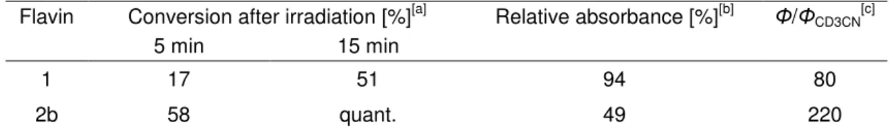
Table 2-7 Photooxidation of 4-methoxybenzyl alcohol to 4-methoxybenzaldehyde in CD3CN/D2O (1:1) catalyzed by 1 and 10-arylflavin 2b.
Flavin Conversion after irradiation [%][a] Relative absorbance [%][b] Φ/ΦCD3CN[c]
5 min 15 min
1 17 51 94 80
2b 58 quant. 49 220
[a] Conditions: calcohol=4x10⁻³ molL-1, cflavin=4x10⁻⁴molL-1, irradiation with 1 W LED (lmax=450 nm), T=25 °C, monitoring by 1H NMR. [b]
Relative absorbance of the reaction mixture at =443 nm after irradiation for 10 min relative to the absorbance at the beginning of the experiment. [c] Relative values of the quantum yields in CD3CN/D2O (1:1) relative to the quantum yields in pure CD3CN (Table 6).
Significantly enhanced quantum yields (by a factor of 80) were reported for the oxidation of 4-methoxybenzyl alcohol catalyzed by 1 by changing the solvent from pure acetonitrile to acetonitrile/water (1:1).[27,52] In the case of arylflavin 2b, the effect of water content on the quantum yield is even higher and a factor of 220 was reached (Table 2-7).
Consequently, quantitative conversion was observed after only 15 minutes of photooxidation catalyzed by 2b, whereas only 37 % conversion was achieved in pure CD3CN after 90 minutes (compare Table 2-7 with Table 2-6, entry 3). Unfortunately, the decomposition of 2b was also relatively fast in CD3CN/D2O (1:1; see Table 7 and supporting information).
18
2.4 Conclusion
10-Arylisoalloxazines 2a–f were prepared as potentially nonaggregating flavin photocatalysts by condensation of the appropriately substituted aminouracils 5a–f with nitrosobenzene. The investigation of their structures in the crystalline phase confirms that 10-arylflavins 2 have no structural prerequisites to interact by means of strong π-π interactions and to form stacks in a similar manner to simple flavin molecules, which is caused by steric hindrance of the substituted phenyl ring oriented perpendicularly to the flavin skeleton. X-ray diffraction studies also revealed that N-H···O hydrogen bonding dominates in the crystals of 2a–d. Blocking the N3 position with a methyl group in 2f inhibits the formation of N-H···O bonds; instead, C-H···O hydrogen bonds and weak π-π interactions shape the structure of the molecules in the solid state. The significantly lower tendency of flavins 2a–f to aggregate in acetonitrile was confirmed by 1H-DOSY NMR spectroscopic experiments. Nevertheless, it was shown that there is no direct correlation between the steric demand of the substituents in 2a–f and the aggregation numbers, which is probably due to the contributions of other noncovalent interactions; for example, the N-H···O hydrogen bonds in the case of 2a–e or dispersion forces between the bulkier substituents in the case of 2d and 2e.
Flavins 2b–f are far more effective photocatalysts for the photooxidation of 4-methoxybenzyl alcohol than tetra-O-acetyl riboflavin (1). The observed quantum yield of this oxidation in the presence of 2b (the best photocatalyst among 10-arylflavins 2) exceeds that of 1 by almost one order of magnitude. Unfortunately, the increased reactivity of 2 is accompanied by their lower photostability. Although the conversions of the photooxidation reactions of 4-methoxybenzyl alcohol in acetonitrile catalyzed by flavins 2b–f are not quantitative, they are among the most active flavins tested so far in the photooxidation reactions of benzyl alcohols. The efficiency of flavins 2 can be significantly enhanced by water as reported for 1.[27,52]
The results show that the efficiency of a flavin photocatalyst can be altered and improved by changing the structural elements that influence aggregation properties. However, there is no simple correlation as to how intermolecular interactions affect the ability of flavins to mediate the photooxidation of 4-methoxybenzyl alcohol. Although π-π interactions decrease the activity of flavin photocatalysts, the effect of hydrogen bonding seems to be positive. Therefore π–π interactions and hydrogen bonding should both be taken into account in the design of the structure of new flavins for photocatalysis.
Additionally, photophysical properties (e.g., the quantum yields of singlet and triplet flavin excited state formation) are influenced by substitution.
19
2.5 Experimental Details
NMR spectra were recorded on a Varian Mercury Plus 300 (299.97 and 75.44 MHz for
1H and 13C, respectively), Bruker Avance 300 (300.13 and 75.03 MHz for 1H and 13C, respectively), Bruker Avance 400 (400.13 and 100.03 MHz for 1H and 13C, respectively), and Bruker Avance 600 (600.13 and 150.03 MHz for 1H and 13C, respectively) spectrometers. Chemical shifts are given in ppm with the residual solvent or tetramethylsilane (TMS) as an internal standard. Coupling constants are reported in Hz.
UV/Vis spectra were recorded on a Varian Cary 50 spectrophotometer and fluorescence spectra on a Varian Cary Eclipse fluorescence spectrophotometer. TLC analyses were carried out on DC Alufolien Kieselgel 60 F254 and on DC Silicagel 60 RP-18 F254s (Merck). Preparative column chromatography separations were performed on silica gel Kieselgel 60 0.040–0.063 mm (Merck). Melting points were measured on a Boetius melting point apparatus or SRS MPA100 OptiMelt and are uncorrected. Elemental analyses (C, H, N) were performed on a Perkin–Elmer 240 analyzer. MS spectra were recorded on a ThermoQuest Finnigran TSQ 7000 mass spectrometer in tandem with a Janeiro LC system. HPLC analyses were carried out on an Ingos HPLC System (column:
Phenomenex Luna 5u Silica, 150-4.6 mm) with a UV/Vis spectrophotometric detector.
The starting materials and reagents were purchased from Sigma–Aldrich, Eurorad (deuterated solvents: CDCl3, [D6]DMSO, CD3CN), and Lach-Ner (acetonitrile, propan-2-ol, and n-heptane for HPLC). The solvents were purified and dried by using standard procedures.[85]
Tetra-O-acetyl riboflavin (1),[86] 6-chlorouracil (4a),[87,88] 6-chloro-3-methyluracil,[9] and nitrosobenzene[89] were prepared according to the reported procedures. The experimental details about the synthesis and characterization of 6-aminouracils 5a–f are provided in the supporting information.
Synthesis of 10-arylisoalloxazines 2a–f: Nitrosobenzene and the substituted 6-arylaminouracil 5 were dissolved in acetic acid/acetic anhydride (1:1, 10 mL). The reaction mixture was heated under reflux and stirred for 1.5 h (monitoring by TLC analysis with dichloromethane/methanol (10:1) as the mobile phase). The solvent was evaporated under reduced pressure and the crude product was purified by column chromatography with dichloromethane/methanol as the eluent (10:1 for 2a–c and 2f; 8:1 for 2d and 2e) or/and by recrystallization from ethanol. The resulting isoalloxazine derivative was dried in vacuo.
10-Phenylisoalloxazine (2a): Following the general procedure, aminouracil 5a (0.68 g, 3.35 mmol) and nitrosobenzene (1.00 g, 10.4 mmol) were heated to reflux to yield
20
10-phenylisoalloxazine (2a) as a green-yellow powder (0.32 g, 33 %). M.p. 215 °C; 1H NMR (400 MHz, [D6]DMSO, 25 °C, TMS): =6.75 (dd, J(H,H)=8.5, 0.8 Hz, 1H; Ar-H), 7.44 (dd, J(H,H)=5.2, 3.2 Hz, 2H; Ar-H), 7.85–7.54 (m, 5H; Ar-H), 8.19 (dd, J(H,H)=8.1, 1.3 Hz, 1H; Ar-H), 11.43 ppm (s, 1H; NH); 13C NMR (100 MHz, [D6]DMSO, 25 °C, TMS):
=116.71, 125.93, 127.78, 129.75, 130.26, 131.33, 134.00, 134.69, 136.05, 139.46, 151.68, 155.46, 159.47 ppm; UV/Vis (CH3CN): max( )=335 (6200), 436 nm (8900 mol-1dm3cm-1); MS (ESI): m/z (%): 291 (100) [M+H]+; 581 (32) [2M+H]+; HRMS (ESI): m/z calcd for C16H10N4O2 [M+H]+: 291.08765; found: 291.08764; elemental analysis calcd (%) for C16H10N4O2 : C 66.20, H 3.47, N 19.30; found: C 66.24, H 3.15, N 18.86.
10-(2’,6’-Dimethylphenyl)isoalloxazine (2b): Following the general procedure, aminouracil 5b (360 mg, 1.56 mmol) and nitrosobenzene (500 mg, 4.67 mmol) were heated to reflux to yield 10-(2’,6’-dimethylphenyl)isoalloxazine (2b). The pure product was obtained after recrystallization from ethanol as an orange powder (115 mg, 23 %).
M.p. 350 °C (decomp); 1H NMR (400 MHz, CDCl3, 25 °C, TMS): =1.93 (s, 6H; CH3), 6.80 (dd, J(H,H)=8.5, 1.0 Hz, 1H; Ar-H), 7.29 (d, J(H,H)=7.7 Hz, 2H; Ar-H), 7.40 (dd, J(H,H)=8.1, 7.1 Hz, 1H; Ar-H), 7.67–7.57 (m, 1H; Ar-H), 7.71 (ddd, J(H,H)=8.6, 7.2, 1.6 Hz, 1H; Ar-H), 8.38 (dd, J(H,H)=8.1, 1.5 Hz, 1H; Ar-H), 8.83 ppm (s, 1H; NH);
13C NMR (100 MHz, CDCl3, 25 °C, TMS): =17.81, 116.19, 127.26, 129.79, 130.57, 133.09, 133.58, 134.38, 135.98, 136.47, 138.64, 150.46, 155.15, 159.18 ppm; UV/Vis (CH3CN): max( )=330 (7000), 437 nm (10 000 mol-1dm3cm-1); MS (ESI): m/z (%): 319 (100) [M+H]+; 637 (82) [2M+H]+; HRMS (ESI): m/z calcd for C18H14N4O2 [M+Na]+: 341.10090; found: 341.10087; elemental analysis calcd (%) for C18H14N4O2 : C 67.91, H 4.43, N 17.60; found: C 67.91, H 4.29, N 17.72.
10-(2’,6’-Diethylphenyl)isoalloxazine (2c): Following the general procedure, aminouracil 5c (405 mg, 1.56 mmol) and nitrosobenzene (500 mg, 4.67 mmol) were heated to reflux to yield 10-(2’,6’-diethylphenyl)isoalloxazine (2c). The pure product was obtained after recrystallization from ethanol as an orange powder (110 mg, 20 %). M.p. 350 °C (decomp); 1H NMR (400 MHz, CDCl3, 25 °C, TMS): =1.06 (t, J(H,H)=7.6 Hz, 6H; CH3), 2.07 (dq, J(H,H)=15.1, 7.5 Hz, 2H; CH2), 2.24 (dq, J(H,H)= 15.2, 7.6 Hz, 2H; CH2), 6.78 (dd, J(H,H)=8.5, 1.1 Hz, 1H; Ar-H), 7.36 (d, J(H,H)=7.7 Hz, 2H; Ar-H), 7.52 (t, J(H,H)=7.7 Hz, 1H; Ar-H), 7.65–7.59 (m, 1H; Ar-H), 7.69 (ddd, J(H,H)=8.6, 7.3, 1.5 Hz, 1H; Ar-H), 8.37 (dd, J(H,H)=8.1, 1.5 Hz, 1H; Ar-H), 8.96 ppm (s, 1H; NH); 13C NMR (100 MHz, CDCl3, 25 °C, TMS): =13.41, 23.85, 116.76, 127.21, 127.46, 130.87, 132.55, 132.97, 133.84, 135.91, 136.17, 138.59, 139.49, 151.05, 155.14, 159.24 ppm; UV/Vis
21 (CH3CN): max( )=331 (7000), 434 nm (10 100 mol-1dm3cm-1); MS (ESI): m/z (%): 347 (100) [M+H]+; 693 (70) [2M+H]+; HRMS (ESI): m/z calcd for C20H18N4O2 [M+Na]+: 369.13220; found: 369.13216; elemental analysis calcd (%) for C20H18N4O2 : C 69.35, H 5.24, N 16.17; found: C 69.20, H 5.20, N 16.55.
10-(2’,6’-Diisopropylphenyl)isoalloxazine (2d): Following the general procedure, aminouracil 5d (200 mg, 0.70 mmol) and nitrosobenzene (224 mg, 2.09 mmol) were reacted to yield 10-(2’,6’-diisopropylphenyl)isoalloxazine (2d). The pure product was obtained after recrystallization from ethanol as an orange powder (50 mg, 19 %). M.p.
350 °C (decomp); 1H NMR (400 MHz, CDCl3, 25 °C, TMS): =0.97 (d, J(H,H)=6.8 Hz, 6H; CH3), 1.15 (d, J(H,H)=6.8 Hz, 6H; CH3), 2.16 (m, 2H; CH), 6.82 (dd, J(H,H)=8.5, 1.0 Hz, 1H; Ar-H), 7.40 (d, J(H,H)=7.8 Hz, 2H; Ar-H), 7.75–7.50 (m, 3H; Ar-H), 8.37 (dd, J(H,H)=8.1, 1.3 Hz, 1H; Ar-H), 8.79 ppm (s, 1H; NH); 13C NMR (100 MHz, CDCl3, 25 °C, TMS): = 23.84, 24.09, 29.18, 117.20, 125.54, 127.22, 130.65, 131.34, 132.97, 134.44, 135.87, 138.47, 144.52 ppm; UV/Vis (CH3CN): max( )=330 (6200), 436 nm (8900 mol-1dm3cm-1); MS (ESI): m/z (%): 375 (100) [M+H]+; 749 (24) [2M+H]+; HRMS (ESI): m/z calcd for C22H22N4O2 [M+Na]+: 397.16350; found: 397.16345; elemental analysis calcd (%) for C22H22N4O2: C 69.57, H 5.92, N 14.96; found: C 69.86, H 6.06, N 15.25.
10-(2’-tert-Butylphenyl)isoalloxazine (2e): Following the general procedure, aminouracil 5e (390 mg, 1.50 mmol) and nitrosobenzene (483 mg, 4.50 mmol) were heated to reflux to yield 10-(2’-tert-butylphenyl)isoalloxazine (2e). The pure product was obtained after recrystallization from ethanol as an orange powder (65 mg, 13 %). M.p. 300 °C (decomp); 1H NMR (400 MHz, CDCl3, 25 °C, TMS): =1.12 (s, 9H; CH3), 6.83 (dd, J(H,H)=8.6 and 0.9 Hz, 1H; Ar-H), 6.91 (dd, J(H,H)=7.9 and 1.4 Hz, 1H; Ar-H), 7.42 (m, 1H; Ar-H), 7.58–7.51 (m, 1H; Ar-H), 7.61 (m, 1H; Ar-H), 7.71 (m, 1H; Ar-H), 7.76 (dd, J(H,H)=8.2 and 1.3 Hz, 1H; Ar- H), 8.34 (dd, J(H,H)=8.2 and 1.3 Hz, 1H; Ar-H), 8.80 ppm (s, 1H; NH); 13C NMR (100 MHz, CDCl3, 25 °C, TMS): =31.75, 36.70, 118.24, 127.03, 128.73, 129.34, 130.76, 131.15, 132.81, 135.49, 135.62, 135.77, 138.16, 146.29, 152.66, 154.80, 159.11 ppm; UV/Vis (CH3CN): max( )=332 (7000), 437 nm (9900 mol-1dm3cm-1);
MS (ESI): m/z (%): 347 (100) [M+H]+; 693 (60) [2M+H]+; HRMS (ESI): m/z calcd for C20H18N4O2 [M+Na]+: 369.13220; found: 369.13213; elemental analysis calcd (%) for C20H18N4O2 : calcd C 69.35, H 5.24, N 16.17; found: C 68.93, H 5.62, N 16.42.
10-(2’,6’-Dimethylphenyl)-3-methylisoalloxazine (2f): Following the general procedure, aminouracil 5f (100 mg, 0.41 mmol) and nitrosobenzene (200 mg, 1.87 mmol) were reacted to yield isoalloxazine 2f. The pure product was obtained after recrystallization
22
from ethanol as an orange powder (60 mg, 44 %). M.p. 350 °C (decomp); 1H NMR (400 MHz, CDCl3, 25 °C, TMS): =1.92 (s, 6H; CH3), 3.52 (s, 3H; CH3), 6.86–6.73 (m, 1H; Ar-H), 7.30 (d, J(H,H)=7.6 Hz, 2H; Ar-H), 7.40 (d, J(H,H)= 7.4 Hz, 1H; Ar-H), 7.64-7.56 (m, 1H; Ar-H), 7.68 (dd, J(H,H)=8.5 and 1.4 Hz, 1H; Ar-H), 8.39 ppm (dd, J(H,H)=8.1 and 1.4 Hz, 1H; Ar-H); 13C NMR (100 MHz, CDCl3, 25 °C, TMS): =17.66, 28.87, 115.84, 126.79, 129.60, 130.38, 132.81, 132.85, 133.30, 134.42, 135.87, 135.94, 137.93, 148.73, 155.81, 159.70 ppm; UV/Vis (CH3CN): max( )=321 (5500), 427 nm (6500 mol-1dm3cm-1); MS (ESI): m/z (%): 333 (100) [M+H]+; 665 (86) [2M+H]+; HRMS (ESI): m/z calcd for C19H16N4O2 [M+H]+: 333.13460; found: 333.13457; elemental analysis calcd (%) for C19H16N4O2 : C 69.35, H 5.24, N 16.17; found: C 69.20, H 5.20, N 16.55.
X-ray diffraction studies: Single crystals of 2a, 2b, 2d, and 2f suitable for X-ray analysis were prepared by slow evaporation of the solvent from solutions of 2a (2.6 mg, 0.009 mmol), 2b (1.6 mg, 0.005 mmol), 2d (4.4 mg, 0.012 mmol), and 2f (1.0 mg, 0.003 mmol) in ethanol (1.46, 1.00, 0.50, and 0.20 mL, respectively). A single crystal of 2c was prepared by slow cooling of a solution of 2c (3.2 mg, 0.009 mmol) in ethanol (0.50 mL) from 60 °C to ambient temperature. X-ray diffraction data for yellow-to-ruby crystals of flavin derivatives 2a–d and 2f were measured at 170 K on a four-circle CCD diffractometer Gemini of Oxford Diffraction Ltd. with graphite monochromated CuKa radiation ( = 1.5418 Å). Data reduction including empirical absorption correction by using spherical harmonics were performed with CrysAlis-Pro (Oxford Diffraction). The crystal structure was solved by the charge-flipping method using the program Superflip[90]
and refined with the Jana2006 program package by full-matrix least-squares technique on F. The non-hydrogen atoms were refined anisotropically and the hydrogen atoms were positioned geometrically and refined by using the riding model. The molecular- structure plots were prepared by using ORTEP III, and the intermolecular interactions were viewed in Mercury.[91] Selected data for 2a–d and 2f are collected in the supporting information.
CCDC-887842 (2c), CCDC-887843 (2a), CCDC-887844 (2b), CCDC-887845 (2d), and CCDC-887846 (2f) contain the supplementary crystallographic data for this paper. These data can be obtained free of charge from The Cambridge Crystallographic Data Centre via www.ccdc.cam.ac. uk/data-request/cif.
1H-DOSY NMR: The 1H-DOSY NMR spectroscopic measurements were conducted on a Bruker Avance 600 spectrometer (600.13 Hz) equipped with a triple-resonance broadband inverse (TBI) 31P/13C selective probe. Temperature stability was ensured by a