Research Collection
Review Article
Membrane lipids and transporter function
Author(s):
Stieger, Bruno; Steiger, Julia; Locher, Kaspar P.
Publication Date:
2021-05-01 Permanent Link:
https://doi.org/10.3929/ethz-b-000467223
Originally published in:
Biochimica et Biophysica Acta (BBA) - Molecular Basis of Disease 1867(5), http://doi.org/10.1016/
j.bbadis.2021.166079
Rights / License:
Creative Commons Attribution-NonCommercial-NoDerivatives 4.0 International
This page was generated automatically upon download from the ETH Zurich Research Collection. For more
information please consult the Terms of use.
BBA - Molecular Basis of Disease 1867 (2021) 166079
Available online 19 January 2021
0925-4439/© 2021 The Authors. Published by Elsevier B.V. This is an open access article under the CC BY-NC-ND license
(http://creativecommons.org/licenses/by-nc-nd/4.0/).
Membrane lipids and transporter function
Bruno Stieger
a,*, Julia Steiger
a, Kaspar P. Locher
baDepartment of Clinical Pharmacology and Toxicology, University Hospital Zurich, University of Zurich, 8091 Zurich, Switzerland
bDepartment of Biology, Institute of Molecular Biology and Biophysics, ETH Zurich, 8093 Zurich, Switzerland
A R T I C L E I N F O Keywords:
Transport protein Lipids
Plasma membrane composition Human lipid homeostasis
A B S T R A C T
Transport proteins are essential for cells in allowing the exchange of substances between cells and their envi- ronment across the lipid bilayer forming a tight barrier. Membrane lipids modulate the function of trans- membrane proteins such as transporters in two ways: Lipids are tightly and specifically bound to transport proteins and in addition they modulate from the bulk of the lipid bilayer the function of transport proteins. This overview summarizes currently available information at the ultrastructural level on lipids tightly bound to transport proteins and the impact of altered bulk membrane lipid composition. Human diseases leading to altered lipid homeostasis will lead to altered membrane lipid composition, which in turn affect the function of trans- porter proteins.
1. Introduction
Cellular membranes are of critical importance for all kingdoms of life as they form the boundaries between different compartments within a cell and between cells. The evolutionary origin of membranes is elusive and a matter of debate [1], but modern life has three essential interde- pendent building blocks: metabolic machineries, genetic and/or tem- plate information and compartmentalization maintained by membranes [2]. The compartmentalization allows highly divergent biological pro- cesses such as supplying energy by mitochondria and “waste” processing by lysosomes to occur in parallel in a cell. Moreover, membranes sepa- rate units of life from their environment. As the conditions within as well as outside of compartments are constantly changing, membranes need to be able to transmit information between compartments as well as exchanging substances between compartments. The basic constituents of biological membranes are lipids, proteins and sugars in form of gly- colipids and post-translationally modified proteins [3]. The basic model of a biological membrane is the unit membrane introduced by Robertson as reviewed in [4] and consists of a triple-layered structure with a hy- drophobic core between two hydrophilic surfaces. The accepted model of a unit membrane today is the so called “fluid mosaic model” intro- duced by Singer and Nicolson in 1972 [5], which is taking into account the lateral mobility of lipids as well as proteins in membranes. The glycerophospholipids are the main lipid constituent and are arranged in a tail-to-tail configuration. The hydrophobic core of the lipid bilayer of biological membranes formed by the fatty acids is providing the barrier
function of biological membranes [6,7]. The possibilities of lipid anal- ysis have made tremendous progress in the recent past, both in the area of instrumentation as well as in the area of computational approaches for data handling and analysis [8,9]. Today, there are thousands of different lipids known [10]. This fascinating diversity of lipids allows cells and consequently organisms to respond to changes in homeostasis as well as inputs from the environment. Post-translational modification of transporters and its functional implications has been studied inten- sively [11]. However, the knowledge on the role and impact of mem- brane lipids on transporter function is not yet fully explored. The aim of this overview therefore is to give an overview on findings on lipids tightly bound to transporters from protein structural studies and on the current knowledge on the impact of membrane lipid composition on transporter function.
2. Membrane lipid composition
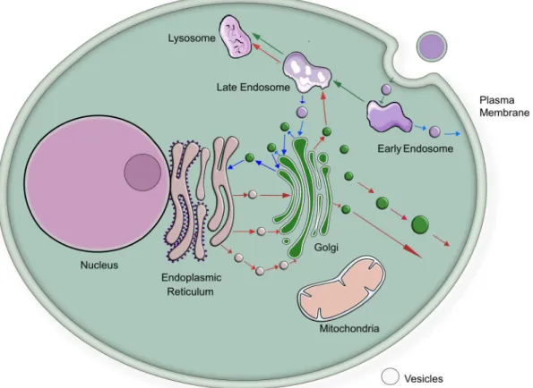
As different membranes (also within the same cell) have different functions, the lipid composition of membranes varies considerably [6,7,12]. In brief [13] (Fig. 1): Within an eukaryotic cell, the nuclear membrane fences the genetic blueprint off from the rest of the cell and at the same time forms a continuum with the endoplasmic reticulum. The endoplasmic reticulum is the synthetic site for membrane and secreted proteins as well as an important organelle involved in cellular responses to stress [14,15]. The endoplasmic reticulum is also the major organelle for lipid biosynthesis [6,7]. While cholesterol synthesis starts at the
* Corresponding author at: University Hospital Zurich, Department of Clinical Pharmacology and Toxicology, 8091 Zurich, Switzerland.
E-mail address: bruno.stieger@uzh.ch (B. Stieger).
Contents lists available at ScienceDirect
BBA - Molecular Basis of Disease
journal homepage: www.elsevier.com/locate/bbadis
https://doi.org/10.1016/j.bbadis.2021.166079
Received 15 August 2020; Received in revised form 12 December 2020; Accepted 7 January 2021
endoplasmic reticulum, cholesterol is rapidly leaving this organelle and is following the biosynthetic path of membranes to the cell surface, where it is found at a higher relative amount [16] (Fig. 2). Hence, the endoplasmic reticulum can be viewed as the assembly site of cellular membranes. The endoplasmic reticulum is in exchange with other cellular organelles by a brisk traffic of membrane vesicles moving from or to the endoplasmic reticulum [17] (Fig. 1). This vesicular flux is complemented by phospholipid transfer proteins mediating also ex- change of phospholipids between cellular compartments [18,19]. The Golgi apparatus puts the finish on newly synthesized membrane proteins and is also a repair system for membrane proteins of the plasma mem- brane [20]. The plasma membrane is the final destination for many membrane proteins and lipids. Functionally, the lipid composition of subcellular membranes needs to adapt from the endoplasmic reticulum to the plasma membrane from a biosynthetic to a barrier function [21].
This route of newly synthesized proteins and lipids from the endo- plasmic reticulum to the plasma membrane has a counterpart (Fig. 1):
Endocytosis is directed in an opposite direction from the plasma mem- brane towards the interior of the cell [22]. In polarized cells, endocytosis also continues into transcytosis, which is a vesicular route crossing cells [23]. The membrane flux through these routes with opposing and intersecting directions requires extensive sorting and regulation [24].
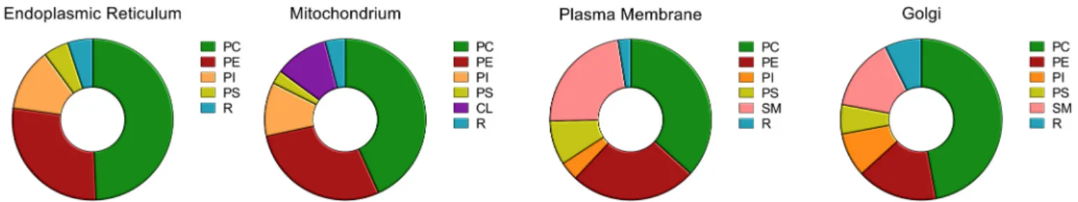
These very different functions of subcellular membranes are reflected by their different lipid compositions [6,7,25] (Fig. 3). At a higher level, this complex pattern of lipid specificity is topped at the level of organisms by different lipid compositions of different cell types fulfilling very diverse functions [26].
Lipids have been defined “as hydrophobic or amphipathic small molecules that may originate entirely or in part by carbanion-based condensations of thioesters (fatty acyls, glycerolipids, glycer- ophospholipids, sphingolipids, saccharolipids, and polyketides) and/or by carbocation-based condensations of isoprene units (prenol lipids and
sterol lipids)” [27] and can be divided into eight categories. Membrane lipids are distinguished into four [28] or three [7] categories glycer- ophospholipids, sphingolipids, sterols and glycolipids as minor compo- nent. Glycerophospholipids are derived from glycerol 3-phosphate, while sphingolipids are derived from sphingosine. The prototypic structure of sterols is cholesterol, which is also the major sterol found in mammalian membranes. Glycolipids are glycerophospholipids and sphingolipids containing sugars and are typically found in the outer leaflet. The large variety of different lipids [10,27] originates from are large number of different fatty acids and their combination as sub- stituents of the major lipid backbones.
Fig. 1. Schematic view of membrane flow in eukaryotic cells. Red arrows indicate flux of membrane vesicles from the endoplasmic reticulum towards the plasma membrane. Blue arrows indicate flux of membrane vesicles from the cell membrane towards the endoplasmic reticulum. These routes are not intended to imply a direct route of individual vesicles through the entire pathway. Green arrows indicate flux of vesicles in the endocytic compartment.
Fig. 2.Steady state distribution of cholesterol in major cellular organelles. ER:
endoplasmic reticulum, Mito: mitochondria, ES LS: endosomal and lysosomal compartment, PM: plasma membrane. Data are taken from [171].
3. Lipids tightly bound to membrane proteins
Lipids interact at different levels with membrane proteins [29]
(Fig. 4): Annular or boundary lipids are viewed as a single shell of lipids directly interacting with the membrane protein. This interaction may be very defined, e.g. by lipids found in clefts on the surface of the protein, occupying space between transmembrane helices or “sitting” on hy- drophobic protein surfaces. Lipids may also exert an interaction with membrane proteins at distance, which can be observed as different molar lipid to protein ratios in a membrane [29]. Studying lipids tightly bound to membrane proteins requires structural studies at a resolution sufficient to identify lipids or at least identification of lipids of highly purified membrane proteins by analytical tools. Studying the impact of bulk membrane lipids necessitates the isolation of highly purified frac- tions of the membranes of interest followed by lipid analysis.
The basic definition of solute transport is movement of a dissolved molecule from one compartment to another compartment, which is separated from each other by a biological membrane. The term solute implies that the molecule of interest is in a solution, which in biology is aqueous. Transport in biology is with rare exceptions (e.g. O2) mediated by transmembrane proteins. Parenthetically it is currently debated whether aquaporins act as proteins facilitating O2 movement across biological membranes [30]. Solute transport proteins are classified into SLC transporters, primary active ATP-dependent transporters (P-type ATPases and ABC transporters) and channels [31]. Investigating the direct interaction of lipids with transporters requires detailed knowl- edge on the protein structure. Detailed structural information on membrane proteins started to emerge in the mid1980s, when the crystal structure of bacteriorhodopsin was resolved [32,33]. Bacteriorhodopsin can be viewed as a transporter, as it uses the energy of light for moving protons against a concentration gradient across a bacterial membrane.
Following the transport definition given above, ion channels are trans- port proteins [31]. Consequently, the first structure of a bacterial membrane protein (porin) acting as a channel was published in 1991 [34] followed by the ferrichrome receptor/transporter [35]. In 1998 the structure of a bacterial homologue of a mammalian potassium channel
was unveiled. [36]. The P-type ATPase Ca2+-ATPase, which mediates Ca2+transport against a steep concentration gradient, was the first primary active transporter known structurally [37]. The first ABC transporter architecture was published in 2002 [38]. The first major facilitator structures were published in 2003 [39] including the co- transporter lactose permease [40]. These early structural studies of membrane proteins did not address the issue of lipids tightly bound to the investigated proteins.
The human genome encodes for more than 90 P-type ATPases [41].
The crystal structure of mammalian of (Na++K+)-ATPase was reported to contain a phosphatidylcholine tightly bound to an α-helix [42]. Later it was observed that the structure of (Na++K+)-ATPase crystalized from the rectal gland of spiny dogfish contained at the same position a tightly bound cholesterol, which was carried to the structure determination of the purified protein from the native transporter in the tissue used for isolation [43].
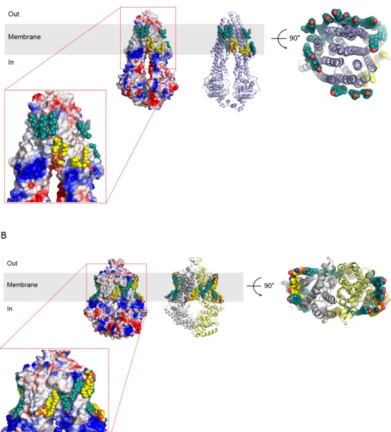
In human tissues, 48 ABC proteins are expressed, most of which are transporters [44]. Several laboratories published structures of MDR1 (also called P-glycoprotein). To the best of our knowledge, only one of the publications addressed the issue of lipids tightly bound to human MDR1 [45]. These authors observed at the level of the outer leaflet a ring of ordered cholesterol (Fig. 5A). At the level of the inner leaflet, tightly bound lipids were identified as phosphatidylethanolamines as well as cholesterol. A potential functional role of the tightly bound phospho- lipids in MDR1 function is supported by findings that phosphatidyleth- anolamine strongly stimulates the ATPase activity of partially purified MDR1 in a reconstituted system [46]. Whether the tightly bound cholesterol molecules are indeed solely responsible for the modulation of the ATPase and transport activity of MDR1 [47,48] remains to be experimentally demonstrated. Further, an additional model for the role of cholesterol in MDR1 transport has been proposed: the so called cholesterol fill-in model takes the rather large substrate binding site of MDR1 into account and suggests that cholesterol, by “filling” the bind- ing pocket in addition to a small substrate exerts the observed stimula- tion [49]. The cryogenic electron microscopy (cryo-EM) structure of ABCG2 indeed contained two cholesterol molecules located at the sub- strate binding site of this multidrug exporter [50]. Comparable to MDR1, ABCG2 is also surrounded by a belt of tightly bound phospho- lipids, namely five phosphatidylethanolamines as well as five choles- terol molecules (Fig. 5B) [51]. Here, the lipids are located at the outer surface of the homodimer, but not between the subunits. It is premature at this state of knowledge to make detailed statements about the inter- action of the tightly bound lipids with these two ABC transporters, but in both structures, some of the lipids are clearly observed in indentations at the protein surface. It should also be noted that no lipids intercalating with the transmembrane helices were identified in these two proteins (Fig. 5). A tight interaction of lipids with ABC transporters is not restricted to the plasma membrane. The structure of the mitochondrial transporter ABCB10 contains tightly bound cardiolipin, a lipid restricted to mitochondria [52]. Taken together, there is now plenty of structural evidence available that transport proteins utilizing ATP as energy source contain tightly bound phospholipids and cholesterol.
Fig. 3. Steady state distribution of lipids in major cellular organelles. PC: phosphatidylcholine, PE: phosphatidylethanolamine, PI: phosphatidylinositol, PS: phos- phatidylserine, R: remaining lipids. Data are taken from [6,171].
Fig. 4. Schematic view of membrane lipid interaction with transporters. Lipids can interact with transporters as tightly bound lipids (marked in blue) or via the bulk lipid composition (blue arrow).
Fig. 5. Phospholipids and cholesterol bound to ABCB1 (A) and to ABCG2 (B). The structures of the transporters are shown either as electrostatic surface potentials (ESPs) or as ribbon diagrams. ESPs are colored for negative partial charges red and blue for positive partial charges. Tightly bound cholesterol molecules and phosphatidylethanolamine are shown in sphere representation. Cholesterol is colored green, whereas phosphatidylethanolamine is colored yellow. The structures are taken from [45] for MDR1 and from [51] for ABCG2.
Currently, over 450 transporters belonging to the SLC superfamily of transporters are known in humans [54]. The structure of the Drosophila melanogaster dopamine transporter (DAT; SLC6A3) displays a tightly bound cholesterol molecule in a groove between TM5 and TM7 [55].
Similarly, the transporter mediating the transport of the human neuro- transmitter serotonin (SERT; SLC6A4) binds tightly cholesterol hemi- succinate, which in vivo may be a binding site for cholesterol [56]. A tightly bound cholesterol hemisuccinate was also identified in the human glutamine transporter (ASCT2, SLC1A5) [57]. The recent cryo- EM structure of the human L-type amino acid transporter (SLC7A5) in complex with SLC3A2 shows several tightly bound cholesterol mole- cules, which may also be involved in the interaction of the two subunits of this transporter [58]. The crystal structure of the mitochondrial ADP/
ATP exchanger from beef heart has been shown tightly binding two cardiolipins, a lipid only found in mitochondria [59] and in a follow up crystal structure three cardiolipin molecules [60]. Hence, tightly bound lipids are not only “simple” but also complex glycerophospholipds. The list of bacterial transporters containing tightly bound lipids is consid- erable [61] and using mass spectrometry as a new tool for the investi- gation of the interaction membrane proteins with tightly bound lipids offers a new and exciting possibility for expanding this list further [28].
TMEM16K is a lipid scramblase residing in the endoplasmic reticulum and its function requires a transient, tight interaction with phospho- lipids. Interestingly, this scramblase also contains tightly bound, yet unidentified lipids at the boundary of the protein and the membrane of the endoplasmic reticulum [53].
Channels are the third class of transporters and over 310 genes coding for channels are currently known in humans [31]. Potassium inwardly rectifying (Kir) channels interacts with phosphatidylinositol 4,5-bisphosphonate (PIP2), which in turn acts as an agonist on these channels [62]. The determination of the X-ray crystal structure of chicken Kir 2.2 bound with a short-chain PIP2 derivative revealed that one PIP2 binds to each of the four subunits [63]. The binding site is located at the interface between the transmembrane and the cytoplasmic domain and binding of PIP2 induces a large change in conformation by tethering the cytoplasmic domain to the transmembrane domain, which activates the channel. The ammonia channel (AmtB) from Escherichia coli was found binding eight phosphatidylglycerol molecules, while a ninth molecule could not be definitively identified [64]. Cryo-EM analysis of the human voltage-gated Nav1.4/α1 sodium channel revealed densities of four phospholipid molecules bound to the pore- forming α-subunit [65]. The authors interpreted three additional linear densities traversing fenestrations of the pore subunit as lipids.
Similarly, the cryo-EM structure of a human voltage gated calcium channel, Cav3.1, which was engineered as a corresponding splice variant of a rat Cav.1 lacking the linker between domains I and II also identified transverse lipids in the pore domain [66]. Bacterial voltage-gated so- dium channels (NaChBac) are likely evolutionary ancestors of mammalian voltage-gated sodium channels [67] and also contain mul- tiple tightly bound lipids at the transmembrane domain [68]. Transient receptor channel TRPM8 has been studied in detail and is a channel requiring PIP2 for action. [69].
Taken together, as cryo-EM technology nurtures a rapidly expanding list of transport and other membrane protein structures [70,71], the information of tightly bound lipids to such transporters is rapidly growing as well. For example, the purified transporter associated with antigen processing (TAP) (ABCB2/ABCB3) was found to be associated with several different phospholipids [72].
4. Lipid dependent modulation of transport protein activity As outlined above, the identification of tightly bound lipids to transport proteins as well as other membrane proteins requires knowl- edge of the structure of the proteins of interest and their direct sur- roundings. This information should be complemented and detailed to the atomic level of highly purified membrane proteins by the application
of mass spectrometry. Since both approaches require highly specialized and sophisticated equipment as well as technically challenging pro- cedures, the impact of lipid modulation on the function of membrane proteins like transporters is so far often investigated at the bulk level where membrane lipid analysis is combined with a functional assay using the same membrane preparation.
The investigation of the role of lipids on membrane protein function started by modifying the lipid composition of the membrane in which the protein of interest is expressed. (Na++K+)-ATPase is a P-type ATPase and as such a prototypic primary active transporter, which is in addition key for providing the energy source for secondary active transporters [73]. The activity of (Na++K+)-ATPase of erythrocytes from rats fed with different fat diets was found to depend on erythrocyte membrane fatty acid composition [74] as well as on cholesterol content [75] of red cell membranes. Working with (Na++K+)-ATPase from bovine kidney, a bell-shaped dependence of the activity of this transporter on membrane cholesterol content was reported [76]. A study working with shark rectal gland (Na++K+)-ATPase reconstituted in proteoliposomes revealed that the enzymatic activity was both a function of the cholesterol content and the acyl chain length of the phospholipids [77,78]. In the absence of cholesterol the maximal turnover rate was at an acyl chain length of 22, while in the presence of 40 mol% cholesterol, the acyl chain length for maximal turnover rate decreased to 18. Another study found the dependence of the ATPase activity from porcine kidney on membrane thickness by a method known to alter membrane thickness [79]. How- ever, the thickness of the membrane was not experimentally determined in this study. While is seems intuitive that increasing acyl chain length increases membrane thickness, this has not been verified experimentally as a general rule. Rather it was observed that the area occupied by a phospholipid molecule increases more than the thickness of the bilayer when the acyl chain length is increased [80]. In addition, it should be kept in mind that the hydrophobic surface of a membrane protein exerts a force on the lipid bilayer and can induce local curvatures in the lipid bilayer. Lipids in the bilayer on the other hand can counteract such structural changes and thereby exert a force on the membrane protein [81,82]. Consequently, there is a complex and mutual biophysical interaction between membrane lipids and membrane proteins.
P-type ATPases reside in all cellular membranes [83]. Reconstitution of sarcoplasmic reticulum Ca2+-ATPase into proteoliposomes demon- strated a dependence of its transport activity on the phosphatidyletha- nolamine content [84]. The effects of lipids on Ca2+-ATPase were found to be cooperative: The activity of this transporter is lower when recon- stituted in a phosphatidylethanolamine membrane than in a membrane composed of phosphatidylcholine. However, in a mixture of the two phospholipids, the portion of phosphatidylethanolamine needs to exceed 80 mol% to reduce the ATPase activity of the reconstituted Ca2+- ATPase [85]. However, at contents higher than 75% of phosphatidyl- ethanolamine, the lipid forms a hexagonal (II) phase, which may, by mechanisms other than protein-lipid interactions, affect the activity of Ca2+-ATPase. In addition to affecting the activity [86], the chain length of phosphatidylcholine affects the stoichiometry of Ca2+ binding to sarcoplasmic reticulum Ca2+-ATPase [87]. These lipid effects are not restricted to the isoform of Ca2+-ATPase expressed in the sarcoplasmic reticulum. Ca2+-ATPase purified from human erythrocyte membranes and reconstituted into proteoliposomes is also regulated by various phospholipids, which affect both, Vmax and Km [88].
ABC transporters are another very important superfamily of primary active transporters [31]. MDR1 is known to be very sensitive to its lipid environment, as this transporter loses its activity if deprived from lipids during purification [89,90]. Drug binding is also depending on the type of phospholipids used [91]. Drug binding to purified MDR1 recon- stituted in proteoliposomes shows a bell-shape dependence of relative cholesterol content [92]. Depleting cells expressing MDR1 from cholesterol reduced the activity of MDR1 using different substrates [93–95]. It is interesting to note that the ATPase activity of MDR1 reconstituted into proteoliposomes is lower in comparison to
reconstitution into nanodiscs [96]. The exact reason for the higher basal ATPase rate of MDR1 in nanodiscs is unknown. Hence, the specific ac- tivity of MDR1 in both configurations does not differ. In 2007, three laboratories reported that the activity of ABCG2 is stimulated by an increased membrane cholesterol content [97–99]. Using cholesterol- poor insect cell membranes [100] in conjunction with sterol loading, this stimulation was most pronounced, if not specific for cholesterol:
ergosterol loading tended to reduce ABCG2 transport activity while sitosterol and hydrocortisone had no effect [99]. This sterol-dependence of transporter activation is different in MDR1, where next to cholesterol stigmasterol, sitosterol and campesterol stimulate ATPase activity [49].
Cholesterol did not affect the Km values of ABCG2, but stimulated Vmax
about 20-fold for methotrexate and stimulated Vmax for estradiol-17β- glucuronide about 15-fold [99]. Another group also found no effect of cholesterol loading on Km values, but an about 25-fold Vmax stimulation for esterone-3-sulfate and an about 8-fold Vmax stimulation for prazosin transport [97]. Starting from isolated canalicular membrane vesicles from mouse liver, cholesterol depletion showed a reduction of the transport activity of mBSEP and of mMRP2, illustrating again a stimu- latory effect of cholesterol on these two ABC transporters [102]. The effect of cholesterol on the transport activity of the bile salt export pump BSEP was investigated using four different bile salts as substrate and three species: human, rat and mouse. As for ABCG2, BSEP of all three species was only affected by an increased Vmax but not at the Km for all bile salts tested after cholesterol loading Sf9 insect cell vesicles [103].
Mutations in ABCB11 coding for BSEP lead to progressive cholestasis, which progresses often to severe liver disease [104] and polymorphisms in the ABCB11 gene may lead to acquired cholestasis [105]. Often, BSEP variants are functionally characterized upon expression in insect cells.
As insect cell membrane vesicles are a convenient tool for functionally characterizing ABC transporters [106], we investigated the impact of cholesterol on the function of BSEP variants [107]. The p.444A and the wild type BSEP variants did not change affinity to taurocholate, but had both an increased Vmax in high cholesterol insect membrane vesicles.
The p.E297G and the p.R432T variants can cause cholestasis in patients and display practically no transport activity in wild type insect cell membrane vesicles. These two variants displayed clearly measureable transport activity after cholesterol loading. However they did not come near the transport activity of the wild type BSEP in cholesterol loaded insect cell vesicles [107]. These findings suggest that changes of cholesterol levels in the canalicular membrane are not an additional susceptibility factor for acquired (or inherited) cholestasis. This result obtained for BSEP variants could also indicate that reconstituting BSEP (as well as other hepatocellular ABC transporters) in lipid membranes of different cholesterol content for structural studies may not induce gross structural alterations. Studies using HepG2 cells and a functional assay for MRP2 showed that in this expression system MRP2 activity was dependent on cholesterol [101]. In parallel to the kinetic BSEP study, we also investigated the effect of membrane cholesterol on kinetic proper- ties of MRP2. For all substrates, the Vmax was increased by cholesterol loading [107]. Interestingly, the effects on the affinity were substrate dependent: For small substrates, loading with cholesterol turned allo- steric transport kinetics to regular Michaelis-Menten type kinetics, while for large substrates, the transporter showed in both conditions regular kinetics. In this context, it is interesting and important to note that insect Sf9 cells contain endogenous ABC transporter activities, which become unmasked after cholesterol loading [108]. Two potential candidate genes belonging to the ABCC family are found in Sf9 cells and could explain the observed substrate pattern: SfABCC2 and SfABCC3. Hence, in all expression systems, careful controls are needed when studying the impact of membrane lipid composition on (ABC) transporter activity.
The transporter associated with antigen processing (TAP) is a hetero- dimer composed of ABCB2 and ABCB3 and resides in the endoplasmic reticulum [109]. Functional reconstitution of this heterodimer expressed in the yeast Pichia pastors shows a lipid dependence whereby the transporter was not active in phosphatidylcholine [72].
Interestingly, addition of cholesterol inhibited TAP activity, which may be explained by the relatively low cholesterol content of the endo- plasmic reticulum [16] (Fig. 2). Cystic fibrosis transmembrane conductor (ABCC7) functions as an ATP-gated chloride channel [110].
Investigation of the purified and reconstituted protein showed a clear dependence of the ATPase activity on cholesterol [111].
To our knowledge, the effect of sphingolipids on the activity of ABC transporters has not been investigated in detail. Depleting murine neu- roblastoma Neuro-2a cells from sphingolipids did not result in an alteration of MRP1 activity as assessed by a fluorescein efflux assay [112]. In the same cell line, depletion of gangliosides did also not affect the activity of MDR1 and MRP1 [113]. We found that sphingomyelin loading of membranes with low and with high cholesterol content did not affect the activity of MRP2 and BSEP (Steiger and Stieger, unpublished).
Tetraethylammonium is a substrate of human organic cation trans- porters of the SLC22A and SLC47A families [114]. Loading cholesterol to brush border membrane vesicles isolated from rat kidney resulted in both, a reduced dissipation rate of a H+gradient and an increased intrinsic tetraethylammonium uptake (in the absence of a pH gradient) [115]. This can be interpreted that one (or more) of the organic cation transporters are modulated by membrane cholesterol content. An alternate interpretation of these findings is an altered stability of sub- strate gradients over the membrane of the vesicles. Reconstitution ex- periments of bovine cardiac sarcolemmal Na+/Ca2+ exchanger demonstrated that the addition of plasmalogen to the proteoliposomes massively stimulates the activity of this exchanger [116].
Depleting cholesterol from membranes of HEK 293 cells expressing the rat serotonin transporter 1 (rSRT1) reduced rSRT1 transport activity [117]. Replenishing membrane steroid content with cholesterol, but not with ergosterol, 5-cholestene or pregnenolone restored the transport activity, suggesting a specific interaction of cholesterol with this trans- porter. Depletion of cholesterol from HEK293 cells expressing rat NTCP (SLC10A1) led to an increased taurocholate uptake by the latter [118].
This suggests that this transporter is modulated by membrane choles- terol content. A caveat remains as in the same paper the subcellular localization of NTCP was altered depending on the cholesterol content, with a stronger membrane fluorescence after cholesterol depletion. Own unpublished data show that the transport activity of rNTCP expressed in cholesterol poor insect Sf9 cells decreases with addition of cholesterol, which is opposite to the published finding. Interestingly, the highly homologous intestinal bile salt transporter ASBT (SLC10A2), if expressed in HEK293 cells was inhibited by cholesterol depletion [119].
Currently, it remains unclear whether the opposing effects of membrane cholesterol content are an intrinsic property of the two closely related transporters or whether they are due to technical issues. The finding with NTCP is reminiscent of a study of the Glucose transporter GLUT1 expressed in M07e cells, where cholesterol depletion leads to a trans- location of GLUT1 to the plasma membrane accompanied by an increased D-glucose transport activity [120]. This example shows that modulation of plasma membrane cholesterol content in cells may not only affect the function of membrane transporters but also the subcel- lular localization of transporters. Any altered expression level at the plasma membrane will lead to altered transport rates.
If HEK293 cells expressing the neutral amino acid transporter LAT1 (SLC7A5) are depleted in cholesterol, the Vmax of L-DOPA transport is decreased, whereas the Km remains unchanged [121]. Based on results obtained from LAT1 purification in the presence and absence of cholesterol and in analogy to DAT1 and SERT1, the authors concluded that the function of LAT1 critically depends on tightly bound cholesterol molecules. Similarly, the transport activity of OCT2 (SLC22A2) expressed in HEK293 cells was reduced after cholesterol depletion [122]. In addition, cholesterol depletion shifted OCT2-mediated trans- port of 1-methyl-4-phenylpyridinium transport from allosteric kinetics to Michaelis-Menten kinetics in this study. To gain more insight on the functional consequences of tightly bound cholesterol on SERT1, this
transporter was expressed in HEK293-MSR cells [123]. In a binding assay, cholesterol depletion shifted the conformation of SERT1 towards a more inward-facing conformation.
Cardiolipin is a dimeric anionic phospholipid and is in eukaryotes almost exclusively found in the inner mitochondrial membrane [124]. It could be demonstrated in genetically modified yeast that cardiolipin is essential for the stability and function of the mitochondrial Ca2+uni- porter [125]. This role of cardiolipin is further supported by the clinical manifestation of the Barth syndrome, displaying among others with cardiomyopathy and skeletal muscle myopathy. Barth syndrome is an X- linked disorder with an inherited defect in the biosynthesis of car- diolipin [126].
Like other transmembrane proteins, ion channels are also modulated by bulk lipid composition of membranes. With the rapid progress made by cryo-EM structure determination, investigation of the lipid interac- tion with channels as wells as other transport proteins is now not only focusing on the impact of bulk lipid composition but also on tightly bound annular lipids and these areas are rapidly moving together, as outlined by a series of recent overviews [127–133].
In summary, experiments manipulating bulk membrane lipid composition either in cells or when reconstituting transporters into proteoliposomes have provided ample evidence that (bulk) membrane lipid composition modulates the activity and properties of transport proteins. This effect is not restricted to the lipid class but also, in the case of phospholipids, to acyl chain composition. It is important to note that the effect of cholesterol on transporter activity may be complex. The cholesterol (sterol) to lipid ratio affects lipid dynamics in membranes [134,135]. However, for some transporters, an alteration of activity was not observed for all sterols tested, but was found to be sterol specific.
This implies that in addition to alterations of membrane lipid dynamics a specific interaction of sterols with transporters is relevant, which was, indeed, observed in cryo-EM studies. Having now the first high resolu- tion structures of lipid transporters [136,137] or translocators [53,138–140], it will be interesting to see whether structural features of these lipid transporters reveal information on structural requirements for the tight binding of annular lipids in transmembrane transporters as well as other transmembrane proteins. Needless to say that such infor- mation will also be obtained once the resolution of membrane proteins can be further increased.
5. Disease-mediated lipid changes
In Western society, overweight and obesity are of epidemic extent and rising [141]. This condition may lead to nonalcoholic fatty liver disease [142]. Nonalcoholic fatty liver disease is a liver pathology ranging from steatosis to steatohepatitis, which may progress to fibrosis and ultimately cirrhosis [143]. As it is difficult obtaining human liver tissue for analytical and functional studies, the number of studies using human plasma samples clearly exceeds the number of studies working with human liver tissue [142]. It was for example observed in liver bi- opsies that the ratio of free cholesterol to phosphatidylcholine is significantly increased in patients with nonalcoholic fatty liver disease or nonalcoholic steatohepatitis [144]. Lipidomic analysis of liver bi- opsies from patients with nonfatty liver disease demonstrated that different stages can be discriminated [145]. A lipidomic analysis in conjunction with a random forests based machine learning of liver bi- opsies from patients with nonalcoholic fatty liver disease and from pa- tients with nonalcoholic steatohepatitis led to the identification of a specific lipid signature of 32 lipids allowing the discrimination of the different forms of fatty liver diseases from controls [146]. The changes observed at the level of individual phospholipids are complex, some phospholipids tend to be increased and some tend to be decreased in patients relative to control samples. In addition, the lipid composition between non-steatotic and steatotic tissue areas is very different [147].
A zonal differentiation of lipid patterns is not only found in diseased human livers but also in biopsies from healthy individuals [148]. It was
shown that the clearance of mebrofenin is significantly reduced in pa- tients with nonalcoholic steatohepatitis in comparison to control sub- jects [149]. Mebrofenin is a substrate for organic anion uptake transporters and of MRP2 [150,151]. A quantitative proteomic analysis of human liver biopsies showed a significantly lower expression of OATP1B1, OATP1B3 as well as MRP2 [152]. This example shows that the impact of altered membrane lipid composition in diseased states on transporter function is not easily delineated from human studies, as in parallel to altering membrane lipid composition, diseases also affect the protein expression levels of transporters. So far no common pattern on altered transport protein expression in biopsies from humans with different forms and degrees of liver diseases has emerged [153]. The interpretation of human pharmacokinetic studies at the level of molec- ular mechanisms is further complicated as not only transporters, but also drug metabolizing enzymes display altered protein expression in different forms of nonalcoholic fatty liver disease [154].
Statins are inhibitors of cholesterol biosynthesis and are widely prescribed drugs for the treatment of patients with hypercholesterole- mia. Statins are generally well tolerated but are known to lead to myopathy as the most common adverse event [155]. The exact molec- ular mechanism leading to statin-induced myopathy is still discussed [156]. Biophysical changes of membrane properties may be involved.
Treatment of HEK293T cells with simvastatin led to a reduction of the cholesterol to phospholipid ratio, but did not change membrane fluidity [157]. This study did however observe an increase in phosphatidyleth- anolamine and its plasmalogen. To what extent this finding can be extrapolated to myocytes remains open. It is accepted that statins may affect plasma membrane microdomains (also called lipid rafts) [158].
Treatment of patients with statins was found to cause a reduction of erythrocyte and platelet membrane cholesterol and an increased activity of (Na++K+)-ATPase [159–161]. However, conflicting data on choles- terol content of erythrocytes have been published [162].
Viral infections, for example with hepatitis viruses, affect over two billion people globally [163]. Hepatitis viruses are one example of many different viruses affecting not only protein expression but also lipid homeostasis including membrane lipid composition after host cell infection [164]. Such lipid changes might alter the function of mem- brane proteins such as transporters. Viral infection has also been demonstrated to alter transporter expression, e.g. in liver [165], but it should be kept in mind that transporter expression shows a large interindividual variability [166].
6. Conclusion
Technical advances in lipidomics as well in the determination of structures of membrane proteins have provided convincing evidence for an important role of membrane lipids in affecting both the structure and the function of transport proteins. Evidence is emerging that changes in lipid composition in vivo (either due to diseases or to dietary habits) may have consequences on the disposition of drugs. It remains chal- lenging to differentiate between altered transporter numbers and altered transporter function, as quantification of transporters requires taking biopsies from the organs of interest, which is generally not possible with humans. As erythrocytes also express transport proteins [167,168] and are easily available from patients, transport protein levels could be determined by quantitative, targeted proteomics. Such a surrogate approach might be useful for an estimation of transporter expression levels in individual patients. To the best of our knowledge, such an approach has so far not been validated in a study involving humans. In parallel to in vivo transport (pharmacokinetics) quantification, the number of transporter substrates suitable for imaging in humans is constantly increasing [169,170]. Combing in vivo methods with in vitro experiments will certainly advance our knowledge on the impact of membrane lipid composition and of tightly bound lipids on the modu- lation of transport protein function and its role in health and disease.
Declaration of competing interest The authors have no conflicts of interest.
Acknowledgements
This work was supported by grants # 310030_155563 to BST and # 310030_189111 to KPL from the Swiss National Science Foundation.
References
[1] M.M. Hanczyc, P.A. Monnard, Primordial membranes: more than simple container boundaries, Curr. Opin. Chem. Biol. 40 (2017) 78–86.
[2] K. Ruiz-Mirazo, C. Briones, A. de la Escosura, Prebiotic systems chemistry: new perspectives for the origins of life, Chem. Rev. 114 (1) (2014) 285–366.
[3] H. Watson, Biological membranes, Essays Biochem. 59 (2015) 43–69.
[4] J.D. Robertson, Unit membranes: a review with recent new studies of experimental alterations and a new subunit strucutre in synaptic membranes, in:
M. Locke (Ed.), Cellular Membranes in Development, Acadmeic Press, New York, 1964, pp. 1–81.
[5] S.J. Singer, G.L. Nicolson, The fluid mosaic model of the structure of cell membranes, Science 175 (4023) (1972) 720–731.
[6] G. van Meer, D.R. Voelker, G.W. Feigenson, Membrane lipids: where they are and how they behave, Nat Rev Mol Cell Biol 9 (2) (2008) 112–124.
[7] T. Harayama, H. Riezman, Understanding the diversity of membrane lipid composition, Nat Rev Mol Cell Biol 19 (5) (2018) 281–296.
[8] T. Züllig, H.C. Kofeler, High resolution mass spectrometry in lipidomics, Mass ¨ Spectrom. Rev. (2020), https://doi.org/10.1002/mas.21627 [Online ahead of print].
[9] T. Züllig, M. Trotzmüller, H.C. K¨ofeler, Lipidomics from sample preparation to data analysis: a primer, Anal. Bioanal. Chem. 412 (10) (2020) 2191–2209.
[10] S. Subramaniam, E. Fahy, S. Gupta, M. Sud, R.W. Byrnes, D. Cotter, A.
R. Dinasarapu, M.R. Maurya, Bioinformatics and systems biology of the lipidome, Chem. Rev. 111 (10) (2011) 6452–6490.
[11] L.C. Czuba, K.M. Hillgren, P.W. Swaan, Post-translational modifications of transporters, Pharmacol. Ther. 192 (2018) 88–99.
[12] C. Klose, M.A. Surma, K. Simons, Organellar lipidomics–background and perspectives, Curr. Opin. Cell Biol. 25 (4) (2013) 406–413.
[13] G. Blobel, Protein targeting (Nobel lecture), Chembiochem 1 (2) (2000) 86–102.
[14] G.K. Voeltz, M.M. Rolls, T.A. Rapoport, Structural organization of the endoplasmic reticulum, EMBO Rep. 3 (10) (2002) 944–950.
[15] J.H. Lin, P. Walter, T.S. Yen, Endoplasmic reticulum stress in disease pathogenesis, Annu. Rev. Pathol. 3 (2008) 399–425.
[16] M.S. Bretscher, S. Munro, Cholesterol and the Golgi apparatus, Science 261 (5126) (1993) 1280–1281.
[17] H. Stenmark, Rab GTPases as coordinators of vesicle traffic, Nat Rev Mol Cell Biol 10 (8) (2009) 513–525.
[18] S. Cockcroft, P. Raghu, Phospholipid transport protein function at organelle contact sites, Curr. Opin. Cell Biol. 53 (2018) 52–60.
[19] Y. Yang, M. Lee, G.D. Fairn, Phospholipid subcellular localization and dynamics, J. Biol. Chem. 293 (17) (2018) 6230–6240.
[20] M.G. Farquhar, G.E. Palade, The Golgi apparatus (complex)-(1954–1981)-from artifact to center stage, J Cell Biol, 91 (3 Pt 2) (1981) 77s–103s.
[21] J.C. Holthuis, A.K. Menon, Lipid landscapes and pipelines in membrane homeostasis, Nature 510 (7503) (2014) 48–57.
[22] H. Riezman, P.G. Woodman, G. van Meer, M. Marsh, Molecular mechanisms of endocytosis, Cell 91 (6) (1997) 731–738.
[23] K.E. Mostov, M.H. Cardone, Regulation of protein traffic in polarized epithelial cells, Bioessays 17 (2) (1995) 129–138.
[24] R. Sannerud, J. Saraste, B. Goud, Retrograde traffic in the biosynthetic-secretory route: pathways and machinery, Curr. Opin. Cell Biol. 15 (4) (2003) 438–445.
[25] D. Casares, P.V. Escriba, C.A. Rossello, Membrane lipid composition: effect on membrane and organelle structure, function and compartmentalization and therapeutic avenues, Int. J. Mol. Sci. 20 (9) (2019).
[26] A.A. Spector, M.A. Yorek, Membrane lipid composition and cellular function, J. Lipid Res. 26 (9) (1985) 1015–1035.
[27] E. Fahy, S. Subramaniam, R.C. Murphy, M. Nishijima, C.R. Raetz, T. Shimizu, F. Spener, G. van Meer, M.J. Wakelam, E.A. Dennis, Update of the LIPID MAPS comprehensive classification system for lipids, J. Lipid Res. 50 (Suppl) (2009) S9–14.
[28] J.R. Bolla, M.T. Agasid, S. Mehmood, C.V. Robinson, Membrane protein-lipid interactions probed using mass spectrometry, Annu. Rev. Biochem. 88 (2019) 85–111.
[29] M.F. Brown, Soft matter in lipid-protein interactions, Annu. Rev. Biophys. 46 (2017) 379–410.
[30] J.J. Zwiazek, H. Xu, X. Tan, A. Navarro-Rodenas, A. Morte, Significance of oxygen transport through aquaporins, Sci. Rep. 7 (2017) 40411.
[31] M.A. Hediger, B. Clemencon, R.E. Burrier, E.A. Bruford, The ABCs of membrane transporters in health and disease (SLC series): introduction, Mol. Asp. Med. 34 (2–3) (2013) 95–107.
[32] J. Deisenhofer, O. Epp, K. Miki, R. Huber, H. Michel, X-ray structure analysis of a membrane protein complex. Electron density map at 3 A resolution and a model
of the chromophores of the photosynthetic reaction center from Rhodopseudomonas viridis, J. Mol. Biol. 180 (2) (1984) 385–398.
[33] J. Deisenhofer, H. Michel, Nobel lecture, The photosynthetic reaction centre from the purple bacterium Rhodopseudomonas viridis, EMBO J 8 (8) (1989) 2149–2170.
[34] M.S. Weiss, U. Abele, J. Weckesser, W. Welte, E. Schiltz, G.E. Schulz, Molecular architecture and electrostatic properties of a bacterial porin, Science 254 (5038) (1991) 1627–1630.
[35] K.P. Locher, B. Rees, R. Koebnik, A. Mitschler, L. Moulinier, J.P. Rosenbusch, D. Moras, Transmembrane signaling across the ligand-gated FhuA receptor:
crystal structures of free and ferrichrome-bound states reveal allosteric changes, Cell 95 (6) (1998) 771–778.
[36] D.A. Doyle, J. Morais Cabral, R.A. Pfuetzner, A. Kuo, J.M. Gulbis, S.L. Cohen, B.
T. Chait, R. MacKinnon, The structure of the potassium channel: molecular basis of K+conduction and selectivity, Science 280 (5360) (1998) 69–77.
[37] C. Toyoshima, M. Nakasako, H. Nomura, H. Ogawa, Crystal structure of the calcium pump of sarcoplasmic reticulum at 2.6 A resolution, Nature 405 (6787) (2000) 647–655.
[38] K.P. Locher, A.T. Lee, D.C. Rees, The E. coli BtuCD structure: a framework for ABC transporter architecture and mechanism, Science, 296 (5570) (2002) 1091–1098.
[39] Y. Huang, M.J. Lemieux, J. Song, M. Auer, D.N. Wang, Structure and mechanism of the glycerol-3-phosphate transporter from Escherichia coli, Science 301 (5633) (2003) 616–620.
[40] J. Abramson, I. Smirnova, V. Kasho, G. Verner, H.R. Kaback, S. Iwata, Structure and mechanism of the lactose permease of Escherichia coli, Science 301 (5633) (2003) 610–615.
[41] M.H. Saier Jr., C.V. Tran, R.D. Barabote, TCDB: the Transporter Classification Database for membrane transport protein analyses and information, Nucleic Acids Res. 34 (Database issue) (2006) D181–D186.
[42] J.P. Morth, B.P. Pedersen, M.S. Toustrup-Jensen, T.L. Sorensen, J. Petersen, J.
P. Andersen, B. Vilsen, P. Nissen, Crystal structure of the sodium-potassium pump, Nature 450 (7172) (2007) 1043–1049.
[43] T. Shinoda, H. Ogawa, F. Cornelius, C. Toyoshima, Crystal structure of the sodium-potassium pump at 2.4 A resolution, Nature 459 (7245) (2009) 446–450.
[44] M. Dean, T. Annilo, Evolution of the ATP-binding cassette (ABC) transporter superfamily in vertebrates, Annu. Rev. Genomics Hum. Genet. 6 (2005) 123–142.
[45] A. Alam, J. Kowal, E. Broude, I. Roninson, K.P. Locher, Structural insight into substrate and inhibitor discrimination by human P-glycoprotein, Science 363 (6428) (2019) 753–756.
[46] C.A. Doige, X. Yu, F.J. Sharom, The effects of lipids and detergents on ATPase- active P-glycoprotein, Biochim. Biophys. Acta 1146 (1) (1993) 65–72.
[47] P.D. Eckford, F.J. Sharom, Interaction of the P-glycoprotein multidrug efflux pump with cholesterol: effects on ATPase activity, drug binding and transport, Biochemistry 47 (51) (2008) 13686–13698.
[48] F.J. Sharom, Complex interplay between the P-glycoprotein multidrug efflux pump and the membrane: its role in modulating protein function, Front. Oncol. 4 (2014) 41.
[49] Y. Kimura, N. Kioka, H. Kato, M. Matsuo, K. Ueda, Modulation of drug-stimulated ATPase activity of human MDR1/P-glycoprotein by cholesterol, Biochem. J. 401 (2) (2007) 597–605.
[50] N.M.I. Taylor, I. Manolaridis, S.M. Jackson, J. Kowal, H. Stahlberg, K.P. Locher, Structure of the human multidrug transporter ABCG2, Nature 546 (7659) (2017) 504–509.
[51] S.M. Jackson, I. Manolaridis, J. Kowal, M. Zechner, N.M.I. Taylor, M. Bause, S. Bauer, R. Bartholomaeus, G. Bernhardt, B. Koenig, A. Buschauer, H. Stahlberg, K.H. Altmann, K.P. Locher, Structural basis of small-molecule inhibition of human multidrug transporter ABCG2, Nat. Struct. Mol. Biol. 25 (4) (2018) 333–340.
[52] C.A. Shintre, A.C. Pike, Q. Li, J.I. Kim, A.J. Barr, S. Goubin, L. Shrestha, J. Yang, G. Berridge, J. Ross, P.J. Stansfeld, M.S. Sansom, A.M. Edwards, C. Bountra, B.
D. Marsden, F. von Delft, A.N. Bullock, O. Gileadi, N.A. Burgess-Brown, E.
P. Carpenter, Structures of ABCB10, a human ATP-binding cassette transporter in apo- and nucleotide-bound states, Proc. Natl. Acad. Sci. U. S. A. 110 (24) (2013) 9710–9715.
[53] S.R. Bushell, A.C.W. Pike, M.E. Falzone, N.J.G. Rorsman, C.M. Ta, R.A. Corey, T.
D. Newport, J.C. Christianson, L.F. Scofano, C.A. Shintre, A. Tessitore, A. Chu, Q. Wang, L. Shrestha, S.M.M. Mukhopadhyay, J.D. Love, N.A. Burgess-Brown, R. Sitsapesan, P.J. Stansfeld, J.T. Huiskonen, P. Tammaro, A. Accardi, E.
P. Carpenter, The structural basis of lipid scrambling and inactivation in the endoplasmic reticulum scramblase TMEM16K, Nat. Commun. 10 (1) (2019) 3956.
[54] M.D. Pizzagalli, A. Bensimon, G. Superti-Furga, A guide to plasma membrane solute carrier proteins, FEBS J. (2020), https://doi.org/10.1111/febs15531 [Online ahead of print].
[55] A. Penmatsa, K.H. Wang, E. Gouaux, X-ray structure of dopamine transporter elucidates antidepressant mechanism, Nature 503 (7474) (2013) 85–90.
[56] J.A. Coleman, E.M. Green, E. Gouaux, X-ray structures and mechanism of the human serotonin transporter, Nature 532 (7599) (2016) 334–339.
[57] X. Yu, O. Plotnikova, P.D. Bonin, T.A. Subashi, T.J. McLellan, D. Dumlao, Y. Che, Y.Y. Dong, E.P. Carpenter, G.M. West, X. Qiu, J.S. Culp, S. Han, Cryo-EM structures of the human glutamine transporter SLC1A5 (ASCT2) in the outward- facing conformation, eLife, 8, 2019.
[58] Y. Lee, P. Wiriyasermkul, C. Jin, L. Quan, R. Ohgaki, S. Okuda, T. Kusakizako, T. Nishizawa, K. Oda, R. Ishitani, T. Yokoyama, T. Nakane, M. Shirouzu, H. Endou, S. Nagamori, Y. Kanai, O. Nureki, Cryo-EM structure of the human L- type amino acid transporter 1 in complex with glycoprotein CD98hc, Nat. Struct.
Mol. Biol. 26 (6) (2019) 510–517.