Plum and Nielsen: Acid phosphatase in Spielmeyer-Vogt-Batten lymphocytes 645 J. Clin. Chem. Clin. Biochem.
Vol. 15,1977, pp. 645-648
Acid Phosphatase Activity in Lymphocytes from Patients with Spielmeyer-Vogt-Batten's Syndrome By C M. Plum
The Biochemical Research Laboratory, Kolonien Filadelfia, DK-4293 Dianalund, Denmark, and
A. Nielsen
Hie Institute of Genetic Biology, University of Copenhagen, Tagensvej 14, DK-2200 Copenhagen N, Denmark
(Received April, 16/July 27,1977)Summary: The purpose of the investigation presented was to study whether the lymphocytes from patients with Spielmeyer-Vogt-Batten's syndrome deviate from normal with respect to acid phosphatase activity. The distribution of the activity seems to show that the patients with Spielmeyer-Vogt-Batteri*s syndrome can be divided into two groups, viz. one in which the values are concentrated around the normal level, and another with increased values.
Aktivität der sauren Phosphatase in Lymphocyten von Patienten mitSpielmeyer-Vogt-Batten-Syndrom Es wurde untersucht, ob die Lymphocyten von Patienten mit Spielmeyer-Vogt-Batten-Syndrom von denen von Normalpersonen bezüglich der sauren PhospKatase abweichen. Die Verteilung der Aktivität der sauren Phosphatase scheint zu zeigen, daß die Patienten mit Spielmeyer-Vogt-Batten-Syndiom in zwei Gruppen eingeteilt werden können: eine, bei der die Werte um den Normalbereich konzentriert sind und eine andere mit erhöhten Werten.
Introduction
The first case of juvenile amaurotic idiocy was reported by Stengel (1) in the Norwegian journal "EYR" in 1826.
He described how all four children in a family succes- sively became blind, imbecile and epileptic, and died at the age of about 20 years.
This clinical picture was later described by Batten (2), Mayou (3), Spielmeyer (4) and Vogt (5).
Spielmeyer+VogtrBatterfs syndrome is also called neu- ronal ceroid lipofuscinosis and is a progressive encephalo- pathy.
The first signs of the disease are seen after an apparently symptom-free period extending over several years, usually at the age of (4)-6-8-(l 0) years.
The predominant clinical feature is visual loss associated with pigmentary degeneration of the retina, intellectual deterioration, extrapyramidal signs and spasticity are also common features, and epileptic seizures appear in the late stages of the disease.
Morphological changes in the white blood cells are of great significance, von Bagh & Hortling (6) drew attention
to the occurrence of vacuolated lymphocytes. In a com- prehensive study on 37 patients, Rayner (7) further showed that this trait behaves as zMendelian dominant, being present in parents as well as in two thirds of the unaffected siblings.
Stubbe-Teglbjaerg&Plum (8) were unable to find any relation between the number of vacuolated lymphocytes and the duration of the disease. On the other hand, they demonstrated wide variations in the number of vacuo- lated lymphocytes in the patients from week to week, without any relation to the clinical condition.
Many attempts have been made to elucidate the chemical structure of the vacuolated lymphocytes.
von Bagh & Hortling (6) studied vacuolated lymphocytes in smears stained by the May-Grunwald-Giemsa method.
It is obvious that their preparations could not show any
lipids although they may have been present. Thiebaut
et al. (9) were unable to stain the vacuoles or their con-
J. Clin. Chem. Clin. Biochem. / Vol. 15,1977 / No. 11646
Plum and Nielsen: Acid phosphatase in Spielmeyer-Vogt-Batten lymphocytestents. JuliaOj Canelas &Long (10) reported only negative findings both in stainings with Sudan Black and PAS. Plum & Stubbe-Teglbjasrg (11) did not find a reaction to lipid stains in vacuolated lymphocytes, but some positive reactions for ribonucleic acid. Kivalo &
Stjernvall (12) reported an electron-microscopic study of lymphocytes from patients with juvenile amaurotic idiocy. They found that the vacuoles seemed to consist of inclusions of very poorly absorbing fluid and gas.
These findings were confirmed by Plum & Stubbe- Teglbjasrg(l3).
Spiegel-Adolf et al. (14) performed some histochemical studies on smears. Oil-red 0 and Sudan Black stains gave to a certain extent similar results. With both stains there was a number of vacuoles containing orange-red or black granules, but the majority of the vacuoles appeared empty.
Storti et al. (15) advanced the theory that the locali- zation of the phosphatases in the cells is closely related to the lipid content and the amounts of polysaccharides and nucleic acid. As the metabolism of lipids, etc. is abnormal in Spielmeyer-Vogt-Batterfs syndrome, we studied the amount of acid phosphatase in the lympho- cytes, which present an abnormal picture in this disease, The purpose of the present work was to investigate whether lymphocytes from patients with Spielmeyer·
Vogt-Batterie syndrome deviate from normal with respect to acid phosphatase activity.
Using Gomorfs staining method (16) for the determin- ation of acid phosphatase in tissues, Rabinowich &
Andreucci (17) and Storti et al. (15) demonstrated the occurrence of acid phosphatase activity in bone-marrow cells, whereas they failed to reveal any activity in periph- eral blood cells.
Other investigators (Haight &Rossiter (18), Valentin &
Beck (15), Rozenzajn et al. (20)), who employed bio- chemical methods, were able to demonstrate the presence of acid phosphatase activity in cells both from the bone marrow and peripheral blood.
Finally, Löffler & Berghoff (21) demonstrated acid phosphatase activity both in bone-marrow and peripheral blood cells.
In the study reported below, two methods were used in the determination of acid phosphatase activity in lymphocytes, viz. (I) a purely histochemical examination of the cells in peripheral blood smears, and (II) isolation of the lymphocytes followed by chemical determination of the phosphatase activity.
years), 15 hospitalized patients with epilepsy (9 men and 6 women aged 17-35 years, randomly selected) and 15 hospitalized patients with Spielmeyer-Vogt-Batten's syndrome (7 men and 8 women aged 12-23 years).
All the patients with Spielmeyer-Vogt-Batterfs syndrome were admitted to Kolonien Filadelfia, where they had stayed for at least 12 months before the study The reason for their admission was that they could no longer remain in their homes or in other institutions because their disease was so far advanced that it was necessary to give them the care which can be provided only in a specialized hospital like ours.
The smears were stained by the method described by Pearse (8) as modified by Rozenzajn et al. (6).
The evaluation of the smears was graded on a 0-4 scale as follows:
Score Result of staining 0 No stained granules
1 1-2 medium-sized or 1-5 small granules 2 2-4 large or 4-8 medium-sized granules 3 4-8 large or 8-16 medium^sized granules 4 Number of granules exceeding those for score 3 The scores thus obtained for 100 consecutive neutrbphil granu- locytes or 100 lymphocytes were then added and used in the determination.
Results
Table 1 and figure 1 show the values obtained. It is seen that wide interindividuäl variations in the activity, both in the neutrophil granulocytes and lymphocytes, were observed in the three groups.
Tab. 1. Acid phosphatase activity in neutrophil granulocytes and lymphocytes calculated on the basis of positive cells (see text).
N Mean S.D.
Neutrophil granulocytes
Controls 20 206 42 Epilepsy 15 193 47 Spielmeyer-Vogt-Batten's syndrome 15 199 60 Lymphocytes
Controls 20 61 21 Epilepsy 15 73 31 Spielmeyer-Vogt-Batten's syndrome 15 89 24 N, number of individuals studied; S.D., standard deviation.
No difference in the activity of acid phosphatase in the granulocytes was demonstrated in the groups studied, whereas there was a statistically significant increase in the activity in the lymphocytes from the patients with Spielmeyer-Vogt-Batterfs syndrome as compared with the controls.
Histochemical Determination of Acid Phosphatase in Granulocytes and Lymphocytes in Peripheral Blood Smears
Material and technique
We studied freshly prepared smears from 20 normal individuals (10 men and 10 women, hospital staff members aged 20-35
Chemical Determination of Acid Phosphatase Activity in Lymphocytes
Whereas the histochemical method (I) gives only a qualitative expression of the phosphatase activity in the cells, a quantitative determination of the activity can be performed by biochemical methods.
J. Clin. Chem. Clin. Biochem. / Vol. 15,1977 / No. 11
Plum and Nielsen: Acid phosphatase in Spielmeyer-Vogt-Batten lymphocytes 647
300 - A
250 -
200
οα
A AA A A
§150
to
100
50
η
_ A B A ·
" *
• •
-
Neutrophil Granulocytes -
I I I
A
*
A
ι
A1
AiA
1
Lymphocytes a
•B
n
π
• 0D
I
•
t
•8
I
•••
••
|
Fig. 1. Distribution of acid phosphatase in neutrophil granu- locytes and lymphocytes.
Ordinate: Mean score for positive cells from each individual.
Abscissa: C, controls; E, epilepsy; S, Spielmeyer- Vogt·
Batten's syndrome.
Material and methods
In this part of the study, the control group consisted of 36 members of the hospital staff (18 men and 18 women), while the two study groups comprised 25 patients with epilepsy (15 men and 10 women) and all hospitalized patients with Spiel·
meyer- Vogt-Batten's syndrome (4 men and 7 women). The latter group of patients were some of those studied by method I, while only a few of the patients with epilepsy were used in both parts of the study. Most of the subjects in the control group were the same in both parts of the study.
However, it should be noted that the samples used for methods I and II were not taken on the same day in the individuals studied.
The blood samples were taken in the morning after an overnight fast.
Preparation of lymphocyte suspensions
The separation of the cells, based on their specific gravity, was performed with the aid of "Ficoll-Paqye" (Pharmacia Fine Chemicals, Sweden).
The phosphatase activity was determined by the method of Bessey et al. (22).
The amount of catalytic-phosphatase (amount of enzyme) is expressed in international enzyme units (U), l U being equal to the amount which catalyses the hydrolysis of 1 μπιρί of p- nitrophenyl phosphate, corresponding to the formation of 1 μιηοΐ of p-nitrophenol per minute. The enzyme catalytic con- centration is expressed in /1 at 37 °C.
Results
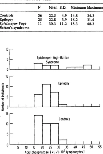
The results are shown in table 2 and figure 2. It is seen that there was no distinct difference between the levels of acid phosphatase activity in the lymphocytes from the control group and the patients with epilepsy.
On the other hand, the group of patients with Spiel- meyer-Vogt-Batterfs syndrome revealed an increase in the mean phosphatase activity, but the variations were so wide that the increase in the activity was not signif- icant at the 5% level.
Tab. 2. Acid phosphatase activity (U) in lymphocytes calculated on the basis of 104 cells.
N Mean S.D. Minimum Maximum Controls 36 22.5 4.9 14.8 34.3 Epilepsy 25 22.8 3.9 16.2 31.4 Spielmeyer-Vogt- 11 30.3 11.2 18.3 48.3 Batten's syndrome
Number of individuals D cn CD en ο on o JA CD un CD
_ Spie
, , r~
Γ-
Ι 1
Imeyer- Vogt- Batten Syndrome
ι 1 1 1 l
Epilepsy
1 ι ι Ι ι
Controls
1 1 1 1 1 Acid phosphatase [ k U /l ·104 lymphocytes)
Fig. 2. Histograms showing the distribution of acid phos- phatase in the three groups studied.
Conclusion
The studies of the acid phosphatase activity in the lymphocytes show that the mean activity is higher in the patients with Spielmeyer-Vogt-Batten's syndrome, although it does not significantly deviate from normal.
The distribution of the activity seems to show that the patients with Spielmeyer- Vogt-Batterfs syndrome can be divided into two groups, viz. one in which the values are concentrated around the normal level, and another with
J. Clin. Chem. Clin. Biochem. / Vol. 15,1977 / No. 11
648
Plum and Nielsen: Acid phosphatase in Spielmeyer-Vogt-Batten lymphocytesincreased values. The values observed in the two groups seem to be independent of the duration of the disease.
The group with increased activity included a pair of siblings.
This study was supported by a grant from the Scientific Foundation, Kolonien Filadelfia, and is part of the research work carried out by the Danish Research Group for Spielmeyer-
Vogt-Batten's Disease and Related Disorders.
References
1. Stengel, E. (1826), EYR, et medicinsk Tidsskrift 1, 347-352.
2. Batten, F. E. (1903), Trans. Ophthalmol. Soc. U. K. 23, 386-390.
3. Mayou, M. S. (1904), Trans. Ophthalmol. Soc. U. K. 24, 142-145.
4. Spielmeyer, W. (1905), Neurol. Ctrbl. 24, 620-621.
5. Vogt, H. (1905), Mschr. Psychiat. Neurol. 18,161-171, 310-357.
6. von Bagh, K. V. & Hortling, H. (1948), Nord. Med. 38, 1072-1076.
7. Rayner, S. (1962), Juvenile amaurotic idiocy in Sweden.
Uppsala Institute for Medical Genetics, 107 pp. (Ohlssons Bogtryckeri, Lund, Sweden 1962).
8. Stubbe-Teglbjasg, H. P. & Plum, C. M. (1955), Acta Psychiatr. Neurol. Scand. 30, 327-341.
9. Thiebaut, E., Waitz, R., Rohmer, F., Brini, A. & Israel, L.
(1954), Rev. Neurol. 90, 235-238.
10. Juliäo, V. T., Canelas, A. M. & Long, N. A. (1956), Arch.
Neuropsiquiat. S. Paulo 14,136-157.
11. Plum, C. M. & Stubbe-Teglbjxrg, H. P. (1961), Acta Neurol.
Scand. 37, 243-281.
12. Kivalo, E. & Stjernwall, L. (1958), Ann. Pediat. Fenn. 4, 25-29.
13. Plum, C. M. & StubberTeglbjaerg, H. P. (1960), Ann.
Pediat. Fenn. 6,17-20.
14. Spiegel-Adolf, M., Baird, H. W., Szekely, E. G. & Coleman, H. Si (I960), Confinia Neurological, 343-354.
15. Storti, E., Perugini, S. & Rossi, V. (1953), Medicina 3, 333-355.
16. Gompri, G. (1941), Arch. Pathol. 32,189-193.
17. Rabinovitch, M. & Andreucci, D. (1949), Blood 4, 580-586.
18. Haight, W. F. & Rossiter, R. J. (1950), Blood 5, 267-272.
19. Valentine, W. N. & Beck, W. S. (1951), J. Lab. Clin. Med.
38, 39-42.
20. Rozenzajn, L., Marshak, G. & Efrati, P. (1963), Acta Haematol. 30, 310-314.
21. Loftier, H. & Berghoff, W. (1962), Klin. Wochenschr. 40, 363-367.
22. Pearse, A. G. E. (1960), Histpchemistry, Churchill, London p. 882.
23. Bessey, 0. A., Lowry, 0. H. & Brock, M. J. (1964), J. Biol.
Chem. 164, 321^325.
Claus Munk Plum,
The Biochemical Research Laboratory Kolonien Füadelfia,
DK 4293 Dianalund, Denmark
J. Clin. Chem. Clin. Biochem. / Vol. 15,1977 / No. 11