Article
SOX9 Knockout Induces Polyploidy and Changes Sensitivity to Tumor Treatment Strategies in a Chondrosarcoma Cell Line
Sabine Stöckl
1,*, Georg Lindner
2, Shushan Li
1, Philipp Schuster
2, Sebastian Haferkamp
3, Ferdinand Wagner
4,5, Peter M. Prodinger
6,7, Gabriele Multhoff
8, Melanie Boxberg
9,
Axel Hillmann
10, Richard J. Bauer
11and Susanne Grässel
11
Department of Orthopaedic Surgery, Experimental Orthopaedics, Centre for Medical Biotechnology (ZMB/Biopark 1), University of Regensburg, 93053 Regensburg, Germany;
Shushan.Li@stud.uni-regensburg.de (S.L.); Susanne.Graessel@klinik.uni-regensburg.de (S.G.)
2
Institute of Microbiology and Hygiene, University Hospital Regensburg, 93053 Regensburg, Germany;
Georg1.Lindner@klinik.uni-regensburg.de (G.L.); Philipp1.Schuster@klinik.uni-regensburg.de (P.S.)
3
Department of Dermatology, University Medical Center Regensburg, 93053 Regensburg, Germany;
Sebastian.Haferkamp@klinik.uni-regensburg.de
4
Department of Pediatric Surgery, Dr. von Haunersche’s Children’s Hospital, LMU, 80337 Munich, Germany;
Ferdinand.Wagner@med.uni-muenchen.de
5
Department of Orthopaedic Surgery, Campus Großhadern, LMU, 81377 Munich, Germany
6
Department of Orthopaedic Surgery, Klinikum Rechts der Isar, Technical University of Munich (TUM), 81675 Munich, Germany; Peter.Prodinger@khagatharied.de
7
Department of Trauma Surgery and Orthopaedics, Krankenhaus Agatharied, 83734 Hausham, Germany
8
Center for Translational Cancer Research (TranslaTUM), Radiation Immuno Oncology Group, Klinikum Rechts der Isar, Technical University of Munich (TUM), 81675 Munich, Germany; gabriele.multhoff@tum.de
9
Department of Pathology, Technical University of Munich (TMU), 80333 Munich, Germany;
melanie.boxberg@tum.de
10
Department of Sarcomas and Musculoskeletal Tumors, Barmherzige Brüder Hospital, 93049 Regensburg, Germany; Axel.Hillmann@barmherzige-regensburg.de
11
Department of Oral and Maxillofacial Surgery, Center for Medical Biotechnology, University Hospital Regensburg, 93053 Regensburg, Germany; richard.bauer@ukr.de
* Correspondence: sabine.stoeckl@ukr.de
Received: 21 September 2020; Accepted: 12 October 2020; Published: 15 October 2020
Abstract: As most chemotherapeutic drugs are ineffective in the treatment of chondrosarcoma, we studied the expression pattern and function of SOX9, the master transcription factor for chondrogenesis, in chondrosarcoma, to understand the basic molecular principles needed for engineering new targeted therapies. Our study shows an increase in SOX9 expression in chondrosarcoma compared to normal cartilage, but a decrease when the tumors are finally defined as dedifferentiated chondrosarcoma (DDCS). In DDCS, SOX9 is almost completely absent in the non-chondroid, dedifferentiated compartments. CRISPR/Cas9-mediated knockout of SOX9 in a human chondrosarcoma cell line (HTB94) results in reduced proliferation, clonogenicity and migration, accompanied by an inability to activate MMP13. In contrast, adhesion, apoptosis and polyploidy formation are favored after SOX9 deletion, probably involving BCL2 and survivin. The siRNA-mediated SOX9 knockdown partially confirmed these results, suggesting the need for a certain SOX9 threshold for particular cancer-related events. To increase the efficacy of chondrosarcoma therapies, potential therapeutic approaches were analyzed in SOX9 knockout cells. Here, we found an increased impact of doxorubicin, but a reduced sensitivity for oncolytic virus treatment. Our observations present novel insight into the role of SOX9 in chondrosarcoma biology and could thereby help to overcome the obstacle of drug resistance and limited therapy options.
Int. J. Mol. Sci.2020,21, 7627; doi:10.3390/ijms21207627 www.mdpi.com/journal/ijms
Keywords: SOX9; transcription factor; chondrosarcoma; polyploidy; MMP13; CRISPR/Cas9
1. Introduction
Chondrosarcoma are known to be the second most frequent form of primary bone sarcoma and typically affect the long bones and pelvis [1]. They are generally thought to be relatively resistant to chemotherapy and radiation due to their high content of extracellular matrix, their small percentage of dividing cells and their low vascularity. Wide surgical excision is still the most effective treatment, but in some cases, sufficient resection is not always possible due to the size, localization or metastasizing status of the tumors [2]. Understanding the detailed molecular mechanism of these tumors may provide new treatment targets and might thereby improve the survival rate of patients with chondrosarcoma [3].
The majority (>85%) of conventional chondrosarcomas consist of primary central chondrosarcomas, which are referred to as such due to their central location in the medullary cavity. A small proportion of conventional chondrosarcomas emerge from the bone surface. In most cases, they develop as a result of a malignant transformation in the cartilage cap of an already existing osteochondroma and are therefore termed secondary peripheral chondrosarcomas [4]. In terms of histology, central and peripheral chondrosarcomas are quite similar. Three different grades (I–III) are defined and used as predictors of clinical behavior [5], which can also be supported by analyzing the nuclear DNA content [6]. Dedifferentiated chondrosarcomas (DDCS) are defined as a highly malignant form of chondrosarcoma and have a particularly poor prognosis with 40–85% metastasis rates. They typically show two different histopathological components: a chondrosarcoma or a well differentiated benign chondral lesion directly flanking a high-grade, non-cartilaginous and malignant compound [7,8].
SOX9 (Sex-determining region Y (SRY)-box 9) is the main transcription factor for chondrogenic differentiation during embryogenesis and has a critical role in triggering the expression of chondrogenic genes. Moreover, Sox9 is crucial for the deposition of extracellular matrix (ECM), which consists in cartilage mainly of type II collagen [9] and large proteoglycans. For the normal development and differentiation of mesenchymal stem cells into chondrocytes, a precise timing of induction and the silencing of SOX9 expression are required. Disturbing this regulation implies a high risk of tumor development as SOX9 is involved as an oncogene in various types of cancer, but it can also act as a tumor suppressor in other tumor entities [10,11]. One possible explanation for the versatility of SOX9 could be a combination of post-transcriptional modifications, the type of tissue in which it is expressed and certain binding partners. To date, only very few studies have been published on the detailed role of SOX9 during chondrosarcoma development and progression [12]. The latest studies emphasize the direct crosstalk between SOX9 and a mutation of isocitrate dehydrogenase (IDH) 1.
IDH1 mutations are found in ~50% of all chondrosarcoma patients and are involved in malignant transformation [12]. Thereby, the association between SOX9 and IDH1 highlight the crucial role of SOX9 in chondrosarcoma biology. The objectives of our research study were to determine the expression pattern of SOX9 in chondrosarcoma biopsies from different grades and thus to understand the role of SOX9 during the process of chondrosarcoma development, progression and dedifferentiation.
The analysis of chondrosarcoma cells after the knockdown and knockout of SOX9 was performed to determine the subsequent molecular impact on the carcinogenic properties, downstream targets of SOX9 and therapeutic resistance mechanisms.
2. Results
2.1. Increased SOX9 Gene Expression in G1, G2 and G3 Chondrosarcoma, but Not in Dedifferentiated Chondrosarcoma
To assess the expression of SOX9 in chondrosarcoma, we performed a meta-analysis using different
open source databases. Thereby, we compared SOX9 gene expression in healthy cartilage (26 samples)
Int. J. Mol. Sci.2020,21, 7627 3 of 18
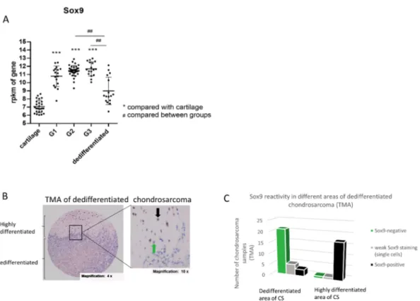
with the expression in grade I (G1) chondrosarcoma (17 samples), grade II (G2) chondrosarcoma (39 samples), grade III (G3) chondrosarcoma (17 samples) and dedifferentiated chondrosarcoma (16 samples). The SOX9 gene expression level was significantly increased in G1, 2 and 3 sarcoma compared to healthy cartilage, but is not changed significantly in dedifferentiated chondrosarcoma compared to the cartilage control. Additionally, G2 and 3 chondrosarcoma revealed an increased SOX9 expression compared to dedifferentiated chondrosarcoma (Figure 1A). We confirmed the data from the meta-analysis in part by isolating RNA from three grade II biopsies and comparing the SOX9 gene expression level with three healthy control cartilage samples (Supplementary Figure S1).
To assess the expression of SOX9 in chondrosarcoma, we performed a meta-analysis using different open source databases. Thereby, we compared SOX9 gene expression in healthy cartilage (26 samples) with the expression in grade I (G1) chondrosarcoma (17 samples), grade II (G2) chondrosarcoma (39 samples), grade III (G3) chondrosarcoma (17 samples) and dedifferentiated chondrosarcoma (16 samples). The SOX9 gene expression level was significantly increased in G1, 2 and 3 sarcoma compared to healthy cartilage, but is not changed significantly in dedifferentiated chondrosarcoma compared to the cartilage control. Additionally, G2 and 3 chondrosarcoma revealed an increased SOX9 expression compared to dedifferentiated chondrosarcoma (Figure 1A). We confirmed the data from the meta-analysis in part by isolating RNA from three grade II biopsies and comparing the SOX9 gene expression level with three healthy control cartilage samples (Supplementary Figure S1).
Figure 1. SOX9 expression in the chondrosarcoma samples. (A) Meta-analysis of SOX9 gene expression in human chondrosarcoma with different grades (G1, G2, G3) and in dedifferentiated chondrosarcoma, in comparison to SOX9 gene expression in healthy cartilage (from non-tumorous cartilage tissue). (B) Immunohistochemical (IHC) staining of SOX9 was performed on a tissue-micro- array (TMA) containing dedifferentiated chondrosarcoma. Representative image of nuclear staining intensity for SOX9 in chondrosarcoma tissue is shown. The black arrow indicates a SOX9-positive cell and the green arrow indicates a SOX9-negative cell. (C) Distribution of SOX9 reactivity in different areas of dedifferentiated chondrosarcoma samples in a TMA reveals a majority of SOX9-negative samples (21/29) in the dedifferentiated part of the tumor tissue and a majority of SOX9-positive samples (17/19) in the highly differentiated samples. rpkm = reads per kilo base per million mapped reads; G = grade; CS = chondrosarcoma; ## p ≤ 0,01; *** p ≤ 0.001.
2.2. SOX9 Protein Expression in Dedifferentiated Chondrosarcoma
Dedifferentiated chondrosarcoma (DDCS), a highly malignant variant of chondrosarcoma, are characterized by a high-grade sarcoma area next to a low-grade chondroid tumor region.
Surprisingly, SOX9 gene expression is not higher in DDCS compared to normal cartilage, as it was the case for G1, 2 and 3 tumors (Figure 1A). Thus, we analyzed SOX9 protein expression and distribution in DDCS by means of a tissue micro array (TMA) of dedifferentiated chondrosarcoma tissue samples from different patients, with immunohistochemical (IHC) staining of SOX9. The tumor
Figure 1. SOX9 expression in the chondrosarcoma samples. (A) Meta-analysis of SOX9 gene expression in human chondrosarcoma with different grades (G1, G2, G3) and in dedifferentiated chondrosarcoma, in comparison to SOX9 gene expression in healthy cartilage (from non-tumorous cartilage tissue).
(B) Immunohistochemical (IHC) staining of SOX9 was performed on a tissue-micro-array (TMA) containing dedifferentiated chondrosarcoma. Representative image of nuclear staining intensity for SOX9 in chondrosarcoma tissue is shown. The black arrow indicates a SOX9-positive cell and the green arrow indicates a SOX9-negative cell. (C) Distribution of SOX9 reactivity in different areas of dedifferentiated chondrosarcoma samples in a TMA reveals a majority of SOX9-negative samples (21/29) in the dedifferentiated part of the tumor tissue and a majority of SOX9-positive samples (17/19) in the highly differentiated samples. rpkm = reads per kilo base per million mapped reads; G = grade;
CS = chondrosarcoma; ## p ≤ 0,01; *** p ≤ 0.001.
2.2. SOX9 Protein Expression in Dedifferentiated Chondrosarcoma
Dedifferentiated chondrosarcoma (DDCS), a highly malignant variant of chondrosarcoma, are
characterized by a high-grade sarcoma area next to a low-grade chondroid tumor region. Surprisingly,
SOX9 gene expression is not higher in DDCS compared to normal cartilage, as it was the case for G1, 2
and 3 tumors (Figure 1A). Thus, we analyzed SOX9 protein expression and distribution in DDCS by
means of a tissue micro array (TMA) of dedifferentiated chondrosarcoma tissue samples from different
patients, with immunohistochemical (IHC) staining of SOX9. The tumor samples were spotted as
triplets and represent different areas of the tumor (complete list in Supplementary Figure S2). Thereby,
a clear difference was observed between SOX9 reactivity in low-grade (highly differentiated and
still cartilaginous) and high-grade, dedifferentiated (non-cartilaginous) parts of the sarcoma samples (Figure 1B). Almost all analyzed chondroid areas of the sarcoma samples were strongly positive for SOX9 (17/19), whereas in the dedifferentiated high-grade parts only three out of 29 samples revealed SOX9 reactivity in single cells (Figure 1C).
2.3. Generation of siRNA-Mediated SOX9 Knockdown and Crispr/Cas9-Mediated SOX9 Knockout Clones According to the data shown in Figure 1, we assumed an important role for SOX9 in tumor development, progression and differentiation/dedifferentiation status, and we hypothesized that the reduction or depletion of SOX9 potentially influences cancerogenic characteristics in chondrosarcoma.
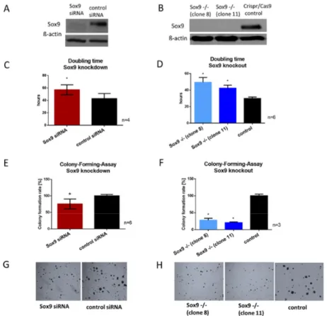
Thus, we transiently reduced SOX9 expression in a chondrosarcoma cell line (HTB94, derived from a G2 chondrosarcoma) via siRNA, leading to a decrease in SOX9 gene expression (Supplementary Figure S3A) and accordingly, to decreased protein expression by 80–90% compared to control cells (non-targeting siRNA = nt) (Figure 2A). In addition to the transient siRNA-based knockdown of SOX9, with the disadvantage of time-limited effects and in order to compare the effects of the total absence of SOX9 with effects of reduced expression, we completely depleted SOX9 via Crispr/Cas9 technology in HTB94 cells. Two single-cell-derived colonies (clones 8 and 11) with complete SOX9 knockout were selected and sequenced (Supplementary Figure S3B) to verify the generated mutations. In the two selected clones, both SOX9 alleles were disrupted by deletion or insertion resulting in a total SOX9 knockout, which was verified via Western blotting (Figure 2B). The SOX9 knockout clones showed slight morphological variations (Supplementary Figure S3C) due to their single cell origin from the typically heterocellular tumor cell line HTB94.
Int. J. Mol. Sci. 2020, 21, x FOR PEER REVIEW 4 of 18
samples were spotted as triplets and represent different areas of the tumor (complete list in Supplementary Figure S2). Thereby, a clear difference was observed between SOX9 reactivity in low- grade (highly differentiated and still cartilaginous) and high-grade, dedifferentiated (non- cartilaginous) parts of the sarcoma samples (Figure 1B). Almost all analyzed chondroid areas of the sarcoma samples were strongly positive for SOX9 (17/19), whereas in the dedifferentiated high-grade parts only three out of 29 samples revealed SOX9 reactivity in single cells (Figure 1C).
2.3. Generation of siRNA-Mediated SOX9 Knockdown and Crispr/Cas9-Mediated SOX9 Knockout Clones
According to the data shown in Figure 1, we assumed an important role for SOX9 in tumor development, progression and differentiation/dedifferentiation status, and we hypothesized that the reduction or depletion of SOX9 potentially influences cancerogenic characteristics in chondrosarcoma. Thus, we transiently reduced SOX9 expression in a chondrosarcoma cell line (HTB94, derived from a G2 chondrosarcoma) via siRNA, leading to a decrease in SOX9 gene expression (Supplementary Figure S3A) and accordingly, to decreased protein expression by 80–90%
compared to control cells (non-targeting siRNA = nt) (Figure 2A). In addition to the transient siRNA- based knockdown of SOX9, with the disadvantage of time-limited effects and in order to compare the effects of the total absence of SOX9 with effects of reduced expression, we completely depleted SOX9 via Crispr/Cas9 technology in HTB94 cells. Two single-cell-derived colonies (clones 8 and 11) with complete SOX9 knockout were selected and sequenced (Supplementary Figure S3B) to verify the generated mutations. In the two selected clones, both SOX9 alleles were disrupted by deletion or insertion resulting in a total SOX9 knockout, which was verified via Western blotting (Figure 2B). The SOX9 knockout clones showed slight morphological variations (Supplementary Figure S3C) due to their single cell origin from the typically heterocellular tumor cell line HTB94.
Figure 2. Proliferation and colony-forming ability after Sox9 knockout and knockdown in HTB94
chondrosarcoma cells. (A) Representative Western blot images of SOX9 after siRNA-mediated SOX9
knockdown and Crispr/Cas9-mediated SOX9 knockout (B). (C,D) SOX9 knockdown and knockout cells
revealed an increase in doubling time and (E,F) a decrease in the ability to form colonies from single
cells cultured in agarose. (G,H) Representative images of colony formation after SOX9 knockdown and
knockout (Magnification 10 × ). Results are the means ± SD; * p ≤ 0.05.
2.4. SOX9 Knockdown and Knockout Impair Proliferation and the Ability to Form Colonies
Since SOX9 is associated with proliferation in various other cell types, we examined the effect of SOX9 inhibition on the growth rate of chondrosarcoma cells. The doubling time of both SOX9 knockdown and knockout cells was significantly extended. After siRNA application, SOX9 knockdown cells had a mean doubling time of 57 h and the control cells, transfected with non-targeting siRNA, a mean doubling time of 43 h (Figure 2C). The two SOX9 knockout clones revealed their average doubling times between 42 and 49 h, whereas the control Crispr/Cas9 clone revealed a mean doubling time of 30 h (Figure 2D).
The ability to form adhesion-independent colonies from a single cell is a hallmark in cancer progression and metastasis potential. Therefore, we performed a colony-forming-assay (CFA) in soft agar after SOX9 reduction and depletion. Both SOX9 knockdown and knockout resulted in a diminished ability to form new colonies from single cells, with a 25% reduction for SOX9 knockdown cells (Figure 2E) and on average a 50–75% reduction for SOX9 knockout cells (Figure 2F). Representative images of the colonies after SOX9 reduction or depletion are shown in Figure 2G,H. The results indicated that SOX9 is essential for maintaining proliferation and also the clonogenicity of chondrosarcoma cells.
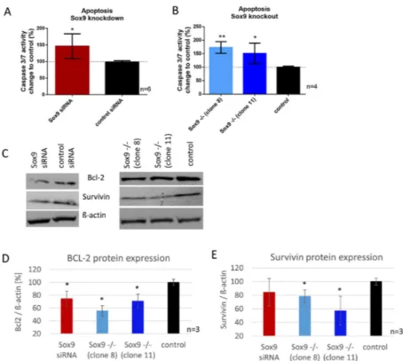
2.5. SOX9 Knockdown and Knockout Increases Apoptosis in Chondrosarcoma Cells Involving BCL-2 and Survivin Expression
Here, we analyzed the apoptotic rate in SOX9 knockdown, knockout and control cells with a caspase 3/7 activity assay and determined the protein expression of two major apoptosis mediators, BCL-2 and survivin. The reduction of SOX9 (knockdown) resulted in a significant increase in caspase 3/7 activity (48%), correlating with increased apoptosis (Figure 3A). SOX9 knockout clone 8 showed 73% and SOX9 knockout clone 11, 48% increase in caspase 3/7 activity (Figure 3B). Protein synthesis of survivin was significantly reduced in both SOX9 knockout clones, and in SOX9 knockdown cells by trend. Comparable results were observed for the anti-apoptosis protein BCL-2. SOX9 knockout clones 8 and 11, as well as SOX9 knockdown cells, showed a significantly reduced BCL-2 protein amount (Figure 3C–E).
Int. J. Mol. Sci. 2020, 21, x FOR PEER REVIEW 6 of 18
Figure 3. Effect of SOX9 knockdown and knockout on apoptosis. (A,B) Increased apoptosis in SOX9 knockdown and knockout cells was measured via Caspase 3/7 activity assay. (C) Western blot analysis of BCL-2 and survivin after SOX9 knockdown and knockout revealed a decrease in both anti- apoptotic proteins. (D,E) Densitometric analysis of 3 independent Western blots are shown. * p ≤ 0.05,
** p ≤ 0.01.
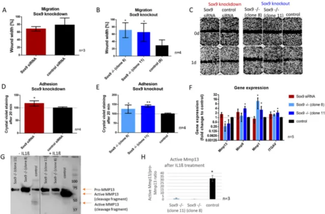
2.6. SOX9 Knockout Increases Cell Adhesion and Decreases Cell Migration Accompanied by the Inability to Activate MMP13
Wound healing assays were performed after SOX9 knockdown and knockout to investigate the migratory ability in those cells. Whereas SOX9 knockdown cells did not show a significant difference in migration compared to control cells (Figure 4A), SOX9 knockout clone 8 and 11 left a significantly wider gap in the cell layer one day after scratch introduction, indicating a reduced migration rate when Sox9 was depleted (Figure 4B). Representative images of the migration assay are shown in Figure 4C. As alterations in migration are potentially related to changes in adhesion capacity, we analyzed the adhesion potential of cells on cell culture dishes 20 min after seeding (Figure 4D,E) and in addition, a panel of migration and adhesion-related genes after SOX9 knockdown and knockout (Figure 4F). Thereby, SOX9 knockdown and knockout cells showed an increased proportion of adherent cells compared to the control (Figure 4D,E). Gene expression analysis after SOX9 knockdown and knockout displayed a significant decrease in MMP13 and an increase in MMP1 and
ITGAV gene expression in both SOX9 knockout clones and an increase in MMP9 in one of the twoclones (Figure 4F). In accordance, the analysis of MMP13 at the protein level, revealed a strongly reduced ability to activate MMP13 after IL-1β treatment in SOX9 knockout cells (Figure 4G,H).
Figure 3. Effect of SOX9 knockdown and knockout on apoptosis. (A,B) Increased apoptosis in SOX9
knockdown and knockout cells was measured via Caspase 3/7 activity assay. (C) Western blot analysis
of BCL-2 and survivin after SOX9 knockdown and knockout revealed a decrease in both anti-apoptotic
proteins. (D,E) Densitometric analysis of 3 independent Western blots are shown. * p ≤ 0.05, ** p ≤ 0.01.
2.6. SOX9 Knockout Increases Cell Adhesion and Decreases Cell Migration Accompanied by the Inability to Activate MMP13
Wound healing assays were performed after SOX9 knockdown and knockout to investigate the migratory ability in those cells. Whereas SOX9 knockdown cells did not show a significant difference in migration compared to control cells (Figure 4A), SOX9 knockout clone 8 and 11 left a significantly wider gap in the cell layer one day after scratch introduction, indicating a reduced migration rate when Sox9 was depleted (Figure 4B). Representative images of the migration assay are shown in Figure 4C.
As alterations in migration are potentially related to changes in adhesion capacity, we analyzed the adhesion potential of cells on cell culture dishes 20 min after seeding (Figure 4D,E) and in addition, a panel of migration and adhesion-related genes after SOX9 knockdown and knockout (Figure 4F).
Thereby, SOX9 knockdown and knockout cells showed an increased proportion of adherent cells compared to the control (Figure 4D,E). Gene expression analysis after SOX9 knockdown and knockout displayed a significant decrease in MMP13 and an increase in MMP1 and ITGAV gene expression in both SOX9 knockout clones and an increase in MMP9 in one of the two clones (Figure 4F). In accordance, the analysis of MMP13 at the protein level, revealed a strongly reduced ability to activate MMP13 after IL-1 β treatment in SOX9 knockout cells (Figure 4G,H).
Int. J. Mol. Sci. 2020, 21, x FOR PEER REVIEW 7 of 18
Figure 4. Effect of SOX9 knockdown and knockout on migration and adhesion. (A,B) Migration (wound healing) assay of SOX9 knockdown and knockout cells showed decreased migration (wider wound) in SOX9 knockout clones 8 and 11 compared to the control. (C) Representative pictures of wound healing assays after SOX9 knockdown and knockout (Magnification 4×). (D,E) Adhesion to cell culture plastic surface increased significantly after SOX9 knockdown and knockout. (F) Gene expression analysis showed a decreased expression of MMP13 and an increased expression of MMP9 and MMP1 and ITGAV after SOX9 knockout. (G) Western blot analysis of MMP13 revealed a hampered ability to activate MMP13 in SOX9 knockout clones after IL-1β stimulation. (H) Densitometric analysis of 3 independent Western blots is shown. Results are the means ± SD; * p ≤ 0.05, ** p ≤ 0.01.
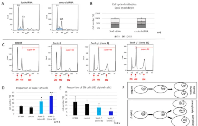
2.7. SOX9 Depletion Induces Polyploidy
We evaluated the effect of reduced and depleted SOX9 expression on the cell cycle of synchronized chondrosarcoma cells. After the SOX9 knockdown, we observed a tendency for a reduced proportion of S-phase cells. On average, control cells had an S-phase proportion of 22%, whereas in the SOX9 knockdown group, less than 17% of the cells were in the S-phase (Figure 5A,B).
Cells without SOX9 (knockout) revealed a significantly increased number of aneuploid and polyploid (super-4N) cells. Hyperdiploidy (47, XX, +7; according to ATCC) was already reported in the original cell line HTB94. In line with this, we determined a proportion of ̴ 20% of super-4N cells (cells with more than a 2-fold chromosome set before mitosis) in both control Crispr/Cas9 cells and untreated HTB94. Interestingly, the depletion of SOX9 resulted in a significant increase in super-4N cells in both SOX9 knockout clones (Figure 5C,D) accompanied by a reduced proportion of cells in the G1-phase (Figure 5E). The 8N-peak especially increased remarkably in the cell clones lacking SOX9, indicating an important role of SOX9 for maintaining genetic stability and inhibiting numerical alterations of chromosomes in chondrosarcoma cells (Figure 5F).
Figure 4. Effect of SOX9 knockdown and knockout on migration and adhesion. (A,B) Migration
(wound healing) assay of SOX9 knockdown and knockout cells showed decreased migration (wider
wound) in SOX9 knockout clones 8 and 11 compared to the control. (C) Representative pictures of
wound healing assays after SOX9 knockdown and knockout (Magnification 4 × ). (D,E) Adhesion to
cell culture plastic surface increased significantly after SOX9 knockdown and knockout. (F) Gene
expression analysis showed a decreased expression of MMP13 and an increased expression of MMP9
and MMP1 and ITGAV after SOX9 knockout. (G) Western blot analysis of MMP13 revealed a hampered
ability to activate MMP13 in SOX9 knockout clones after IL-1β stimulation. (H) Densitometric analysis
of 3 independent Western blots is shown. Results are the means ± SD; * p ≤ 0.05, ** p ≤ 0.01.
2.7. SOX9 Depletion Induces Polyploidy
We evaluated the effect of reduced and depleted SOX9 expression on the cell cycle of synchronized chondrosarcoma cells. After the SOX9 knockdown, we observed a tendency for a reduced proportion of S-phase cells. On average, control cells had an S-phase proportion of 22%, whereas in the SOX9 knockdown group, less than 17% of the cells were in the S-phase (Figure 5A,B).
Int. J. Mol. Sci. 2020, 21, x FOR PEER REVIEW 8 of 18
Figure 5. Cell cycle distribution of SOX9 knockdown and knockout cells. (A) Representative flowcytometric cell cycle analysis of SOX9 knockdown cells and the distribution of cell cycles phases after SOX9 knockdown (B). (C) Representative flowcytometric cell cycle analysis of SOX9 knockout clones. The area of super-4N cells (4N = DNA content after replication and before mitosis) is marked with red lines. (D) The proportion of super-4N cells is significantly increased in SOX9 knockout clones 8 and 11 compared to control. (E) The number of cells in G1-phase was reduced by trend in SOX9 knockout clone 8 and reduced significantly in the SOX9 knockout clone 11 compared to the control.
(F) The development of aneuploidy or polyploidy after the SOX9 knockout. Control cells do not (or to a much smaller extent) exhibit genetic instability and maintain a diploid genome after division, whereas Sox9
−/−cells reveal abnormalities during division leading to aneuploid and/or polyploid cells.
Results are the means ± SD; * p ≤ 0.05; ** p ≤ 0.01.
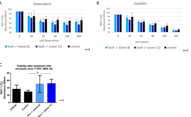
2.8. SOX9 Depletion Alters Sensitivity against Doxorubicin and against Infection with Oncolytic Virus (T- VEC)
We hypothesized that SOX9 knockout cells may show a different sensitivity to therapy approaches due to their difference in functional cancerogenic properties. Therefore, we conducted a WST-1 viability assay to analyze whether SOX9 knockout cells have different sensitivities to doxorubicin and cisplatin. In addition, we performed a WST-1 assay with SOX9 knockout cells after applying a potential new treatment option, infection with an oncolytic virus (T-VEC, approved for melanoma cancer therapy). The viability of the two SOX9 knockout clones decreased significantly after treatment with 50 µM doxorubicin (10–15% reduction) (Figure 6A), whereas cisplatin had no significant effect (Figure 6B). Infection with the oncolytic virus (T-VEC) revealed that SOX9 knockout clone 11 retained significantly more viable cells after T-VEC infection with an MOI (multiplicity of infection) of 10, and SOX9 knockout clone 8 showed the same result by trend (Figure 6C), implying a lower potential efficiency of this anticancer therapy in chondrosarcoma cells without SOX9 expression.
Figure 5. Cell cycle distribution of SOX9 knockdown and knockout cells. (A) Representative flowcytometric cell cycle analysis of SOX9 knockdown cells and the distribution of cell cycles phases after SOX9 knockdown (B). (C) Representative flowcytometric cell cycle analysis of SOX9 knockout clones. The area of super-4N cells (4N = DNA content after replication and before mitosis) is marked with red lines. (D) The proportion of super-4N cells is significantly increased in SOX9 knockout clones 8 and 11 compared to control. (E) The number of cells in G1-phase was reduced by trend in SOX9 knockout clone 8 and reduced significantly in the SOX9 knockout clone 11 compared to the control.
(F) The development of aneuploidy or polyploidy after the SOX9 knockout. Control cells do not (or to a much smaller extent) exhibit genetic instability and maintain a diploid genome after division, whereas Sox9
−/−cells reveal abnormalities during division leading to aneuploid and/or polyploid cells. Results are the means ± SD; * p ≤ 0.05; ** p ≤ 0.01.
Cells without SOX9 (knockout) revealed a significantly increased number of aneuploid and
polyploid (super-4N) cells. Hyperdiploidy (47, XX, +7; according to ATCC) was already reported in the
original cell line HTB94. In line with this, we determined a proportion of ~20% of super-4N cells (cells
with more than a 2-fold chromosome set before mitosis) in both control Crispr/Cas9 cells and untreated
HTB94. Interestingly, the depletion of SOX9 resulted in a significant increase in super-4N cells in both
SOX9 knockout clones (Figure 5C,D) accompanied by a reduced proportion of cells in the G1-phase
(Figure 5E). The 8N-peak especially increased remarkably in the cell clones lacking SOX9, indicating
an important role of SOX9 for maintaining genetic stability and inhibiting numerical alterations of
chromosomes in chondrosarcoma cells (Figure 5F).
2.8. SOX9 Depletion Alters Sensitivity against Doxorubicin and against Infection with Oncolytic Virus (T-VEC)
We hypothesized that SOX9 knockout cells may show a different sensitivity to therapy approaches due to their difference in functional cancerogenic properties. Therefore, we conducted a WST-1 viability assay to analyze whether SOX9 knockout cells have different sensitivities to doxorubicin and cisplatin.
In addition, we performed a WST-1 assay with SOX9 knockout cells after applying a potential new treatment option, infection with an oncolytic virus (T-VEC, approved for melanoma cancer therapy).
The viability of the two SOX9 knockout clones decreased significantly after treatment with 50 µ M doxorubicin (10–15% reduction) (Figure 6A), whereas cisplatin had no significant effect (Figure 6B).
Infection with the oncolytic virus (T-VEC) revealed that SOX9 knockout clone 11 retained significantly more viable cells after T-VEC infection with an MOI (multiplicity of infection) of 10, and SOX9 knockout clone 8 showed the same result by trend (Figure 6C), implying a lower potential efficiency of this anticancer therapy in chondrosarcoma cells without SOX9 expression.
Int. J. Mol. Sci. 2020, 21, x FOR PEER REVIEW 9 of 18