• Keine Ergebnisse gefunden

Generation of an oncolytic adenovirus vector combining three cancer targeting strategies and characterization of a new preclinical model for breast cancer virotherapy

N/A
N/A
Protected

Academic year: 2021

Aktie "Generation of an oncolytic adenovirus vector combining three cancer targeting strategies and characterization of a new preclinical model for breast cancer virotherapy"

Copied!
123
0
0

Wird geladen.... (Jetzt Volltext ansehen)

Volltext

(1)

Generation of an oncolytic adenovirus vector combining three cancer targeting strategies and

characterization of a new preclinical model for breast cancer virotherapy

Inaugural-Dissertation

zur

Erlangung des Doktorgrades Dr.nat.med.

der Medizinischen Fakultät und

der Mathematisch-Naturwissenschaftlichen Fakultät der Universität zu Köln

vorgelegt von Sabrina Brüggemann

aus Köln

DCC Competence Center GmbH, Köln

2012

(2)

Berichterstatterinnen: Prof. Dagmar Knebel-Mörsdorf PD Dr. Hildegard Büning

Tag der letzten mündlichen Prüfung: 11.07.2012

(3)

Dedicated to my parents, my sisters, my boyfriend and Jochen († 2010).

(4)

Table of content

1. Introduction __________________________________________________ 1

1.1 Breast cancer __________________________________________________________________ 1 1.1.1 Pathophysiology of breast cancer __________________________________________________ 1 1.1.2 Breast cancer treatments _________________________________________________________ 2 1.2 Gene therapy for breast cancer ___________________________________________________ 4

1.3 Adenovirus ____________________________________________________________________ 6 1.3.1 Adenoviral structure ____________________________________________________________ 6 1.3.2 Adenoviral infection pathway _____________________________________________________ 8 1.4 Oncolytic virotherapy of breast cancer ____________________________________________ 10 1.4.1 Different targeting strategies _____________________________________________________ 12 1.4.2 Animal models for oncolytic virotherapy ___________________________________________ 18 1.5 The Syrian hamster animal model ________________________________________________ 19 1.5.1 Induction of breast cancer in female Syrian hamsters __________________________________ 20

Aim of Thesis ___________________________________________________ 22 2. Materials ___________________________________________________ 23

2.1 Bacterial Strains _______________________________________________________________ 23 2.1.1 Media for cultivation of bacteria __________________________________________________ 23 2.2 Eukaryotic cells _______________________________________________________________ 24 2.2.1 Cell Culture Media _____________________________________________________________ 26 2.3 Nucleic Acids _________________________________________________________________ 27 2.3.1 Synthetic Oligonucleotides ______________________________________________________ 27 2.3.2 Plasmids used to generate the Ad5/3 CXCR4 UTR ____________________________________ 28 2.3.3 Plasmids used for the standard curve of the qPCR ____________________________________ 28 2.3.4 Transfection reagents ___________________________________________________________ 28 2.3.5 RT-PCR _____________________________________________________________________ 28 2.3.6 Loading dyes and DNA Markers __________________________________________________ 29 2.4 DNA Preparation ______________________________________________________________ 29

2.5 RNA Preparation and cDNA Synthesis ____________________________________________ 29

2.6 Viruses ______________________________________________________________________ 30

2.7 Proteins ______________________________________________________________________ 30

2.7.1 Enzymes _____________________________________________________________________ 30

(5)

2.7.3 Antibodies ___________________________________________________________________ 31 2.8 Staining reagents ______________________________________________________________ 31

2.9 Solutions and Buffers __________________________________________________________ 32

2.10 Chemicals and reagents _________________________________________________ 34

2.11 Machines used in this study ______________________________________________ 35

3. Methods ____________________________________________________ 36

3.1 Molecular Biology _____________________________________________________________ 36 3.1.1 Generation of the triple-level targeted adenovirus (Ad5/3 CXCR4 UTR) ___________________ 36 3.1.2 Quantitative polymerase chain reaction (qPCR) ______________________________________ 38 3.2 Cell Biology___________________________________________________________________ 39 3.2.1 Cultivation of Cell lines _________________________________________________________ 39 3.2.2 Adenoviral infection of cell lines __________________________________________________ 39 3.2.3 Transfection of Ad5/3 CXCR4 UTR vector into HEK-293 cells _________________________ 39 3.2.4 Titer determination of Adenoviral stocks ___________________________________________ 40 3.2.5 Defining oncolytic activity ______________________________________________________ 41 3.2.6 Quantifying viral cell entry, transcription and replication _______________________________ 42 3.2.7 Half-life determination of E1A mRNA _____________________________________________ 43 3.2.8 Growth characterizations ________________________________________________________ 43 3.2.9 Anchorage independence determination ____________________________________________ 43 3.3 Biochemistry __________________________________________________________________ 44 3.3.1 Preparation of protein extracts ____________________________________________________ 44 3.3.2 SDS-PAGE and Immunoblotting __________________________________________________ 44 3.4 In vivo experiments ____________________________________________________________ 46 3.4.1 Establishment of an in vivo syngeneic tumor model ___________________________________ 46

4. Results _____________________________________________________ 47

4.1 Triple-level targeted oncolytic Adenovirus, combining transductional, transcriptional and translational targeting __________________________________________________________ 47

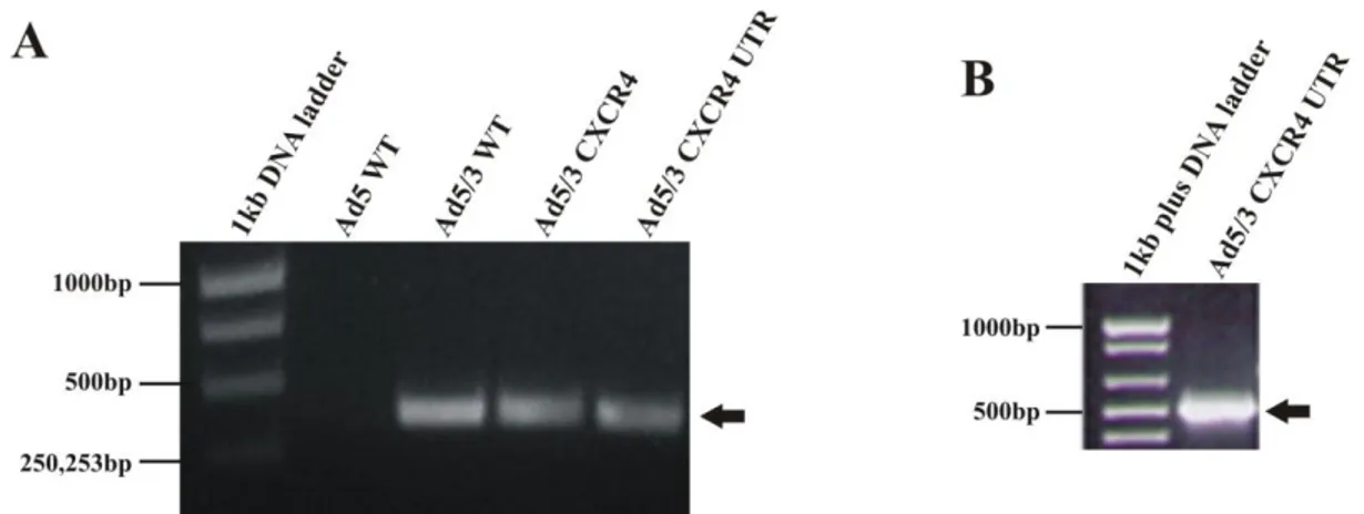
4.1.1 Incorporation of Ad3 fiber knob, human CXCR4 promoter and rat 5’UTR-sequence of FGF-2 mRNA into Ad5 genome ________________________________________________________ 48

4.1.2

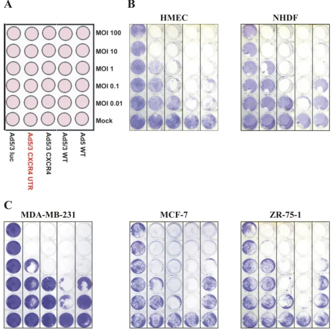
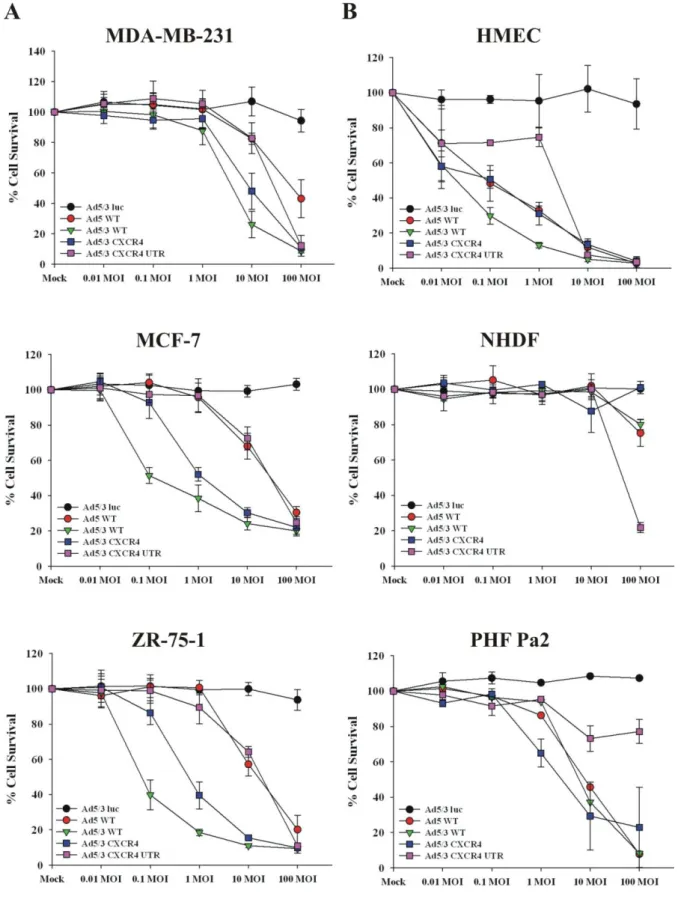
Ad5/3 CXCR4 UTR gains differential oncolytic activity in breast cancer cells compared to normal

cells in vitro __________________________________________________________________ 49

4.1.3 E1A protein expression is reduced in Ad5/3 CXCR4 UTR infected cells ___________________ 54

4.1.4 Replication efficiency is decreased in Ad5/3 CXCR4 UTR infected MDA-MB-231 cells ______ 55

4.1.5 Different titration method does not reveal inaccurate relative Ad titers ____________________ 57

(6)

4.1.6 Viral cell entry of Ad5/3 CXCR4 UTR is comparable to the one of Ad5/3 WT and Ad5/3 CXCR4 ____________________________________________________________________________ 58 4.1.7 eIF4E protein expression is not dysregulated by Ad3 fiber knob binding ___________________ 60 4.1.8 Phosphorylation of 4E-BP1 is delayed in Ad5/3 CXCR4 UTR infected breast cancer cells _____ 61 4.1.9 Delayed expression of E1A and pTP of Ad5/3 CXCR4 UTR in MDA-MB-231 cells ________ 63 4.1.10 E1A mRNA half life is not altered in Ad5/3 CXCR4 UTR infected MDA-MB-231 cells ______ 66 4.1.11 Efficient cytotoxicity of Ad5/3 CXCR4 UTR in HEK-293 cells __________________________ 68 4.2 Characterization of the Syrian hamster breast cancer cell line_________________________ 72 4.2.1 Syrian hamster breast cancer cells induce tumor formation in syngeneic Syrian hamsters ______ 73 4.2.2 HMAM5 cells displayed anchorage independent growth and 24 h doubling time during

exponential growth phase _______________________________________________________ 75 4.3 Replication efficiency of oncolytic Ads on HMAM5 cells _____________________________ 78 4.3.1 Ad5 ∆24 RGD displays a significant E1A mRNA and protein expression in HMAM5 cells ____ 79 4.3.2 Viral replication of Ad5 ∆24 RGD in HMAM5 is significantly increased compared to Ad5 WT 81 4.3.3 Enhanced oncolytic activity of Ad5 ∆24 RGD in HMAM5 cells _________________________ 82

5. Discussion __________________________________________________ 84

5.1 Construction of an oncolytic adenovirus containing transductional, transcriptional and translational targeting __________________________________________________________ 84

5.2 Establishment of a Syrian hamster breast cancer cell line ____________________________ 90

6. References __________________________________________________ 94

7. List of figures and tables ______________________________________ 107

8. Abstract ___________________________________________________ 108

9. Zusammenfassung __________________________________________ 110

10. Abbrevations _______________________________________________ 112

11. Danksagungen _____________________________________________ 114

12. Eidesstattliche Erklärung _____________________________________ 115

13. Lebenslauf _________________________________________________ 116

(7)

1. Introduction

1.1 Breast cancer

Despite early detection methods and advanced conventional treatments, breast cancer is one of the most frequently diagnosed malignancies and the second leading cause of cancer deaths among women worldwide (DeSantis et al., 2011; Jemal et al., 2011; Thomas et al., 2006). The chance of being diagnosed for women is 1 in 8 ( http://seer.cancer.gov/csr/1975_2009_pops09/ ).

Every year more than 192,000 women are diagnosed as positive for breast cancer in the United States (Jemal et al., 2010); the incidence rate for breast cancer is still high in the United States as well as Australia/ New Zealand, and Western and Northern Europe (Jemal et al., 2011).

Since the last 25 years, breast cancer death rates decreased in several European countries and Northern America due to early detection methods. However, the number of estimated new cases of breast cancer in 2012 is about 226,870 in the United States and the estimated number for breast cancer death is 39,510 (Siegel et al., 2012). According to the Surveillance, Epidemiology, and End Results (SEER) program of the National Cancer Institute, responsible for collecting breast cancer data in the United States, 1 in 3 cancers diagnosed in women is breast cancer (DeSantis et al., 2011). Therefore, novel therapies designed with alternative and complementary strategies are sorely needed.

1.1.1 Pathophysiology of breast cancer

The breast contains 15-20 lobes which radiating from the nipple surrounded by fibrous connective tissue and fats. Each lobe can be separated into several lobules. The lobules supply milk to the ducts and therefore are also referred to as ductual-lobular. The basic structural units of the breast are the lobules which are surrounded by epithelial cells.

The most common breast cancer originates either in the epithelial cells of the inner lining of the

milk ducts (ductal carcinoma) or in the lobules (lobular carcinoma). As with every other

cancers, breast cancer developes from different genetic aberrations or aberrant growth factor

receptor signaling (Madigan et al., 1995; Taylor et al., 2008; Wosikowski et al., 2000; Ziegler

et al., 1993). Furthermore, breast cancer can be inherited, most commonly resulting from a

genetic defect in the DNA repair genes BRCA1 and BRCA2 (breast cancer susceptibility gene

1, 2) (Chappuis et al., 1999; Struewing et al., 1997). Besides inherited genetic aberrations and

(8)

defects, breast cancer can be initiated through environmental factors (Davis and Sieber, 1997;

Madigan et al., 1995; Ziegler et al., 1993).

Development of breast cancer is a multiple process that can be divided into several steps.

Precursor lesions that are benign can be divided into non-proliferative and proliferative lesions.

For example, non-proliferative lesions are duct ectasia or mild hyperplasia of the normal and simple type (Beckmann et al., 1997). Proliferative lesions can be further divided into proliferative phase without atypia and with atypia. Cells found in regions of proliferative changes without atypia show an extreme cell growth without any abnormal cell structure.

Proliferative changes with atypia on the other hand are characterized by regions of cell expansion containing some cells with an abnormal cell structure. Lesions of this type are also called atypical hyperplasia and cells demonstrate a few characteristics of a carcinoma in situ (Beckmann et al., 1997). Carcinoma in situ originated from proliferation of the glandular epithelium demonstrating cellular malignancy and is restricted to a specific area. Carcinoma in situ can be separated in either ductal carcinoma in situ (DCIS) or lobular carcinoma in situ (LCIS). Tumors that aquire the ability to penetrate into the surrounding breast cancer tissue and metastasize to other organs are called invasive breast carcinomas (Lakhani, 1999). The most common type is invasive ductal carcinoma (IDC) (Beckmann et al., 1997).

1.1.2 Breast cancer treatments

Several treatment options for breast cancer have been developed during the last decade. These treatment approaches are divided into surgery, chemo-, radiation-, hormonal and targeted therapy. The breast cancer treatment is chosen depending on stage and type of cancer. Most women diagnosed with breast cancer undergo surgery to remove as much cancer tissue as possible.

Breast conservation surgery is the most common surgery, in which depending on the stage of

the breast tumor, the breast tumor and a small region of tumor surrounding breast tissue are

removed. Segmental or partial mastectomy is another option of surgery during which a larger

section of the tumor surrounding breast tissue is removed. Breast cancer patients with more

advanced tumor stage need to undergo a radical mastectomy, by removing the entire breast as

well as several lymph nodes under the arm. Although, surgery is still the preferred method to

remove a tumor successfully, it also bears some disadvantages. The most common

(9)

complications observed after breast cancer surgery is the collection of serous fluid under the skin, seroma formation and axillary dead space (Pogson et al., 2003), (Hashemi et al., 2004).

Chemotherapy is another option to treat breast cancer. It involves the use of drugs to kill cancer cells, inhibit cancer cell growth or to avoid cancer recurrence. Some of the commonly used drugs for chemotherapy to treat breast cancer patients are adriamycin (also called doxorubicin), cyclophosphamide, and paclitaxel (Ibrahim et al., 2003); (Hutchins et al., 2005).

Usually, chemotherapy can be divided into three different types: 1) Neoadjuvant, given before surgery to shrink the tumor; 2) Adjuvant, given after surgery to prevent recurrence; and 3) Palliativ, given to control cancer in the stage where it is already metastatic. However, treatment with chemotherapeutic reagents induces some mild side effects like fatigue, nausea, hair loss and vomiting and several severe ones such as cardiotoxicity, necrosis of the colon, leukemia and severe infections (Ewertz and Jensen, 2011; Lemieux et al., 2008; Lindley et al., 1999; Sitzia and Huggins, 1998; Yood et al., 2012). The severe side effects can be fatal and cause the death of the breast cancer patient.

Radiation therapy on the other hand exploits high energy X-rays to destroy breast cancer cells or inhibit their proliferation. High energy radiation damages the DNA of cancer cells or forms charged particles like free radicals causing cancer cell killing. There are two types of radiation therapy, external and internal radiation therapy. External radiation therapy is performed with a machine outside of the body whereas internal radiation utilizes radioactive substances injected directly into the tumor or near the cancer occurrence. Radiation therapy is usually given after surgery either alone or in combination with chemotherapeutic reagents to avoid breast cancer recurrence, to kill metastasized breast cancer cells in other organs or residual cancer cells.

Radiation therapy is painless but can induce severe side effects like skin irritation, hair loss, and damage to the salvery glands. Those side effects appear because healthy cells can be harmed during this treatment as well (Rutqvist et al., 2003). One of the severe side effects of radiation therapy is the induction of sarcoma. The occurrence rate is very low, but if sarcoma occurrs after the radiation therapy, it is associated with poor clinical outcomes (Sheth et al., 2012).

Besides chemo- and radiation therapy, hormonal therapy is another treatment utilized to treat

breast cancer by using drugs to inhibit production of steroid hormones that stimulate cancer

cell proliferation. Many breast cancer types express high levels of estrogen and/or

progesterone receptors on their cell surface. Breast cancer patients suffering from tumors with

high expression of estrogen receptors can undergo hormonal therapy. Drugs usually used for

(10)

hormonal therapy include aromatase inhibitors like exemestane (Tomao et al., 2011) and selective estrogen modulators like Tamoxifen (Smith et al., 2000). These drugs block the hormone activity or repress their expression to inhibit breast cancer cell proliferation. Besides killing of cancer cells these drugs show several side effects. For example, treatment with Tamoxifen can cause weight gain, hot flashes, vaginal discharge and severe late side effects such as increasing the risk for blood clots, stroke and invasive endometrial cancer (Perez, 2007). Aromatase inhibitors are most commonly given to postmenopausal women suffering from hormone dependent breast cancer requiring the hormone estrogen to proliferate.

Aromatase inhibitors block the aromatase enzyme that converts androgen to estrogen in the body and therefore, prevent cancer cell proliferation (Gibson et al., 2009). Typical aromatase inhibitor side effects are skeletal fractures, hot flashes and development of osteoporosis (Perez, 2007).

Targeted therapy is another therapeutic treatment of breast cancer employing drugs or monoclonal antibodies to recognize and destroy specific cancer cells without damaging healthy cells. For example, monoclonal antibodies are used to recognize specific cancer cell surface markers or factors that are stimulating the tumor growth. Binding of monoclonal antibodies to their target destroys the cancer cell or inhibits cancer cell proliferation/migration. One example of a therapeutic monoclonal antibody is Trastuzumab that acts by blocking the signal cascade of the growth factor protein HER2. HER2 activates a signal cascade that in return induces proliferation of cancer cells (Higgins and Baselga, 2011). Targeted therapy drugs like tyrosine kinase inhibitors are administered to inhibit tumor growth signaling. Lapatinib, a tyrosine kinase inhibitor, prevents the effect of HER2 protein and other proteins inside the tumor cells. PARP inhibitors on the other hand inhibit DNA repair and may trigger cancer cell death. PARP inhibitors are widely studied for triple negative breast cancer (Higgins and Baselga, 2011). Triple negative breast tumors are lacking the expression of estrogen receptors (ER-), progesterone receptors (PR-) and HER2 (HER2-).

Thus, beside several of breast cancer treatment options currently available, there remains a need for novel treatments to reduce mortality rate of breast cancer.

1.2 Gene therapy for breast cancer

Gene therapy is a therapeutic approach that involves the delivery of genetic material to cancer

or defective cells and its surrounding tissue. The goal of cancer gene therapy is to cause tumor

(11)

cell death or prevent tumor growth. For cells that cause a disease due to a gene defect, gene therapy provides these cells permanently with the missing gene and ideally curing the disease (Cross and Burmester, 2006). Over the last decades, gene therapy became a powerful tool to treat metabolic disorders, cardiovascular diseases and cancer with great success while traditional therapies were limited due to their severe side effects (Kay and Woo, 1994);

(Mulavisala et al., 2001; Rakhmilevich et al., 2004; Zhang et al., 1996). Gene therapy can be categorized into five different groups: 1) genetic immunopotentiation, 2) pro-apoptotic gene therapy, 3) anti-angiogenic gene therapy, 4) mutation compensation, and 5) molecular chemotherapy/suicide gene therapy (Cross and Burmester, 2006).

Genetic immunopotentiation increases the immune response against specific tumor antigens.

Genes that have been analyzed to use for breast cancer immunotherapy are HER-2/neu protein (Disis et al., 1999; Kawashima et al., 1999), carcinoembryonic antigen (CEA) (Kawashima et al., 1999) and MAGE-1 (Toso et al., 1996).

Pro-apoptotic gene therapy encompasses the combination of a therapeutic drug and specific surface cell death molecules like tumor necrosis factor-related apoptosis-inducing ligands (TRAIL) (Griffith et al., 2000; Lin et al., 2002). This combination increases the apoptotic cell death and at the same time it can improve the therapeutic effect in drug-resistant tumor cells.

Anti-angiogenic therapy, on the other hand, uses the knowledge of the role of angiogenesis in tumor cell growth. Angiogenesis facilitates cancer cell growth by providing them with new blood vessels (Atiqur Rahman and Toi, 2003; Marty and Pivot, 2008). Angiostatin and endostatin were found to have an anti-angiogenic function. It was demonstrated that liposomes complexed to plasmids containing angiostatin and endostatin inhibited breast tumor development in nude mice (Chen et al., 1999; Oga et al., 2003). Breast cancer was induced by the injection of either human breast cancer cells or mice breast cancer cells into the mammary fat pats. Injection of liposome:plasmid complexes was performed intratumorally, intravenous or intramuscular (Chen et al., 1999; Oga et al., 2003).

Mutation compensation is based on the idea to restore defects in a cancer cell that are causing

the malignant transformation. For example, one strategy for mutation compensation employs

the replacement of a defected tumor-suppressor gene in cancer cells with a working copy such

as p53 (Chen and Mixson, 1998) or Rb (Wang et al., 1993). Another strategy is to decrease the

tumor cell development by ablating the oncogene function of c-myc (Watson et al., 1991) and

c-fos (Arteaga and Holt, 1996) which are used in breast cancer.

(12)

Suicide gene therapies are designed to treat cancer cells by delivering a gene that can convert a non-toxic prodrug into a toxic form. Besides killing the targeted cancer cells, suicide gene therapy can also affect surrounding cells of the region. This has been termed a bystander effect, providing the opportunity to destroy cancer cells that might have migrated into the tumor surrounding tissue. Clinical trials were performed with erbB-2 (Pandha et al., 1999) and MetXia-P450 (Braybrooke et al., 2005) as suicide gene therapy for breast cancer.

1.3 Adenovirus

Over the past several years, the field of gene therapy has emerged with a broad variety of applications. Throughout this progress, the use of non-viral and viral vectors as delivery vectors has also widened. One of the most promising candidates for gene therapy is the adenovirus (Ad). Ads were first isolated back in the 1950’s and since then their biology has been extensively studied (Hilleman and Werner, 1954; Rowe et al., 1953). Ads have specific features that make them a suitable gene therapy vector (Russell, 2000; Zhang, 1999). First, the generation of high titers required for clinical administration, is no longer a major problem.

Furthermore, the Ad genome is really easy to manipulate and large DNA sequences can be inserted without any complications. In addition, Ads are capable to infect a large variety of dividing and non-dividing cells and they are not integrating their viral genome into host chromosomes. Moreover, they show also a low pathogenicity in humans and are causing only mild symptoms during their infection. All these characteristics of Ads for gene therapy as vectors offer a new therapeutic intervention to target breast cancer.

1.3.1 Adenoviral structure

At present, there are 53 known human Ad serotypes classified into six species (A-F subgroup)

based on their neutralization by specific animal antisera, oncogenic potential and their DNA

GC content (Davison et al., 2003; Madisch et al., 2005). Ads are non-enveloped, icosahedral

viruses that contain a linear double stranded DNA genome of 36,000 base pairs (Rux and

Burnett, 2004). The outer protein layer of the Ad virion is called the capsid that encloses the

DNA-protein core complex. The capsid structure is composed of seven polypeptides: the three

major proteins hexon (II), penton base (III) and fiber (IV) and the minor proteins IIIa, VI, VIII

and IX (Vellinga et al., 2005) as seen in Figure 1. 252 capsomeres are forming the Ad capsid

(13)

composed of pentons and are found at the 12 vertices. Penton capsomeres are formed by pentameric penton bases and a protruding trimeric fiber (Valentine and Pereira, 1965). Penton bases are important for the capsid stabilization and together with the fiber they initiate and mediate viral attachment and internalization into the host cells. All fiber proteins can be divided into three domains: the N-terminal tail domain, the shaft region of variable length, and the C-terminal knob domain. The trimeric structure of the fiber is formed by sequences of the knob and the shaft domain (Rux and Burnett, 2004). Besides pentons and hexons, the capsid structure is stabilized by the minor proteins IIIa, VI, VIII and IX. Furthermore, the capsid contains also a virus-encoded protease (adenain) that cleaves several precursor proteins like VI, VII, VII, X, and terminal protein (TP) and is essential for the assembly of newly infectious viral particles (Russell, 2009).

The viral genome is enclosed by the capsid and associates with the polypeptides V, VII, X

(mu) and TP. This complex builds a condensed core structure. Inverted terminal repeats (ITR)

are located at each end of the Ad genome and each 5’-terminus is associated with the TP. Both

associations are essential for viral genome replication (Russell, 2000). The Ad genome is

separated into regions: the early genes encoding for E1A, E1B, E2A, E2B, E3 and E4; the

delayed early genes encoding IX and IVa2 and the late genes encoding L1-L5. Early gene

products play an essential role in adenoviral transcription, DNA replication, suppression of

host immune response and inhibition of host cell apoptosis whereas the late genes encode for

viral structure proteins which are necessary for viral assembly (Russell, 2000). In addition,

two genes encode for virus-associated RNAs I and II (VAI and VAII RNAs) which have an

important function to inhibit the host protein synthesis during viral replication in infected cells

(O'Malley et al., 1986).

(14)

1.3.2 Adenoviral infection pathway

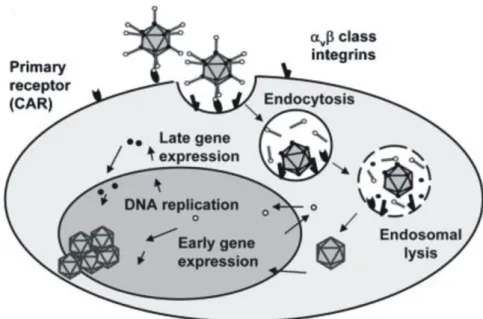
The Ad infection pathway can be separated into several steps: attachment, internalization, endosome escape, viral replication, assembly and lysis. Ad infection of a host cell is initiated by binding of the fiber knob domain to a cellular attachment receptor on the host cell surface.

For most Ad serotypes (except for subgroup B) the primary cellular attachment receptor is the Coxsackie and Adenovirus receptor (CAR) (Bergelson et al., 1997; Roelvink et al., 1998). The expression pattern of CAR on the surface of tissue-specific cells plays a crucial role for the adenoviral tropism. CAR is highly expressed on parenchymal cells in the liver but not exclusively, thus explaining high liver infectivity of Ad vectors upon systemic administration but also the wide Ad vector tropism in vitro (Yee et al., 1996).

The first step of Ad infection is the binding of the Ad fiber knob domain to the N-terminal domain of CAR. Besides the binding of the fiber knob to CAR, a second interaction of virion and the cell surface is essential to initiate viral uptake. The interaction between integrins α

ν

β

3

Figure 1: Adenovirus structure.

General structure of the Ad: displaying hexons, penton bases, fibers and cement proteins of the Ad capsid. The

capsid encompasses the Ad genome and core proteins associated with the Ad genome forming the core of the

Ad. Figure was adapted from (Russell, 2000).

(15)

Ad vector is necessary to mediate internalization of the Ad virions (Mathias et al., 1998;

Wickham et al., 1993.). Binding of integrins is followed by rapid endocytosis of the virions through clathrin-coated vesicles driven by a cytosolic GTPase (Meier et al., 2002; Roelvink et al., 1998; Roelvink et al., 1999). Once the viral particles are taken up by clathrin-coated vesicles, the endosome acidification process is causing conformational changes of the Ad vector capsid resulting in the release of the virions into the cytoplasm (Wang et al., 1998). The process of viral uptake until viral release into the cytosol is completed within 15 min and therefore rapid (Greber et al., 1993).

Next, the virions are translocated to the nuclear pore complex by a microtubule-dependent trafficking. Subsequently on arrival at the nucleus, Ad vectors interact through hexons with the nuclear pore complex and release their genome into the host cell nucleus to start viral replication. Within one hour more than 90% of Ad vectors are transported to the nucleus and about 18 hours post infection transgene expression can be detected reaching a maximum at 48 hours (Greber et al., 1993). The Ad infection pathway is schematically displayed in Figure 2.

After viral genome replication and translation of the viral mRNA transcripts, Ad vectors need

to assemble. Therefore, viral proteins are transported back from the cytoplasm into the nucleus

for particle assembly. Final maturation of Ad vectors is achieved by the adenain protease that

cleaves the precursor polypeptides of VI, VII, VIII, Mu and TP to obtain infectious virion

particles. In the final step of the Ad replication pathway, cells are lysed by the accumulation of

ADP (Ad death protein) (Tollefson et al., 1996) and the cleavage of cellular cytokeratins by the

L3 protease. Cleaved cytokeratins cause an increased susceptibility of the cell to lyse by

disrupting cell integrity (Chen et al., 1993). In addition, in the past several years it has been

suggested that autophagy aids the release of progeny virions (Jiang et al., 2008).

(16)

1.4 Oncolytic virotherapy of breast cancer

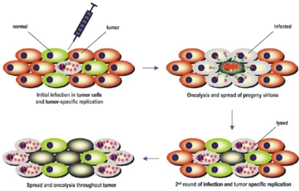
Because of safety concerns, only replication incompetent viruses were used as vehicle vectors in the beginning of gene therapy. However, clinical studies have shown that using replication incompetent viruses resulted in only a minor proportion of tumor cells infected and did not proffer for therapeutical success (Alvarez and Curiel, 1997). Therefore, employing replication competent viruses to treat malignant tumors was referred to as a virotherapy procedure instead of gene therapy (Alemany et al., 2000; Kirn et al., 2001). Virotherapy exploits tumor specific characteristics for a specific cell targeting, viral infection and replication in tumor cells to kill them while leaving healthy cells unaffected (Alemany et al., 2000; Mathis et al., 2005). The principle of virotherapy is based on the idea that a replication competent virus infects a tumor cell and replicates tumor cell specific leading to tumor cell oncolysis and spread of progeny virions to adjacent tumor cells. Ideally, the tumor specific replication is repeated until all tumor cells are destroyed but not the healthy cells (Alemany et al., 2000; Kirn et al., 2001) (Figure 3).

Figure 2: Ad serotype 5 infection pathway.

Illustrated are the principal steps of the adenoviral infection. Binding of the Ad fiber knob to the primary cellular

attachment receptor is followed by the interaction of penton base with integrins. Binding of integrins initiates

viral uptake through clathrin-coated vesicles. In the endosome Ad dissociates in the low pH environment and up

on acidification Ads are dismantled and viral particles are released into the cytosol. Viral particles are then

transported to the nucleus where they release their viral genome and start viral replication. Figure was adapted

from (Kanerva and Hemminki, 2005).

(17)

Besides the promising results of virotherapy in the mouse models, the first results in clinical trials revealed a reduced efficiency of Ads to target cancer cells (Douglas et al., 2001; Vasey et al., 2002). It was shown that expression of CAR on the surface of the targeted cell type was a limiting factor. Most tumor types including breast carcinoma displayed a reduced expression of CAR on their cell surface compared to normal cells (Li et al., 1999; Shayakhmetov et al., 2002; Zabner et al., 1997). This was leading to an unspecific targeting of normal cells instead of cancer cells (Glasgow et al., 2006). Ad serotype 2 (Ad2) and Ad serotype 5 (Ad5) are used as vectors for oncolytic virotherapy by the majority of investigators because those two serotypes are the ones which genomes were first sequenced and efficiently studied.

Theretofore, modifying the native Ad tropism by altering the Ad fiber structure, the transductional targeting, was developed. Moreover, the Ad targeting can be changed by modifying the Ad replication efficiency on the genetic level. Transcriptional targeting is really important because Ad serotype 5 (Ad5) is able to replicate in a wide variety of cell types. To avoid Ad replication in normal healthy cells, the Ad5 vector can be genetically modified to replicate only tumor specific by incorporating a tumor specific promoter into the Ad genome.

In addition to transductional and transcriptional targeting, a third targeting strategy,

Figure 3: Oncolytic virotherapy of replicative viruses.

A replication competent virus infects the target cell, replicates tumor specific and kills the tumor cell by lysis.

Progeny virions are spread and can infect neighboring tumor cells. This infection cycle is repeated until all

tumor cells are destroyed leaving the healthy cells uninfected. Figure was adapted from (Mathis et al., 2005).

(18)

translational targeting, was recently established. Ad replication is restricted by controlling the initiation of E1A mRNA translation.

1.4.1 Different targeting strategies

1.4.1.1 Transductional targeting

A successful oncolytic virotherapy depends on the efficient binding of the Ad5 vector to the target cancer cells. Low expression of CAR on most cancer types including breast cancer was reported by several investigators to be responsible for reduced infectivity of Ad5 (Glasgow et al., 2006; Li et al., 1999; Shayakhmetov et al., 2002; Zabner et al., 1997). CAR is usually expressed on healthy cells because CAR can induce cell to cell contact, a phenotype that is lost in malignant tumors (Walters et al., 2002). Furthermore, it was shown that Ad vectors were rapidly interacting with factors of the blood system and accumulated in the liver upon intravenous injection (Shayakhmetov et al., 2004). Thus, to provide effective Ad delivery to targeted cells, a large dose of Ad5 was necessary to administer which could induce toxicity.

To circumvent low Ad5 infection efficiency and high accumulation of Ad5 in the liver, the transductional targeting was developed. This targeting strategy alters the native Ad5 tropism to re-target Ad5 vectors to cancer cells. Transductional targeting can be divided into two different categories: 1) genetic targeting and 2) physical targeting (Glasgow et al., 2006).

The concept of adapter-based targeting of Ad vectors is based on the idea that the adapter is forming a molecular bridge between adenoviral vectors and cell receptors. Adapters are composed of bispecific molecules and are used to bypass the native Ad5 CAR tropism. This targeting strategy was explored on the fact, that Ad attachment and entry are two separate steps. Thus, altering the Ad attachment receptor is not changing the entry pathway because entry of Ad vectors is initiated by the binding of cell surface integrins to the Ad penton base (Mathias et al., 1998; Wickham et al., 1993). Bi-specific antibodies, antibody fragments (Fab)- peptide ligand conjugates, Fab antibody conjugates, recombinant fusion proteins (composed of Fabs and peptide ligands), and chemical conjugates between Fabs and cell-specific ligands (Glasgow et al., 2006) are employed as adapters to retarget Ad vectors.

The first successful retargeting of Ad vectors to cancer cells overexpressing folate receptor was

established by using a bispecific conjugate that contained an anti-knob neutralizing Fab that

was chemically associated to folate (Douglas et al., 1996). Besides this study several other

(19)

Fab-ligand conjugates targeted against several cell surface markers such as EGFR (Miller et al., 1998), CD40 (Tillman et al., 1999) and EpCAM (Haisma et al., 1999) have been published to successfully retarget Ad vectors. However, adapter-based delivery systems have a big disadvantage. They are more complex in their kinetics and pharmacodynamics, and their stability has not been investigated in humans so far. That is why a one-component system is preferable to administer into humans for clinical trials (Glasgow et al., 2004).

The second strategy of transductional targeting employs genetical modification of the Ad capsid to circumvent the native CAR-based tropism without destroying molecular functions that are essential for accurate viral replication. Upon basic understanding of Ad attachment, the Ad fiber knob domain became the major site for modifications. Genetical modifications of the Ad capsid can be further divided into three categories: 1) ligand incorporation into the fiber knob, 2) de-knobbing of the fiber and 3) fiber-pseudotyping (Glasgow et al., 2004).

Ligand incorporation into the Ad fiber knob domain was shown to successfully retarget Ad vectors to specific cells. Integrating a polylysine motif (pK7) into the C-Terminus of the fiber knob increased the infection efficiency of tumor cells upon high affinity binding to poly anions like heparansulfate proteoglycans (Wickham et al., 1996). Another integration side for ligands is the HI-loop located in the fiber knob domain. It is possible to incorporate up to 83 amino acids without inhibiting or influencing the adenoviral replication (Belousova et al., 2002).

Integration of an Arg-Gly-Asp (RGD)-motif into the HI-loop demonstrated an enhanced binding to integrins and retargeting the modified Ad vector to tumor cells like in ovarian cancer cells (Dmitriev et al., 1998).

De-knobbing of Ad includes the displacement of the entire fiber and substitution with a

chimeric knobless fiber. This targeting strategy was developed upon the investigation that Ads

without a fiber could be generated (Falgout and Ketner, 1988; Von Seggern et al., 1999). Fiber

knob-deleted Ads ablate the native CAR-tropism and can be retargeted to different cell types

by incorporating a ligand into the knobless fiber. The maintainance of the fiber trimerization is

of main importance for this type of modification because otherwise the modified Ad is unable

to assemble correctly and attach to the receptor. Incorporation of a trimeric CD40 ligand

resulted in a 100-fold improvement of transduction to human dendritic cells (Belousova et al.,

2002). In addition, it is also possible to insert targeting ligands into the capsid proteins like

hexon or pIX. A successful insertion of a RGD-motif in the hypervariable region 5 of the

hexon protein showed an increased fiber-independent targeting to low-CAR expressing

vascular smooth muscle cells (Vigne et al., 1999).

(20)

Another category to retarget Ad vectors is fiber-pseudotyping in which the whole fiber or only the fiber knob domain of serotype 5 is exchanged with one of a different human Ad serotype.

It was shown that subgroups B and D of human Ads do not use CAR as their primary cellular attachment receptor. CD46, CD80 and CD86 have been identified as cellular attachment receptors for subgroup B Ads like Ad serotype 3, 35 and 7 (Fleischli et al., 2007; Short et al., 2004; Sirena et al., 2004). Binding of Ad3 to these receptors has been brought into question, because several investigators failed to confirm these receptors for Ad3 (Marttila et al., 2005;

Segerman et al., 2003). Recently, Wang et al. published that Desmoglein-2 is the cellular attachment receptor of Ad3 (Wang et al., 2011). Desmoglein-2 is a transmembrane glycoprotein which binds calcium and is a member of the cadherin protein family (Steinberg et al., 1987).

The fiber chimera Ad5/3, in which fiber knob of serotype 5 was exchanged with the one of serotype 3, was first established by Krasnykh and colleagues (Krasnykh et al., 1996). Ad5/3 ablated CAR binding in the same manner as Ad3 by itself. Studies including this fiber chimera demonstrated an increased targeting of Ad to the low-CAR ovarian cancer cell lines (Kanerva et al., 2002; Kanerva et al., 2002). In addition, the Ad3 serotype has the same biodistribution, liver toxicity and clearance profile as Ad5 serotype. This suggests that Ad3 has a good safety profile, an important feature for human clinical administration (Kanerva et al., 2002).

1.4.1.2 Transcriptional targeting

In addition to alter the native Ad tropism with transductional targeting, there is still a need to enhance the cancer cell specific Ad replication due to the wide variety of Ad5 wild type (WT) tissue replication efficiency. Transcriptional targeting involves the genetic modification of the Ad genome to restrict Ad replication to a specific cancer cell type. This method involves the use of a human tumor- or tissue-specific gene promoter (TSP) exchanged with the endogenous E1A gene promoter. Since Ad virions can broadly infect multiple cell types, Ad replication can be restricted by inserting a TSP in place of the E1A promoter.

The E1A protein is needed for initiating Ad transcription and replication and is therefore

expressed very early after Ad infection. Incorporation of a TSP upstream of the E1A gene is

leading to a promoter-controlled Ad replication in the cells where the TSP is active (Pelka et

al., 2008). Ads that are genetically modified to restrict Ad replication are named conditionally

replicating adenoviruses (CRAds).

(21)

In the last decade, a great number of TSPs were identified for several cancer types. One of the first TSP that was identified and shown to be over expressed in a number of pancreatic, gastric and lung cancers was the carcinoembryonic antigen (CEA) promoter (Tanaka et al., 1996). It was shown that upon the insertion of the CEA promoter liver toxicity after intravascular Ad administration was reduced compared to a control vector (Tanaka et al., 1996).

Moreover, several TSP have been reported to be highly activated in breast cancer cells such as CXCR4 (Zhu et al., 2004), survivin (Zhu et al., 2004) and DF3/MUC1 (Ring et al., 1997).

Recent publications suggested a role of the CXCR4/SDF-1α signaling to play a role in progression of numerous cancer types (Moore, 2001; Murakami et al., 2002). In particular, a study performed by Muller et al. revealed that CXCR4 was highly expressed in breast cancer cells and metastases while expression in their normal counterparts was low (Muller et al., 2001). Therefore, several groups utilized the CXCR4 promoter and exchanged it with the E1A promoter to successful restrict Ad replication to a specific cancer type such as breast cancer (Haviv et al., 2004; Stoff-Khalili et al., 2005; Zhu et al., 2004).

Besides limiting the Ad replication by controlling the E1A expression through a TSP, another procedure to restrict Ad replication is to delete specific base pairs in the Ad genome. Upon the deletion of specific base pairs, Ad requires tumor-specific cellular factors to restore Ad replication. The first deletion mutant CRAd was ONYX-015 carrying two deletions in the E1B gene, encoding for a 55 kDa protein (Bischoff et al., 1996). Usually the functional protein binds p53, inactivates it and induces S-phase entry upon Ad infection. Mutating two sites in the E1B gene, Ad virions can only replicate in cells that possess an aberrant p53 pathway (Bischoff et al., 1996).

Another deletion mutant CRAd is Ad5 ∆24 that carries a 24 base pair deletion in the constant region 2 (CR2) of the E1A gene (Bischoff et al., 1996). This region is necessary to bind retinoblastoma (Rb) protein to induce S-phase entry. CRAds containing the ∆24bp deletion can only replicate efficiently in cells where this interaction is not necessary because cells are defective in the Rb-p16 pathway (Bischoff et al., 1996).

1.4.1.3 Translational targeting

Besides transductional and transcriptional targeting, there is a clear need for additional and new

targeting strategies to optimize the tumor specific Ad replication. Transcriptional targeting

bears the disadvantage that most TSPs demonstrate a low background activity in normal and

(22)

liver tissues. This background activity could induce problems during clinical studies by increasing the Ad hepatotoxicity. Therefore, there is a clear need for additional and new targeting strategies to optimize the tumor specific Ad replication.

Recently, Stoff-Khalili et al. introduced the translational targeting strategy to the virotherapy field (Stoff-Khalili et al., 2008). Translational targeting involves the insertion of a control element upstream of the E1A gene to restrict E1A mRNA translation to a specific cell type. In cancer cells, translational regulation has become a well-recognized mechanism contributing to the neoplastic phenotype. In normal cells eIF4E protein concentration is low, it is bound to the 4E-Binding Protein (4E-BP) and gets inactivated and therefore, translation initiation is regulated. Phosphorylation of 4E-BP results in the release and activation of eIF4E causing the formation of the translation initiation complex and translation initiation itself (De Benedetti and Graff, 2004). Clearly, the eukaryotic initiation factor 4E (eIF4E) is a key player in translational regulation and is critical to the genesis and/or progression of cancer cells based on the fact that it is overexpressed in the majority of solid tumors studied to date (DeFatta et al., 1999; Miyagi et al., 1995; Rosenwald et al., 1999; Stoff-Khalili et al., 2008). In this context, increased expression of eIF4E is leading to elevation of basic fibroblast growth factor (FGF-2) (Kevil et al., 1995) and vascular endothelial growth factor (VEGF) protein synthesis (Kevil et al., 1996). The mRNAs of those two proteins have a long and complex GC-rich structure, named also “weak mRNA”. The structure of weak mRNAs reduces the translation efficiency due to time that the translation initiation complex needs to scan the weak mRNA for the start codon. Therefore, weak mRNAs are usually poorly translated in normal cells due to the limited translation initiation whereas strong mRNAs have a short UTR sequence and are highly translated into proteins.

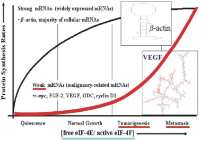
Besides the significance of eIF4E in the early changes of a primary tumor, it has been identified as one of the eight genes which when increased, exhibit a molecular signature of metastatic potential (Ramaswamy et al., 2003). In tumorigenic cells and during cellular transformation the concentration of free eIF4E is increased (DeFatta et al., 1999), causing an enhanced translation of weak mRNAs in cancer cells as seen in Figure 4 (De Benedetti and Graff, 2004). Translational targeting was realized by incorporating of the 5’UTR sequence of rat fibroblast growth factor 2 (FGF2) producing a weak mRNA upstream of the E1A gene.

This incorporation is then restricting the E1A mRNA translation to the cells with high

concentration of free eIF4E (Stoff-Khalili et al., 2008).

(23)

1.4.1.4 Combination of targeting strategies

Although, an enhancement of tumor specificity was gained by every single targeting strategy, all targeting strategies have some disadvantages. Hence, combining targeting strategies was reasonable to increase the safety for infection and replication in normal cells. The feasibility of combining either transductional and transcriptional or transcriptional and translational regulation strategies (dual-level targeting CRAd) for the key goal of oncolytic adenovirus replicative specificity has recently been established (Stoff-Khalili et al., 2008; Stoff-Khalili et al., 2007). Both dual-level viruses showed a specific gain in safety of normal cells and controlled viral replication (Stoff-Khalili et al., 2008; Stoff-Khalili et al., 2007), suggesting that the combination of several targeting strategies in a single CRAd is enhancing cancer cell specific replication and safety of normal cells. The US Food and Drug Administration is still anxious about the specific replication of tropism modified Ads due to the possibility of infecting a wide range of normal cells carrying the receptor to which the modified virus can bind (Yamamoto and Curiel, 2010). Consequently, there is still a need to even increase the cancer cell specific replication due to the fact that several TSPs show a background activity in several cell types. Furthermore, translational restriction of E1A would be even more effective, if the virus would be targeted directly to a specific cancer cell type.

Figure 4: mRNA translation efficiency in tumor progression.

Protein synthesis rate of weak mRNAs is increased in a proportional manner with increasing free eIF4E

concentration and tumor progression. Modified from De Benedetti and Graff (De Benedetti and Graff, 2004).

(24)

1.4.2 Animal models for oncolytic virotherapy

After a successful engineering of an Ad vector and gaining cancer cell specificity in vitro, a newly created Ad vector needs to be investigated in an adequate in vivo animal model. Thus far, the most common animal model used to analyze oncolytic efficiency of newly generated Ad vectors is the human xenograft model in immunodeficient mice (Yamamoto and Curiel, 2010). Disruption of foxn1 gene in nude mice is causing deficiency in B-cell development and T-cell activity. Nude mice are usually used as an animal model for oncolytic virotherapy because the immunosuppressed mice allow human cancer cells to from tumors and to treat the developed tumors with different anti-cancer treatments (Davis and Fang, 2005). Certainly, the reliability on data received from this animal model is questionable because the immune system and the associated inflammation are impaired in nude mice. Furthermore, the species specific Ad replication is a disadvantage for the nude mouse model (Ginsberg et al., 1991; Paielli et al., 2000; Rustia and Shubik, 1979). It was shown, that mice are poorly permissive for human Ad replication leading to a limited possibility for investigating human Ad replication in normal organs and hepatotoxicity resulting from virus replication (Duncan et al., 1978; Ginsberg et al., 1991; Hjorth et al., 1988).

Recently, the “Tissue-Slice-Model” was introduced using three-dimensional precision tissue slices from human tissues to evaluate preclinical oncolytic vectors. Tissue slices can be obtained from normal liver and tumor tissue by the Krumdieck tissue slicer (Krumdieck et al., 1980). The “Tissue-Slice-Model” represents an active metabolic system displaying the heterogeneity of the investigated tissue. It was shown, that human CRAds can successfully replicate on those tissue slices and hepatotoxicity of CRAds can be investigated on liver tissue slices (Kirby et al., 2004; Stoff-Khalili et al., 2006, Stoff-Khalili et al., 2007; Stoff-Khalili et al., 2005). This newly invented technique makes it possible to analyze CRAds pre-clinical efficiently and fast (Kirby et al., 2004; Stoff-Khalili et al., 2006). Unfortunately, analyzing the interaction of CRAds with the immune system is impossible with the “Tissue-slice-model”.

Hence, other pre-clinical animal models are necessary that are permissive to Ad replication and immunocompetent.

It was reported in the past, that Cotton rats’ species Sigmodon hispidus is permissive for the replication of several human respiratory viruses including Ad5 (Niewiesk and Prince, 2002).

Cotton rats were shown to allow Ad5 replication in lungs, nasal passages and cornea and

display the same pathology seen in Ad5 infected-humans (Ginsberg et al., 1989; Pacini et al.,

1984; Prince et al., 1993; Tsai et al., 1992). Based on these findings, the cotton rat animal

(25)

model was employed to investigate in vivo spread and toxicity of Ad5 based vectors (Rojas- Martinez et al., 1998). Recently, the cotton rat model was introduced as a semi permissive animal model in the field of oncolytic virotherapy by establishing transplantable cotton rat tumor cell lines (Steel et al., 2007; Toth et al., 2005). Both groups reported that Ad5 replication and spread to neighboring tumor cells is possible in the generated cotton rat cancer cell lines in vitro and in vivo. Consequently, the cotton rat tumor model was suggested as an animal model to determine replication efficiency, hepatotoxicity and immune response of the Ad5 vectors for oncolytic therapy of tumors. However, a big disadvantage of the use of cotton rats is the difficulty in handling them because they are more aggressive and bite frequently (Niewiesk and Prince, 2002).

Besides the cotton rat model and the tissue slice model, another method to analyze the safety profile of Ads and the immune response, is the exploitation of conditionally replicative canine Ads to investigate in spontaneous dog osteosarcoma treatment (Hay, 2003; Hemminki et al., 2003). This model offers the possibility to gain valuable results about oncolytic activity and toxicity in a natural host and transferable to human application. Nevertheless, employing the canine model is expensive and a connection to veterinarian facilities is essential (Ternovoi et al., 2005), reducing the availability of the canine model for most investigators.

Lately, Thomas et al. introduced the Syrian hamster as a permissive immunocompetent animal model to analyze oncolytic Ad5 vectors (Thomas et al., 2006).

1.5 The Syrian hamster animal model

Previously, the Syrian hamster (Mesocricetus auratus) has been introduced to be permissive

for human Ad5 replication on the contrary to mainly other species examined (Eggerding and

Pierce, 1986; Ginsberg et al., 1991; Silverstein and Strohl, 1986). In cancer cell lines of this

animal Ad5 is competent to infect those cells, replicate in them and spread from cell-to-cell

(Bortolanza et al., 2007; Thomas et al., 2006; Ying et al., 2009). In addition, it was shown that

the adenovirus has important antitumor efficacy after injection in Syrian hamster tumors in

vivo (Spencer et al., 2009). Significantly, the Ad replicates not only in the tumors, but also in

the lungs, liver and other organs (Thomas et al., 2006). Therefore, the “Recombinant DNA

Advisory Committee (RAC)” has suggested using the Syrian hamster model to further

optimize adenovirotherapy and characterize/identify the basic mechanisms in tumor killing

(Lichtenstein et al., 2009).

(26)

To date, several established hamster cancer cell lines are available to study oncolytic CRAds but not for all cancer cell types (Bortolanza et al., 2007; Thomas et al., 2006). Recently, Spencer et al. developed a new pancreatic carcinoma model to study CRAds in the Syrian hamster (Spencer et al., 2009). In this study transplantable tumors of the Syrian hamster from the National Cancer Institute were employed to establish a permanent pancreatic cancer cell line. It was shown, that the Syrian hamster pancreatic carcinoma (SHPC9) cell line was permissive for Ad5 replication and could induce tumor growth upon subcutaneous injection into the hamsters (Spencer et al., 2009).

Nonetheless, up to now no Syrian hamster breast cancer cell line is available because hamsters are somehow resistant to mammary cancer and only two studies have been reported to induce mammary tumors in hamsters (Della Porta, 1961; Rustia and Shubik, 1979).

1.5.1 Induction of breast cancer in female Syrian hamsters

In the past, several different studies of carcinogenesis and cancer chemoprevention have been performed using the Syrian hamster as animal model except for mammary carcinogenesis. The hamster cheek pouch model is one of the studies that was performed in Syrian hamster (Gimenez-Conti, 1993). This model uses the polycyclic aromatic hydrocarbon 7,12- dimethylbenz[a]anthracene (DMBA) applied either as an initiating agent in combination with a tumor promoter such as benzoyl peroxide or as a total carcinogen. Usually, squamous cell carcinomas containing a mutation in the Ha-ras gene are developed after this treatment (Gimenez-Conti, 1993). Furthermore, it was reported that diethyl-stilbestrol (DES) treatment of Syrian hamsters leads to the development of renal carcinomas (Devanesan et al., 2001). Based on this treatment result, Laurent et al. developed a Syrian hamster renal carcinoma cell line (Laurent et al., 1999). In addition to the development of renal carcinomas, Syrian hamsters are capable of developing cholangiocarcinomas, pancreatic cancer and bile duct hyperplasia after exposition to aromatic amines like N-nitrosobis(2-oxopropyl)amine (BOP) (Kaneko et al., 2004; Miyauchi et al., 2000). Pancreatic cancer on the other hand can also be introduced by injecting two to three rounds of ethionine-methionine-BOP into the hamsters and feed them a choline-deficient diet (Mizumoto et al., 1988).

To date, only a few studies reported the development of mammary tumors (Della Porta, 1961;

Rustia and Shubik, 1979), but none could establish a breast cancer cell line so far. Hence, it

was of great interest to establish a breast cancer cell line that could be used to evaluate the

(27)

potency of newly designed oncolytic vectors alone or in combination with chemotherapy or

radiation therapy. Therefore, Coburn et al. induced mammary tumor formation in female

Syrian hamsters by exposing the hamster to the carcinogen N-methyl-N-nitrosurea (MNU)

(Coburn et al., 2011). This study was based on the induction of mammary tumors in rat upon

MNU treatment (Steele et al., 1994). Treatment with MNU induced mammary tumors in

female Syrian hamster, but only in a limited number (Coburn et al., 2011). Histology of these

primary mammary tumors showed abnormal ducts containing secretory products separated by

poorly differentiated epithelial cells (Coburn et al., 2011). Mammary tumors and premalignant

lesions (ADH) were stained positive for HER-2/neu and pancytokeratin, indicating that the

lesions and tumors were of epithelial cell and mammary origin (Coburn et al., 2011).

(28)

Aim of Thesis

Virotherapy employing conditionally replicating adenoviruses (CRAds) represents a promising tool for a wide array of neoplastic diseases and especially to treat breast cancer. Ideally, cancer specific replication of CRAds results in virus mediated oncolysis of infected tumor tissues and release of the progeny virions capable to further propagate in surrounding tumor cells but not in those of normal tissues. Critical for a therapeutic index is selective killing of tumor cells while avoiding killing normal cells. It was recently shown in our group that combination of targeting strategies (dual-level CRAds) increase the tumor on/liver off ratio for breast cancer cells (Stoff-Khalili et al., 2008; Stoff-Khalili et al., 2007).

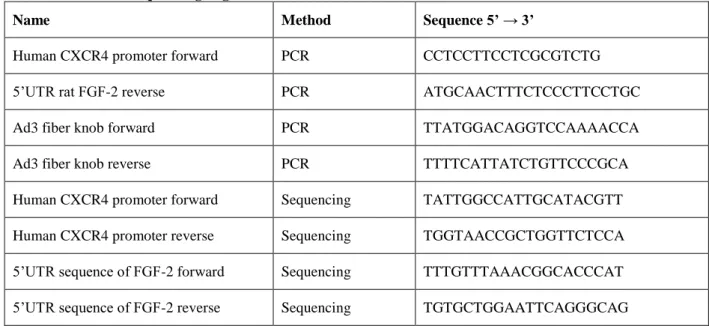
Based on these findings, the first aim of this study was to generate a new CRAd combining three targeting strategies that are used in oncolytic virotherapy to further increase breast cancer specific replication. For this purpose, it was planned to construct a novel 5/3 fiber chimeric (transductional targeting) virus containing the human tumor-specific promoter CXCR4 as a transcriptional control element and the 5’-untranslated region (5’-UTR) of rat FGF-2 (Fibroblast Growth Factor-2) mRNA as a translational control element upstream of the E1A gene. Background replication efficiency and breast cancer specificity were determined by using cytotoxicitiy assays, quantitative realtime polymerase chain reaction (qRT-PCR) and immunoblot analyses comparing infected breast cancer cells to normal cells.

However, the preclinical evaluation of human Ad vectors has been limited due to the poor

replication of human Ads in mice. In this regard, the Syrian hamster has been lately introduced

as an immunocompetent and Ad replication permissive animal model. Due to the lack of a

breast cancer model in the Syrian hamster to evaluate newly constructed oncolytic Ad vectors,

our group recently succeeded in inducing mammary tumors in female Syrian hamsters. Thus,

the second aim of this study was to characterize the first Syrian hamster breast cancer cell line

for its cancerous and tumorigenic character. Furthermore, this cell line was investigated

whether it is a suitable in vitro cell line to evaluate oncolytic Ad vectors for their cancer

specificity. Therefore, several established CRAds such as Ad5 ∆24 RGD and Ad5/3 WT were

used to determine their infection and replication efficiency in this newly established Syrian

hamster breast cancer cell line. The final goal is to use the Syrian hamster as a novel breast

cancer model for preclinical evaluation that is immunocompetent and permissive for human

Ad5 replication.

(29)

2. Materials

2.1 Bacterial Strains

BJ 5183 (Stratagene, Agilent Technologies, La Jolla USA)

This bacterial strain is a recombinant competent strain in which the recombination between a vector containing the adenoviral genome and a transfer vector containing the gene of interest takes place only if both vectors share appropriate regions of homology.

Genotyping endA1 sbcBC recBC galK met thi-1 bioT hsdR (Strr) (Hanahan, 1983).

XL10 Gold (Stratagene, Agilent Technologies, La Jolla, USA)

The bacterial strain XL10 Gold is used commonly for transformation of large DNA molecules as the adenoviral genome with high efficiency. Genotyping: TetrD(mcrA)183 D(mcrCB- hsdSMR-mrr)173 endA1 supE44 thi-1 recA1 gyrA96 relA1 (lac Hte [F´ proAB lacIqZDM15 Tn10Tetr) Amy Camr].

2.1.1 Media for cultivation of bacteria

LB-Medium LB-Agar

Trypton 10 g/l Trypton 10 g/l

Yeast extract 5 g/l Yeast extract 5 g/l

NaCl 8 g/l NaCl 8 g/l

adjust pH to 7.5 with NaOH Agar 15 g/l

→ add H

2

O up to 1 liter adjust pH to 7.5 with NaOH

→ add H

2

O up to 1 liter

(30)

2.2 Eukaryotic cells

A549 (ATCC, CCL-185

TM

)

A549 is a human lung carcinoma cell line, isolated from a lung carcinoma of a 58 year old Caucasian man in 1972.

Hamster Fibroblasts

Fibroblasts were dissected from the lower back a Syrian hamster as a normal control cell line (Kindly provided by Dr. M. Mathis, LSU Health Sciences Center, Shreveport, USA; Present work).

HEK-293 (ATCC, CRL-1573™)

HEK-293 cells were isolated from human embryonic kidneys of a healthy aborted fetus.

This cell line expresses the left end of the adenoviral genome of serotype 5 and is excellent for titrating human adenoviruses. The left arm sequence of the Ad5 genome was incorporated through transfection into the human chromosome 19.

HMAM4 spindle and round

This hamster breast cancer cell line was developed by MNU-treatment in the Syrian Hamster. (Kindly provided by Dr. M. Mathis, LSU Health Sciences Center, Shreveport, USA Present work)

HMAM5

Hamster breast cancer cell line was developed by MNU-treatment in the Syrian Hamster. (Kindly provided by Dr. M. Mathis, LSU Health Sciences Center, Shreveport, USA Present work)

HMEpC – Human mammary epithelial cells (PromoCell, C-12650)

HMEpC were isolated from human adult mammary glands and stained positive for cytokeratin.

MCF-7 (ATCC, HTB-22™)

This breast cancer cell line was developed from the mammary glands from an

adenocarcinoma of a 69-years old Caucasian woman. Growth of MCF-7 cells is

inhibited by tumor necrosis factor alpha (TNF alpha). Furthermore, MCF-7 cells

express the estrogen receptor on their surfaces.

(31)

MDA-MB-231 (ATCC, HTB-26™)

MDA-MB-231 cells were obtained from the mammary glands from an adenocarcinoma of a 51-years old Caucasian woman. MDA-MB-231 cells express the WNT7B oncogene and are immortalized.

MDA-MB-231/N (Meric et al., 2000)

MDA-MB-231 cells were stable transfected with a control plasmid pSV-neo.

MDA-MB-231/E1A (Meric et al., 2000)

MDA-MB-231 cells were stable transfected with pE1A-neo that was carrying the WT E1A gene of Adenovirus serotype 5.

NHDF - Normal human dermal fibroblasts (PromoCell, C-12300 juvenile foreskin) To isolate NHDFs the dermis of juvenile foreskin was used from different locations like the breast, the thighs, the face and the abdomen. This primary cell line is not immortalized and can be maximal passaged 16 times.

PHF PA2 und PHF 25M

Human fibroblasts isolated from human cutaneous skin. (Kindly provided by Dr. B.

Akgül, University of Cologne)

SK-BR-3 (ATCC, HTB-30™)

This cell line is a breast cancer cell line, derived from the mammary glands of a 46 year old Caucasian woman. The cell line is immortalized and over expresses the HER2/c- erb-2 gene product (Hudziak RM, et al. Monoclonal antibodies directed to the Her2 receptor. US Patent 5,677,171 dated Oct 14 1997).

ZR-75-1 (ATCC, CRL-1500™)

ZR-75-1 is a breast cancer cell line established from the mammary glands of a ductal

carcinoma of a 63-years old Caucasian woman.

Abbildung

Figure 1: Adenovirus structure.
Figure 2: Ad serotype 5 infection pathway.
Figure 3: Oncolytic virotherapy of replicative viruses.
Figure 4: mRNA translation efficiency in tumor progression.
+7

Referenzen

ÄHNLICHE DOKUMENTE

Direct comparison of various cell lines and measles virus strains has only been performed by (Scott and Choppin, 1982), so far, who documented the highest yields using Vero

Because CA15-3 was more frequently obtained for the more recently conducted trials, we found that the median follow-up and percentage of patients with recurrence was greater

Expression of E545K and H1047R mutant p110α in the immortalized but non-transformed mammary epithelial cell line MCF10A increased the PI3K kinase activity, allowed

Bei Patientinnen mit einem deutlich erhöhten Risiko für die Entstehung einer Os- teoporose oder bei bekannter Osteoporose sollte eine entspre- chende Medikation entsprechend

Table 5 Odds ratios (ORs) and 95% confidence intervals (CIs) for the associations between age, parity, body mass index (BMI), family history, and levels of the Nottingham

(ii) the PROGNOSTIC collection ( N ¼ 826), including node negative patients with early breast cancers not treated with any systemic therapy until relapse; (iii) the TAM

The aims of this article were therefore (a) to describe the process behind a free, not-for-profit commu- nity-based therapeutic exercise and education programme (TEEP) for BCS in

(2) changes in cell fate by epithelial-mesenchymal transition (EMT) including a transforming growth factor beta (TGF β )-mediated switch of E-cadherin to N-cadherin expression