R E S E A R C H A R T I C L E Open Access
Genetic engineering of Pyrococcus furiosus to use chitin as a carbon source
Martina Kreuzer, Karolin Schmutzler, Ingrid Waege, Michael Thomm and Winfried Hausner*
Abstract
Background:Bioinformatic analysis of the genes coding for the chitinase inPyrococcus furiosusandThermococcus kodakarensisrevealed that most likely a one nucleotide insertion inPyrococcuscaused a frame shift in the chitinase gene. This splits the enzyme into two separate genes, PF1233 and PF1234, in comparison toThermococcus
kodakarensis. Furthermore, our attempts to grow the wild type strain ofPyrococcus furiosuson chitin were negative.
From these data we assume thatPyrococcus furiosusis most likely unable to use chitin as a carbon source. The aim of this study was to analyzein vivoif the one nucleotide insertion is responsible for the inability to grow on chitin, using a recently described genetic system forPyrococcus furiosus.
Results:A marker-less genetic system forPyrococcus furiosuswas developed using simvastatin for positive selection and 6-methylpurine for negative selection. Resistance against simvastatin was achieved by overexpression of the hydroxymethylglutaryl coenzyme A reductase gene. For the resistance to 6-methylpurine the hypoxanthine-guanine phosphoribosyltransferase gene was deleted. This system was used to delete the additional nucleotide at position 1006 in PF1234. The resulting chitinase in the mutant strain was a single subunit enzyme and aligns perfectly to the enzyme fromThermococcus kodakarensis. A detailed analysis of the wild type and the mutant using counted cell numbers as well as ATP and acetate production as growth indicators revealed that only the mutant is able to use chitin as a carbon source. An additional mutant strain containing a reduced chitinase version containing just one catalytic and one chitin-binding domain showed diminished growth on chitin in comparison to the mutant containing the single large enzyme.
Conclusions:Wild typePyrococcus furiosusis most likely unable to grow on chitin in the natural biotope due to a nucleotide insertion which separates the chitinase gene into two ORFs, whereas a genetically engineered strain with the deleted nucleotide is able to grow on chitin. The overall high sequence identity of the two chitinases betweenP. furiosusandT. kodakarensisindicates that this mutation occurred very recently or there is still some kind of selection pressure for a functional enzyme using programmed +/−1 frameshifting.
Background
Chitin is the second most abundant polysaccharide on earth after cellulose. It is the major component of the exos- keletons of insects, the shells of crustaceans and of fungal cell walls [1]. Chitin consists of N-acetylglucosamine subu- nits which are linked byβ-1,4-glycosidic bonds. The deg- radation of chitin is catalyzed by chitinases which hydrolyze these β-1,4-glycosidic bonds. Based on amino acid sequence similarity, chitinases have been classified into the glycoside hydrolases families 18 and 19 [2]. Family 18 chitinases contain a multidomain structure and are
widely distributed in all domains of life. The common fea- tures of these enzymes are catalytic domains which con- sists of a (βα)8(TIM barrel) fold with a conserved DXDXE motif and chitin-binding domains (ChBD) which are involved in the binding to the substrate [3,4].
Most chitinases described so far have been found in the eukaryal and the bacterial domains [5]. Within the domain ofArchaea, only ten euryarchaeal chitinases and one cre- narchaeal have been identified so far [6]. Most of these archaeal enzymes have been only annotated by sequence comparison. Experimental data about the activity and the structure of the enzymes are limited to the genera of Halobacterium,ThermococcusandPyrococcus[7-10]. The first and best characterized archaeal chitinase was from
* Correspondence:Winfried.Hausner@biologie.uni-regensburg.de
Lehrstuhl für Mikrobiologie und Archaeenzentrum, University of Regensburg, Regensburg 93053, Germany
© 2013 Kreuzer et al.; licensee BioMed Central Ltd. This is an Open Access article distributed under the terms of the Creative Commons Attribution License (http://creativecommons.org/licenses/by/2.0), which permits unrestricted use, distribution, and reproduction in any medium, provided the original work is properly cited.
Thermococcus kodakarensis [7,11]. A mutational analy- sis revealed that the enzyme possesses two catalytic do- mains (A and B) and three ChBDs [7]. The N-terminal catalytic domain A functions as an exochitinase and liberates diacetyl-chitobiose. The C-terminal catalytic domain B acts as an endochitinase which produces N-acetyl-chitooligosaccharides of various length, which could be further hydrolyzed to diacetyl-chitobiose by the N-terminal catalytic domain A. Further degradation to N-acetylglucosamine is the result of a concerted action of diacetyl-chitobiose deacetylase and exo-β-D-glucosa- minidase [12].
In contrast to the single chitinase of Thermococcus kodakarensis the chitin-degrading enzymes of Pyrococcus furiosus are encoded by two open reading frames ChiA (PF1234) and ChiB (PF1233) which are separated by 37 nucleotides [8]. The gene product of PF1233 has a ChBD and a catalytic domain which is closely related to the T. kodakarensis catalytic domain B. The structure of this catalytic domain was determined in detail by NMR and X-ray analysis [13-15]. These data also indicate an endochi- tinase activity for PF1233 very similar to T. kodakarensis, however in contrast to the extracellular T. kodakarensis enzyme, the separated P. furiosus enzyme has no signal peptide at the N-terminal region. This would mean that the P. furiosusenzyme is intracellular and the substrates have to be imported into the cell.
A few years ago, Oku and Ishikawa suggested a different explanation for this observation: In principle, P. furiosus has also a single chitinase gene likeT. kodakarensis, but a one nucleotide insertion at position 1006 in PF1234 caused a frame shift which resulted in the separation of the chitinase gene into two genes. Furthermore, their attempts to growP. furiosuson chitin failed [16]. This re- sult is in perfect agreement with previous growth experi- ments on chitin which also failed [17,18]. So far, there is only one report thatP. furiosus is able to use chitin as a carbon source [8].
To reveal this issue, we used a genetic system for P. furiosuswhich allows the removal of the one nucleotide insertion in PF1234 to redesign the chitinase to a single enzyme [19]. Growth experiments with the wildtype and the mutant clearly demonstrate that the wild type strain of P. furiosus is - in contrast to the mutant with the rede- signed chitinase - unable to efficiently use chitin as the main carbon source.
Methods
Strains and growth conditions
P. furiosus was cultivated at 90°C in SME medium, as described previously [20]. For the growth of Pyrococcus strain MUR27Pf with the deleted xanthine-guanine phosphoribosyltransferase gene (xgprt) the medium was supplemented with 6 mM guanosine monophosphate
(Sigma, St. Louis, USA). For solidification, gelrite was added to a final concentration of 1%. The antibiotic sim- vastatin (Toronto Research Inc., Toronto, Canada) was dissolved in ethanol and 6-methylpurine (Sigma, St. Louis, USA) was dissolved in water. Both supplements were ster- ilized by filtration. SME-chitin medium was supplemented with 0.5% colloidal chitin, 0.025% yeast extract and 0.025%
peptone.
For preparation of colloidal chitin 20 g of chitin powder (practical grade, from shrimp shells; Sigma, St. Louis, USA) were mixed with at least 200 ml 37% HCl (pre- cooled to 4°C) and stirred for 1 h at 4°C [21]. The suspen- sion was poured into 1 l of H2O (pre-cooled to 4°C) and was filtered through paper filter (311853, Schleicher and Schüll, Dassel, Germany). The filtrate was washed three times with 1 l of H2O, resuspended in 1 l of H2O and neu- tralized by the addition of NaOH until pH 7.0. The sus- pension was filtered and washed with 3 l of H2O to deionize the chitin. The resulting suspension was filtered and a part was dried to determine the content of liquidity.
Escherichia coli strain DH5αwas used as a host strain for plasmid constructions and was cultivated at 37°C in Luria-Bertani (LB) medium. For the selection of trans- formants, ampicillin was added at 100 μg ml-1 to the medium.
General DNA manipulations and plasmid constructions Restriction enzymes and DNA polymerases for PCR reac- tions were purchased from NEB (Ipswich, USA). Plasmid DNA and DNA fragments from agarose gels were isolated using a WizardW Plus SV Miniprep DNA Purification System or WizardW SV Gel and PCR Clean-Up System from Promega (Mannheim, Germany). DNA sequencing was performed by Seqlab (Göttingen, Germany). Genomic DNA from P. furiosus wild type and genectically engi- neered strains was isolated as described previously [19].
The plasmid pMUR37 was created to enable a markerless deletion of PF1950 (xgprt). It contained three DNA fragments which were joined by single overlap extension PCR reactions: An upstream region of PF1950 (primers Pf1950single_mi_fus-F 5´ -aacagaagtttaagccttcgaagaattggga agagggaga-3´ and Pf1950ml_up_fus1500bp_R 5´ -gcttttt ccttatccactacttatatgaccgcaggtattc-3´ ), a downstream region of PF1950 (primers Pf1950ml_mi-fus1500bp_F 5´ -gaatacc tgcggtcatataagtagtggataaggaaaaagc-3´ and Pf1950_hr_do_R 5´ - gttgaaacagttgcaactcttgg-3´ ) and for the selection with simvastatin the resistance cassette (primers Pf1950sin- gle_up_BamHI_F 5´ -gggcccggatccgggcatttcatcattttt-3´ and Pf1950single_up_fus_R 5´ -tctccctcttcccaattcttcgaaggcttaaac ttctgtt-3´ ) as described previously [19]. The fused fragment was hydrolyzed with BamHI and SacI and was ligated into the corresponding sites of pUC19.
For the construction of the plasmids pMUR47 and pMUR50, a modified pUC19 plasmid with an additional
AscI recognition sequence within the multiple cloning site was used. For both constructs two PCR fusion products were ligated using a common NotI restriction site and inserted into the modified vector using AscI and SbfI re- striction sites.
In the case of pMUR47 the first fusion PCR product with the deleted nucleotide was created using the follow- ing two primer pairs: (Pf1234_AscI_F 5´ -atcgaaggcgcgc ctgctcggtattgtgcttgc-3´ /Pf1234_Del_Fus_R 5´ -tttatcttc taattcggcttgatc-3´ ) and (Pf1233_Del_Fus_F 5´ -ataaaaaa gagtatctcctaactgcagc-3´ / Pf1233_NotI_A_R 5´ -ggtgca gcggccgctggagttggtgatggtgttg-3´). The second fusion PCR product consisted of a two-gene resistance cassette which was needed for the selection-counter-selection system. The resistance cassette contained a gdh pro- moter, thehmgCoA reductase from T. kodakarensis, the region coding for the xgprt (PF1950) and the histone A1 terminator sequence of P. furiosus [19]. The first part was amplified using the primers SimV_NotI_F 5´
-gatgcgcggccgcgggcatttcatcatttttatgaactttgatgaacg-3´ and SimV_Rv 5´ -tcaccctagaaaaagataagcc-3´ . For the second part, the primer pair Pf1950_F_fus1233A 5´ -gcttatctttt tctagggtgacctgggatccaattaccg-3´ and Pf1233_SbfI_R 5´
-atacggcctgcaggttggagtgggtgtggg-3´ was used. Both PCR products were combined with single-overlap extension PCR.
Plasmid pMUR50 was constructed by combination of a PCR product containing the upstream region up to the signal peptide region of PF1234 (primers Pfup1234_As- cI_B_F 5´ -atacgaggcgcgccaactccaatttccctgagc-3´ and Pf SP_up1234_ R 5´ -ggccgatactggatagaatagagatat-3´ ) and a PCR product coding for the catalytic domain of PF1233 (primers Pf SP_1233_fus_F5´ -tatccagtatcggccactacccc tgtcccag-3´ and Pf1233_NotI_B_R 5´ -cctaatgcggccgctag aggaattgagcctgc-3´ ). The resulting PCR product was com- bined with the PCR-amplified resistance cassette using primer pair Pf1950_NotI_F 5´ -tagcatgcggccgctcaccctaga aaaagataagcc-3´ and PfhmgCoA_SbfIR 5´ -gataggcctgcag ggggcatttcatcatttttatg-3´ and inserted into the modified vector as mentioned before. The resulting constructs were verified by DNA sequencing.
Transformation ofP. furiosus
Standard heat shock transformation of P. furiosus was performed as described previously [19]. To obtain the marker-less mutant MUR27Pf, circular plasmid DNA of pMUR37 was used and the corresponding transformants were selected with 10μM simvastatin in SME-starch li- quid medium at 85°C for 48 h. Pure cultures of the intermediate mutant MUR27Pf_i were obtained by plat- ing the cells on solidified medium in the presence of 10 μM simvastatin. The integration of the plasmid into the genome by single cross-over was verified by analyzing corresponding PCR products.
Cultures of the correct intermediate mutant were washed with medium under anaerobic conditions to re- move the simvastatin. In detail, 1.5 ml of a grown cul- ture were centrifuged in an anaerobic chamber for 4 min at 6,000g and resuspended in fresh culture medium without simvastatin. This procedure was repeated three times. For the counter selection the cultures were plated in the presence of 50 μM 6-methylpurine to induce a second homologous recombination step to recycle the selection marker and to eliminate integrated plasmid sequences. In the case of mutant MUR23Pf linearized plasmid pMUR47 was used for the transformation. The genotype of the final mutants was confirmed by PCR and Southern blot experiments.
Growth analysis on chitin medium
For a more detailed analysis of the growth behavior of Pyrococcuson chitin, bottles with 200 ml chitin medium were used and incubated at 90°C. Cell numbers were analyzed with a Thoma counting chamber (0.02-mm depth; Marienfeld, Lauda-Königshofen, Germany) using a phase-contrast microscope.
For the preparation of cell extracts to measure the ATP content, 0.5 ml of Pyrococcus cultures was centrifuged (3 minutes, 10,000g). The cell pellet was washed three times in 0.8 ml PBS buffer, resuspended in 200 μl PBS and treated with glass beads using a FastPrep-24 (M. P. Biomedicals, Irvine, CA) for cell lysis. After centri- fugation (10,000g for 3 minutes at 4°C), the ATP amount in the supernatants was quantified by a luciferin/luciferase assay (FluoProbes, Interchim, Montluçon Cedex, France) using a portable tube luminometer (Junior LB 9509, Berthold Technologies, Bad Wildbad, Germany) according to the instruction manual.
The amount of acetate in the culture supernatant was analyzed using an enzymatic acetate determination kit (R-biopharm, Darmstadt, Germany). For quantification 0.5 ml of aPyrococcusculture was centrifuged and used as indicated in the operating guidelines.
Results and discussion
A detailed sequence analysis of the P. furiosus chitinase genes around the gene split of PF1234 and PF1233 con- firmed the suggestion of Oku and Ishikawa [16] that a one nucleotide insertion at position 1006 in PF1234 caused a frame shift which resulted in the generation of a stop codon after a stretch of ten amino acids (Figure 1).
The second chitinase gene PF1233 starts 37 bp down- stream of PF1234. RT-PCR experiments indicate that both genes were separately transcribed for growth on chitin as well as on tryptone-containing medium lacking chitin [8]. Nevertheless, it is difficult to localize a strong promoter signal upstream of PF1233. Furthermore, a detailed comparison with the corresponding region of
A
B
Figure 1(See legend on next page.)
the single chitinase gene from T. kodakarensis clearly indicates that the deletion of an adenine residue at pos- ition 1006 allows a much better sequence alignment be- tween P. furiosusand T. kodakarensiswithin this region (Figure 1).
To exclude the possibilities that the presence of this additional nucleotide is caused by a sequencing error or is only present in theP. furiosusstrain used for the gen- ome sequencing project [22] we ordered a newP. furio- sus strain (DSM 3638) from the DSMZ and confirmed the presence of this additional nucleotide by sequencing of a corresponding PCR product (data not shown).
In vitroexperiments using an artificial recombinant chiti- nase from P. furiosus, heterologously expressed in E. coli,
indicate that the single enzyme constructed by the deletion of the additional nucleotide is much more active than the separated wild type enzymes [16]. To investigate if this find- ing from the in vitroexperiments could be confirmed by in vivo data we used a modified version of the recently developed genetic system forP. furiosusto delete this add- itional nucleotide in the genome [19].
As the genetic system described so far is based on a shuttle vector and did not allow the modification of genes within the genome we started the genetic modification of P. furiosus with the establishment of a selection/counter selection (pop-in/pop-out) system according to a genetic system which was recently described for T. kodakarensis [23,24]. Santangelo et al. demonstrated that the deletion of
(See figure on previous page.)
Figure 1Comparison of chitinases fromP. furiosusandT. kodakarensis.(A) Schematic models of catalytic and chitin binding domains of both chitinases, the single enzyme fromT. kodakarensisand the split version fromP. furiosus. (B) Alignment of the amino acid sequences. In the case ofP. furiosusthe corresponding DNA sequence was modified by the deletion of the adenine at position 1006. The details of this region were shown in the box within the alignment. The upper part within the box shows the original DNA sequence ofP. furiosustogether with the corresponding amino acid sequence in the region of the putative nucleotide insertion of PF1234. The lower part shows the sequence with the deleted nucleotide. For sequence comparison, the amino acid sequence of the corresponding chitinase region ofT. kodakarensisis also shown and identical amino acids between both,T. kodakarensisandP. furiosus, are shadowed in black. The position with the additional nucleotide is underlined. The putative stop codon is marked by an asterisk.
genomic DNA
marker-less mutant MUR27Pf MUR27Pf_i pMUR37
positive selection with simvastatin for intermediate mutant
negative selection with 6-methylpurine marker-less mutant resistance cassette
TK0914
TK0914
TK0914 PF1951/1952
PF1951/1952
PF1951/1952
PF1951/1952
PF1951/1952 PF1951/1952
PF1951/1952 PF1949
PF1949
PF1949
PF1949 PF1949
PF1949
PF1949 PF1950
PF1950 PF1948
PF1948
PF1948
PF1948 PF1948
PF1948
PF1948
PF1950
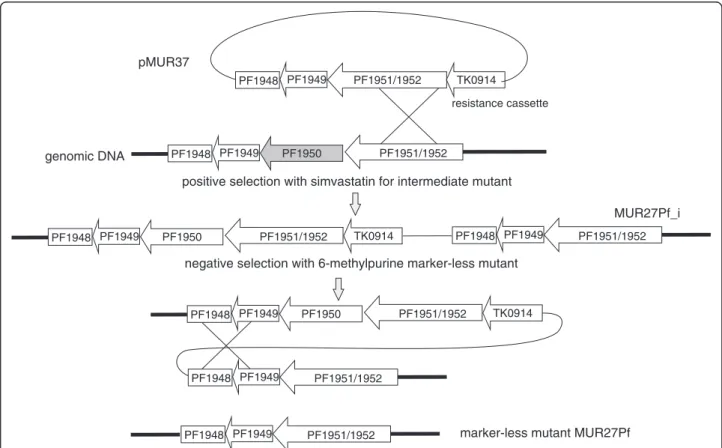
Figure 2Schematic drawing for the construction of marker-less strain MUR27Pf.Plasmid pMUR37 was obtained by overlapping PCR. It contained 1-kb upstream and downstream regions of PF1950 and the resistance cassette TK0914 for selection with the antibiotics simvastatin.
After transformation the intermediate mutant MUR27Pf_i was isolated and characterized. An additional negative selection with 6-methylpurine produced strain MUR27Pf with the marker-less deletion of PF1950 (shadowed in grey).
the xgprt gene (TK0664) confers resistance to 6-methyl- purine. First experiments to inactivate the corresponding gene PF1950 inP. furiosusindicated that this gene can be also used for counter selection experiments inP. furiosus (data not shown).
To establish a deletion mutant for PF1950 inP. furiosus, plasmid pMUR37 was constructed (Figure 2). This plasmid contains about 400 bp upstream and 1500 bp downstream sequence of PF1950 followed by thehydroxymethylglutaryl coenzyme A(hmg-CoA)reductasegene fromT. kodakaren- sis(TK0914). The expression of this gene under the control of a stronggdhpromoter conveys resistance to the antibio- tics simvastatin [19,25]. The resulting plasmid pMUR37 was integrated into the wild typeP. furiosusby single cross- over into the homologous genomic region of PF1951/
PF1952 by transformation (Figure 2). A successful inter- mediate mutant (MUR27Pf_i) was selected based on the re- sistance toward simvastatin and further characterized by PCR analysis (data not shown). For the marker-less deletion of PF1950 a negative selection with 6-methylpurine was performed to induce a second recombination event which enabled the removal of the complete plasmid together with the resistance cassette (pop-out mechanism). The resulting P. furiosusmutant with the deletion of PF1950 (MUR27Pf)
was verified by PCR and Southern blot experiments (data not shown). Further characterization of MUR27Pf revealed that the presence of 6 mM guanine monophosphate in the medium improved the growth of this strain (data not shown). Furthermore, in combination with the positive sim- vastatin selection marker the new strain enables marker- less gene modifications within the genome ofP. furiosus.
In the next step we used the new strain MUR27Pf for the redesign of the chitinase to a single enzyme. For the deletion of the additional adenine nucleotide at position 1006 within PF1234 plasmid pMUR47 was constructed.
This plasmid contains a two-gene resistance cassette con- sisting of the hmg-CoA reductase from T. kodakarensis (TK0914) and the xgprt (PF1950) from P. furiosus (Figure 3). Both genes were transcribed together under the control of the gdh promoter of T. kodakarensis and the terminator of the histoneA1gene [19]. This fragment was fused by single-overlap extension PCR with a part of PF1233 on one side and with the modified region of PF1233/PF1234 containing the sequence with the deleted nucleotide at position 1006 on the other side. After cloning inE. coliand sequence verification the resulting construct pMUR47 was hydrolyzed with restriction enyzmes to re- move the vector DNA and used for transformation of
pf1235 Delta A 1006
pf1233
pf1233
Delta A 1006 pf1234 pf1233
resistance cassette Delta A 1006
pf1234
pf1233
pf1235
tk0914 pf1950
pf1234 pf1233
pf1234 pf1235 pf1232
pf1233 pf1234
AAAAAA pf1232
pf1233
pf1232
AAAAAAA WT
genomic DNA pMUR47
positive selection with simvastatin
negative selection with 6-methylpurine
MUR23Pf
MUR23Pf_i pf1233
resistance cassette tk0914 pf1950
resistance cassette tk0914 pf1950
pf1235 ChiA
ChiB
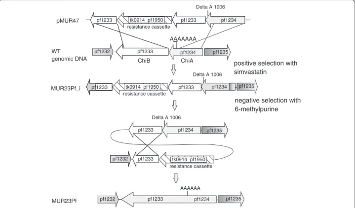
Figure 3Cloning strategy for the mutant MUR23Pf.Plasmid pMUR47 was constructed by overlapping PCR. It contained the chitinase sequence with the deleted nucleotide at position 1006 with corresponding upstream and downstream regions for homologous recombination together with the resistance cassette consisting of TK0914 for selection with the antibiotics simvastatin and PF1950 for the counter selection with 6-methylpurine. After transformation with linearized pMUR47 the intermediate mutant MUR23Pf_i was isolated and characterized. The negative selection with 6-methylpurine resulted in strain MUR23Pf with the redesigned chitinase.
P. furiosusby homologous recombination via double cross- over (Figure 3). Transformants with the integrated DNA fragment were selected with the antibiotic simvastatin.
After characterization of the intermediate mutant a nega- tive selection with 6-methylpurine was performed to create the marker-lessP. furiosusMUR23Pf strain with the single chitinase gene.
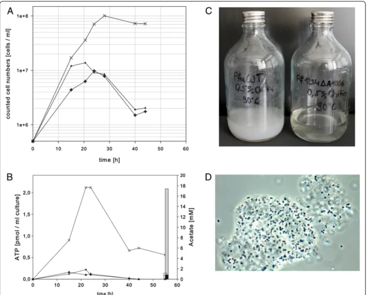
After verification of the resulting mutant MUR23Pf by PCR and Southern blot experiments (data not shown), the mutant strains, MUR23Pf and MUR27Pf, and the wild type of P. furiosus were grown in SME medium in the presence of 0.5% colloidal chitin, 0.025% yeast extract and 0.025% peptone. The mutant with the single chitinase reached a maximum cell dens- ity of 1 × 10⁸ ml-1 after approximately 28 h and
remained stable in the stationary phase for at least add- itional 20 h (Figure 4A). In contrast, the wild type and the strain with the deletion of the xgprtgene reached a ten-fold lower cell density of about 1 × 10⁷ml-1after 28 h and further incubation resulted in a strong decrease in the cell density (Figure 4A). The quantification of the ATP level revealed that the ATP concentration of the mutant with the redesigned chitinase was also about 10-fold higher than the ATP concentration of the con- trol strains (Figure 4B). The determination of the acet- ate concentration after 55 h incubation indicated a strong increase almost up to 18 mM acetate in the medium of the strain with the modified chitinase, whereas the media of the control strains exhibited ace- tate concentrations below 1 mM (Figure 4B).
Figure 4Growth behavior in SME medium with 0.5% colloidal chitin at 90°C.(A) Growth curves of wild typeP. furiosus(plus signs), mutant MUR27Pf (triangles) and mutant MUR23Pf (crossings) with the redesigned chitinase as a single enzyme. (B) Growth analysis of thePyrococcus strains by quantification of the ATP level (left axis). In addition, the final acetate concentration in the medium was analyzed after 55 h incubation (right axis). (C) Documentation of the incubated bottles after x h incubation,P. furiosuswild type is shown on the left side and mutant MUR23Pf on the right side. (D) Phase contrast microscopic picture of the mutant strain MUR23Pf grown on chitin.
up pf1234 pf1233
pf1233 C-Term
pf1234 SP
pf1235 pf1232
pf1232 WT
genomic DNA pMUR50
positive selection with simvastatin
negative selection with 6 methylpurine
MUR24Pf MUR24Pf_i
resistance cassette tk0914 pf1950 resistance cassette
tk0914 pf1950
pf1235 ChiA
ChiB
SP
plasmid pf1233 C-Term
up pf1234 pf1233
C-Term SP
resistance cassette tk0914 pf1950 plasmid
pf1233 ChiB
pf1233 ChiB
pf1233 SP C-Term
up pf1234 SP
pf1235 pf1234 SP
ChiA
pf1234 SP ChiA
pf1235
Figure 5Schematic drawing for the construction of strain MUR24Pf.Plasmid pMUR50 possesses a modified chitinase construct together with the resistance cassette as used for the construction of plasmid pMUR47. After transformation with circular pMUR50 the intermediate mutant MUR24Pf_i was isolated and characterized. The negative selection with 6-methylpurine resulted in strain MUR24Pf with a modified version of the chitinase.
Figure 6Growth curves (left axis) and final acetate concentrations (right axis) of thePyrococcusstrains MUR23Pf (crossings) and MUR24Pf (triangles).
This strong difference in the observed acetate concen- trations clearly indicates that the genetically engineered P. furiosusstrain uses chitin as a carbon source. Chitin is highly acetylated and we therefore assume that acetate is released from the chitin during growth of P. furiosus. In comparison, in an experiment with similar cell density and incubation time grown on cellobiose as substrate, an acet- ate concentration of about 4 mM was observed [26]. Due to the insolubility of the chitin and the fact that many cells stick on the chitin it was very difficult to report reliable OD values, but the visual inspection of the incubated bottles of the wild type and the mutant strain clearly demonstrated chitin degradation (Figure 4C). The bottle containing the wild type after an incubation of 55 h still exhibited a milky turbidity due to the insolubility of the chitin. In contrast, the amount of insoluble chitin in the bottle with the genet- ically engineered P. furiosus strain was considerably reduced (Figure 4C). Figure 4D shows a phase contrast microscopic picture of the mutant strain. It can be clearly seen that the cells are attached to the chitin particles.
The experiments presented so far clearly indicate that the P. furiosus strain with the redesigned chitinase can grow much better on colloidal chitin than the wild type strain. In this context it is interesting to note the overall high se- quence identity of the two chitinases between P. furiosus andT. kodakarensis. This indicates that this mutation oc- curred very recently or there is still some kind of selection pressure for a functional enzyme. It is possible thatP. furio- sus is able to synthesize the chitinase as a single enzyme using programmed +/−1 frameshifting which enables the ribosome to bypass the frameshift [27]. This situation was recently described for the expression of thefucA1gene in Sulfolobus acidocaldarius. However, in this case the expres- sion efficiency is only about 5% in comparison to a gene without the programmed frameshift [27]. To exclude or to confirm the idea of programmed frameshifting additional experiments will be necessary. It is not possible to use avail- able proteomics data ofP. furiosus to support this idea as the available proteomics data do not match the chitinases.
Furthermore, first attempts to use purified enzyme for a detailed mass spectrometry-based analysis were compli- cated by the finding that the enzyme is most likely the target of intensive proteolytic cleavage activity (data not shown).
To analyze if we could further stimulate the growth of P. furiosus on chitin we constructed an additional mu- tant. Oku and Ishikawa also demonstrated in in vitro experiments that a reduced recombinant version of the chitinase consisting of the chitin binding and the cata- lytic domain B had a higher activity as the whole enzyme [16]. To analyze if this in vitroresult with heterologous expressed proteins could be confirmed in vivo, the add- itional plasmid pMUR50 was created (Figure 5). It con- tained the resistance cassette, the catalytic and the chitin
binding domain of PF1233, the signal peptide sequence of PF1234 and the corresponding upstream sequence for homologous recombination by single cross-over. After se- quence verification the plasmid was used to create the marker-less P. furiosusmutant MUR24Pf using the same strategy as described before (Figure 5).
We also performed growth experiments using similar conditions as outlined above with the strain containing the ChiB domain (Figure 6). For comparison, the data of the mutant strain MUR23Pf with the redesigned chitinase as a single enzyme were included. The maximum cell density of the strain with the ChiB domain was reduced from 1 × 10⁸ml-1to 6.5 × 10⁷ml-1. Furthermore, the sta- bility of the cells in the stationary phase was lower and the acetate concentration in the medium was reduced by one third in comparison to the strain with the redesigned complete chitinase (Figure 6). Taken together, the data clearly indicate that the P. furiosusstrain with the rede- signed complete chitinase as a single enzyme had a growth advantage compared to the strain with the smaller version containing the ChiB domain. This result did not confirm thein vitrodata from Oku and Ishikawa and is therefore a nice example that sometimesin vitroresults do not match within vivoresults [16].
Conclusions
Our data demonstrate that wild type P. furiosusis most likely unable to use chitin as a main carbon source due to a one nucleotide insertion which splits the chitinase into two separate enzymes. In contrast, a genetically engi- neered strain with the deleted nucleotide is able to grow on chitin. The overall high sequence identity of the two chitinases between P. furiosus and T. kodakarensis indi- cates that this mutation occurred very recently or there is still some kind of selection pressure for a functional en- zyme using programmed +/−1 frameshifting [27]. As the later one resulted in a very low expression level of the chit- inase, we conclude thatP. furiosusdoes not rely on chitin as a major carbon source in the natural biotope.
Abbreviations
ChBD: Chitin binding domain;fucA1:α-fucosidase;gdh: Glutamate
dehydrogenase;hmg-CoA: Hydroxymethylglutaryl-Coenyzme A;P:Pyrococcus;
PCR: Polymerase chain reaction;T:Thermococcus;Xgprt: Hypoxanthine- guanine phosphoribosyltransferase.
Competing interests
The authors declare that they have no competing interests.
Authors’contributions
MK participated in designing the data and carried out the experimental work on the chitinase mutants. KS established the marker-less system. IW was involved to set up the experimental work. WH and MT coordinated and supervised the research and wrote the manuscript. All authors read and approved the final manuscript.
Acknowledgements
We thank Renate Richau for technical assistance and Emma Gagen for critical reading of the manuscript. The work was supported by the Deutsche Forschungsgemeinschaft TH 422/11-1.
Received: 16 November 2012 Accepted: 4 February 2013 Published: 7 February 2013
References
1. Gooday GW:The ecology of chitin degradation. New York: Plenum Press;
1990:387–430.
2. Henrissat B, Davies G:Structural and sequence-based classification of glycoside hydrolases.Curr Opin Struct Biol1997,7(5):637–644.
3. Synstad HB, Gåseidnes S, van Aalten DMF, Vriend G, Nielsen JE, Eijsink VGH:
Mutational and computational analysis of the role of conserved residues in the active site of a family 18 chitinase.Eur J Biochem2004,
271(2):253–262.
4. Funkhouser JD, Aronson NNJ:Chitinase family GH18: evolutionary insights from the genomic history of a diverse protein family.BMC Evol Biol2007, 7:96–112.
5. Li H, Greene LH:Sequence and structural analysis of the chitinase insertion domain reveals Two conserved motifs involved in chitin-binding.PLoS One2010,5(1):e8654.
6. Staufenberger T, Imhoff JF, Labes A:First crenarchaeal chitinase found in Sulfolobus tokodaii.Microbiol Res2012,167(5):262–269.
7. Tanaka T, Fukui T, Imanaka T:Different cleavage specificities of the dual catalytic domains in chitinase from the hyperthermophilic archaeon Thermococcus kodakaraensis KOD1.Biol Chem2001,276(38):35629–35635.
8. Gao J, Bauer MW, Shockley KR, Pysz MA, Kelly RM:Growth of
hyperthermophilic archaeon Pyrococcus furiosus on chitin involves two family 18 chitinases.Appl Environ Microbiol2003,69(6):3119–3128.
9. Andronopoulou E, Vorgias CE:Isolation, cloning, and overexpression of a chitinase gene fragment from the hyperthermophilic archaeon Thermococcus chitonophagus: semi-denaturing purification of the recombinant peptide and investigation of its relation with other chitinases.Protein Express Purif2004,35(2):264–271.
10. Hatori Y, Sato M, Orishimo K, Yatsunami R, Endo K, Fukui T, Nakamura S:
Characterization of recombinant family 18 chitinase from extremely halophilic archaeon Halobacterium salinarum strain NRC-1.Chitin and Chitosan Res2006,12:201.
11. Tanaka T, Fujiwara S, Nishikori S, Fukui T, Takagi M, Imanaka T:A unique chitinase with dual active sites and triple substrate binding sites from the hyperthermophilic archaeon Pyrococcus kodakaraensis KOD1.
Appl Environ Microbiol1999,65:5338–5344.
12. Tanaka T, Fukui T, Fujiwara S, Atomi H, Imanaka T:Concerted action of diacetylchitobiose deacetylase and exo-beta-d-glucosaminidase in a novel chitinolytic pathway in the hyperthermophilic archaeon Thermococcus kodakaraensis KOD1.J Biol Chem2004,279:30021–30027.
13. Nakamura T, Mine S, Hagihara Y, Ishikawa K, Uegaki K:Structure of the catalytic domain of the hyperthermophilic chitinase from Pyrococcus furiosus.Acta Crystallogr F2007,F63:7–11.
14. Nakamura T, Mine S, Hagihara Y, Ishikawa K, Ikegami T, Uegaki K:Tertiary structure and carbohydrate recognition by the chitin-binding domain of a Hyperthermophilic Chitinase from Pyrococcus furiosus.J Mol Biol2008, 381(3):670–680.
15. Tsuji H, Nishimura S, Inui T, Kado Y, Ishikawa K, Nakamura T, Uegaki K:
Kinetic and crystallographic analyses of the catalytic domain of chitinase from Pyrococcus furiosus- the role of conserved residues in the active site.FEBS J2010,277(12):2683–2695.
16. Oku T, Ishikawa K:Analysis of the hyperthermophilic chitinase from Pyrococcus furiosus: activity toward crystalline chitin.Biosci Biotechnol Biochem2006,70(7):1696–1701.
17. Huber R, Stöhr J, Hohenhaus S, Rachel R, Burggraf S, Jannasch HW, Stetter KO:Thermococcus chitonophagus sp. nov., a novel, chitin-degrading, hyperthermophilic archaeum from a deep-sea hydrothermal vent environment.Arch Microbiol1995,164:255–264.
18. Driskill LE, Kusy K, Bauer MW, Kelly RM:Relationship between glycosyl hydrolase inventory and growth physiology of the Hyperthermophile Pyrococcus furiosus on carbohydrate-based media.Appl Environ Microbiol 1999,65(3):893–897.
19. Waege I, Schmid G, Thumann S, Thomm M, Hausner W:Shuttle vector- based transformation system for Pyrococcus furiosus.Appl Environ Microbiol2010,76:3308–3313.
20. Fiala G, Stetter KO:Pyrococcus furiosus sp. nov. represents a novel genus of marine heterotrophic archaebacteria growing optimally at 100°C.
Arch Microbiol1986,145:56–61.
21. Reichenbach H, Dworkin M:The order Cytophagales (with addenda on the genera Herpetosiphon, Saprospirs and Fexithrix). InThe Prokaryotes.
Volume 1. Edited by Starr MP,et al. New York: Springer Verlag;
1981:356–379.
22. Maeder DL, Weiss RB, Dunn DM, Cherry JL, González JM, DiRuggiero J, Robb FT:Divergence of the hyperthermophilic archaea Pyrococcus furiosus and P. horikoshii inferred from complete genomic sequences.
Genetics1999,152(4):1299–1305.
23. Sato T, Fukui T, Atomi H, Imanaka T:Improved and versatile
transformation system allowing multiple genetic manipulations of the hyperthermophilic archaeon Thermococcus kodakaraensis.Appl Environ Microbiol2005,71(7):3889–3899.
24. Santangelo TJ, Cubonová L, Reeve JN:Thermococcus kodakarensis genetics: TK1827.Appl Environ Microbiol2010,76(4):1044–1052.
25. Matsumi R, Manabe K, Fukui T, Atomi H, Imanaka T:Disruption of a sugar transporter gene cluster in a hyperthermophilic archaeon using a host-marker system based on antibiotic resistance.J Bacteriol2007, 189(7):2683–2691.
26. Basen M, Sun J, Adams MW:Engineering a hyperthermophilic archaeon for temperature-dependent product formation.mBio2012,
3(2):e00053–12.
27. Cobucci-Ponzano B, Rossi M, Moracci M:Translational recoding in archaea.
Extremophiles2012,16(6):793–803.
doi:10.1186/1472-6750-13-9
Cite this article as:Kreuzeret al.:Genetic engineering ofPyrococcus furiosusto use chitin as a carbon source.BMC Biotechnology201313:9.
Submit your next manuscript to BioMed Central and take full advantage of:
• Convenient online submission
• Thorough peer review
• No space constraints or color figure charges
• Immediate publication on acceptance
• Inclusion in PubMed, CAS, Scopus and Google Scholar
• Research which is freely available for redistribution
Submit your manuscript at www.biomedcentral.com/submit