Central modulators of human pain:
Effects of oxytocin, exam stress, breathing exercises and transcranial magnetic stimulation
Kumulative Inaugural-Dissertation zur Erlangung der Doktorwürde
der Philosophischen Fakultät II (Psychologie, Pädagogik und Sportwissenschaft) der Universität Regensburg,
vorgelegt in englischer Sprache von Matthias Zunhammer
aus Traunwalchen am 28.05.2014.
Die Arbeit entstand in Betreuung durch Prof. Dr. Mark W. Greenlee,
Lehrstuhl für Experimentelle Psychologie, Philosophische Fakultät II der Universität Regensburg.
Regensburg 2014
Erstgutachter: Prof. Dr. Mark W. Greenlee Zweitgutachter: Prof. Dr. Andreas Mühlberger
PR E F A C E
This cumulative dissertation comprises four studies investigating pain processing within the human brain. A short overview of these studies is provided on page 7. From here on, the four studies are referred to as Studies 1, 2, 3 and 4 in chronological order, starting with the latest. All studies have been published in peer-reviewed journals within the last four years. All manuscripts are reproduced in their last pre-print version with permission from the publishers. All manuscripts were adapted to the formatting and orthography recommended by the Publication Manual of the American Psychological Association (2010) for consistency.
The four separate reference sections were integrated into one bibliography at the end of the thesis. Tables, figures and appendices were renumbered. Otherwise the manuscripts were not changed.
A number of people have made this work possible. They are given due credit on page 5. The contributions of co-authors to the four studies are further detailed on page 8. My colleagues and I have completed a number of related studies during my doctoral studies, which are not included in this dissertation, but worth mentioning:
• In parallel with Study 1, we investigated the effects of oxytocin on emotional pain modulation and its brain correlates. The study is currently in preparation for publication.
• In parallel with Study 2, we assessed the effects of exam stress on sleep and legal drug consumption. The results have been submitted for publication, but do not match the topic of this dissertation and were therefore not included.
• Before Study 3, we completed and published an experiment testing the effects of acupuncture on motor system excitability (Zunhammer, Eichhammer, Franz, Hajak, &
Busch, 2012). Main contributor was Dr. med. Johanna Franz, who also wrote her doctoral thesis on the topic.
EI D E S S T A T T L I C H E VE R S I C H E R U N G
Ich erkläre hiermit an Eides Statt, dass ich die vorliegende Arbeit ohne unzulässige Hilfe Dritter und ohne Benutzung anderer als der angegebenen Hilfsmittel angefertigt habe. Die aus anderen Quellen direkt oder indirekt übernommenen Daten und Konzepte sind unter Angabe der Quelle gekennzeichnet.
Bei der Auswahl und Auswertung des Materials haben mir die auf der Seite 8 aufgeführten Personen in der jeweils detaillierten Weise unentgeltlich geholfen. Weitere Personen waren an der inhaltlich-materiellen Erstellung der vorliegenden Arbeit nicht beteiligt. Insbesondere habe ich hierfür nicht die entgeltliche Hilfe von Vermittlungs- beziehungsweise Beratungsdiensten (Promotionsberater oder anderer Personen) in Anspruch genommen. Niemand hat von mir unmittelbar oder mittelbar geldwerte Leistungen für Arbeiten erhalten, die im Zusammenhang mit dem Inhalt der vorgelegten Dissertation stehen.
Die Arbeit wurde bisher weder im In- noch im Ausland in gleicher oder ähnlicher Form einer anderen Prüfungsbehörde vorgelegt.
Ich versichere an Eides Statt, dass ich nach bestem Wissen die reine Wahrheit gesagt und nichts verschwiegen habe. Vor Aufnahme der obigen Versicherung an Eides Statt wurde ich über die Bedeutung der eidesstattlichen Versicherung und die strafrechtlichen Folgen einer unrichtigen oder unvollständigen eidesstattlichen Versicherung belehrt.
Die Eidesstattliche Erklärung zu dieser Dissertation wurde in Regensburg am 14.05.2014 von Matthias Zunhammer gegeben und vom Fakultätsverwalter Peter Grimm aufgenommen. Die signierte Eidesstattlichen Versicherung wird vom Dekanat der Fakultäten für Sprach-, Literatur- und Kulturwissenschaft sowie Psychologie, Pädagogik und Sportwissenschaft verwahrt.
DA N K S A G U N G
Über mehr als vier Jahre wurde ich von meinem Doktorvater Prof. Mark W. Greenlee und meinem Mentor am Klinikum Prof. Peter Eichhammer auf den Weg in die akademische Welt unterstützt und gefördert. Dafür möchte ich Ihnen aus ganzem Herzen danken. Für die ständige Erreichbarkeit, die schnellen Korrekturen, die Kongressteilnahmen und den vertrauensvollen Spielraum.
Berthold Langguth möchte ich für den Start in die Welt der Versuchsplanung, der akademischen Publikationen und der aufregenden Kongressreisen danken, genau so Volker Busch, Katharina Rosengart und Anton Beer für die vielen Diskussionen und die entscheidende Hilfe mit der verflixten Methodik. Besonderer Dank gilt Sandra Geis, Hanna Eberle, Sonja Blumenstock und Johanna Franz, die mir im Rahmen ihrer Abschlussarbeiten/Praktika geholfen und so viele Stunden Arbeitszeit geopfert haben. Für die bitter nötige Hilfe bei der Beantragung einer klinischen Studie nach Arzneimittelgesetz danke ich: Florian Zeman und dem Zentrum für Klinische Studien am Uniklinikum Regensburg, Ralph Heimke-Brinck von der Apotheke des Uniklinikums Erlangen, Jörg Pfeiffer von der Hausapotheke des Bezirksklinikums, Zaven Orfalian und Vincenco Giordano von der Firma Sigma Tau, sowie Prof. Dr. Thomas Baghai.
Dank an dieser Stelle auch den weit über 200 Probanden, die das Vertrauen aufgebracht haben im Rahmen dieser Arbeit an einer Schmerzstudie teilzunehmen. Der Studienstiftung des deutschen Volkes danke ich für das viele Geld; ohne sie wäre ich mittendrin ohne Finanzierung dagestanden und hätte viele inspirierende Menschen nicht kennengelernt. Zuletzt danke ich meinen Eltern für die Liebe und für die geduldigen Antworten auf das ständige „Warum?“.
CO N T E N T S
Preface 3
Eidesstattliche Versicherung 4
Danksagung 5
Abstract 7
Contributions 8
Abbreviations 9
Introduction 12
Rationale and aims Background
Study 1 21
Title: Effects of intranasal oxytocin on thermal pain in healthy males — a randomized fMRI study
Authors: Zunhammer, M, Geis, S, Busch, V, Greenlee MW, Eichhammer, P.
Status: Accepted for publication by Psychosomatic Medicine, as of Oct 16th, 2014.
© 2014 American Psychosomatic Society, Lippincott Williams & Wilkins.
Reprinted with permission.
Study 2 42
Title: Somatic symptoms evoked by exam stress in university students: the role of alexithymia, neuroticism, anxiety, and depression.
Authors: Zunhammer, M, Eberle, H, Eichhammer, P, Busch, V.
Status: Published 2013 in PLOS ONE, 8(e84911).
© Authors retain copyright.
Study 3 60
Title: Do cardiorespiratory variables predict the antinociceptive effects of deep and slow breathing?
Authors: Zunhammer, M, Eichhammer, P, Busch, V.
Status: Published 2013 in Pain Medicine, 14(6), 843–54.
© 2013 Wiley periodicals Inc.: Reprinted with permission.
Study 4 76
Title: rTMS over the cerebellum modulates temperature detection and pain thresholds through peripheral mechanisms
Authors: Zunhammer, M, Busch V, Griesbach, F, Landgrebe M, Hajak, G, Langguth, B.
Status: Published 2011 in Brain Stimulation, 4(4), 210–7.e1.
© 2011 Elsevier Inc. Reprinted with permission.
Concluding Discussion 87
On measuring pain
Discussion of methods and study designs used Discussion of results and outlook
Bibliography 98
AB S T R A C T
The available means to control human pain are insufficient, novel mechanisms of pain modulation must be explored and understood. This cumulative dissertation comprises four studies, which explored potential means to modulate pain in the central nervous system. An overview on the current understanding of pain, its basic mechanisms, and its known modulators is provided.
Study 1 tested if a high intranasal dose of the neuro-hypophyseal hormone oxytocin affected perception and processing of thermal pain in 36 healthy male volunteers. Experimental pain thresholds were obtained and a functional Magnetic Resonance Imaging experiment with ratings of noxious heat was conducted. Oxytocin was found to reduce ratings of perceived heat intensity and amygdala activity. Both effects were small and independent of temperature. Although the hypothesis of an antinociceptive effect of oxytocin could not be confirmed, the study provides first evidence for effects of oxytocin on thermal stimulus processing.
Study 2, a longitudinal questionnaire study, examined the effects of a period of academic exam stress on bodily symptoms in 150 students. Various symptoms of pain, as well as gastro-intestinal and autonomic complaints were found to increase during exam stress. Neuroticism, but not alexithymia, trait anxiety, or depression explained symptom increases under exam stress. Study two offers the first comprehensive, quantitative description of bodily symptoms under exam stress. Neuroticism was identified as a potential predisposing personality factor for the occurrence of bodily symptoms under stress.
Study 3 aimed at elucidating physiological mechanisms behind the proposed antinociceptive effects of slow breathing exercises in 20 healthy participants. Breathing frequency, heart rate variability, and hyperventilation were not found to predict changes in experimental pain thresholds or heat pain ratings. A correlation between heart rate at baseline and pain ratings could be observed, confirming that autonomic nervous system function and pain are intertwined.
Study 4 explored and tested if and how repetitive transcranial magnetic stimulation (rTMS) over the cerebellum affected thermal pain thresholds in two separate experiments. Although pain- relieving effects of cerebellar rTMS could be found in a first experiment, the second experiment showed that these effects were driven by peripheral mechanisms and/or the placebo effect. The study highlights the importance of proper experimental control conditions when investigating central modulators of pain.
In a concluding discussion, the current methodology of pain research is reviewed. The methods used for this dissertation are discussed and limitations are identified; findings are summarized and further directions of research are highlighted.
CO N T R I B U T I O N S
Contributors
Study 1 Effects of intranasal oxytocin on thermal pain in healthy males — a randomized fMRI study
Type Double-blind clinical trial; three sessions within 36 participants Study idea: Zunhammer, Eichhammer
Study design: Zunhammer, Eichhammer, Greenlee Data acquisition: Zunhammer, Geis, Busch
Statistical analysis: Zunhammer Manuscript writing: Zunhammer
Manuscript revision: Geis, Busch, Eichhammer, Greenlee Study supervision: Eichhammer, Greenlee
Study 2 Somatic symptoms evoked by exam stress in university students: the role of alexithymia, neuroticism, anxiety, and depression.
Type Longitudinal survey; three time-points within 150 participants Study idea: Zunhammer, Busch, Eichhammer
Study design: Zunhammer, Busch Data acquisition: Zunhammer, Eberle Statistical analysis: Zunhammer Manuscript writing: Zunhammer
Manuscript revision: Eberle, Busch, Eichhammer Study supervision: Busch, Eichhammer
Study 3 Do cardiorespiratory variables predict the antinociceptive effects of deep and slow breathing?
Type Experimental study; four sessions within 20 participants Study idea: Zunhammer, Busch, Eichhammer
Study design: Zunhammer, Busch Data acquisition: Zunhammer Statistical analysis: Zunhammer Manuscript writing: Zunhammer Manuscript revision: Busch, Eichhammer Study supervision: Busch, Eichhammer
Study 4 rTMS over the cerebellum modulates temperature detection and pain thresholds through peripheral mechanisms
Type Experimental study; two experiments with 10 + 12 participants
Study idea: Langguth
Study design: Langguth, Griesbach (part one), Zunhammer (part two) Data acquisition: Griesbach (part one), Zunhammer (part two)
Statistical analysis: Zunhammer, Langguth
Manuscript writing: Zunhammer (~60%), Langguth (~40%)
Manuscript revision: Langguth, Griesbach, Landgrebe, Hajak, Busch Study supervision: Langguth
AB B R E V I A T I O N S
3-T 3-Tesla
AICc Akaike’s Information Criterion for finite sample sizes ANS autonomic nervous system
AR1 first-degree autoregressive model ATP adenosine triphosphate
BDI-II Beck’s Depression Inventory, revised version BOLD blood-oxygen dependent
Bonf. Bonferroni (correction procedure for multiple comparisons)
°C degree Celsius CA California CCK cholecystokinin
CDT cold detection threshold CGRP calcitonin gene-related peptide CI confidence interval
cm centimeter
CNS central nervous system CO2 carbon-dioxide
CPT cold detection threshold CSF cerebrospinal fluid DIS disorders reported
DLPFC dorsolateral prefrontal cortex DNIC diffuse noxious inhibition controls doi digital online identifier
DSB deep and slow breathing
DSM-IV Diagnostic and Statistical Manual of Mental Disorders 4th edition e.g. exempli gratia, for example
ECG electrocardiogram EPI echo-planar imaging
EUDRA-CT European Clinical Trials Database
F F-value; standardized deviation from reference (Fisher-Snedecor distribution)
f female
FIR finite impulse response
fMRI functional Magnetic Resonance Imaging FoV Field of View
FWE family-wise error
FWHM full-width at half maximum GABA gamma-aminobutyric acid GLM general linear model
h hour
HPT heat detection threshold HRF hemodynamic response function
Hz Hertz
i.e. id est, that is
I.U. international unit (WHO standard unit for hormone dose based on a biological effect)
ICC intra-class correlation coefficient
ICD-10 International Classification of Diseases 10th revision IL Illinois
KCNK potassium channel subfamily K
L left
LASSO-
PCR least absolute shrinkage and selection operator-regularized principal components regression
m meter
m male
MANOVA multivariate analysis of variance MES medically explained symptom
mg milligram
min minute
ml milliliter mm millimeter
MNI Montreal Neurological Institute
MP-RAGE Magnetization Prepared Rapid Gradient Echo MR magnetic resonance
MSO maximum stimulator output MUS medically unexplained symptom
n number of, (number of cases in subsample) N number of, (number of cases in total sample) NEO-FFI Big-Five Personality Interview
noDIS no disorders reported (factor level of DIS in analysis) noMES no evidence for MES (factor level of MES in analysis) NTS nucleus of the solitary tract
p p-value; probability of obtaining a statistic at least as extreme as the observed
p. page
PAG periaqueductal gray
pCO2 carbon-dioxide partial pressure PET positron emission tomography PFC prefrontal cortex
pH "power of hydrogen", unit for acidity/basicity (hydronium ion concentration) PLOS Public Library of Science
pp. pages
PSQ-20 Perceived Stress Questionnaire 20-item version PSQ-30 Perceived Stress Questionnaire 30-item version QST Quantitative Sensory Testing
R right
r Pearson correlation coefficient
rMS repetitive magnetic stimulation (of peripheral tissue) RMSSD root mean square of successive differences (in R-R-interval) ROI region of interest
RR R-wave-to-R-wave interval of the cardiac rythm rTMS repetitive transcranial magnetic stimulation
s second
s.p.a. Società per Azioni
SD standard deviation
SDRR standard deviation of the RR-interval SEM standard error of mean
SI, SII somatosensory cortex I and II, also: primary and secondary somatosensory cortex SOMS-7d Screening for Somatoform Symptoms 7-day version
SPECT single photon emission computed tomography SPM statistical parametric mapping
STAI-G-X1 State-Trait Anxiety Inventory – State Form, German Version STAI-G-X2 State-Trait Anxiety Inventory – Trait Form, German Version
t t-value; standardized deviation from reference (Student's t-distribution) T1 longitudinal (spin-lattice relaxation time
T2* transverse (spin-spin) relaxation time, including magnetic field inhomogeneity TAS-20 Toronto-Alexithymia Scale 20-item version
TE echo-time
TENS transcutaneous electrical nerve stimulation TGF-beta tumor growth factor beta
TMS transcranial magnetic stimulation TR time of repetition
TRAAK outdated acronym; synonym for: potassium channel subfamily K TREK outdated acronym; synonym for: potassium channel subfamily K TrkA Tropomyosin related kinase A
TRP transient receptor potential (channel)
TRPA1 transient receptor potential channel ankyrin subtype 1 TRPM8 transient receptor potential channel melastatin subtype 8 TRPV1-4 transient receptor potential vanilloid channel subtype 1-4 TSA thermo-sensory analyzer
UK United Kingdom
USA United States of America VAS Visual Analog Scale
WDT warmth detection threshold WMA World Medical Association
yesDIS stable past or chronic disorders (factor level of DIS in analysis) yesMES evidence for MES (factor level of MES in analysis)
Z Z-value; standardized deviation from reference (normal distribution) α Type-I (or alpha) error level. see: p
β unstandardized parameter estimate (in regression analysis), but also: Type-II error level (in power analysis)
Δ deviation in
τ Kendall’s τ-b coefficient of non-parametric correlation
IN T R O D U C T I O N
Rationale, aims and hypotheses
All studies of this thesis have a common motivation: The World Health Organization (2011) ranks “back and neck pain” as the 4th leading cause of disability adjusted life years in Europe.
Chronic pain is a burden for the individual sufferer and the social welfare systems. Most patients are diagnosed with idiopathic pain, i.e. pain of unknown physiological cause (McMahon & Wall, 2013, pp. 233–247). A considerable proportion of surgery patients suffer from chronic pain long after the acute recovery phase (Perkins & Kehlet, 2000). The available pharmacological and non- pharmacological treatments are limited in their effectiveness and leave many non-responders with insufficient relief of pain (Moore, Derry, & Wiffen, 2013). Despite evidence suggesting that chronic pain is influenced by behavior, beliefs, emotions, motivation, and stress (Gatchel, Polatin, & Kinney, 1995; Wiech & Tracey, 2013) the psychological aspects of pain are complex and insufficiently understood. In this situation the study of central pain modulators can help to comprehend the mechanisms of pain and thus help to develop new and better treatments. The present dissertation is concerned with mechanisms of pain modulation from a neuropsychiatric perspective and a focus on stress-related phenomena. Four means of pain modulation were investigated:
• Study 1, the main study of this thesis, was employed to test a novel pharmacological approach: Intranasal oxytocin has been shown to modulate stress, anxiety, and social cognition in a multitude of studies (Guastella & MacLeod, 2012; Kubzansky, Mendes, Appleton, Block, & Adler, 2012). Oxytocin was repeatedly found to affect amygdala activity (Bethlehem et al., 2013). Moreover, Rash, Aguirre-Camacho, & Campbell, (2013) recently suggested that oxytocin might have antinociceptive actions, based on a review of animal and human results.
Study 1 tested if the hormone oxytocin, given as a nasal spray has antinociceptive effects.
Effects of oxytocin within the brain were investigated by functional magnetic resonance imaging (fMRI). We hypothesized that intranasal oxytocin should:
a) increase noxious thermal pain thresholds;
b) decrease ratings of noxious heat intensity and/or unpleasantness;
c) alter blood oxygen dependent (BOLD) signal changes related to the processing of the thermal stimulus on whole brain level;
d) alter stimulus processing in the amygdala.
• Study 2 focused on the phenomenon of somatization, i.e. bodily complaints in response to psychosocial stress: This questionnaire survey investigated how academic exam stress impacts on reports of pain and other everyday symptoms in university students. In addition, personality factors contributing to the development of symptoms were assessed.
Our aim was to provide a detailed quantitative description of somatic symptom increases under exam stress. We hypothesized that symptoms of pain and other bodily complaints would increase during an exam period and return to baseline after a period without exams.
We further aimed to compare the personality traits alexithymia, neuroticism, trait anxiety and depression with respect to their ability to explain increases in somatization under exam stress.
According to the stress-alexithymia hypothesis, a stronger association of somatization symptoms with alexithymia than with neuroticism, trait anxiety and depression was expected.
• Study 3 followed up hypotheses on the mechanisms of slow breathing, a widespread alternative treatment for stress and pain control (Martin et al., 2012; Zautra et al., 2010): It was experimentally tested if breathing exercises modulate pain perception via breathing frequency, hyperventilation, or cardiac autonomic control, as suggested by Chalaye, Goffaux, Lafrenaye, & Marchand (2009). According to this hypothesis, measures of heart rate variability, such as the standard deviation of heart rate, were expected to explain a significant amount of variance in thermal pain thresholds and/or pain ratings.
• Study 4 aimed to modulate pain perception experimentally by stimulating the cerebellum with repetitive Transcranial Magnetic Stimulation (rTMS). This non-invasive brain stimulation technique was previously shown to modulate thalamo-cortical excitability (Fierro et al., 2007).
A two-step study was performed: Cerebellar target regions and frequencies of rTMS were explored for potential antinociceptive effects in a first experiment. Based on the results of the first experiment, it was hypothesized that 1 Hz stimulation over the lateral cerebellum would significantly decrease thermal detection and pain thresholds, compared to sham stimulation over the cerebellum and 1 Hz verum stimulation over the peripheral neck. A second experiment was then performed to test this hypothesis.
Before these four studies are presented in detail, background information on the current knowledge on the processing of acute pain is provided. Information with relevance to the four studies of this dissertation will be highlighted in grey boxes.
Background
The concept of pain anno 2014
Pain is defined as “an unpleasant sensory and emotional experience associated with actual or potential tissue damage, or described in terms of such damage” (International Association for the Study of Pain, 2002, p. 210). This definition implies that pain is not only a sensory but also an affective phenomenon with multiple dimensions, to which motivation, memory, and other cognitive processes must be added (Wiech & Tracey, 2013). The definition further underscores the subjective nature of pain, “which is always a psychological state, even though we may well appreciate that pain most often has a proximate physical cause” (International Association for the Study of Pain, 2002, p. 210). This definition pays regard to the fact that many disorders of pain are characterized by the lack of a detectable physiological cause and that “resultant pain is not necessarily related linearly to the nociceptive drive or input” (Tracey & Mantyh, 2007). Pain ultimately is a product of the brain and its function.
Peripheral nociception
The current definition of pain emphasizes the psychological nature of pain, but also acknowledges its usual bottom-up causes. Especially the understanding of the latter has advanced within the last decade:
Transducer proteins are the sensors detecting harmful mechanical, thermal, and chemical signals on cellular level (for review see: Dubin & Patapoutian, 2010). These proteins are ion channels, located in the cellular membranes of so-called nociceptors, i.e. neurons dedicated to the perception of pain (for review see: Woolf & Ma, 2007). Transducer proteins are specifically located in the highly branched unmyelinated endings of nociceptors (Dubin & Patapoutian, 2010). When activated, transducer proteins generate transient membrane potentials, which can cause neuronal discharge of the nociceptor (Dubin & Patapoutian, 2010). Accordingly, the main family of pain transducers was named “transient receptor potential” (TRP) channel family (Julius, 2013). The existence of a machinery specific to pain detection rebuts the historical “intensity theory of pain” (Prescott & Ratté, 2012), which claimed that pain results from an
“overstimulation” of somatosensory neurons rather than a distinct sensory machinery (e.g.
Darwin, 1796; for review see: Mendell, 2013).
Studies 1, 3 and 4 involved experimental heat and cold stimulation in the painful range. While the stimulation procedures were used to examine central pain processes, the following known TRP-family members were likely involved in mediating the observed effects in the periphery (for review see: McMahon & Wall, 2013, pp. 1–47):
The “vanilloid” TRP channel subtypes TRPV3 and TRPV4 show optimal response characteristics below 43 °C and therefore might be responsible for the detection of non-noxious heat (Schepers & Ringkamp, 2010). Heat above this temperature is known to activate the subtypes TRPV1 and TRPV2, which are acknowledged transducers of heat pain (Julius, 2013;
McMahon & Wall, 2013, pp. 1–47). For non-noxious cold the candidate transducers are the TRPV4 (again), and the “melastatin” TRP subtype TRPM8 (Schepers & Ringkamp, 2010). The
“ankyrin” subtype TRPA1 has been suggested as the transducer for cold pain temperatures below 17 °C (Schepers & Ringkamp, 2010). Other proteins may play a role in noxious hot and
cold transduction as well, but are less understood, e.g. the transducer proteins of the potassium channel subfamily K (KCNK, also known as TREK/TRAAK), who have been shown to contribute to thermal pain perception, (Dubin & Patapoutian, 2010).
Injuries and burns induce local sensitization within the skin. Immune cells, mast cells, keratinocytes, as well as nociceptors (Dubin & Patapoutian, 2010) of affected tissues release a noxious “soup” (Woolf & Ma, 2007), consisting of substances such as: hydroxonium ions (low pH), Adenosine triphosphate (ATP), glutamate, serotonin, bradykinin, interleukins, neurotrophins and prostaglandins (Mense, 2009). While some of these chemicals can directly activate transducers proteins and elicit nociceptor activation, most of them have a local sensitizing effect, especially when acting in combination (Julius, 2013; McMahon & Wall, 2013, pp. 1–47). The repeated and prolonged experimental thermal stimulation in Studies 1, 3 and 4 might have caused such sensitization.
Primary nociceptive neurons
Nociceptors are pseudo-unipolar afferent neurons with soma located in the dorsal-root ganglia of the spinal cord that detect noxious signals within their receptive field and convey them to the central nervous system (CNS, Dubin & Patapoutian, 2010; McMahon & Wall, 2013, pp. 2–11).
Two morphologically different nociceptive fiber classes can be found in humans: Small, myelinated nociceptive axons are classified as “Aδ-type”; they have conduction velocities between 5 and 30 m/s. Nociceptors with unmyelinated axons are classified as “C-type” and have relatively slow conduction velocities between 0.4 and 1.4 m/s (Dubin & Patapoutian, 2010). Both fiber types have been subject to several sub-classifications by researchers, according to target, function, and molecular features (McMahon & Wall, 2013, pp. 2–11). For brevity only the most prevalent sub-classifications are presented: Cutaneous nociceptors innervating the skin and visceral nociceptors innervating internal organs are target-specific sub-classifications (McMahon
& Wall, 2013, pp. 2–11). Examples of function-specific sub-classifications are “mechano- insensitive”, “heat-sensitive”, “heat-insensitive”, “cold-sensitive”, “cold-insensitive” and
“polymodal” Aδ- and C-nociceptors (Ringkamp et al., 2001; Schepers & Ringkamp, 2010).
Importantly, a mechano-sensitive C-fiber subtype not involved in nociception, but in the detection of gentle/social touch has been described (Olausson, Wessberg, Morrison, McGlone,
& Vallbo, 2010). The main molecular classification divides Aδ-, as well as C-fibers, into
“peptidergic” and “non-peptidergic” subgroups (Woolf & Ma, 2007). The peptidergic neurons contain neuropeptides like Substance P, calcitonin gene-related peptide (CGRP), and somatostatin; they are ontogenetically dependent of the Tropomyosin related kinase A (TrkA) pathway (Woolf & Ma, 2007). The non-peptidergic neurons do not produce neuromodulatory signaling-peptides in significant amounts and are ontogenetically dependent on the expression of a receptor tyrosine kinase “Ret” (Woolf & Ma, 2007). Differences of peptidergic and non- peptidergic nociceptors in terms of target tissue innervation, signal relay in the spine, and transducer protein expression have been described, but are still a matter of ongoing research.
(McMahon & Wall, 2013, pp. 6–7; Woolf & Ma, 2007) The fact that these classifications overlap and that these overlaps differ between model organisms (mouse, rat, cell culture, human), tissue types (glabrous skin, hairy skin, muscles vessels, viscerae), and even stimulation protocols (fast and slow ramping stimuli, high and low stimulus intensities) leads to a confusing picture (McMahon & Wall, 2013, pp. 2–11). An illustrative example is the often cited notion that Aδ- fibers convey the “fast and sharp early pain” and C-fibers convey the “slow and burning pain”
(Campbell & LaMotte, 1983): It is much less noted that the original report found this distinction
only for the hairy skin of the lower arm, but not for the glabrous skin of the hand (Campbell &
LaMotte, 1983). Further, the distinction of “fast and early” and “slow and burning” was based on the transduction velocity of the fibers (Campbell & LaMotte, 1983). However, there are both slow- and fast-adapting C-fibers and Aδ-fibers, as well as Aδ-fibers with delayed-onset.
(McMahon & Wall, 2013, pp. 2–11; Schepers & Ringkamp, 2010; Treede, Meyer, Raja, &
Campbell, 1995)
The experimental thermal stimulation in Studies 1, 3 and 4 most likely activated both Aδ- and C-fibers. Nociceptive signaling during the thresholding procedures may have been dominated by fibers with no latency, and low activation thresholds, since the stimuli involved were of short duration and low intensity. Slow-adapting, late onset fibers might have contributed to the pain rating procedures used in Studies 1 and 3, which involved prolonged stimulation. As discussed above, the relative contributions of Aδ- and C-fiber (sub-)types to the observed effects are difficult to judge. However, the use of repeated stimuli and of pre-conditioning stimuli for the pain rating procedures might have promoted C-fiber signaling over Aδ-fiber signaling according to Hashmi & Davis (2008).
Spinal pain processing and descending brainstem control
Aδ-fibers and C-fibers from the periphery do not project directly to the brain via axonal collaterals, like non-nociceptive afferents (McMahon & Wall, 2013, pp. 77–84), but connect with neurons in the dorsal horn of the spine1: Nociceptive C-fibers mainly terminate in laminae I and II of the dorsal horn, while nociceptive Aδ-fibers mainly terminate in laminae I, II and V; visceral nociceptors tend to connect to deeper laminae (McMahon & Wall, 2013, pp. 77–84). Within the dorsal horn, primary nociceptors pass their signals on to interneurons and/or projection neurons (secondary nociceptive neurons) by releasing the excitatory neurotransmitter glutamate and signaling peptides such as Substance P (Millan, 2002;
Todd, 2010). The interneurons accomplish an immediate, spinal-level pre-processing of the incoming nociceptive information (Todd, 2010). Around 30 % of them release inhibitory (GABA, glycine) neurotransmitters, while the others are thought to release excitatory (glutamate) neurotransmitters (Todd, 2010). The interneurons modulate the activity of primary nociceptive neurons, projection neurons, and other interneurons (McMahon & Wall, 2013, pp. 87–88). The axons of the projection neurons decussate to the contralateral spinal hemisphere and then project to the brain via the anterolateral system (Kandel, Schwartz, Jessell, Siegelbaum, & Hudspeth, 2013, pp. 493–495). It is still a matter of dispute if the information from different nociceptor subtypes is relayed to the brain in a “labeled lines” fashion (specificity theory), or if information from different nociceptors is combined at spinal level first (combinatory theory, for a discussion see: Prescott & Ratté, 2012)
A number of brainstem nuclei, such as the periaqueductal gray (PAG), the nucleus of the solitary tract (NTS), and the nucleus cuneiformis receive direct input from secondary nociceptive neurons (for review see: Todd, 2010; Tracey & Mantyh, 2007). These and other brainstem regions can provide modulatory feedback to the spinal level, e.g. modulation of spinal presynaptic primary and postsynaptic secondary nociceptor activity can be achieved by descending
1 Cranial nerves terminate in homologous regions of the trigeminal nucleus of the brainstem.
projections from the nucleus raphe (serotonin), the locus coeruleus (norepinephrine), and most importantly the rostral ventro-medial medulla (GABA, for review see: Millan, 2002).
The majority of projection neurons relay their information to the ventral posterio-lateral thalamus and other posterior thalamic nuclei (Millan, 2002; Gauriau & Bernard, 2004). These thalamic nuclei have been suggested to convey the sensory-discriminative domain of pain (Gauriau & Bernard, 2004). Further, intra-laminar nuclei of the thalamus receive nociceptive input and were suggested to mediate arousal and motivation (Gauriau & Bernard, 2004). Besides the thalamus, two other forebrain structures receive direct input from secondary nociceptive neurons: The amygdala and hypothalamus (Millan, 2002). These connections are thought to mediate pain-related emotional and autonomic reactions, consistent with the central roles of these regions in anxiety and bodily homeostasis (Millan, 2002).
The magno- and parvo-cellular nuclei of the paraventricular hypothalamus are the main source of oxytocin in the body (Lee, Macbeth, Pagani, & Young, 2009). From here, oxytocinergic neurites project to the posterior pituitary gland, diverse brain regions and the spinal cord (Lee, Macbeth, Pagani, & Young, 2009). The projections to the spinal cord have been suggested to be involved in anti-nociception in animal studies (Millan, 2002; Rash et al., 2013). Following intranasal application, oxytocin and other neuropeptides were found in increased amounts within the cerebrospinal fluid at spinal level (Born et al., 2002; Striepens et al., 2013). The modulation of spinal nociceptive signalling therefore poses one potential mechanism of action of intranasal oxytocin in Study 1.
Stress is partly mediated by the autonomic nervous system (ANS), with its antagonistic sympathetic and parasympathetic branches. Especially the sympathetic branch is known as a potent modulator of pain perception (Millan, 2002). Of the brain regions known to control the sympathetic branch, almost all receive direct or indirect nociceptive input from spinal levels: The hypothalamus, the NST, the parabranchial nucleus, the locus coeruleus, and the ventrolateral medulla (compare: Kandel, Schwartz, Jessell, Siegelbaum, & Hudspeth, 2013, pp. 1069–1076 and Millan, 2002). These regions can directly modulate pain perception via descending noradrenergic fibers (locus coeruleus) and via controlling the spinal intero-mediolateral cell column, which in turn modulates spinal pain processing via noradrenergic signaling (Millan, 2002). Further, sympathetic effectors can impact on pain via systemic effectors, e.g. epinephrine release from the adrenal medulla, which can modulate inflammatory processes at peripheral and visceral sites (Dhabhar, 2009). These and other interactions of ANS control and pain processing might underlie the symptoms reported by our participants in Study 2.
The nervus vagus is a main source of pre-ganglionic parasympathetic effector neurons (Kandel, Schwartz, Jessell, Siegelbaum, & Hudspeth, 2013, pp. 1069–1076). Its pre-ganglionic axons originate from a system of brainstem nuclei: (1) the dorsal vagal motor nucleus (2) the nucleus ambiguus, (3) the NTS, and (4) the spinal part of the trigeminal nucleus (Kandel et al., 2013, p 1025). The NTS not only receives secondary nociceptor input, but is also part of the dorsal respiratory group and gathers information from pulmonary stretch receptors (Kandel et al., 2013, p 1032). Due to the anatomical co-localization a modulatory effect of breathing on pain- related processes in the NTS seems plausible. Moreover, breathing exercises were hypothesized to modulate ANS activity through cardiac vagal control (Chalaye et al., 2009), which is coordinated centrally in the dorsal vagal motor nucleus and the nucleus ambiguus. Study 3 was
designed to test whether breathing frequency, or autonomic cardiac control are related to the proposed efficacy of breathing exercises as a behavioral intervention for pain relief.
Pain processing in the forebrain
Corresponding to the multidimensional nature of pain, not one, but several cerebral regions are thought to underlie pain processing (Tracey, 2008). The thalamus distributes nociceptive information to the cortex. Main targets of nociceptive input from the thalamus are the insula, the somatosensory cortices I and II (SI, SII), as well as the cingulate cortex (McMahon & Wall, 2013, pp. 111–128). Further, prefrontal areas–e.g. the dorsolateral prefrontal cortex (DLPFC)–
supplementary motor areas, basal ganglia, and the cerebellum (Duerden & Albanese, 2013) are thought to play a role in pain processing. All of these regions could be identified by neuroimaging studies (for review see: Apkarian, Bushnell, Treede, & Zubieta, 2005; Duerden & Albanese, 2013) and were termed in the past as the “pain matrix” (May, 2007). Attempts have been made to establish functional subdivisions of the pain matrix (May, 2007; Iannetti & Mouraux, 2010): e.g.
brain regions have been divided into a “medial pain system” (medial/intralaminar thalamic nuclei, anterior cingulate cortex, amygdala, and hypothalamus) and a “lateral pain system” (ventral posterolateral thalamus, SI, and SII). The medial system has been suggested to mediate affective, motivational and somatic aspects of pain, while the lateral system has been suggested to process discriminative and sensory aspects of pain (May, 2007; Iannetti & Mouraux, 2010). The insula was proposed to be part of both systems (May, 2007). However, the term pain matrix has been criticized: According to Iannetti & Mouraux (2010) the concept is ill defined and has been interpreted differently by different authors. Iannetti & Mouraux (2010) particularly question if all brain regions identified in neuroimaging studies are pain-specific and necessary for the pain experience, as implied by the term “pain matrix”. This view is supported by a meta-analysis of Cauda et al. (2012).
Nevertheless, for most regions commonly associated with pain in neuroimaging studies specific functional hypotheses have been proposed: SI and SII are thought to process location, controllability, and intensity of painful and non-painful somatosensation (Helmchen, Mohr, Erdmann, Binkofski, & Büchel, 2006; Mazzola, Isnard, & Mauguière, 2006; McMahon & Wall, 2013, pp. 112-118). The insula has been suggested to serve as a monitor of bodily states and to be involved in magnitude estimation per se (Sterzer & Kleinschmidt, 2010). The insula has also been suggested to specifically code pain intensity and contribute to affective pain processing (Baliki, Geha, & Apkarian, 2009). The posterior cingulate cortex has been suggested to coordinate immediate motoric responses to pain via the supplementary motor area, whereas the anterior cingulate cortex is thought to be involved in affective pain processing and pain-related decisions (McMahon & Wall, 2013, pp. 112-118). Finally, higher cognitive modulation of the pain experience has been located in the DLPFC and the orbitofrontal cortex (Krummenacher et al., 2010; Wiech & Tracey, 2013).
Modulators of central pain processing
“Central pain modulation”, in the sense of this thesis, is an umbrella term covering any internal and external mechanism affecting the processing of pain in the CNS. The term includes external and internal pharmacological, as well as cognitive, affective, and autonomic mechanisms. Since the means of central pain modulation are tantamount, only a few examples for established effectors on central pain processing are provided here.
The most prominent pharmacological modulators of central pain may be opiates, cannabinoids and substance P, which have been shown to play a relatively specific role in pain processing (McMahon & Wall, 2013, pp. 375–401). However, many “classic” neurotransmitters, like glutamate, GABA, glycine, dopamine, serotonin, and acetylcholine are also an integral part of the pain signaling pathways (McMahon & Wall, 2013, pp. 375–401). Further, hormones, neuromodulators and immune signals, such as vasopressin, oxytocin, cholecystokinin (CCK), TGF-beta, and histamine have been shown to have a modulatory effect on pain (Millan, 2002).
Most of the substances have been found to influence pain processing at multiple levels of the nociceptive pathway: the periphery, the spine, the brainstem, and/or the forebrain (McMahon &
Wall, 2013, pp. 375–401; Millan, 2002).
Examples for cognitive modulators of pain experience are attention, expectation (Buhle, Stevens, Friedman, & Wager, 2012), the placebo effect (Krummenacher, Candia, Folkers, Schedlowski, &
Schönbächler, 2010), hypnotic suggestions (Rainville, Carrier, Hofbauer, Bushnell, & Duncan, 1999), (perceived) control over pain (Helmchen, Mohr, Erdmann, Binkofski, & Büchel, 2006;
Salomons, Johnstone, Backonja, Shackman, & Davidson, 2007), social context (Aslaksen, Myrbakk, Høifødt, & Flaten, 2007; Jackson, Iezzi, Chen, Ebnet, & Eglitis, 2005), catastrophizing, memory, motivations, and goals (Read & Loewenstein, 1999). Prefrontal areas, especially the anterior cingulate cortex, DLPFC (Krummenacher et al., 2010), and the orbitofrontal cortex are considered to be involved in these higher order, top-down modulatory influences on pain (for review see: Wiech & Tracey, 2013).
The affective modulation of pain has been extensively studied, e.g. by using facial pictures (Heckel et al., 2011), scenes (Meagher, Arnau, & Rhudy, 2001), film sequences (Loggia, Mogil, &
Bushnell, 2008), odors (Villemure & Bushnell, 2009), music (Roy, Peretz, & Rainville, 2008), and emotional narratives (Mayer, Allen, & Beauregard, 1995). Generally emotional content of negative valence has been found to increase pain, while positive emotional material has been found to decreased pain (Wiech & Tracey, 2009). However, when occurring in high intensities, negative emotions, anxiety, and stress have also been reported to exert an antinociceptive effect, which might be an advantageous adaption for fight-or-flight situations (Wiech & Tracey, 2009).
Oxytocin has been shown to modulate emotional processing (Di Simplicio, Massey-Chase, Cowen, & Harmer, 2009; Domes et al., 2009) and emotional processing is known to be a central component of pain (Wiech & Tracey, 2009). Oxytocin has repeatedly been shown to be a modulator of pain in animals (Rash et al., 2013). Spinal mechanisms, as well as opioid effects, have been hypothesized (Rash et al., 2013). Interestingly, oxytocin receptors are found in high densities within several pain processing regions in human brain samples (compare: Duerden &
Albanese, 2013 and: http://human.brain-map.org, last seen: 01.03.2014, for reference see:
Hawrylycz et al. 2012). Main motivation for Study 1 was that more controlled human trials investigating the role of oxytocin in pain are necessary, since there are only a few preliminary human studies on the topic (for review see: Rash et al., 2013).
Psychosocial stress has been shown to exacerbate experimental pain (Crettaz et al., 2013) and might be an important factor in the development of chronic pain (Van Houdenhove, 2000).
Besides the effects of stress on the ANS, higher brain functions like stress coping (Koh, Choe, Song, & Lee, 2006), attention to bodily states (for review see: Van Damme, Legrain, Vogt, &
Crombez, 2010), and personality traits (Pud, Eisenberg, Sprecher, Rogowski, & Yarnitsky, 2004) might be involved. Therefore one aim of Study 2 was to identify personality traits predicting the
occurrence of symptoms under stress.
Study 3 aimed to determine if and how breathing exercises affect pain processing at brainstem level. However, alternative hypotheses had to be considered, which involved higher brain function: Distraction and attention-related processes, the placebo effect (Buhle et al., 2012), and relaxation (Busch et al., 2012) had to be considered as alternative explanations for the effects of breathing exercises on pain perception.
The cerebellum has been found to be involved in pain processing, but its exact role is still cryptic (Moulton, Schmahmann, Becerra, & Borsook, 2010). Study 4 explored whether repetitive rTMS over the cerebellum could modulate experimental pain thresholds. A preceding study showed that cerebellar rTMS affects measures of thalamo-cortical excitability (Langguth et al., 2008). As the thalamo-cortical connections are an integral part of the pathway for pain, it was hypothesized that rTMS over the cerebellum might influence pain perception.
ST U D Y 1
Effects of intranasal oxytocin on thermal pain in healthy males — a randomized fMRI study
Matthias Zunhammer, Sandra Geis, Volker Busch, Mark W. Greenlee, Peter Eichhammer
This is a pre-copy-editing, author-produced version of an article accepted for publication in Psychosomatic Medicine (16.10.2014) after peer review. © 2014 American Psychosomatic Society, Lippincott Williams & Wilkins.
Reprinted with permission.
Abstract
OBJECTIVE: Intranasal oxytocin has been shown to affect human social and emotional processing, but its potential to affect pain remains elusive. This randomized, placebo-controlled, double-blind, crossover trial investigated the effect of intranasal oxytocin on the perception and processing of noxious experimental heat in 36 healthy male volunteers.
METHODS: Thermal thresholds were determined according to the Quantitative Sensory Testing (QST) protocol. A functional Magnetic Resonance Imaging (fMRI) experiment including intensity and unpleasantness ratings of tonic heat was used to investigate the effects of oxytocin within the brain.
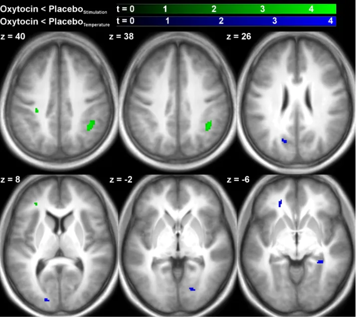
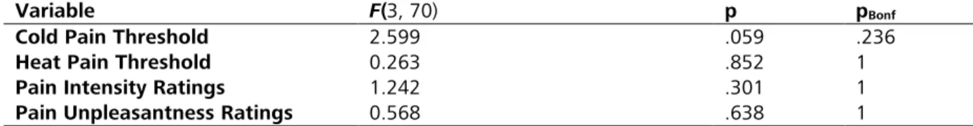
RESULTS: Thirty participants entered analysis. Intranasal oxytocin had no significant effect on thermal thresholds, but significantly (t = -2.06, p = .046) reduced heat intensity ratings during fMRI. The effect on intensity ratings was small (-3.46 points on a 100-point visual analog scale, 95% CI [-6.86; -0.07]) and independent of temperature. No effects of oxytocin on stimulus- or temperature-related processing were found at the whole-brain level at a robust statistical threshold. A ROI analysis indicated that oxytocin caused small but significant decreases in left (-0.045 %, 95% CI [-0.087, -0.003], t = -2.19, p = .037) and right (-0.051 %, 95% CI [-0.088, -0.014], t = -2.82, p = .008) amygdala activity across all temperatures.
CONCLUSIONS: The present study provides evidence for a significant, but subtle inhibitory effect of oxytocin on thermal stimulus ratings and concurrent amygdala activity. Neither of the two effects significantly depended of temperature, therefore the hypothesis of a pain-specific effect of oxytocin could not be confirmed.
Introduction
Anti-nociceptive effects of oxytocin have been reported by nearly 30 non-human, but only a few human studies (Rash, Aguirre-Camacho, & Campbell, 2013). Only four studies have investigated the effects of oxytocin on human pain using the convenient nasal application route, which has been established as a safe (MacDonald et al., 2011) and effective method to increase oxytocin concentrations in the central nervous system (Born et al., 2002; Neumann, Maloumby, Beiderbeck, Lukas, & Landgraf, 2013; Striepens et al., 2013). Singer et al. (2008) were the first to test the potential of intranasal oxytocin to alter empathic responses to pain. Although this hypothesis could not be confirmed, they found that oxytocin reduced amygdala reactivity in response to pain in a small sub-sample. Using an experimental model of placebo analgesia, Kessner et al. (2013) showed that intranasal oxytocin enhances the placebo effect. However, no general anti-nociceptive effect of intranasal oxytocin was evident within their large healthy sample. Mameli et al. (2014) explored the potential of oxytocin as an adjunctive analgesic in a small sample of fibromyalgia patients with negative results. Rash & Campbell (2014) recently found that intranasal oxytocin reduces behavioral and physiological reactions in response to cold- pressor pain.
Various effects and neural correlates of intranasal oxytocin on the processing of stress and emotion have been identified, yet most neuroimaging studies reported oxytocin effects on the amygdala, a key region for anxiety processing (Bethlehem, Baron-Cohen, van Honk, Auyeung, &
Bos, 2014; Bethlehem, van Honk, Auyeung, & Baron-Cohen, 2013). For example, intranasal oxytocin was found to attenuate amygdala responses related to threatening scenes (Kirsch et al., 2005) and to conditioned fear of faces (Petrovic, Kalisch, Singer, & Dolan, 2008). Interestingly, a recent meta-analysis found 17 neuroimaging studies that report increased amygdala activity in response to experimental pain, which suggests that the amygdala plays a role in acute pain processing (Simons et al., 2014).
The potential of neuropeptides such as oxytocin as an adjunctive treatment of chronic pain is of considerable interest. Nevertheless, the potential of intranasal oxytocin to affect pain processing is understudied (Rash et al., 2013). The present trial aimed to investigate the effect of oxytocin on experimental pain perception and processing. The established Quantitative Sensory Testing (QST) protocol was used to test effects of oxytocin on noxious and non-noxious thermal thresholds. Further, a functional Magnetic Resonance Imaging (fMRI) experiment with tonic heat pain stimulation and Visual Analog Scale (VAS) ratings of heat intensity and unpleasantness was employed to measure oxytocin effects on experimental pain perception and processing.
Based on the above-mentioned studies we hypothesized that intranasal oxytocin should A) increase noxious thermal pain thresholds;
B) decrease VAS ratings of noxious heat intensity and/or unpleasantness;
C) cause detectable blood oxygen level dependent (BOLD) signal changes related to thermal stimulus processing on whole brain level;
D) alter painful stimulus processing in the amygdala.
Methods
Trial information
The present study was approved by the ethics committee of the University of Regensburg (Approval Number: 11-111-0322) and the responsible federal medical agency. It was registered in a clinical trial registry (EUDRA-CT Number: 2009-015115-40) and conforms to the Declaration of Helsinki (59th WMA General Assembly, 2009). Written informed consent was obtained from every participant. All measures took place at the MRI facilities of the University of Regensburg at the Bezirksklinikum Regensburg between November 2012 and April 2013.
Study design
This is a placebo-controlled, double-blind, crossover trial. The random allocation sequence was generated by the Center for Clinical Trials at the University of Regensburg. According to this sequence, the Hospital Pharmacy of the University of Erlangen labeled and numbered the nasal sprays and corresponding emergency wrappers. At study inclusion, participant were assigned sequential participant numbers by author PE and hereby allocated to Group A or B at random.
Group A received placebo and Group B received oxytocin at Visit 1; Group A received oxytocin and Group B received placebo at Visit 2. Both groups completed an additional training visit (Visit 0) at study inclusion. A period of ≥ 7 days between all visits was observed to minimize carry-over effects.
Participants
Healthy right-handed male volunteers between 18 to 50 years of age were eligible for trial participation. Participants were recruited by advertisement at the University of Regensburg, aiming at a sample size of 36. The sample size was chosen to optimize statistical power within the limits of the devoted resources. Participants received a compensation of 10 Euros per hour.
Exclusion criteria were surveyed in a structured interview at inclusion. Exclusion criteria were:
Allergies against any ingredients of the trial medication; past or present cardiac, major internal, neurological, psychiatric, hormonal, or chronic conditions; acute infections; recent surgery; recent use of illicit drugs, psychotropics, or analgesics; alcohol addiction; conditions incompatible with fMRI safety. Participants were required to abstain from alcohol and caffeinated beverages at least 24 hours and 12 hours before visits, respectively.
Procedure
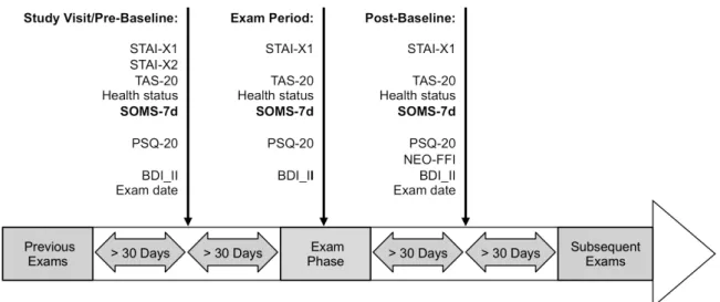
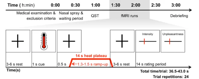
Figure 1.1: Time schedule of experimental procedures (upper row) and schematic overview of a typical trial within the fMRI block design (lower row). The temperature applied during the 14-second heat plateau ranged from 44.7 °C to 47.5° C in steps of 0.4 °C. Abbreviations: fMRI: functional Magnetic Resonance Imaging, QST: Quantitative Sensory Testing.
An overview of the experimental procedures is provided in Figure 1.1. Visits were scheduled between 8:00 h and 20:00 h, always at the same time of day (± 1h) within participants. Each visit started with a medical examination and a re-evaluation of inclusion and exclusion criteria.
Subsequently, participants received a dose of 32 IU oxytocin, or placebo, applied as four puffs of 0.1 ml per nostril. The dose was chosen according to Singer et al., (2008). The oxytocin and the placebo spray only differed in the absence of oxytocin in the placebo. Both sprays had the formulation of Syntocinon Spray (Sigma Tau, Rome, Italy). Participants self-administered the nasal spray under supervision by a physician, according to recommendations by Guastella et al.
(2013). Testing began after a waiting period of 40 minutes, consistent with the expected peak CSF concentration of nasally applied neuropeptides (Born et al., 2002). During the waiting period, participants completed questionnaires and were given standardized instructions for the following testing procedures. Then, thermal thresholding according to the QST protocol (20 min) was performed outside of the MRI scanner, followed by two fMRI runs. At debriefing, the occurrence and severity of 18 typical side effects was recorded on a 5-point numeric rating scale ranging from 0 (side effect absent) to 4 (severe). Additionally, each participant was asked to guess if he received placebo or oxytocin.
Monitoring mood
The Profile of Mood States (POMS, Pollock, Cho, Reker, & Volavka, 1979), an adjective rating scale instrument with 65 items, was used to monitor mood changes over the course of the experiment. The POMS total score reflects mood disturbance in general, while sub-scales allow for the monitoring of “tension-anxiety”, “depression-dejection”, “anger-hostility”, “fatigue- inertia”, “vigor-activity”, and “confusion-bewilderment”. The POMS was administered three times: At the beginning of each session, after nasal spray application (near the end of the waiting period), and at the end of each session. At each time-point the participants were asked to rate their “momentary” state.
Quantitative sensory testing (QST)
Participants were seated in upright position outside the MRI chamber with their arms comfortably resting on a cushioned tabletop. Thermal thresholding was performed on the volar surface of the left lower arm, 5 cm proximal from the wrist crease. Thermal stimuli were applied using a Thermosensory Analyzer II (Medoc, Israel) and a MR-safe 30 x 30 mm thermode, kept in place by an elastic strap. All thresholding procedures were performed in the presence of the same experimenter (MZ), with no other person or distractors present. Visual, auditory, or social clues indicating the onset of stimulation were precluded by the experimental setup. Written instructions were read aloud to the participant at each session. Cold (CDT) and warmth (WDT) detection thresholds, as well as cold (CPT) and heat (HPT) pain thresholds were retrieved in this order, according to the established QST protocol (Magerl et al., 2010; Rolke et al., 2006). For each measure five repetitions were obtained. The first stimulus of each measure was defined as a trial stimulus and discarded from analysis; thresholds were defined as the mean of the last four stimuli.
fMRI—general information:
After QST, participants underwent two separate fMRI experiments. Again, stimulation was applied to the volar surface of the left lower arm. Now the thermode location was 10 cm, or 15 cm proximal from the wrist crease. The thermode was moved in-between the fMRI runs to avoid skin damage. The order of fMRI runs and the order of thermode locations were balanced across participants and medication conditions. Only one of the two fMRI experiments was part of the present study, the other experiment will be reported elsewhere (Zunhammer et al., unpublished observations).
A 3-Tesla Allegra Head Scanner (Siemens, Germany) equipped with a single channel head coil was used for MRI. Functional volumes were obtained with a T2*-weighted Echo-Planar Imaging (EPI) sequence (TR = 2000 ms, TE = 30 ms, interleaved slicing, flip angle = 90 º, 3 × 3 x 3.5 mm voxel size, including a 16 % slice-gap, FoV = 192 × 192 mm), covering the full brain in 34 horizontal slices co-planar to the anterior and posterior commissure. The first five volumes of each run were discarded to account for T1-saturation effects. In addition a T1-weighted high-resolution structural head volume with 160 sagittal slices was obtained at Visit 1, using a Magnetization Prepared Rapid Gradient Echo (MP-RAGE) sequence (TR = 2250 ms, TE = 2.6 ms, flip angle = 9 º, 1 x 1 x 1 mm voxel size, FoV = 256 × 256 mm).
Presentation 14.9 for Windows (Neurobehavioral Systems Inc., USA) was used to display stimuli, retrieve ratings, and log events. Visual stimuli were presented at a resolution of 1024 x 768 pixels and a frame-rate of 60 Hz on a screen attached to the head-end of the coil. Participant could see the entire display via a mirror attached to the head coil. The thermal stimulation equipment was the same as for the QST.
MR images were pre-processed and analyzed with SPM 8. Volumes were corrected for slice- timing differences using the middle slice as a reference; event onsets were adjusted accordingly.
Volume time series were re-aligned and re-sliced to the first volume to account for head motion, using SPM’s rigid body transformation with 4th degree B-spline interpolation. Time series were screened for excessive head motion events using ArtRepair (Mazaika, Hoeft, Glover, & Reiss, 2009). Anatomical images were co-registered to the mean realigned functional image, segmented using SPM’s MNI (Montreal Neurological Institute) tissue probability maps, and re-sampled at 2 x 2 x 2 mm. Realigned functional images were normalized to MNI space using the parameters retrieved from the segmentation procedure, preserving signal concentrations (“unmodulated”).
![Figure 1.2 a and b: Oxytocin effects on visual analog scale ratings of heat. Parameter estimates indicated that intensity ratings (β = -3.46, 95% CI [-6.86, -0.07], t = -2.06, p = .046) significantly decreased during oxytocin sessions, compared to placeb](https://thumb-eu.123doks.com/thumbv2/1library_info/5650168.1693852/30.892.114.789.462.698/oxytocin-parameter-estimates-indicated-intensity-significantly-decreased-oxytocin.webp)