Arthritis and cholinergic transition of sympathetic nerve fibers
(Arthritis und die cholinerge Umwandlung sympathischer Nervenfasern)
DISSERTATION ZUR ERLANGUNG DES DOKTORGRADES DER NATURWISSENSCHAFTEN (DR.RER.NAT)
DER FAKULTÄT CHEMIE UND PHARMAZIE DER UNIVERSITÄT REGENSBURG
vorgelegt von
Hubert Werner Stangl aus
Regensburg
im Jahr 2015
Promotionsgesuch eingereicht am: 03.07.2015
Die Arbeit wurde angeleitet von: Prof. Jens Schlossmann (Lehrstuhl für Pharmakologie und Toxikolo- gie, Universität Regensburg) in Kooperation mit Prof. Rainer H. Straub (Labor für experimentelle Rheumatologie und Neuroendokrinoimmunologie, Universitätsklinikum Regensburg)
Index
INDEX ...
ABBREVIATIONS ...
1 INTRODUCTION ... 1
1.1 RHEUMATOID ARTHRITIS... 1
1.1.1 Prevalence and current models of pathology ... 1
1.1.2 Diagnosis and current treatment of RA ... 3
1.2 THE PERIPHERAL NERVOUS SYSTEM IN RA ... 7
1.3 CHOLINERGIC TRANSITION OF SYMPATHETIC NERVE FIBERS ... 13
1.4 HYPOTHESIS AND STUDY AIM ... 16
2 PATIENTS, MATERIALS AND METHODS ... 17
2.1 OVERVIEW OF CHEMICALS AND LABWARE ... 17
2.2 PATIENTS AND TISSUE SAMPLES ... 21
2.3 ANIMALS, ARTHRITIS INDUCTION AND SAMPLE COLLECTION ... 22
2.4 HISTOLOGY AND IMMUNOFLUORESCENCE STAINING ... 23
2.5 GENERATION OF OSTEOCLAST PROGENITOR CELLS ... 24
2.6 IN-VITRO EXPERIMENTS WITH SYMPATHETIC GANGLIA, CO-CULTURE WITH OSTEOCLAST PROGENITORS, IMMUNOFLUORESCENCE STAINING AND IMAGE ANALYSIS ... 26
2.7 ISOLATION OF DRAINING LYMPH NODE CELLS ... 30
2.8 GENE EXPRESSION PROFILING OF OSTEOCLAST PROGENITOR CELLS ... 30
2.9 PROTEOME PROFILING OF OSTEOCLAST PROGENITOR CELLS ... 31
2.9.1 Proteome profile of osteoclast progenitor supernatants ... 32
2.9.2 ELISA measurements of osteoclast progenitor supernatants... 32
2.10 GENETIC ANALYSIS OF THE CHOLINERGIC GENE LOCUS ... 33
2.11 PRESENTATION OF DATA AND STATISTICAL ANALYSIS ... 34
3 RESULTS ... 35
3.1 DENSITY OF CATECHOLAMINERGIC TYROSINE HYDROXYLASE-POSITIVE AND CHOLINERGIC VESICULAR
ACETYLCHOLINE TRANSPORTER-POSITIVE NERVE FIBERS IN MICE ... 35
3.2 DENSITY OF SYMPATHETIC TH-POSITIVE, CHOLINERGIC VACHT-POSITIVE AND VIP-POSITIVE NERVE FIBERS IN OSTEOARTHRITIS AND RHEUMATOID ARTHRITIS ... 38
3.2.1 Innervation of finger joints ... 38
3.2.2 Innervation of knee synovial tissue of OA and RA patients ... 41
3.2.3 Expression of the alpha-7 subtype-containing nicotinic acetylcholine receptor in fibroblast like synoviocytes ... 42
3.3 INDUCTION OF THE CHOLINERGIC PHENOTYPE OF NERVE FIBERS IN MICE SYMPATHETIC GANGLIA .. 42
3.3.1 Co-culture experiments ... 43
3.3.2 Stimulation of sympathetic ganglia ... 45
3.4 GENE EXPRESSION LEVELS OF OSTEOCLAST PROGENITOR CELLS ... 47
3.4.1 Gene expression in osteoclast progenitor cells from healthy mice ... 49
3.4.2 Gene expression in osteoclast progenitor cells from arthritic mice ... 49
3.4.3 Clustering of selected genes in osteoclast progenitor cells ... 49
3.5 PROTEOME ANALYSIS OF OSTEOCLAST PROGENITOR CELLS ... 51
3.5.1 Proteome profiler results ... 51
3.5.2 ELISA results of supernatants from osteoclast progenitor cells ... 54
3.6 ANALYSIS OF PROMOTER REGIONS IN THE CHOLINERGIC GENE LOCUS IN THE MURINE AND HUMAN GENOME ... 61
3.6.1 Detection of steroid binding sites in the promoter region of the murine vesicular acetylcholine transporter gene ... 61
3.6.2 Detection of steroid binding sites in the promoter region of the human vesicular acetylcholine transporter gene ... 61
4 DISCUSSION ... 62
4.1 CATECHOLAMINERGIC AND CHOLINERGIC NERVE FIBERS IN MOUSE LIMBS DURING EXPERIMENTAL ARTHRITIS ... 62
4.1.1 Plasticity of sympathetic nerve fibers in arthritis and other inflammatory conditions ... 62
4.1.2 Cholinergic nerve fibers in mouse limbs during arthritis ... 64
4.2 CHOLINERGIC AND SYMPATHETIC ARTICULAR INNERVATION IN PATIENTS WITH OA AND RA ... 67
4.3 TRANSITION OF NERVE FIBERS FROM A CATECHOLAMINERGIC TO A CHOLINERGIC PHENOTYPE IN VITRO ... 70
4.3.1 Transition in the co-culture system of sympathetic ganglia ... 71
4.3.2 Identification of possible transition factors of osteoclast progenitor cells via gene expression analysis ... 72
4.3.3 Identification of possible transition factors of osteoclast progenitor cells by proteome analysis 83 4.3.4 Identification of possible transition factor DNA binding sites by genomic analysis ... 85
4.3.5 Transition of sympathetic ganglia by stimulation with single molecules ... 86
5 CONCLUSION ... 87
6 APPENDIX ... 88
6.1 LIST OF LITERATURE ... 88
6.2 LIST OF FIGURES ... 109
6.3 ABSTRACT ... 111
6.4 ACKNOWLEDGEMENTS ... 112
Abbreviations
ACh: acetylcholine
AChE: acetylcholine esterase, ACh degrading enzyme
AChR: acetylcholine receptor
ACPA: anti-citrullinated protein antibody
BGN: biglycan
BMM: bone marrow derived macrophage
catecholamine: neurotransmitter with a catechol group
catecholaminergic: signaling involving catecholamines as neurotransmitters
CCL: chemokine (C-C motif) ligand
CCR: chemokine receptor
CGRP: calcitonin gene related peptide
ChAT: choline acetyltransferase, ACh synthesizing enzyme cholinergic: signaling involving ACh and/or VIP as neurotransmitters
ChT: high affinity choline transporter
CIA: collagen type II-induced arthritis
CNS: central nervous system
CNTF: ciliary neurotrophic factor
CT-1: cardiotrophin 1
CXCL: chemokine (C-X-C motif) ligand
DBH: dopamine beta hydroxylase, enzyme in catecholamine synthesis DMARD: disease modifying anti-rheumatic drug
DMMB: 1,9-dimethyl-methylene blue
ECM: extracellular matrix
ELISA: enzyme-linked immunosorbent assay
ERK: extracellular signal-regulated kinase
FLS: fibroblast like synoviocyte
gp130: glycoprotein 130
HE: hematoxylin-eosin
HLA: human leukocyte antigen
IL: interleukin
JAK: janus kinase
LIF: leukemia inhibitory factor
MAPK: mitogen activated kinase
MCP-1: monocyte chemoattractant protein-1
MCP-3: monocyte chemoattractant protein-3
M-CSF: macrophage colony stimulating factor
MIP-1a: macrophage inflammatory protein-1a
MIP-2a: macrophage inflammatory protein-2a
MMP: matrix metalloproteinase
mRNA: messenger ribonucleic acid
nAChR: nicotinic acetylcholine receptor
NE: norepinephrine
NFКb: nuclear factor kappa b
NGF: nerve growth factor
NPY: neuropeptide Y
OA: osteoarthritis
OCP: osteoclast progenitor
OSF-2: osteoblast specific factor
OSM: oncostatin M
p.i.: post immunization
parasympathetic: belonging to the parasympathetic nervous system
Phox2a: paired-like homeobox 2a
PNS: peripheral nervous system
RA: rheumatoid arthritis
RANK: receptor activator of nuclear factor kappa b RANKL: receptor activator of nuclear factor kappa b ligand
RANTES: regulated upon activation, normally T-cell expressed and presumably secreted
RF: rheumatoid factor
RGDS: arginine-glycine-aspartate-serine tetrapeptide Satb2: special AT-rich sequence binding protein 2
SDF-1: stromal derived factor 1
SLC18A3: solute carrier family member 18 member 3
SNS: sympathetic nervous system
SP: substance P
STAT: signal transducer and activator of transcription sympathetic: belonging to the sympathetic nervous system
TH: tyrosine hydroxylase, enzyme in catecholamine synthesis TIMP-1: tissue inhibitor of metalloproteinase 1
TNF: tumor necrosis factor
V$GRE.02: glucocorticoid receptor IR3 site
V$GREF: glucocorticoid responsive and related elements V$PRE.01: progesterone receptor binding site
VAChT: vesicular acetylcholine transporter
VIP: vasoactive intestinal peptide
VMAT: vesicular monoamine transporter
VPAC1: vasoactive intestinal peptide receptor type 1 VPAC2: vasoactive intestinal peptide receptor type 2
α7nAChR: α7-subtype containing nicotinic acetylcholine receptor
αAR: α-adrenoceptor
βAR: β-adrenoceptor
1.1 Rheumatoid arthritis
1
1 Introduction
1.1 Rheumatoid arthritis
1.1.1 Prevalence and current models of pathology
Rheumatoid arthritis (RA) is a chronic inflammatory disease (CID) which mainly affects small interdigi- tal, typically the proximal interphalangeal, metacarpophalangeal and wrist joints in hands, the metatar- sophalangeal joints in the feet, and the knee joint (Aletaha et al., 2010; McInnes & Schett, 2011).
Worldwide, about 1% of the population is diagnosed with RA (Firestein, 2003). If left untreated, pro- gression of RA leads to articular damage, typically expressed by symptoms like swelling and reoccur- ring pain at morning, and ultimately disability of affected joints, rendering this disease a socio- economical threat to patients and the whole population (Aletaha et al., 2010; McInnes & O'Dell, 2010;
McInnes & Schett, 2011). Moreover, due to the systemic and self-sustaining inflammatory nature of RA, the risk of developing secondary diseases related to the cardiovascular system, and insulin re- sistance, metabolic syndrome, osteoporosis, fatigue and depression increases, all possible reasons for an overall increased mortality observed in RA (Aletaha et al., 2010; McInnes & Schett, 2011).
In the last few decades genetic association studies revealed that rheumatoid arthritis is in fact an auto- immune disease, based on the identification of many risk alleles which alone or in synergy with envi- ronmental triggers (especially smoking) increase susceptibility of developing autoimmune mechanisms and eventually disease outbreak (McInnes & Schett, 2011): An important discovery was that the pres- ence of circulating autoantibodies, like rheumatoid factor (RF) and ACPA (anti-citrullinated protein an- tibody) which are diagnostic markers for RA, is not only highly associated with the human leukocyte antigen (HLA)-DRB1 gene but even more so with a certain amino acid sequence (QKRAA motif) within the HLA-DRB1 protein, a major histocompatibility complex class II (MHCII) molecule (Firestein, 2003;
McInnes & O'Dell, 2010; McInnes & Schett, 2011). Further it was demonstrated that the conversion of arginine residues to citrulline residues in endogenous proteins by exogenous noxae like smoking or exposition to certain pathogens, is highly associated with antigen presentation to T cells via HLA- DRB1 and production of autoantibodies like RF or ACPA which are directed against these modified endogenous proteins (Firestein, 2003; McInnes & O'Dell, 2010; McInnes & Schett, 2011). These ob- servations suggest that the adaptive immune system is a key player in the accrual of auto-reactivity of the immune system in RA (Firestein, 2003; McInnes & O'Dell, 2010; McInnes & Schett, 2011). Howev- er, it was found that this could not be the sole reason for the pathogenesis, since there are RA patients
1.1 Rheumatoid arthritis
2
who do not display expression of circulating antibodies like RF or ACPA, although disease severity is mostly more intense in RF- or ACPA-positive patients (McInnes & Schett, 2011). There is strong evi- dence that the innate immune system is as well involved in the pathogenesis of RA since antagonizing of certain cytokines like tumor necrosis factor (TNF) or interleukin (IL)-6, which are mainly produced by cells of the innate immune system like macrophages, has been proven to be highly effective in the therapy of many RA patients (Firestein, 2003; McInnes & O'Dell, 2010; McInnes & Schett, 2011; van Vollenhoven, 2009). In general, leukocytes, cells of both innate and adaptive immune system, are attracted to the inflamed joint by a chemotactic gradient and eventually migrate into the joint (Firestein, 2003; McInnes & Schett, 2011). Enhanced adhesion of these immune cells to the vessels near the joint then promotes accumulation and invasion into inflamed tissues adjacent to these vessels (Firestein, 2003; McInnes & Schett, 2011). The synovial membrane is a special connective tissue sur- rounding a joint and producing the synovial fluid, which has the purpose of lubrication and exchange of nutrients and metabolites in chondrocytes of articular cartilage (Haywood & Walsh, 2001; Levick, 1995). In RA, the synovial membrane typically displays signs of inflammation (synovitis) including pro- liferation, increased angiogenesis, altered lymphatic vessels and activation of the endothelium which facilitates the attraction and accumulation of circulating leukocytes (McInnes & Schett, 2011). This has been shown by the accumulation of macrophages, mast cells and natural killer (NK) cells, T and B-cell aggregates in the synovial membrane as well as neutrophil granulocytes in the synovial fluid (Firestein, 2003; McInnes & Schett, 2011). Together with activated fibroblast like synoviocytes (FLS), these immune cells generate a self-sustaining pro-inflammatory milieu by producing pro-inflammatory cytokines like TNF, IL-1b, IL-6 and IL-17, reactive oxygen and nitrogen species, and matrix degrading proteases like matrix metallo-proteinases (MMPs). Since anti-inflammatory and inhibitory mechanisms especially signaling of regulatory T (Treg) cells, endogenous tissue inhibitors of metalloproteinases (TIMPs) and inhibitory cytokines increasingly fail during pathogenesis of RA, this local inflammatory milieu prevails and ultimately leads to breakdown of articular cartilage by invasive FLS and activity of MMPs thereby forming typical aggressive, pannus like structures (Firestein, 2003; McInnes & Schett, 2011). In addition to the degradation of articular cartilage, also erosions of articular bone are typical features of active RA, which seem to occur already soon after diagnosis and are detected in most RA patients (McInnes & Schett, 2011). Erosions of bone are uniquely mediated by osteoclasts, a special- ized multinucleated cell type, which arises from the monocyte /macrophage linage of cells and is pre- sent in circulating blood and in the bone marrow (Herman et al., 2008a; Udagawa et al., 1990). Under the influence of certain locally enhanced cytokines, especially M-CSF (macrophage colony stimulating
1.1 Rheumatoid arthritis
3
factor) and RANK (receptor activator of nuclear factor kappa B) ligand, and further augmented by TNF, IL-1, IL-6, IL-17, these cells fuse and ultimately differentiate into active multinucleated bone resorbing osteoclasts (Herman et al., 2008a). Under physiological conditions, bone turnover is tightly balanced between bone resorption by osteoclasts and bone formation by osteoblasts (Herman et al., 2008a). In RA however, this homeostasis is disturbed by local conditions, which favor generation and activity of osteoclasts and simultaneously inhibit activity of bone forming osteoblasts (Herman et al., 2008a;
McInnes & Schett, 2007; Schett, 2011). Continuous resorption of bone can lead to a direct contact between the normally mutually separated bone marrow and the synovial fluid which contains activated immune cells, invasive FLS and inflammatory cytokines, leading to inflammation of the bone marrow (osteitis) (McInnes & Schett, 2011). However, there is still debate whether synovitis or osteitis and subsequent bone resorption precede initial steps of RA pathogenesis (Kleyer & Schett, 2014;
McQueen & Naredo, 2011; Schett & Firestein, 2010).
Eventually, erosions of articular bone, degradation of articular cartilage and an expanding, destructive inflamed hypertrophic synovial membrane including an aggressive pannus lead to a loss of joint integ- rity and function, pain and immobility (Firestein, 2003; McInnes & Schett, 2011). The high systemic inflammatory load in established active RA further increases the risk of developing secondary, espe- cially cardiovascular diseases and ultimately increases mortality (McInnes & O'Dell, 2010; McInnes &
Schett, 2011). Hence, a diagnosis as early as possible is of upmost importance, distinguishing joints typically affected by osteoarthritis (OA) from joints typically affected by RA and further differentiating RA subgroups, as this has an impact on the treatment and disease development (Aletaha et al., 2010;
McInnes & O'Dell, 2010; McInnes & Schett, 2011).
1.1.2 Diagnosis and current treatment of RA
The current RA classification criteria of the American College of Rheumatology (ACR, formerly Ameri- can Rheumatism Association) and the European League Against Rheumatism (EULAR) emphasize the importance of early diagnosis in order to initiate also an early aggressive treatment, ideally achiev- ing remission of disease in the shortest possible period of time (Aletaha et al., 2010). Diagnosis first involves the definite recognition of swelling in joints caused by inflammation in the synovial membrane (synovitis) and possibly erosions in articular bone which occur early in RA (Aletaha et al., 2010;
McInnes & Schett, 2011). Detection of these alterations should be confirmed by x-ray radiography, ultrasound, or magnet resonance imaging (MRI) (Aletaha et al., 2010). Definite synovitis and/or ten- derness in at least one joint that can not be explained better otherwise renders a patient eligible for
1.1 Rheumatoid arthritis
4
diagnosis of RA (Aletaha et al., 2010). To diagnose a possible RA status of such an individual patient, an objective scoring further involves the number of affected joints, detection of auto-antibodies RF and/or ACPA (see 1.1.1), abnormalities in erythrocyte sedimentation rates and levels of C-reactive protein (CRP), and duration of observed symptoms (Aletaha et al., 2010). While these criteria include the latest knowledge for the best and most early diagnosis of RA, many other scoring systems for evaluating current RA disease activity in patients have evolved in order to compare different treat- ments and to monitor and ensure efficacy of treatment (McInnes & O'Dell, 2010). Some of these are relative scores, describing the percentage of improvement (e.g. ACR20, 20% improvement), which renders them valuable in clinical trials but unsuitable in clinical practice (McInnes & O'Dell, 2010).
Others are of absolute nature and highly accurate (DAS, disease activity score) (McInnes & O'Dell, 2010). Therefore, and owing to the fact that disease activity should be monitored regularly to ensure appropriate treatment, several other means of disease activity assessment are being used in practice, which do not require special equipment and can be acquired in a fast and relatively easy manner (McInnes & O'Dell, 2010).
It has been widely accepted that upon diagnosis initial and first line treatment of RA should rapidly begin with a monotherapy using disease modifying anti-rheumatic drugs (DMARDs) which includes most importantly methotrexate (MTX), leflunomide, sulfasalazine, hydroxychloroquin and glucocorti- coids (McInnes & O'Dell, 2010; van Vollenhoven, 2009). Methotrexate and leflunomide are regarded as anti-metabolites with MTX antagonizing purine synthesis and leflunomide antagonizing pyrimidine synthesis, which is considered especially effective in lymphocytes (van Vollenhoven, 2009). The anti- inflammatory action of MTX is at least in part thought to be mediated by signaling via adenosine (van Vollenhoven, 2009). Although both drugs, MTX and leflunomide, display similar efficacy to safety rati- os, MTX became the most widely used DMARD, probably also due to better compliance and lower costs (van Vollenhoven, 2009). Breakthrough in therapy for DMARDs was also due to the fact that DMARDs can be combined in therapy if needed, and that certain combinations are superior to MTX monotherapy (van Vollenhoven, 2009). According to current guidelines and several studies, initial therapy should start off early with administration of MTX only or MTX combined with low dose gluco- corticoids (Aletaha et al., 2010; McInnes & O'Dell, 2010), rendering MTX as the ‘anchor drug’ in mono- and combinational therapy (Aletaha et al., 2010; Pincus et al., 2003). In cases in which RA patients do not respond to MTX or adverse side effects are observed, alternatives like leflunomide or sulfasala- zine, an anti-inflammatory drug should be tried in monotherapy, before a step up to certain combina-
1.1 Rheumatoid arthritis
5
tions of DMARDs is required (McInnes & O'Dell, 2010; van Vollenhoven, 2009). Other DMARDs like gold containing drugs, ciclosporin A (an immunosuppressive agent), certain antibiotics, and azathio- prine (a cytostatic drug) are still used in certain cases but due to a higher risk of adverse side effects they are not considered first line medications (McInnes & O'Dell, 2010; van Vollenhoven, 2009). The anti-malaria drug hydroxychloroquine, which seems to interfere with antigen presentation, was shown to be effective especially in combination with MTX or sulfasalazine, and to decrease the risk of devel- oping diabetes, while displaying only relatively mild adverse effects (McInnes & O'Dell, 2010; van Vollenhoven, 2009). The use of glucocorticoids has experienced a renaissance as a first line DMARD since it was found that administration of low doses during initial treatment is effective in limiting joint damage in RA and has the advantage of a relatively fast response, in contrast to most other DMARDs (McInnes & O'Dell, 2010; van Vollenhoven, 2009). However, it is still controversial whether or how fast administration of glucocorticoids in combination with MTX or alternative DMARDs should be tapered off in order to avoid adverse effects of glucocorticoids or be kept up to achieve remission of RA in the long run, also paying respect to the inadequate release of endogenous glucocorticoids (cortisol in humans, corticosteron in rodents) in arthritis (McInnes & O'Dell, 2010; Straub et al., 2013; van Vollenhoven, 2009).
With gaining knowledge about the cellular and molecular signaling pathways in RA in recent decades, new classes of substances were developed and approved for treating RA. The need for new sub- stances emerged due to a growing number of patients who were not responding to mono or combina- tional therapy at all, experienced adverse effects, or response to a combination of several DMARDs was inadequate (McInnes & O'Dell, 2010; van Vollenhoven, 2009). The relatively new class of sub- stances, currently simply referred to as ‘biologicals’ due to their antibody or protein related molecular structure and (micro-)biological way of production (van Vollenhoven, 2009), mainly exploits its efficacy by interference with cytokine signaling pathways which play a major role in activation of leukocytes, synovial fibroblasts and osteoclasts (McInnes & Schett, 2011). Since TNF plays a central role in acti- vating many cells and inflammatory pathways relevant to RA (Firestein, 2003; McInnes & Schett, 2007; McInnes & Schett, 2011; Schett et al., 2013) several antibodies to TNF (adalimumab, certoli- zumab, golimumab, infliximab) and a fusion receptor (etanercept) have been approved for treatment of RA (McInnes & O'Dell, 2010; van Vollenhoven, 2009) and have shown efficacy even in other chronic inflammatory disorders (Schett et al., 2013). Interestingly, all TNF inhibitors display similar efficacy to risk ratios (McInnes & O'Dell, 2010). The broad success and the high efficacy highlight the crucial
1.1 Rheumatoid arthritis
6
roles of TNF in these cytokine networks and led to development of further molecules targeting the IL-6 pathway (tocilizumab, an anti-IL-6 receptor antibody) and IL-1 pathway (anakinra, an IL-1 receptor antagonist), due to both cytokines being important in signaling of leukocytes, synovial fibroblasts, and also osteoclasts (McInnes & Schett, 2007; Schett, 2011). Yet, anakinra proved only modest efficacy in the treatment of RA but seems to be more efficient in treating other inflammatory diseases like juvenile idiopathic arthritis and gout (McInnes & Schett, 2007; Schett et al., 2013; van Vollenhoven, 2009).
Moreover, also specific inhibition of B-cells with the help of a monoclonal antibody directed against CD20 (rituximab), which is believed to decrease production of auto-antibodies like RF and ACPA, and, hence, is especially useful in respective serum positive patients, has been approved (McInnes &
Schett, 2007; McInnes & O'Dell, 2010; van Vollenhoven, 2009). Also specific inhibition of T-cell co- stimulation, a critical step of T-cell activation, by the CTLA-4 ligand abatacept has shown efficacy, e.g.
in patients resistant to anti-TNF therapy (McInnes & Schett, 2007; McInnes & O'Dell, 2010; van Vollenhoven, 2009).
Existing general guidelines and the treat-to-target of remission principle suggest a step up, escalating therapy regime in which after failure of mono or combination therapy with DMARDs, first TNF inhibiting biologicals should be used or added, followed by more specialized agents like tocilizumab, rituximab and abatacept if needed or anti-TNF therapy fails (McInnes & O'Dell, 2010; van Vollenhoven, 2009).
Effective and safe therapy has to be routinely monitored and adjusted if needed depending on each patient’s individual medical records, which includes presence and treatment of co-morbidities, pres- ence of serum auto-antibodies like RF or ACPA, efficacy or failure of DMARD mono or combination therapy, and step-up therapy with biologicals including possible adverse effects (McInnes & O'Dell, 2010; van Vollenhoven, 2009).
Recent research focused on intracellular signal transduction pathways, especially, on targets like Ja- nus kinases (JAKs) and tyrosine kinases, which belong to cell surface receptors that trigger such in- tracellular signaling cascades often involved in activating expression of pro-inflammatory mediators (McInnes & Schett, 2011). This led to the recent approval of tofacitinib, a molecule which is thought to inhibit JAK1 and subsequent STAT (signal transducer and activator of transcription) 1 and 3 signaling, and was shown to decrease gene expression of MMPs and chemokines in synovial fibroblasts of RA patients (Boyle et al., 2014). Despite promising preclinical results, p38-mitogen activated protein ki- nase (MAPK) inhibitors, and the phosphodiesterase (PDE)-4 inhibitor apremilast, which inhibits the degradation of the intracellular signaling molecule cyclic adenosine-mono-phosphate (cAMP) and,
1.2 The peripheral nervous system in RA
7
thus, TNF production, displayed disappointing results in clinical studies with RA patients (McInnes &
Schett, 2011; Genovese et al., 2015; McCann et al., 2010; McInnes & O'Dell, 2010). Further, two dis- advantages of biologicals especially regarding TNF inhibitors remain: Compared to DMARDs they are costly and adverse effects include serious, potentially fatal infections and an increased risk of develop- ing lymphoma and other malignancies (McInnes & Schett, 2007; McInnes & O'Dell, 2010; van Vollenhoven, 2009). Other potential drug targets still under trial include B-cell activating factors, the pro-inflammatory interleukin 17, RANK ligand, and other kinases (McInnes & Schett, 2011).
Due to developing therapy resistance after a certain time or adverse effects seen in some patients under treatment with DMARDs and biologicals, there still remains need for exploiting further pathways involved in RA pathogenesis (Lipsky, 2009; McInnes & O'Dell, 2010; van Vollenhoven, 2009). Such attempts also include targeting the peripheral nervous system (PNS), particularly since activation of certain (α7 subunit containing) nicotinic acetylcholine receptors (α7nAChR) in the periphery was sug- gested to elicit anti-inflammatory pathways (Waldburger et al., 2008; Wang et al., 2003) and these receptors were discovered on the surface of macrophages in the spleen (Rosas-Ballina et al., 2011) and on fibroblasts and macrophages in the synovium (Forsgren, 2012; van Maanen et al., 2009b;
Waldburger et al., 2008; Westman et al., 2009). Exact mechanisms of observed effects are still unclear as proposed models have changed several times (reviewed in (Martelli et al., 2014a)) and hence are subject of debate (Martelli et al., 2014a; Martelli et al., 2014b; Nance & Sanders, 2007; Pongratz &
Straub, 2013; Straub et al., 2013). However, pharmacological stimulation of α7nAChR and electrical stimulation of the vagus nerve, the anatomically biggest nerve of the cholinergic PNS, has shown promising results in experimental arthritis (Levine et al., 2014; van Maanen et al., 2009a) and it was suggested that exploiting these pathways might be a therapy option in treatment of RA (Koopman et al., 2011; Koopman et al., 2014; Levine et al., 2014; van Maanen et al., 2009c).
1.2 The peripheral nervous system in RA
Apart from a disturbed signaling of the CNS and the endocrine system with inadequate composition of sex hormones and inadequate provision of endogenous glucocorticoids during arthritis (Straub et al., 2013; Del Rey et al., 2010; Wolff et al., 2014; Straub, 2014), it has been long established that the pe- ripheral nervous system (PNS) locally modulates outbreak and progression of RA (Levine et al., 1987;
Pongratz & Straub, 2013). RA typically displays a symmetrical involvement of arthritic joints in the extremities, meaning that with increasing number of affected joints over time mostly also bilateral af- fection of extremities is observed (Aletaha et al., 2010; Firestein, 2003). The most striking proof of an
1.2 The peripheral nervous system in RA
8
involvement of the PNS in RA is the observation that patients who suffer from hemiplegia display RA mostly in the non-paralyzed hand, whereas the hemiplegic hand is typically spared from RA (Keyszer et al., 2004). To verify this clinical observation, sciatic and femoral nerves were severed in an animal model of experimental arthritis and indeed, denervated limbs were protected from developing arthritis (Stangenberg et al., 2014). Yet, the exact mechanisms like an involvement of the microvasculature and the immune system, as suggested by the interesting findings of Stangenberg et al., and how they are controlled by the PNS still remain to be further elucidated (Rabquer & Koch, 2014; Schaible &
Straub, 2014).
However, drawing more specific conclusions from these observations can be potentially misleading because hemiplegia and experimental transection of limb nerves most likely affects all different kinds of nerves since the PNS is divided into different parts according to its function: The sensory nervous system which mainly transmits pain, inflammatory related stimuli and danger signals from the periph- ery to the brain (afferent function), the parasympathetic nervous system, which is often referred as
‘rest and digest’ promoting nervous system, but the role of which in arthritis is still relatively unclear, and its counterpart the sympathetic nervous system, which in regards to arthritis was shown to in- fluence blood flow, vascular permeability and local immune processes (reviewed in (Pongratz &
Straub, 2013; Schaible & Straub, 2014)). The sympathetic nervous system can be further subdivided into an (nor)-adrenergic catecholaminergic and a cholinergic branch, based on which neurotransmit- ters convey the signals in the post-ganglionic part of the connection between CNS and periphery (Ernsberger & Rohrer, 1999). The main neurotransmitters present in the sensory nervous system and important for the efferent function of sensory nerves are substance P (SP) and calcitonin gene related peptide (CGRP) (Pongratz & Straub, 2013), and detection of SP is commonly used to identify sensory nerve fibers. Important neurotransmitters of the (nor)-adrenergic part of the sympathetic PNS are norepinephrine (NE, also known as noradrenaline) and its co-transmitters neuropeptide Y (NPY) and adenosine (Pongratz & Straub, 2013; Straub, 2012). Detection of sympathetic nerve fibers containing these neurotransmitters is commonly accomplished by staining tyrosine hydroxylase (TH) or dopamine beta hydroxylase (DBH), as these two proteins are key enzymes in the biosynthesis of the catechola- mines dopamine and NE, and hence are expressed in sympathetic nerves of CNS and PNS (Tekin et al., 2014). Cholinergic parts of the PNS convey signals via the main cholinergic and eponymous neu- rotransmitter acetylcholine (ACh) and vasoactive intestinal peptide (VIP), a typical co-transmitter of cholinergic nerve endings (Ernsberger & Rohrer, 1999). These nerves are commonly detected by
1.2 The peripheral nervous system in RA
9
staining the ACh synthesizing enzyme choline acetyltransferase (ChAT), the vesicular acetylcholine transporter (VAChT) or VIP protein (Duong et al., 2002; Eiden et al., 2004; Erickson et al., 1994;
Ernsberger & Rohrer, 1999; Guidry & Landis, 1998; Schafer et al., 1998).
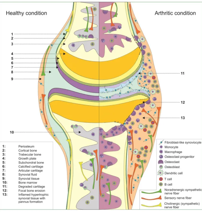
Interestingly, all of these systems including the respective neurotransmitters and their receptors are present in bone (Figure 1) and take part in the control of physiological bone turnover differently influ- encing bone formation by osteoblasts and bone resorption by osteoclasts (Aitken et al., 2009;
Bjurholm et al., 1988; Eimar et al., 2013; Elefteriou, 2005; Hill & Elde, 1991; Hohmann et al., 1986;
Imai & Matsusue, 2002; Lerner & Persson, 2008; Persson & Lerner, 2011; Suzuki et al., 1998).
Figure 1. Schematic drawing of a joint in physiological, healthy condition (left) and during arthritis (right). Involved cells and the different parts of the peripheral nervous system are highlighted.
1.2 The peripheral nervous system in RA
10
In RA however, this neuronal control is disturbed, and activity of osteoclasts is enhanced also due to inflammatory cytokines, leading to characteristic focal bony erosions (Braun & Zwerina, 2011; McInnes
& Schett, 2007; Schett, 2011). Further, presence of sympathetic noradrenergic nerve fibers and senso- ry nerve fibers has been also demonstrated in the synovial tissue of humans and mice (reviewed in (Pongratz & Straub, 2013)). However, so far not much is known about a possible direct innervation by cholinergic nerve fibers in joints, although expression of VAChT-positive nerve fibers was recently shown in femora from mice (Bajayo et al., 2012) and rats (Lips et al., 2014) in addition to VIP contain- ing nerve fibers in bone (Asmus et al., 2000; Bjurholm et al., 1988; Hill & Elde, 1991; Hohmann et al., 1986; Sisask et al., 1996) and synovial tissue of the rat (Bjurholm et al., 1990). Moreover, in addition to the α7nAChR protein, mRNA transcripts of several parts of the cholinergic machinery like ChAT or the high affinity choline transporter (ChT), were shown to be expressed in the synovial tissue of arthritis patients (Beckmann et al., 2015; Forsgren, 2012). However, no direct innervation by the PNS was shown, and the authors concluded that in the joint a local cell based, non-neuronal cholinergic system (NNCS) might be present (Beckmann & Lips, 2013), similar to such a system, which was shown by Kawashima and coworkers to exist in leukocytes (Kawashima & Fujii, 2004; Kawashima et al., 2012).
Further and in contrast to noradrenergic sympathetic innervation, nothing is known about a possible cholinergic innervation of joints in mice and humans during the course of arthritis.
Importantly, innervation by peripheral nerves is not static per se but under certain circumstances shows neuroplasticity (Pongratz & Straub, 2013). This phenomenon has been demonstrated in the joints of arthritic mice and RA patients, which during arthritis display a loss of sympathetic nerve fibers while sensory innervation increases (Pongratz & Straub, 2013). These observations have been mainly addressed to the action of elevated levels of the unspecific nerve growth factor NGF and semaphorins 3C and F, which are nerve repellent factors specific for noradrenergic sympathetic nerve fibers (Aloe et al., 1993; Fassold et al., 2009; Miller et al., 2004). Interestingly, a local loss of sympathetic nora- drenergic nerve fibers is not exclusive to arthritic joints but has been shown also in other inflammatory settings like in the colon wall of Crohn’s disease patients (Straub et al., 2008a), in chronic Charcot foot in diabetes (Koeck et al., 2009), in the myocardium after heart failure (Parrish et al., 2010), in inflamed pancreatic islands of diabetic rats (Mei et al., 2002), and in secondary lymphoid organs of arthritic rats and mice (Lorton et al., 2005; Lorton et al., 2009; Straub et al., 2008b).
Regarding the relationship between inflammation and the PNS during arthritis, a time-dependent, bi- modal action was demonstrated for the noradrenergic sympathetic PNS as it acts pro-inflammatory in
1.2 The peripheral nervous system in RA
11
early stage experimental arthritis and anti-inflammatory in later stages of arthritis (Harle et al., 2005;
Harle et al., 2008). In contrast to this early pro-inflammatory effect, which was attributed to enhanced cell mobilization, action of certain pro-inflammatory T-cells and antibody production by B-cells, it is still relatively unclear how this late anti-inflammatory effect might be conveyed since noradrenergic sympa- thetic nerve fibers are seemingly lost (Pongratz & Straub, 2013). This has the consequence that, due to lower concentrations of NE and higher receptor affinity of NE towards α-AR (adrenoceptors), also receptor signaling changes from a predominantly β-AR based signaling, which may act pro- and anti- inflammatory, to a predominantly α-AR based signaling, thought to elicit mainly pro-inflammatory ef- fects (Pongratz & Straub, 2013). It was suggested that this late anti-inflammatory effect at least in part might be due to locally emerging catecholamine-producing cells, which partially substitute input from sympathetic nerve endings (Jenei-Lanzl et al., 2015; Capellino et al., 2012; Miller et al., 2000; Miller et al., 2002), and an elevated production of the anti-inflammatory cytokine interleukin 10 in B-cells (Pongratz et al., 2012; Pongratz & Straub, 2013).
Efferent signaling of sensory nerve fibers and its main neurotransmitters SP and CGRP is generally regarded as pro-inflammatory in arthritis (Lorton et al., 2000; Uematsu et al., 2011) as they induce general inflammatory symptoms like vasodilatation, reddening and edema (Pongratz & Straub, 2013), proliferation of T and B cells (Laurenzi et al., 1989; Payan et al., 1983) and in the case for SP stimu- lates osteoclast formation and activity (Kojima et al., 2006; Lerner & Persson, 2008; Wang et al., 2009).
Anti-inflammatory effects of cholinergic signaling have been described for the activation of nicotinic acetylcholine receptors, especially by activation of the α7nAChR on immune cells and synoviocytes (Kawashima et al., 2012; van Maanen et al., 2009a; van Maanen et al., 2010; Waldburger et al., 2008;
Wang et al., 2003; Yoshikawa et al., 2006), but signaling of ACh via certain muscarinic receptors can also elicit pro-inflammatory pathways (Eglen, 2006; Fujii et al., 2003; Fujii et al., 2008; Kawashima et al., 2012). Further, anti-inflammatory effects have been reported for VIP (Delgado et al., 2001;
Delgado et al., 2003; Delgado & Ganea, 2008; Gonzalez-Rey et al., 2006; Gonzalez-Rey & Delgado, 2008). Since presence of these systems has been identified in arthritis (Delgado et al., 2008a; van Maanen et al., 2009b; Waldburger et al., 2008; Westman et al., 2009), consequently it was suggested not to enhance general cholinergic signaling via ACh, but to specifically target the α7nAChR and VIP receptors to exploit their anti-inflammatory potential (Bencherif et al., 2011; Delgado et al., 2002;
1.2 The peripheral nervous system in RA
12
Delgado et al., 2008b; Delgado & Ganea, 2008; Gonzalez-Rey et al., 2007; Koopman et al., 2011; van Maanen et al., 2009c).
In general however, whether signaling of different parts of the PNS results in activation of pro- or anti- inflammatory pathways, critically depends on local neurotransmitter concentration provided by the respective nerve endings and the locally present different neurotransmitter receptors (Pongratz &
Straub, 2013), both of which themselves may change during developmental stages (Sisask et al., 1996) and diseases such as RA (Pongratz & Straub, 2013).
1.3 Cholinergic transition of sympathetic nerve fibers
13
1.3 Cholinergic transition of sympathetic nerve fibers
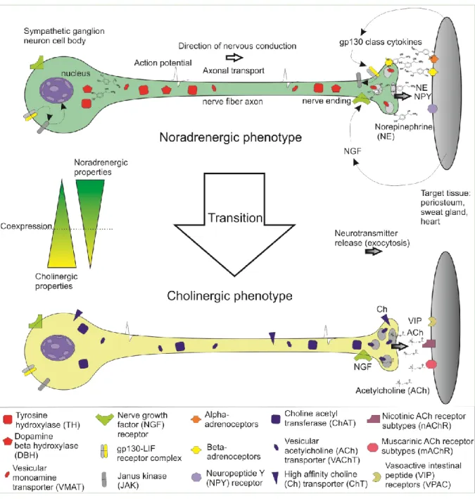
Under certain circumstances noradrenergic sympathetic nerve fibers are able to change towards a cholinergic phenotype of sympathetic nerve fibers (see Figure 2), the process of which is mostly re- ferred to as sympathetic transition or cholinergic transition / differentiation of sympathetic nerve fibers and neurons (reviewed in (Ernsberger & Rohrer, 1999)).
Figure 2. Scheme of catecholaminergic-to-cholinergic transition of sympathetic nerve fibers.
This phenomenon was first described by Landis and co-workers, who were investigating the innerva- tion of developing sweat glands (Landis & Keefe, 1983; Landis et al., 1988), and was later also found in the periosteum (Asmus et al., 2000; Asmus et al., 2001), and in the myocardium (Kanazawa et al.,
1.3 Cholinergic transition of sympathetic nerve fibers
14
2010; Parrish et al., 2010; Yamamori et al., 1989) (reviewed in (Kimura et al., 2012). Molecular mech- anisms of these phenomena are highly complex and several pathways involved in these processes have been studied: Generally, there seem to be target-independent (initial induction of cholinergic properties) and target-dependent (later trans-differentiation after target contact) pathways which can drive the acquisition of cholinergic properties in sympathetic neurons (Apostolova & Dechant, 2009;
Stanke et al., 2006). Regarding sweat glands, there are studies supporting both, a target-dependent pathway, in which sympathetic neurons show cholinergic properties only after target contact (Guidry &
Landis, 1998), or an early target-independent pathway, suggesting a neuronal co-expression of both noradrenergic and cholinergic properties before reaching the sweat glands (Schütz et al., 2008).
While in the heart leukemia inhibitory factor (LIF) was identified as the responsible factor driving this transition (Yamamori et al., 1989), the exact nature of the sweat gland specific transition factor still remains to be determined as several promising candidate cytokines belonging to the glycoprotein-130 (gp130) family of cytokines like LIF, ciliary neurotrophic factor (CNTF) and cardiotrophin-1 (CT-1) have already been ruled out. This was demonstrated in vivo by anti-cytokine treatment or mice lacking ge- netic information for one or more candidate cytokines. Both approaches did not inhibit the sympathetic transition in sweat glands, which is a hint for the existence of a further yet undiscovered cytokine (Francis et al., 1997; Habecker et al., 1995; Francis & Landis, 1999; Rao & Landis, 1990; Rao et al., 1993). Still, the molecule that induces sympathetic transition in sweat glands seems to act via the gp130/ LIF-receptor-β receptor complex and, thus, could be a novel member of the gp130 cytokine class (Habecker et al., 1997; Apostolova & Dechant, 2009). While it was established that signaling via the gp130/LIF-receptor-β and following activation of the JAK/STAT3 (Janus Kinase, signal transducer and activator of transcription) pathway plays a central role for the transition (Bamber et al., 1994;
Geissen et al., 1998; Habecker et al., 1997), there are also studies reporting secretory responsiveness in gp130 deficient mice and cholinergic reactivity in sympathetic neurons not induced by the LIF- receptor β. This raised the question whether secretory response is solely mediated via cholinergic signaling or if possible remnants of catecholaminergic signaling still can elicit a response (Stanke et al., 2000; Stanke et al., 2006).
Other pathways than the classical LIF-receptor-β/STAT activation influencing cholinergic development have been discovered: Loy et al. and Apostolova et al. demonstrated that the p38-mitogen activated kinases (p38-MAPK) subtypes α and β independently from the JAK/STAT pathway promote cholinergic properties by activating the nuclear matrix protein Satb2. Upon activation, Satb2 binds to the choliner-
1.3 Cholinergic transition of sympathetic nerve fibers
15
gic gene locus and up-regulates expression of cholinergic markers while at the same time down- regulating expression of catecholaminergic properties (Apostolova et al., 2010; Loy et al., 2011). To add further complexity, it was also shown that in most sympathetic cholinergic neurons, which are positive for VIP, the GDNF (glial cell-line derived neurotrophic factor) family receptor GFR-alpha2 is co-expressed. In their respective target tissues, namely sweat glands and periosteum, gene expres- sion of NRTN (neurturin), a ligand for GFR-alpha2 was detected. In GFR-alpha2 knock-out mice, in- nervation of sweat glands as such was unchanged but the density of cholinergic fibers was drastically reduced and even completely absent in the periosteum (Hiltunen & Airaksinen, 2004). Therefore, NRTN signaling via GFR-alpha2 also seems to play an important role in the acquisition and mainte- nance of cholinergic innervation in sweat glands. Additionally, the growth factors NGF (nerve growth factor) and NT-3 (neurotrophin-3), which are typically released by the target tissues critically influence induction, regulation and survival of a sympathetic catecholaminergic phenotype (Francis et al., 1999;
Francis & Landis, 1999). NT-3 signaling additionally seems to be influenced by the p75NTR receptor (Brennan et al., 1999). Even for neurotransmitter molecules, such as catecholamines, an involvement in sympathetic transition of sweat glands was shown: While a direct activation of adenylyl cyclase (AC), an important signaling component after G-protein coupled receptor (GPCR) activation, by for- skolin supports the process of the sympathetic transition to a cholinergic phenotype, a blockade of alpha and beta adrenoceptors inhibits the latter. Seemingly, the sympathetic axons release NE, trig- gering the up-regulation and release of the transition factor from the sweat glands, which in turn elicits the neuron to transform itself into a neuron with a cholinergic phenotype. In the same study, the sweat gland derived transition factor activity was absent in sympathectomized mice compared to control mice (Habecker & Landis, 1994). In a further study, TH deficient mice were treated with L-DOPA (the pro- genitor of dopamine in catecholamine synthesis), which increased the number of active sweat glands in these mice (Tian et al., 2000). The role of acetylcholine has as well been investigated: Treatment of young rats with atropine, a nonselective ACh antagonist or with a selective antagonist against the muscarinic (gland) subtype of ACh receptors delayed the development of secretory responsiveness and hence also sympathetic transition to a cholinergic phenotype. Treatment of adult rats with atropine resulted in a complete loss of function. When rats were selectively denervated, the following loss of secretory responsiveness could be rescued by pilocarpine, a muscarinic ACh receptor agonist (Grant et al., 1995).
1.4 Hypothesis and study aim
16
1.4 Hypothesis and study aim
This study’s aim was to elucidate such a possible transition of sympathetic nerve fibers to nerve fibers with cholinergic properties in highly inflamed tissues of arthritic mice and in synovial tissue from pa- tients with arthritis, based on following hypothesis: The observed loss of TH in sympathetic catechol- aminergic nerve fibers during arthritis is in fact not due to a loss of the entire sympathetic nerve fiber, but instead due to a sympathetic transition of the latter to a cholinergic phenotype.
The rationale for this thesis is founded on the well described loss of sympathetic nerve fibers in arthri- tis (see 1.2.) and the fact that such a loss of sympathetic nerve fibers is also being observed in tissues like developing sweat glands and the periosteum that show the phenomenon of sympathetic transition (Asmus et al., 2000; Asmus et al., 2001; Landis et al., 1988; Landis, 1996). Furthermore, the presence of cytokines capable of inducing this transition, like LIF (see above) and oncostatin M (OSM) (Rao et al., 1992), has been demonstrated in the synovium of RA patients ((Hui et al., 1997; Lotz et al., 1992) reviewed in (Richards, 2013).
This possible transition mediated by cytokines like LIF might resemble another part in the concept of an “anti-inflammatory cholinergic pathway” (Wang et al., 2003), which is also being proposed for treatment of arthritis (see 1.2). The exact mechanisms of this pathway(s), however, especially the in- volvement and connection between noradrenergic sympathetic nerves in secondary lymphoid organs like the spleen, the cholinergic vagus nerve (Borovikova et al., 2000; Koopman et al., 2014; Rosas- Ballina et al., 2011; Wang et al., 2003), and the anti-inflammatory mechanisms of α7nAChR activation still remain controversial (reviewed in (Martelli et al., 2014a; Martelli et al., 2014b; Pongratz & Straub, 2013)).
2.1 Overview of chemicals and labware
17
2 Patients, materials and methods
2.1 Overview of chemicals and labware
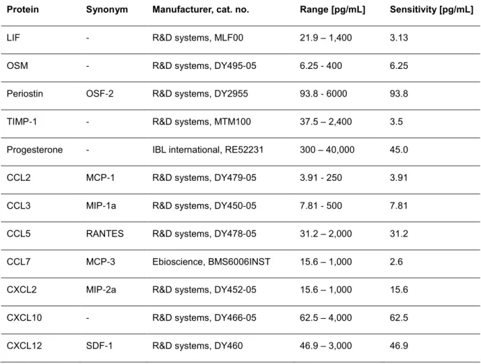
An overview of the devices, chemicals and antibodies in use for this study is given in table 1, table 2 and table 3, respectively. Details of the patients under study and of ELISA kits for proteome analysis are given in separate tables (tables 4, 5, and 6, respectively).
Table 1. Devices in use for this study.
Device Company Details Purpose
iMark Microplate reader, Mi- croplate reader imaging soft- ware
Bio-Rad, USA Microplate manager soft- ware version 6.2
ELISA measurement and analysis
Fluorescence microscope,
Visiview imaging software Leica, Germany Vis- itron GmbH, Germa- ny
- Immunofluorescence
microscopy, bright field microscopy
Axiovision fluorescence mi- croscope, Axiovision imaging software
Zeiss, Germany Axiovision software ver- sion 4.8
Immunofluorescence microscopy, bright field microscopy
STEMI, stereo microscope Zeiss, Germany - Dissection, ganglia prepa- ration
Nanodrop 2000 spectropho-
tometer Thermo scientific,
USA Software ver-
sion 1.4.2 Cell culture, BMM, RNA quantification
Bioanalyzer 2100 series Agilent Technologies,
USA B.02.08.SI648
(SR2) Cell culture BMM, RNA quality analysis
ChemiDoc Imager, ChemiDoc Imager software Image Lab
Bio-Rad, USA Image Lab
version 4.01
Cytokine profiling, imag- ing, image analysis
Cryotome Leica biosystems,
Germany - Histology
Ultra turrax dispersant Ika, Germany T-10 Immunization
Image J Wayne Rasband,
National Institutes of Health, USA
Image J ver-
sion 1.46 Immunofluorescence microscopy, image analy- sis
2.1 Overview of chemicals and labware
18 Table 2. Chemicals in use for this study.
Chemical / Kit Company Cat.no. Purpose
Erythrocyte lysis buffer Qiagen, Germany 79217 Cell culture of BMM Macrophage colony stimulat-
ing factor (M-CSF) Peprotech, USA 315-02 Cell culture of BMM
Culture dish, 35mm Corning, USA 430166 Cell culture of BMM
Cell lifter Corning, USA 3008 Cell culture of BMM
RANK ligand R&D systems, Ger-
many 462-TEC-010 Cell culture of BMM
Acid phosphatase, leukocyte
TRAP kit Sigma Aldrich, USA 387A Cell culture of BMM
Dispase II, neutral protease grade II
Roche Diagnostics, Germany
04942078001 Cell culture of ganglia
Nerve growth factor Sigma Aldrich, USA N0513 Cell culture of ganglia
8well µslide Ibidi, Germany 80826 Cell culture of ganglia
Poly-D-lysine solution Sigma Aldrich, USA 6407 Cell culture of ganglia Leukemia inhibitor factor (LIF) Applichem, Germany A8831 Cell culture of ganglia Progesterone Sigma Aldrich, USA P8783 Cell culture of ganglia Tissue inhibitor of metallopro-
teinase 1 (TIMP-1) R&D Systems, Ger-
many 980-MT Cell culture of ganglia
Culture inserts Ibidi, Germany 80209 Cell culture of ganglia, co- culture
6well culture plate Corning, USA 3516 Cell culture, BMM
Trypan blue Sigma Aldrich, USA T8154 Cell culture, BMM
Minimum essential medium
eagle Sigma Aldrich M4526 Cell culture, BMM
Gene Chip® Mouse Gene 2.0
ST Affymetrix, USA 902118 Cell culture, BMM, gene
expression analysis Cytokine profiler R&D Systems, Ger-
many ARY006 Cell culture, BMM, prote-
ome analysis
RNeasy mini kit Qiagen, Germany 74104 Cell culture, BMM, RNA
isolation Arginine-glycine-aspartate-
serine tetrapeptide (RGDS) Cayman Chemical,
USA 15359 Cell culture, ganglia
2.1 Overview of chemicals and labware
19
Biglycan R&D Systems, Ger-
many 8128-CM Cell culture, ganglia
F12 Nut mix + Glutamax me-
dium Gibco/Life technolo-
gies, USA 31765-027 Cell culture, ganglia RPMI 1640 medium Sigma Aldrich, USA R8758 Cell culture, lymph node
cells
Hematoxylin solution Merck-Millipore, USA 1.09249 Histology, staining Eosin solution Merck-Millipore, USA 1.09844 Histology, staining 1,9-dimethyl-methylene blue
(DMMB)
Sigma Aldrich, USA 341088 Histology, staining
Ethylenediaminetetraacetic acid
Sigma Aldrich, USA ED-1kg Histology, tissue decalcifi- cation
Rapid decalcifying solution
(RDO) Apex Engineering,
USA - Histology, tissue decalcifi-
cation
Tissue Tek Sakura Finetek,
Netherlands 4583 Histology, tissue embed- ding
Formalin Merck-Millipore, USA 1039991000 Histology, Tissue fixation Chicken collagen type II Sigma Aldrich, USA C9301 Immunization
Mycobacterium tuberculosis
H37Ra, heat inactivated Difco Laboratories,
USA 231141 (3114-
33) Immunization
Freund adjuvant, incomplete Sigma Aldrich, USA F5506 Immunization Freund adjuvant, complete Sigma Aldrich, USA F5881 Immunization Bovine collagen type II MD Bioproducts,
Switzerland 804001-sol Immunization Fluorescent mounting medium Dako, Denmark S3023 Immunofluorescence
microscopy
Triton X-100 Sigma Aldrich, USA X-100 Immunofluorescence
microscopy
4',6-Diamidino-2-phenylindol Sigma Aldrich, USA D9542 Immunofluorescence microscopy, cell nuclei staining
Bovine serum albumin Sigma Aldrich, USA A7906 Immunofluorescence microscopy, staining Fetal calf serum Sigma Aldrich, USA F0804 Immunofluorescence
microscopy, staining Donkey serum, donor herd Equitech-BIO Inc.,
USA SD-0100HI Immunofluorescence
microscopy, staining
2.1 Overview of chemicals and labware
20
Goat serum, donor herd Sigma Aldrich, USA G6767 Immunofluorescence microscopy, staining 3,3’ diaminobenzidine (DAB) Vector Labs, USA SK4105 Immunohistochemistry
Table 3. Antibodies in use for this study.
Antibody Company Cat.no. Purpose
Anti-VAChT, rabbit polyclonal Synaptic Systems, Germany
139103 Immunofluorescence microscopy
Anti-TH, guinea pig polyclonal Synaptic Systems,
Germany 213004 Immunofluorescence
microscopy Anti-TH, rabbit polyclonal Chemicon/Millipore,
USA AB152 Immunofluorescence
microscopy
Anti-VIP, rabbit polyclonal Abcam, UK 22736 Immunofluorescence microscopy
Anti-α7nAChR , rabbit poly-
clonal Abcam, UK 10096 Immunohistochemistry
Alexa Fluor 546 labeled goat anti-rabbit antibody
Life Technologies, USA
A-11010 Immunofluorescence microscopy
Alexa Fluor 594 labeled don-
key anti-rabbit antibody Life Technologies,
USA A-21207 Immunofluorescence
microscopy Alexa Fluor 488 labeled goat
anti-guinea pig antibody Life Technologies,
USA A-11073 Immunofluorescence
microscopy Control immunoglobulin frac-
tion, rabbit Dako, Denmark X0903 Immunohistochemistry
Goat anti-rabbit HRP conju-
gated antibody Pierce/Thermo Sci-
entific, USA 32260 Immunohistochemistry
2.2 Patients and tissue samples
21
2.2 Patients and tissue samples
Patients were informed about the studies and gave written consent. The Ethics Committee of the Uni- versity of Regensburg approved the studies (Official reference numbers 13-101-0135 and 13-101- 0031). Synovial tissue was obtained from patients with OA and RA, who underwent knee joint re- placement surgery. Samples from distal or proximal interphalangeal finger joints including bone, carti- lage, and connective tissue like synovial tissue but not dermal skin were obtained from patients with OA and RA who received finger joint arthrodesis surgery. Diagnosis of RA was based on the estab- lished criteria according to the American College of Rheumatology (formerly, the American Rheuma- tism Association) (Arnett et al., 1988). Patients who received finger joint arthrodesis surgery are char- acterized in table 4. Patients who received total knee replacement surgery are described in table 5.
Table 4. Characteristics of patients under study for cholinergic markers VAChT and VIP in finger joints. *One patient was investigated on the left and right side. Data are given as means ± SEM, per- centages in parentheses, and ranges in brackets.
Osteoarthritis of finger joints Rheumatoid arthritis of finger joints
Number 6 4
Age, yr 64.2 ± 11.4 [50-79] 63.8 ± 4.9 [59-70]
Gender male / female 1 / 5 (17/83) 1 / 3 (25/75)
Indication for surgery 6, finger joint arthrodesis 4, finger joint arthrodesis
Affected joint, proximal / distal 5 / 1 3 / 1
Affected side, right / left 5 / 2* 1 / 3
Table 5. Characteristics of patients under study for cholinergic markers VAChT and VIP in synovial tissue of the knee. Data are given as means ± SEM, percentages in parentheses, and ranges in brackets.
Osteoarthritis of the knee Rheumatoid arthritis of the knee
Number 44 24
Age, yr 67.7 ± 8.9 [51-86] 65.9 ± 9.4 [50-84]
Gender male / female 20 / 24 (45/55) 6 / 18 (25/75)
Indication for surgery 44, total knee arthroplasty 24, total knee arthroplasty
Affected side, right / left 23 / 21 13 /11
C-reactive protein, mg/L 2.5 ± 2.1 [0.1-7.2] 9.5 ± 12.4 [0.5-45.0]
2.3 Animals, arthritis induction and sample collection
22
Directly after surgery, samples of knee synovial tissue and finger joint samples were fixed in 3.7%
formalin (Merck-Millipore, Billerica, Massachusetts) for 24 hr. Finger joint samples which partially con- tained bone tissue were decalcified for 7 – 14 d in a solution containing 20% EDTA (Ethylenedia- minetetraacetic acid, Sigma Aldrich) buffered to a pH of 7.2. Both, samples of knee synovial tissue and samples from finger joints were dehydrated for 24 hr in phosphate buffered saline (PBS) containing 20% sucrose. After dehydratation, samples were then transferred into a cylinder made of aluminum foil containing Tissue Tek (Tissue Tek, Sakura Finetek, Zoeterwoude, The Netherlands) and quick frozen floating on liquid nitrogen. The Tissue Tek blocks containing the embedded samples were then stored at -20°C.
2.3 Animals, arthritis induction and sample collection
Male mice (8-10 weeks) were housed in cages, 5 animals per cage, were fed standard laboratory chow and water ad libitum, and exposed to a daily 12 hr light / 12 hr dark cycle (Elevage Janvier, Le Genest St. Isle, France). Male animals were chosen to exclude possible interference of the hormonal cycle in female mice with the outcome of the experiments. C57Bl/6J mice were immunized according to a published protocol (Inglis et al., 2008) (see also below, 2.4, 2.5). Briefly, heat inactivated myco- bacterium tuberculosis H37Ra (Difco Laboratories, Detroit, Michigan) was given to incomplete Freund adjuvant (Sigma Aldrich, St. Louis, USA) at a concentration of 3.3mg/mL (solution A). Chicken colla- gen type II (Sigma Aldrich, St. Louis, USA) was dissolved overnight in 0.1M acetic acid to obtain a solution of 4mg/mL (solution B). Equal amounts of solution A and B were emulsified with the help of a dispersing instrument (T10 Ultraturrax, Ika Werke, Staufen, Germany), and a total of 100µL of this emulsion was intradermally injected into the tail base of each mouse. Analogously, arthritis was also induced in male DBA1/J mice (8-10 weeks of age, Elevage Janvier, Le Genest St. Isle, France) with bovine collagen type II (MD Bioproducts, Egg b. Zurich, Switzerland) emulsified in an equal amount of complete Freund adjuvant (Sigma Aldrich).
All experiments with mice were conducted according to local institutional and governmental regula- tions for animal use (Government of the Oberpfalz, Az. 54-2532.1-25/13 and Az. 54-2532.1-04/13).
Progression of collagen type II-induced arthritis (CIA) was continuously monitored by evaluating clini- cal signs. At days 14, 21, 25, 28, 35, 40, and 55 after immunization, C57Bl/6J mice were killed and limbs were collected. After fixation for 24 hr with 3.7% formalin (Merck-Millipore, Billerica, Massachu- setts), samples were decalcified with rapid decalcifying solution (RDO, Apex Engineering, Illinois).
After 24 hr in PBS containing 20% sucrose, samples were embedded in Tissue Tek (Tissue Tek, Sa-
2.4 Histology and immunofluorescence staining
23
kura Finetek, Zoeterwoude, The Netherlands), quick frozen floating on liquid nitrogen, and stored at - 20°C.
2.4 Histology and immunofluorescence staining
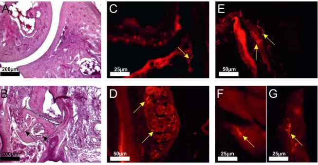
For investigation of neuronal innervation, 10 -12 µm thick cryo-sections were taken with the help of a cryostat (Leica biosystems, Wetzlar, Germany). After air-drying, the sections were rehydrated in PBS for 20 min. Hematoxylin-eosin (HE) staining was performed to evaluate overall structure and, particu- larly, bony erosions in the joints of arthritic mice. Histological scoring of arthritic mouse joints included evaluation of erosions in bone, degradation of cartilage, inflammation of synovial and joint surrounding tissue, and infiltration of immune cells into the joint cavity. In addition, in human finger joint samples, staining with a 0.1% aqueous solution of 1,9-dimethyl-methylene blue (DMMB) was performed to evaluate possible loss of cartilage.
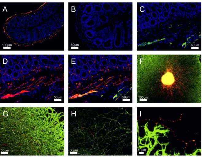
Sections from mice and patients were blocked for 45 min (bovine serum albumin, fetal calf serum, goat serum and 0.3% Triton X-100). After washing with PBS, both sections were incubated for 3-12 hr with a primary antibody against VAChT (cholinergic marker, 1:750, cat.no. 139103, Synaptic Systems GmbH, Göttingen, Germany). In mice, limb sections were incubated with a primary antibody against TH in a separate staining process of neighboring tissue sections (1:500, cat.no. AB152, Chemicon, Temecula, California). In human samples, finger joint and knee synovial sections were incubated with a primary antibody against vasoactive intestinal peptide (VIP) in a separate staining process (1:500, cat.no. 22736, abcam, Cambridge, UK). In human finger joint samples, neighboring sections were also stained for TH in a separate staining process (cat.no. AB152, Chemicon).
After washing, sections were incubated with an Alexa Fluor 546 conjugated secondary antibody (1:500, cat.no. A-11010, goat anti rabbit IgG, Molecular Probes) to achieve immunofluorescent staining of cholinergic (VAChT+ or VIP+) and sympathetic catecholaminergic (TH+) nerve fibers. After an incu- bation time of 90 min, sections were washed, and slides were subsequently mounted with fluorescent mounting medium (Dako, Glostrup, Denmark).
The numbers of TH+ sympathetic catecholaminergic, VAChT+ cholinergic, and VIP+ cholinergic nerve fibers per square millimeter were determined by averaging the number of stained nerve fibers in 17 randomly selected high power fields of view (typical bead chain structure with at least four separated beads along the axon, minimum length 50 µm, determined by a micrometer eyepiece). Positive nerve fiber staining was controlled by incubating tissue without primary antibodies or with immunoglobulin-
2.5 Generation of osteoclast progenitor cells
24
matched control antibodies, which always yielded a negative result. General performance of anti- VAChT and anti-TH antibody was checked by pre-incubation with the respective blocking peptide.
Possible presence of the α7-subtype-containing nicotinic acetylcholine receptor (α7nAChR) in fibro- blast like synoviocytes (FLS) from knee synovial tissue obtained from OA patients was tested by stain- ing formalin fixed FLS with an rabbit polyclonal antibody raised against a peptide containing the amino acids 22-71 from human α7nAChR (cat.no. ab10096, abcam). FLS were first blocked for 45 min (bo- vine serum albumin, fetal calf serum, goat serum and 0.3% Triton X-100) and then treated with a 3%
solution of hydrogen peroxide for 10 min. The stock solution (1mg/mL) of the antibody and a control immunoglobulin fraction from rabbit (cat.no. X0903, Dako, 20mg/mL) were diluted 1:500 and 1:10,000 respectively. FLS were stained overnight at 4° Celsius. After washing with PBS and 0.3% Triton X-100, FLS were incubated in a 1:500 dilution of a secondary goat anti rabbit antibody conjugated with horse- radish peroxidase (HRP) for one hour (cat.no. 32260, Pierce/Thermo Scientific). After washing, cells were then incubated with a solution of 3,3’ diaminobenzidine (DAB) according to the manufacturer’s protocol (cat.no. SK4105, Vector Labs).
2.5 Generation of osteoclast progenitor cells
Generation of osteoclast progenitor (OCP) cells from bone marrow-derived macrophages (BMM) has been described (Takeshita et al., 2000). After sacrifice, femoral and tibial bone marrow was aseptically removed from healthy and arthritic male C57Bl/6J mice, subjected to lysis of erythrocytes (Buffer EL, Qiagen GmbH, Duesseldorf, Germany), and cells were cultured with 20ng/ml murine M-CSF (cat.no.
315-02, Peprotech Inc., Rocky Hill, New Jersey) in a 35mm culture dish (cat.no. 430166, Corning In- corporated, NY, USA). After 2 d, non-adherent cells were washed off with PBS, adherent cells were removed with a cell lifter (cat.no. 3008, Corning Incorporated), and cells were re-suspended in medi- um with 20ng/ml M-CSF and 5ng/ml receptor activator of nuclear factor Kappa-B ligand (RANK ligand, cat.no. 462-TEC-010, R&D Systems, Wiesbaden, Germany). Viability and number of cells were de- termined using the trypan blue exclusion method. In co-culture experiments with sympathetic ganglia 20,000 BMM cells were seeded into each of the second compartment of the insert-equipped 8 well chamber slides (see 2.5 below). For the generation of whole RNA and cell culture supernatants 150,000 cells were seeded into each well of a 6-well culture plate (see also 2.7 and 2.8). Poly- nucleated (nuclei n≥3) and tartrate resistant acidic phosphate-positive cells appeared after 2 - 5 days after seeding and rated as OCP cells (Acid Phosphatase, Leukocyte TRAP Kit, cat.no. 387A, Sigma) (Figure 3).
2.5 Generation of osteoclast progenitor cells
25
In a separate set of experiments, OCPs were also obtained from healthy and arthritic DBA1/J mice to check a possible strain specificity of the co-culture experiments of sympathetic ganglia and OCP cells.
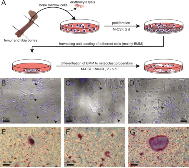
Figure 3. Generation of osteoclast progenitor cells from bone marrow derived macrophages.
A) Diagram delineating the process of obtaining osteoclast progenitors from BMM. B,C,D) Examples of multinucleated (cell nuclei in blue, DAPI stain) osteoclast progenitor cells (arrow heads) after 2 d dif- ferentiation with M-CSF and RANKL, magnification x200. E,F,G) Examples of TRAP-positive (dark red/purple) osteoclast progenitor cells after 2 d differentiation with M-CSF and RANKL, magnification x100. Abbreviations: BMM, bone marrow derived macrophages; M-CSF, macrophage colony stimulat- ing factor; RANKL, receptor activator of nuclear factor Kappa-B ligand; DAPI, 4',6-diamidino-2- phenylindole; TRAP, tartrate resistant acid phosphatase (osteoclast marker).
During the course of generating OCP cells from BMM for gene expression analysis and proteome profiling, two mentionable characteristics were observed: Firstly, the total amount of adherent BMM per animal which was harvested from the bone marrow of two tibiae and two femora and proliferated for two days with the help of M-CSF, was significantly higher in the group of arthritic animals compared to the group of healthy control mice (Figure 4A). Secondly, when BMM were differentiated for five consecutive days compared to 2 d, the size and number of giant multinucleated, TRAP-positive OCP