Neural mechanisms of female aggression:
Implications on the oxytocin and vasopressin systems
DISSERTATION ZUR ERLANGUNG DES DOKOTRGRADES DER NATURWISSENSCHAFTEN (DR. RER. NAT.) DER FAKULTÄT FÜR
BIOLOGIE UND VORKLINISCHE MEDIZIN DER UNIVERSITÄT REGENSBURG
vorgelegt von Vinícius Elias de Moura Oliveira
aus Santo Antônio do Monte, Brasilien
im Jahr 2020
Das Promotionsgesuch wurde eingereicht am:
Die Arbeit wurde angeleitet von: Prof. Dr. rer. nat. Inga D. Neumann
Unterschrift:
Dissertation
Durchgeführt am Institut für Zoologie der Universität Regensburg
Lehrstuhl für Tierphysiologie und Neurobiologie
unter Anleitung von
Prof. Dr. rer. nat. Inga D. Neumann
Table of Contents
T ABLE OF CONTENTS
Summary ... 1
1. General Introduction ... 5
1.1. Aggression ... 5
1.2. Post-weaning social isolation and aggression ... 9
1.3. Neural circuits of aggression ... 10
1.3.1. The lateral septum as a gate for aggressive behavior ... 14
1.4. Neurochemistry and neuroendocrinology of aggression ... 15
1.4.1. The oxytocin and vasopressin systems and their role in modulating aggressive behavior ... 18
1.5. Sex differences in aggression: Female aggression ... 23
1.6. Aims of the present thesis ... 27
1.6.1. Comparing the effects of PWSI on aggression across the sexes ... 27
1.6.2. Evaluating the role of the OXT and AVP systems on female aggression ... 28
Chapter 2: Post-weaning social isolation exacerbates aggression in both sexes and affects the vasopressin and oxytocin system in a sex-specific manner ... 30
2.1. Introduction ... 30
2.2. Material and Methods ... 32
Animals ... 32
PWSI procedure and housing conditions ... 32
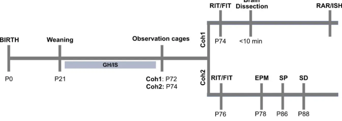
Overview of experiments (Fig 2.1) ... 33
Resident and Female Intruder test ... 34
Elevated Plus-maze ... 35
Social Preference Test ... 35
Social Discrimination Test ... 36
In situ hybridization ... 37
Receptor autoradiography ... 38
Statistical analyses ... 38
2.3. Results... 39
PWSI does not affect body and adrenal weights ... 39
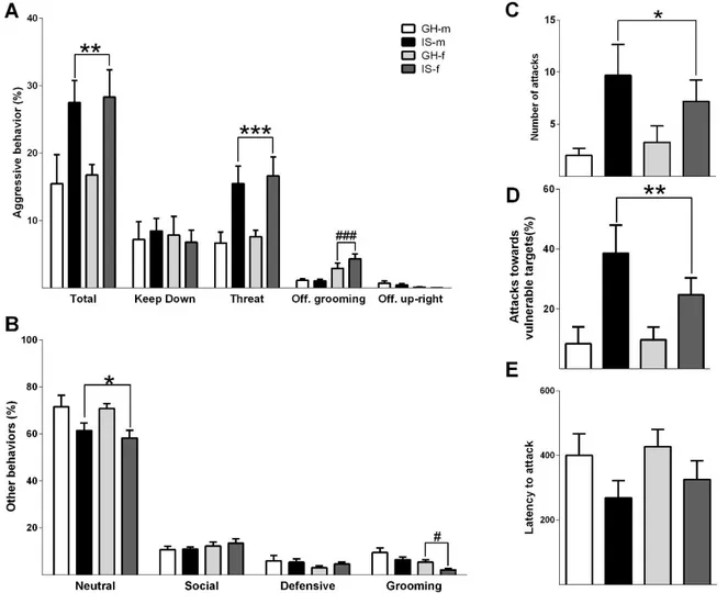
PWSI increases aggression in both male and female rats ... 39
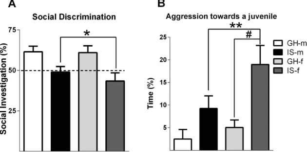
PWSI impairs social discrimination, but not anxiety-like behavior or social preference ... 41
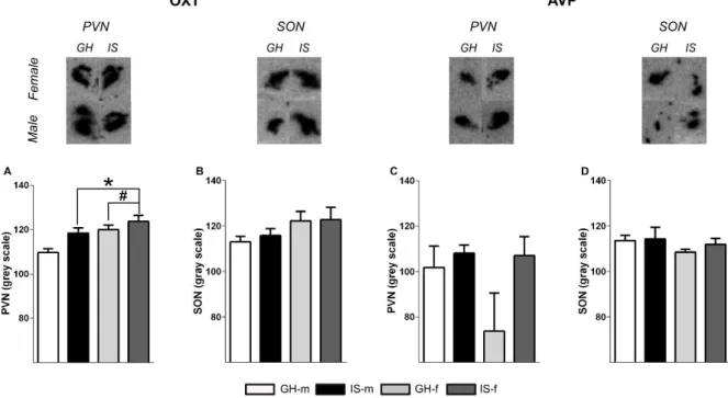
PWSI increases OXT, but not AVP mRNA expression ... 42
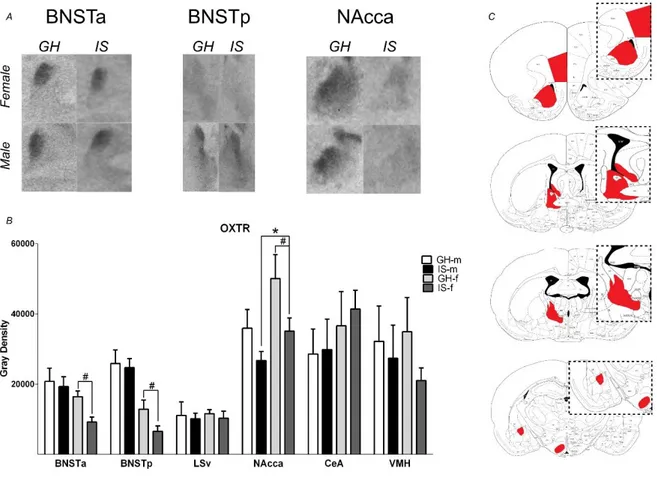
PWSI alters V1a and OXT receptor binding ... 45
2.4. Discussion ... 46
2.5. Conclusion ... 51
Chapter 3: The oxytocin-vasopressin balance within the lateral septum determines aggression levels in virgin female rats ... 52
3.1. Introduction ... 52
3.2. Methods... 55
Ratlines and animal care ... 55
Female Intruder test (FIT) ... 56
Overview of the in-vivo experiments ... 57
Stereotaxic surgery ... 59
Receptor autoradiography ... 60
ELISA for plasma corticosterone ... 61
Radioimmunoassay for OXT and AVP ... 61
Drugs and Viruses ... 61
Patch-Clamp... 62
Perfusion ... 63
Immunohistochemistry ... 63
Statistics ... 65
3.3. Results... 66
Social isolation and training reliably enhanced female aggression ... 66
The high levels of aggression displayed by isolated and trained rats are underlined by high levels of OXT and low levels of V1a receptor binding ... 68
OXT promoted whereas AVP reduced aggression in a central approach ... 70
The pro-aggressive effect of OXT was mediated in the vLS ... 73
AVP exerted anti-aggressive effects within the dLS ... 76
Two distinct subregions of the LS are differently regulated by the activation of OXTRs ... 78
An intrinsic GABAergic circuit within the LS regulates female aggression ... 81
3.4. Discussion ... 84
4.1 General Discussion ... 92
4.2 Outlook for PWSI ... 95
4.3 Outlook for the IST protocol ... 98
4.4 Concluding remarks ... 103
Abbreviation List ... 104
References ... 106
Acknowledgments ... 119
Curriculum Vitae ... 121
Publications ... 123
1
S
UMMARYAggression is defined as a social behavior that has the intention of physically harming a conspecific. In nature, aggressive behavior emerges whenever an individuum, engages in conflict over essential resources for its survival, such as food, water, territory, and mating partners. Thus, aggression may act as an evolutive force controlling populational levels and keeping hierarchy. However, in humans, when expressed out-of-context and in exacerbated manner aggressive behavior becomes disruptive constituting a severe burden on society. This is especially evident in the excessive as well as pathological levels of aggression expressed by individuals suffering from conduct disorder (CD) in childhood and anti-social personality disorder (ASPD) in adulthood. As violent aggressive behavior causes serious damage not only to the victims but also to the perpetrators, scientists have worked throughout the last decades to understand the neurobiological underpinnings of escalated aggression. In order to do so, several rodent models have been established to study aggressive behavior, using mostly males as model organisms whereas females have been rarely studied. Nevertheless, recent evidence shows that women and girls may develop ASPD and CD just like men and boys, respectively. Additionally, there are some new indications that the neurobiology of aggression might be sex-specific in humans and rodents. Thus, new animal models are necessary to understand the neural mechanisms of female aggression.
In my thesis, I first established and utilized two rat models of female aggression
based on different etiologic strategies, namely post-weaning social isolation (PWSI)
was used as a model of early life stress-induced aggression whereas a combination of
social isolation and aggressive training was used to enhance aggression in an
ethological setting in adult rats. These models allowed me to investigate the role of the
Summary
2
brain oxytocin (OXT) and vasopressin (AVP) systems on aggressive behavior. Both neuropeptides are known to regulate social including aggressive behavior in males and lactating females.
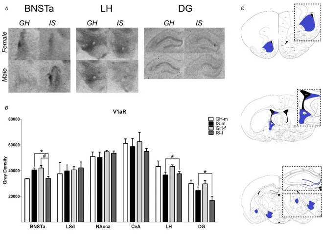
Although aggression is a naturally occurring behavior in animals and humans, exacerbated aggressive behavior may also emerge as a maladaptive response to stress. Especially, early life adverse experiences are known to evoke violent aggressive behavior. Thus, in chapter 2 post-weaning social isolation (PWSI) was used as a reliable model of early life stress-induced (ELS) aggression in order to compare its effects on male and female aggression as well as on the endogenous OXT and AVP systems. My results show that males and females displayed similar levels of aggression independent of the housing conditions and that PWSI increased aggression in both male and female Wistar rats. However, abnormal aggression was displayed in a sex-dependent manner, i.e. females exhibited elevated aggression towards juveniles, whereas males tended to show more attack bites and attacks towards vulnerable body parts. In addition, PWSI also impaired social discrimination in both sexes. From a neurobiological point of view, PWSI decreased OXTmRNA in the paraventricular nucleus of the hypothalamus (PVN) and OXT receptor (OXTR) binding in the nucleus accumbens (NAcc), independent of the sex. Regarding the AVP system, I have found that PWSI rats showed decreased AVP 1a receptor (V1aR) binding in the dentate gyrus (DG) and lateral hypothalamus (LH) independent of sex.
However, the anterior part of the BNST was affected by PWSI in a sex-dependent
manner, i.e. in control conditions, females exhibited higher V1aR binding than males
in this region, but after PWSI females had lower V1aR binding than males. Thus, my
data supports PWSI as a reliable rat model to instigate exaggerated as well as
abnormal aggression not only in males but also in females. In addition, OXTRs in the
3
NAcc and V1aR in the BNSTa, DG, and LH may play a role in the link between PWSI and aggression in rats.
In chapter 3, in order to specifically investigate the role of OXT and AVP on female aggression, social isolation, as well as successive encounters with a same-sex and unknown conspecific (aggression training) (IST), were used to enhance the mild levels os aggression displayed by group-housed (GH) and non-trained females. In comparison to low aggressive GH controls, highly aggressive IST females exhibited elevated levels of OXT and reduced levels of AVP in both CSF and LS in response to a female intruder test (FIT). Furthermore, both OXTR and V1aR binding were decreased in the ventral (vLS) and dorsal (dLS) portion of the LS of IST rats, respectively. Manipulation of both neuropeptide systems using a combination of neuropharmacological, chemo- and optogenetic approaches resulted in dramatic changes in aggression. Elevating OXT availability either centrally or in the vLS of GH rats enhanced aggression. Accordingly, blockade of OXTR, via OXTR antagonist, either centrally or in the vLS decreased aggressive display in IST rats. Regarding the AVP system, synthetic AVP administrated either locally in the dLS or centrally (intracerebroventricular) decreased aggression in IST rats.
Due to the fact that OXT and AVP effects appear to be region- and receptor-
specific, i.e. OXT acted via OXTR in the vLS and AVP acted via V1aR in the dLS, I
decided to verify whether those two neural populations within the LS interact with each
other in a single-cell level after OXTR activation using whole-cell voltage-clamp. OXTR
activation increased GABAergic inhibition of dLS neurons whereas decreased
GABAergic inhibition of vLS neurons. Next, I decided to evaluate whether those
differences in activity were also reflected in vivo after an aggressive encounter, using
pERK as a neural activity marker. Aggression differentially regulated pERK expression
Summary
4
in the LS, i.e. GABAergic neurons in the dLS showed decreased whereas in the vLS showed increased activation after the FIT. In line with that, pharmacological inhibition of the dLS and vLS enhanced and reduced female aggression, respectively. Taken together this part of my thesis shows that the balance between OXT and AVP release within the LS regulates female aggression in a receptor and region-specific manner via modulating GABAergic neurotransmission.
Overall, this thesis shows that females are able to develop escalated as well as
abnormal aggression just like males. In addition, the OXT and the AVP system seem
to be main players in regulating aggressive behavior in female Wistar rats, especially,
regarding their role in controlling aggression by acting on the LS.
5
1. G
ENERALI
NTRODUCTION1.1. A
GGRESSIONSocial aggression is typically defined as a social behavior displayed between conspecifics with the intention of physically harming one another (De Almeida et al, 2005; Koolhaas et al, 2013; Miczek et al, 2001; Nelson and Trainor, 2007). It is expressed by many, if not all animal species including humans, and usually the successful aggressor benefits by gaining access to limited resources, such as food, territory, nests, or mating partners (De Almeida et al, 2005; Nelson and Trainor, 2007).
Further, aggressive behavior may also emerge, as a maladaptive response, whenever animals face challenges to their homeostasis, such as threats and/or stressors (Haller et al, 2014; Sandi and Haller, 2015).
When expressed out-of-context or in an exacerbated manner aggressive behavior becomes disruptive by harming both aggressor and victim. This is especially evident in the pathological levels of aggressive behavior displayed by humans suffering from aggression disorders, such as conduct (CD), anti-social personality (ASPD), and intermittent explosive disorder (De Almeida et al, 2005; Haller, 2016; Nelson and Trainor, 2007). Around 5% and 0.6-3% of the European population are affected by CD and ASPD, respectively. Those patients suffer from a broad spectrum of symptoms including excessive aggression towards others, damaging property, deceitfulness, lack of remorse or guilt, callousness or lack of empathy, shallow or deficient affect, manipulativeness, and egocentrism (DSM-V American Psychiatric Association, 2012;
Freitag et al, 2018; Reynolds and Kamphaus, 2014; Wittchen et al, 2011). Exaggerated
aggression also occurs as a symptom of other psychiatric and neurological disorders,
such as autism, bipolar personality disorder, schizophrenia, post-traumatic stress
disorder (PTSD), and dementia (Nelson and Trainor, 2007). Thus, aggression as
Chapter 1: General Introduction
6
comorbidity or as a disorder constitutes a severe burden to society. In fact, estimations indicate that annually 1.3 million people worldwide die as a consequence of violence, and 126 billion US dollars are spent annually to either prevent or deal with the consequences of violence (WHO, 2014).
In humans, aggressive behavior is mainly classified into two types: instrumental or controlled aggression characterized by having a goal-oriented purpose, and reactive or impulsive aggression known to be a sudden, rather uncontrolled reaction to a stimulus, often related to anger. In general, reactive aggression is related to abrupt and inappropriate aggressive outbursts, as an example, when someone, by accident, punches a co-worker after a passionate argument, this type of aggression is typically linked with intermittent explosive disorder, PTSD and depression-related aggression.
On the other hand, instrumental aggression can be associated with genocide, planned assassination and massive killings, in this case, people act less on impulse and plan their aggressive acts, as an example, we could mention planning for weeks on how to kill the co-worker who punched you after the argument. Patients suffering from CD and ASPD show high levels of instrumental aggression frequently accompanied by a lack of remorse (Viding et al, 2012). Both types of aggression also differ from a physiological point of view, reactive aggression is related to hyper-arousal, i.e. accompanied by high sympathetic activity and cortisol levels, whereas instrumental aggression has been linked to a hypo-arousal phenotype, i.e. low sympathetic activity and cortisol levels (Comai et al, 2012; Haller, 2013; Nelson and Trainor, 2007).
In order to reveal the brain regions and neurobiological mechanisms involved in
aggression regulation, various strategies have been employed in animals. Seminal
studies have used electrical and chemical stimulation of so-called aggression centers
to evoke aggressive phenotypes similar to the ones seen in humans (De Almeida et
al, 2005; Baxter, 1968; Haller, 2013; Potegal, M Blau, A and Glusman, 1981; Potegal
7
et al, 1981), current approaches preferred focusing on the animal’s ethology in order to increase the biological significance of the findings. Therefore, in animals, aggression is usually classified accordingly with the subject’s ethology (territorial, dominance, maternal) (De Almeida et al, 2005; Comai et al, 2012). Especially, territorial aggression has been studied in male rodents for years, using a reliable and consistent paradigm:
the resident-intruder test (RI). This test relies on the fact that male residents will defend their territory against unfamiliar male intruders. Usually, either co-housing with a female over several days or weeks or social isolation are used to instigate the resident’s territoriality and, consequently, aggressive behavior to defend its homecage.
The test consists of releasing a slightly smaller same-sex intruder into the resident's homecage for 10-20 minutes. During this time aggressive behaviors, such as attack bites, threats, chases and tail rattles (mice) as well as dominant behaviors, such as keep down, offensive grooming, offensive up-rights are quantified. In addition, non- aggressive behaviors like non-aggressive social investigation (sniffing), investigation of the homecage, self-grooming and defensive behaviors might be also quantified in order to evaluate, whether the high aggressiveness impacts on other behavioral domains displayed by the residents (Koolhaas et al, 2013; Miczek et al, 2001).
Surprisingly, although territorial aggression has been reported in wild female mice and hamsters (Harmon et al, 2002; Mcdonald et al, 2011; Miczek et al, 2001; Ross et al, 2019; Silva et al, 2010), this behavior has been mostly studied in males, whereas females have been only studied in the context of lactation associated with the prominent display of maternal aggression (see discussion on page 23).
Aiming to mimic pathological and out-of-context aggression seeing in humans,
various rat and mouse models have been established, in order to understand the
neural underpinnings of escalated aggressive behavior. In general, an animal becomes
abnormally aggressive when shows: i) a mismatch between provocation and response,
Chapter 1: General Introduction
8
i.e. attacking in inappropriate situations, as in a neutral arena or showing elevated aggression (excessive attack counts, short-latency to attack, causing severe tissue damage); ii) disregard of species-specific rules, such as attacking juveniles, anesthetized animals and females or attacking vulnerable body parts (head, paws, neck, and belly); iii) insensitivity towards the social signals of the intruder, i.e. sustained aggression despite submissiveness of the opponent or showing an inability in terminating aggression outbursts; iv) “offensive ambiguity”, namely aggressive behavior lacking a normal structure, i.e., failure in signaling attacks from threats, attacking from defensive postures or attacking only smaller intruders (Haller, 2013;
Miczek et al, 2013).
Among the approaches used to develop animal models of abnormal and excessive aggression five main strategies have been successfully described: i) using naturally aggressive animals such as hamsters (Ferris et al, 1997; Harmon et al, 2002;
Potegal et al, 1981), Calfornia mice (Oyegbile and Marler, 2005; Silva et al, 2010) and feral rats (Koolhaas et al, 2013) ii) selecting mice (Caramaschi et al, 2008; Lagerspetz, 1968; Miczek et al, 2013; van Oortmerssen and Bakker, 1981) and rats (Beiderbeck et al, 2012; Koolhaas et al, 2013; Neumann et al, 2010; Walker et al, 2016) based on their levels of aggression in order to have animals showing feral aggression; ii) using ELS such as post-weaning social isolation (Toth et al, 2011), peripubertal stress (PPS) (Marquez et al, 2013) or maternal separation (MS) (Veenema et al, 2006); iii) manipulation of selected biological system to mimic features encountered in highly aggressive patients, such as adrenalectomy (Haller et al, 2004) to induce a state of hypo-CORT, and alcohol consumption (De Almeida et al, 2005; Miczek et al, 2013);
and iv) aggression training, engaging and winning, repeatedly, conflicts have been
implicated in decreasing attack latencies and increasing the number of attacks in
hamsters (Been et al, 2016) and mice (Oyegbile and Marler, 2005; Silva et al, 2010).
9
Especially, ELS models have been repeatedly shown to induce consistent and reliable levels of high aggression in male rats (Haller et al, 2014; Masis-Calvo et al, 2018; Sandi and Haller, 2015). Those models are translationally relevant because they were built on the fact that patients with CD and ASPD as well as offenders, often come from troubled homes and face different types of abuse during their childhood and/or adolescence. In fact, several studies have described that stressful environmental conditions, especially during early life, correlate with aggressive as well as externalizing behavior in humans (Caspi et al, 2002; Dackis et al, 2017; Freitag et al, 2018; Glenn et al, 2013; Haller et al, 2014; Nelson and Trainor, 2007; Sandi and Haller, 2015). Most of the rat studies of ELS support the findings seen in humans (Marquez et al, 2013; Toth et al, 2011; Veenema et al, 2006), showing that an exaggerated stimulation of the hypothalamic-pituitary-adrenal axis (HPA) by stress during early life leads to abnormal aggressive behavior later in life underlined by a hyper-CORT phenotype (Biro et al, 2016; Marquez et al, 2013; Toth et al, 2011, 2012; Veenema et al, 2006; Veenit et al, 2013).
In the present thesis, PWSI has been used as a model of ELS for studying mechanisms of aggression in females in comparison to males. Therefore, I will describe the model in more detail in the next session.
1.2. P
OST-
WEANING SOCIAL ISOLATION AND AGGRESSIONAmong the ELS models of pathological aggression, PWSI seems to have the
strongest effects, because it fulfills the 4 criteria needed to be classified as an abnormal
aggression model as well as presents a robust translational component, psychosocial
deprivation, including neglect and abuse, have been implicated in the development of
ASPD and CD (Dackis et al, 2017; Glenn et al, 2013; Haller, 2016). The protocol
consists of keeping pups (21 days old) either housed in groups (controls) or singly-
housed for 7 weeks after weaning. It is based on the premise that lack of social contact
Chapter 1: General Introduction
10
and the display of play behavior with conspecifics during puberty/adolescence impairs or prevents the development of the rat’s social behavior repertoire, resulting in high and abnormal aggression (Toth et al, 2011).
PWSI induces abnormal aggression in male Wistar rats by increasing attack bites towards vulnerable targets and decreasing signaled attacks, i.e. PWSI rats attack from defensive postures. Conversely, isolated rats exhibit elevated plasma corticosterone (CORT) and autonomic responses during the RI (Toth et al, 2011), characterizing a hyper-arousal type of aggression, which has been linked rather to reactive aggression than to instrumental aggression expressed by ASPD and CD patients (Comai et al, 2012; Nelson and Trainor, 2007).
Concerning the neurobiological mechanisms underlying the high levels of aggression shown by isolated animals, it has been shown that PWSI enhances the neuronal activation of aggression-related regions, such as the hypothalamic-attack area (HAA), the bed nucleus of stria terminalis (BNST), the medial amygdala (MeA) and the orbital frontal cortex (OFC) (Toth et al, 2012). In addition, isolated rats exhibit a thinner right prefrontal cortex (PFC), which was a consequence of reduced glia and dendritic density as well as impaired vascularization of this area (Biro et al, 2016).
Those changes have been related to the finding of decreased levels of the brain- derived neurotrophic factor (BDNF) in the PFC as well as in the MeA of PWSI rats (Mikics et al, 2018).
1.3. N
EURAL CIRCUITS OF AGGRESSIONThroughout the years, neuroscientists have tried to fully characterize the brain
networks involved in aggressive behavior display by using a variety of techniques, such
as neural activity markers (c-fos, pERK, and zif268), chemical and mechanical
lesioning/inhibition of target brain regions, electrophysiological recordings of neuronal
populations during the display of aggression and, recently, calcium imaging in freely
11
moving animals (fiber photometry). All this effort culminated in the establishment of an aggressive behavior neuronal pathway, which completely overlaps with the social behavior network, in agreement with the theory that aggressiveness is an emergent property of social behaviors (Nelson and Trainor, 2007).
In rodents, social cues, including the ones eliciting aggression, are known to be mainly olfactory, therefore, they are initially processed in the main (MOB) and accessory (AOB) olfactory bulbs (Dulac and Torello, 2003; Nelson and Trainor, 2007;
Stowers et al, 2013). In fact, the ablation of either the olfactory bulb by bulbectomy or
of the olfactory nerves strongly impairs aggressive display in male mice (Mucignat-
Caretta et al, 2004) and rats (Bergvall et al, 1991). Also, the existence of aggression
evoking pheromones has been described in male mice (Chamero et al, 2007; Stowers
et al, 2013). After this initial processing, the signals are forwarded to limbic and
hypothalamic regions involved in aggression display such as the BNST, MeA, the
lateral septum (LS), the medial pre-optic area (MPOA), the ventromedial (VMH) and
anterior (AH) hypothalamus (mice), also known as part of the hypothalamic attack-area
or mediobasal hypothalamus (MBH) (rats) (Beiderbeck et al, 2007, 2012; Toth et al,
2012; Trainor et al, 2010a). After, further processing all those signals seem to converge
into the periaqueductal gray matter (PAG), in the midbrain, where the motor outputs
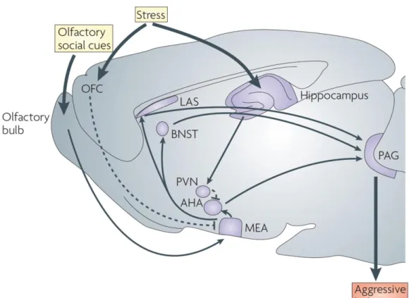
are generated (Nelson and Trainor, 2007) (Figure 1.1).
Chapter 1: General Introduction
12
It is important to highlight the essential role of the HAA in eliciting attacks in male rodents. Early studies have shown that electrical stimulation of the HAA, which anatomically corresponds to several hypothalamic nuclei, leads to irritability and aggression in cats and rats (Brown et al, 1969; Haller, 2013; Kruk et al, 1983).
Additionally, increased activation of the HAA after the RI has been reported, using neural activity markers, in rat models of abnormal aggression (Beiderbeck et al, 2012;
Toth et al, 2012) and in Peryomiscus californicus (Trainor et al, 2010a). Recent experiments using immediate early genes, optogenetics, in vivo electrophysiology and calcium imaging, have shown that a specific population of neurons, expressing the estrogen receptor alpha (ERα) as well as progesterone receptors (PR) within the
Figure 1.1: Neuroanatomical pathways of aggression in the rodent brain. Typically sensorial information arrives in the olfactory bulb and is further processed in the medial amygdala (MEA), the MEA projects to the lateral septum (LAS), bed nucleus of stria terminalis (BNST) and anterior hypothalamus (AHA). These brain areas are known to modulate the periaqueductal gray (PAG) activity, in order to originate the motor patterns need it during aggressive behavior display in rodents.
Other regions such as the hippocampus, orbital frontal cortex (OFC) and the paraventricular nucleus of the hypothalamus (PVN) may also modulated aggressive behavior by acting on these regions, especially under stressful conditions. The OFC is thought to be one of the main inhibitors of aggressive behaviors in mammals (Adapted from Nelson and Trainor, 2007).
13
ventrolateral portion of the ventromedial hypothalamus (VMHvl) work as a switch triggering aggression in solitary male mice independent of pheromone-sensing, gonadal hormones, opponents or social context (Lee et al, 2014; Lin et al, 2011; Yang et al, 2013, 2017). Further, the VMHvl ERαPR-neurons seem to have a pivotal role in aggressive behavior display, not only for controlling attack initiation and termination (Lee et al, 2014; Yang et al, 2013), but also for being responsible for controlling attack motivation/seeking in male mice (Falkner et al, 2016).
In addition to the VMHvl, there are other brain regions that should be drawn to attention for being constantly associated with male aggressive behavior in rodents such as the amygdaloid complex, the nucleus accumbens (NAcc) and the PFC.
Particularly, hyperactivation of the central (CeA) and medial nuclei of the amygdala has
been described in rat models of abnormal aggression (Marquez et al, 2013; Toth et al,
2012), also aromatase-positive neurons in the MeA have been shown to, specifically,
induce aggressive behavior in male mice (Unger et al, 2015). Regarding the
motivational aspects of aggression, the NAcc appears to play a main a role, addiction-
like aggressive behavior has been described in male mice (Golden et al, 2017), this
behavior is, at least, partially underlined by dopamine-sensitive neurons in the NAcc
(Aleyasin et al, 2018; Golden et al, 2019). Accordingly, dopamine release within the
NAcc as well as increased activity of this region have been shown in abnormally and
highly aggressive male rats selectively bred for low anxiety-related behavior (LAB)
during the RI (Beiderbeck et al, 2012). Last, although the PFC has been implicated in
aggression inhibition in rodents (Nelson and Trainor, 2007), its role on it is still
controversial, as both increased (Toth et al, 2012) as well as decreased (Marquez et
al, 2013) neuronal activity has been seen in different subregions of the PFC in rat
models of abnormal aggression. Moreover, direct stimulation of the medial part of PFC
reduces aggression in male mice (Takahashi et al, 2014), whereas stimulation of
Chapter 1: General Introduction
14
specific projections of the PFC to the hypothalamus seems to increase attack counts as well as abnormal attacks in PWSI rats (Biro et al, 2018).
1.3.1. T
HE LATERAL SEPTUM AS A GATE FOR AGGRESSIVE BEHAVIORThe LS consists mainly of GABAergic neurons (~90%) (Alonso et al, 1990) located in different subnuclei, i.e. the dorsal (dLS), intermediate (iLS), ventral (vLS), rostral (rLS) and caudal (cLS) LS, placed in between the lateral ventricles in the rostrodorsal septal region. Although those subnuclei are mostly GABAegic, they are known to co-express different neuropeptides and steroid hormone receptors, such as substance P, neurotensin, encephalin, somatostatin, dynorphin, growth-hormone- releasing hormone, androgen receptors (AR), ERαs, mineralocorticoid receptors (MR), oxytocin receptors (OXTR) and vasopressin 1a receptors (V1aR) in a nuclei- and neuron-specific manner (Risold and Swanson, 1997a; Smith et al, 2017). The main source of inputs to the LS is the hippocampus, although it also receives projections from the brainstem (ventral tegmental area, locus coeruleus and Raphé nucleus) and presents bidirectional connections with the hypothalamus, BNST, preoptic area, and amygdala. Therefore, different neuropeptidergic, as well as monoaminergic terminals, can be found in the LS (DiBenedictis et al, 2017; Risold and Swanson, 1997b).
From a behavioral point of view, the LS has been implicated in several different
types of social behaviors including social anxiety (Zoicas et al, 2014), stress-induced
social avoidance (Guzmán et al, 2013), social memory (Camats Perna and
Engelmann, 2017; Lukas et al, 2011a; Popik et al, 1992) and aggression (Wong et al,
2016). Early case studies have described the role of the septal area on inhibiting
aggression in humans, patients, including women, with septal tumors exhibited
aggression outbursts (septal rage) and increased irritability (Zeman, Wolfgang and
King, 1958). Later studies performed in rodents have shown the same pattern: either
lesion (Potegal, M Blau, A and Glusman, 1981) or pharmacological inhibition
15
(Muscimol-GABA agonist) (Borland et al, 2019) of the LS triggered septal rage and heightened aggression, in hamsters. Accordingly, electrical stimulation of the septum reduced aggression in highly aggressive male hamsters (Potegal et al, 1981). In male rats, a similar picture is seen, where lesions of the LS evoked septal rage (Albert and Chew, 1980). Moreover, reduced activation of the LS (c-fos) was found in highly aggressive LAB rats after an aggressive encounter (Beiderbeck et al, 2007). Recently, the mechanism by which the LS abolishes aggression has been elucidated.
Optogenetic stimulation of LS-GABAergic projections to the VMHvl stopped attacks as well as reduced aggression in male mice (Wong et al, 2016). Intriguingly, new evidence has shown that this pathway is regulated by an intricate polysynaptic LS microcircuit, where GABA neurons in the dLS, under the influence of glutamatergic hippocampal V1bR-positive terminals, inhibit neurons in the vLS, leading to a disinhibition of the VMHvl and subsequently to aggression in male mice.
1.4. N
EUROCHEMISTRY AND NEUROENDOCRINOLOGY OF AGGRESSIONSeveral neurotransmitters, neuropeptides, and hormones have been linked to
aggressive behavior. Regarding the neurotransmitters, especially the monoamines
serotonin (5-HT) and dopamine (DA), but also other neurotransmitters such as
glutamate, GABA, noradrenaline (NA), acetylcholine (ACh) have been studied
extensively (Comai et al, 2012). Drugs targeting both the DA and 5-HT systems such
as haloperidol, risperidone or selective serotonin reuptake inhibitors (SSRIs) have
been used for decades to treat aggressive patients in the clinic; nevertheless, the
efficacy of those treatments is still arguable due to severe side effects (Carrillo and
Ricci, 2009; Comai et al, 2012; Nelson and Trainor, 2007). Those treatments are based
on the hypothesis that especially low levels of 5-HT in the brain might lead to disruptive
aggressive behavior (Comai et al, 2012; Nelson and Trainor, 2007). In contrast to this
hypothesis, low activity of the monoamine oxidase A (MAOA), the enzyme responsible
Chapter 1: General Introduction
16
for degrading the monoamines in the brain, has been associated with high levels of emotional dysfunction and aggression in children with ADHD (Fowler et al, 2009).
However, those results have to be interpreted with caution, because the effect of MAOA on aggression seems to be influenced by environmental conditions like maltreatment in childhood (Caspi et al, 2002; Fowler et al, 2009).
Although animal studies have shown that potentiating serotoninergic neurotransmission was able to reduce aggression in male hamsters (Ferris et al, 1997;
Terranova et al, 2016), abnormally aggressive Wild-Type Groningen (WTG) rats (De Boer and Koolhaas, 2005), and mice (De Almeida and Miczek, 2002; Audero et al, 2013), other studies have shown rather opposite effects in similar models (Audero et al, 2013; Marquez et al, 2013; Mikics et al, 2018). This might be due to the fact that 5- HT receptors can also influence serotonin synthesis and release in the brain by acting on autoreceptors, which makes manipulating this system challenging (Carrillo and Ricci, 2009; Nelson and Trainor, 2007). Regarding the dopaminergic system, recent evidence shows that DA is released in the NAcc of LAB rats during the RI, and in this region, aggression was linked to activation of D2 receptors (Beiderbeck et al, 2012).
Moreover, D1 but not D2 positive neurons in the NAcc shell seem to regulate aggression seeking as well as aggression self-administration in male mice (Golden et al, 2019).
Many studies have tried to link hormonal levels in blood with aggression in
animals and humans. Especially, CORT concentration reflecting the activity of the HPA
axis under basal or stimulated conditions have been associated with pathological
aggression. Intriguingly, both hyper- as well as hypo-CORT levels have been strongly
related to abnormal forms of aggression in different rat models (Masis-Calvo et al,
2018; Sandi and Haller, 2015; Walker et al, 2016). Specifically, heightened CORT
responses to the RI or to an acute stressor have been seen in rat models of high and
17
abnormal aggression induced by ELS protocols such as MS (Veenema et al, 2006), PPS (Marquez et al, 2013), and PWSI (Toth et al, 2011). In agreement rats selected for high CORT responsiveness to restrain-stress exhibited increased levels of aggression in the RI (Walker et al, 2017), and acute inhibition of CORT release via metyrapone decreases attack counts in Wistar rats (Haller et al, 2004). However, abnormal aggression has been also described in Wistar rats after adrenalectomy (Haller et al, 2004). Altogether, this data fits the hypothesis that both low and high arousal states lead to increased aggression and that an aberrant function of the HPA axis underlines exaggerated aggressive behavior (Masis-Calvo et al, 2018; Nelson and Trainor, 2007; Sandi and Haller, 2015).
Sex hormones such as testosterone and estradiol have also been associated
with aggressive behavior display in male rodents. For example, sexual investigation,
i.e. exposition of a male to a receptive female, is known to increase testosterone levels
as well as territoriality and aggressive display in rats (Koolhaas et al, 1980, 2013). In
addition, castration is broadly known to abolish aggression in male rodents (Koolhaas
et al, 1980; Miczek et al, 2001; Nelson and Trainor, 2007). In the brain, aromatized
testosterone acts via estrogen receptors for masculinizing the undifferentiated brain of
males, which contributes to the establishment of male-typical behaviors including
components of mating and aggressive behavior (Lenz et al, 2012). Indeed, the deletion
of ERα (Sano et al, 2016), estrogen receptor β (ERβ) (Nakata et al, 2016) and AR
(Juntti et al, 2010) impairs aggressive behavior in male mice. It is important to highlight
that the activational effects of those receptors are under the influence of environmental
cues and may differ from the organizational effects seen after the deletion of the
receptors (Trainor et al, 2007).
Chapter 1: General Introduction
18
1.4.1. T
HE OXYTOCIN AND VASOPRESSIN SYSTEMS AND THEIR ROLE IN MODULATING AGGRESSIVE BEHAVIORThe nonapeptides Oxytocin (OXT) and arginine vasopressin (AVP) are sister
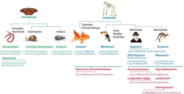
neuropeptides that emerged after the duplication of the single gene coding the peptide
vasotocin in Vertebrata. Interestingly, more than a dozen homologous nonapeptides
have been described among invertebrates and vertebrates, showing how relevant
those peptides are from an evolutive point of view (Jurek and Neumann, 2018) (Figure
1.2). Although OXT and AVP are mainly found in magno- and parvocellular neurons of
the paraventricular (PVN) and magnocellular neurons of the supraoptic (SON) nuclei
of the hypothalamus (Grinevich et al, 2016; Jurek and Neumann, 2018; Koshimizu et
al, 2012), AVP neurons can also be found in several other brain regions such as the
MeA, BNST, MOB, AOB, piriform cortex, MPOA and suprachiasmatic nucleus of
hypothalamus (SCN) (Tobin et al, 2010; De Vries and Panzica, 2006; Wacker and
Ludwig, 2019). Both peptides are released into the capillaries of the neurohypophysis,
reaching the periphery, where they act like hormones. In mammals, hormonal OXT is
known to promote milk-ejection during lactation and the contraction of the myometrium
during labor, whereas hormonal AVP supports water reabsorption in the kidney, and
acts as a vasoconstrictor in the arterial capillaries. Moreover, parvocellular AVP
originated in the PVN acts synergically with corticotrophin-releasing-hormone (CRH)
to regulate corticotrophin (ACTH) secretion in the adenohypophysis (Grinevich et al,
2016; Jurek and Neumann, 2018; Koshimizu et al, 2012).
19
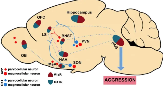
Besides their hormonal role in the periphery, OXT and AVP also act as neuromodulators in the brain, where they are released from either axonal collaterals, terminals in various brain regions, or from soma and dendrites locally in the SON and PVN (Grinevich et al, 2016; Jurek and Neumann, 2018; Ludwig and Leng, 2006). Both peptides change neuronal excitability by binding to their respective receptors. OXT, typically binds to OXT receptors (OXTRs), whereas AVP binds to vasopressin 1a and 1b receptors (V1aRs and V1bRs) (Jurek and Neumann, 2018; Koshimizu et al, 2012), However, cross-activation of each others receptor has been described in vitro (Manning et al, 2012) as well as in vivo (Song et al, 2014; Tan et al, 2019). Moreover, OXT and AVP fibers, as well as OXTR and V1aR, are widespread throughout the rodent brain. Receptor binding is especially found in regions within the social behavior network, such as the amygdala nuclei, LS, BNST, NAcc, hypothalamic nuclei (VMH, AH, LH) and MPOA (DiBenedictis et al, 2017; Smith et al, 2017) (Figure 1.3).
Figure 1.2: OXT and AVP sequences across different Taxa. Nonapeptide sequences of invertebrates are shown in green and vertebrates show in blue and red OXT and AVP, respectively, analogs across the animal kingdom. Each amino acid sequence is initiated by a 19 amino acid signal peptide, followed by the specific nonapeptide sequence depicted above, a processing signal consisting of glycine-lysine-arginine (GKR), and the neurophysin-glycopeptide-COOH-terminus.
Italic/bold amino acids, difference between OXT and AVP; bold amino acids, difference between respective OXT or AVP. (Adapted from Jurek and Neumann, 2018).
Chapter 1: General Introduction
20
Interestingly, in the rat brain, OXTRs and V1aRs seem to be uniquely distributed in different neuronal populations within the same region (Smith et al, 2017; Stoop et al, 2015). This is supposed to be the basis of the antagonistic effects of OXT and AVP on various behaviors. Indeed, in the lateral portion of the CeA, GABAergic neurons expressing OXTR inhibit V1aR-positive neurons in the medial part of the same region via collaterals, this process triggers opposing effects on fear, i.e. OXT decreases it whereas AVP increases it (Huber et al, 2005; Knobloch et al, 2012; Stoop et al, 2015).
Another interesting fact about these systems is their sexual dimorphism in terms of receptor binding, neuronal number, and fiber densities. Male rats present more AVP- positive neurons in the BNST and MeA, and they also exhibit higher fiber density in the
Figure 1.3:The oxytocin (OXT) and vasopressin (AVP) systems in the aggression network.
Scheme shows localization of OXT neurons, in blue, in the paraventricular (PVN) and supraoptic (SON) nucleus of the hypothalamus, as well as AVP neurons, in red, in the PVN, SON, olfactory bulb (OB), medial amygdala (MeA), and bed nucleus of stria terminalis (BNST). OXT receptors (OXTR) and AVP 1a receptors (V1aR) are co-expressed in all regions involved in the aggression network, i.e. BNST, hypothalamic attack area (HAA), hippocampus, lateral septum (LS), MeA, OB and orbitalfrontal cortex (OFC), periaquedutal gray matter (PAG). Projections within the aggression network are shown in dotted blue lines. Blue (OXT) and red (AVP) plain lines show projections to the septum. Purple dotted line indicates inhibitory GABAergic projections from the LS to the HAA (Adapted from Jurek and Neumann, 2018; Nelson and Trainor, 2007; Stoop, 2015 and Swanson, 1997).
21
LS, BNST, MeA and MPOA (DiBenedictis et al, 2017; De Vries and Panzica, 2006).
Regarding the receptors, males showed increased OXTR binding in several regions of the social behavior network, such as posterior BNST (pBNST), MeA and VMH whereas females show increased OXTR binding only the iLS (Smith et al, 2017). On the other hand, V1aR binding is higher in female Wistar rats in the dLS, arcuate nucleus and ventromedial thalamus (Smith et al, 2017).
These sex differences might lead to functional differences. Indeed intracerebroventricular (i.c.v.) application of OXT triggered higher blood oxygen level- dependent (BOLD) activation in males than in female rats (Dumais et al, 2017). In addition, sexual dimorphic actions of the peptides have been reported in social behavior. For instance, endogenous OXT promotes social preference in male (Lukas et al, 2011c), but not in female Wistar rats (Lukas and Neumann, 2014). Also, the application of synthetic OXT into the pBNST was only able to prolong social memory persistence in males, but not females (Dumais et al, 2015). Regarding the vasopressinergic system, sex-dependent effects have been described in the context of aggression. Infusion of synthetic AVP into the AH exacerbates aggression male hamsters, whereas it reduces it in female hamsters (Terranova et al, 2016).
The role of OXT and AVP on modulating social behaviors has been extensively
demonstrated in male rodents, the release of both neuropeptides has been associated
to social behaviors, such as social investigation, social avoidance/ defeat, social
memory, sexual behavior and aggression (Lukas and de Jong, 2017). Focusing
specifically on aggression, there is not much known about how OXT may affect
aggressive behavior in rodents and humans. In humans, conflicting data was found
after intranasal OXT (de Jong and Neumann, 2017), those effects were also influenced
by levels of anxiety (Pfundmair et al, 2018) and sociability, i.e. whether the participants
were from in-groups or out-groups (de Dreu et al, 2012).
Chapter 1: General Introduction
22
In rodents, the effects of OXT on aggression are also controversial, Excessively aggressive male WTG rats showed decreased OXT mRNA in the PVN when compared to low aggressive WTG rats, in those animals OXT mRNA was negatively correlated with offensive behavior in the RI, pointing towards the serenic role of OXT (Calcagnoli et al, 2014a). In addition, synthetic OXT administrated either intranasally (Calcagnoli et al, 2014b), i.c.v (Calcagnoli et al, 2013), or into the CeA (Calcagnoli et al, 2015) was able to reduce aggression in WTG rats, interestingly blockade of the endogenous system via an OXTR antagonist (OXTR-A) had no effect on those animals. Moreover, genetic approaches have also been used to target the OXT system in order to figure its role on intermale aggression, conventional OXTR knockout mice showed increased aggression, whereas conditional knockout mice, which had their OXTRs absent only in the forebrain postnatally, showed normal levels of aggression (Sala et al, 2013). In contrast with those results, the deletion of OXTR specifically in serotoninergic neurons in the raphé nucleus decreased aggression in male mice (Pagani et al, 2015). This shows a regional as well as a developmental-specific effect of the OXTR loss on aggression. The density of OXTRs also seems to be relevant for aggression display in male mice. In another study, OXTR knockouts, but not knockdowns (heterozygous subjects), showed increased aggression. Strikingly, OXT, as well as TGOT (OXTR specific agonist), rescued the social deficits of the knockouts probably by acting on other receptors, presumably V1aRs (Dhakar et al, 2012). In line with that, new evidence has shown anti-aggressive effects of OXT via binding to V1aRs in mice (Tan et al, 2019) and macaques (Jiang and Platt, 2018a).
Literature is also ambiguous about how AVP affects aggression in rodents. In
male Wistar rats, a rise in AVP release was found in the LS of highly aggressive
subjects, whereas low aggressive animals exhibited rising AVP levels in the BNST,
during the RI. Accordingly, AVP levels in the LS are positively correlated with
23
aggression, and blockade of V1aR in the LS, as well as synthetic AVP administration into the BNST, decrease aggression in highly aggressive rats (Veenema et al, 2010).
In male hamsters, pro-aggressive effects of synthetic AVP infusion into the AH have been found as well (Terranova et al, 2016), those effects were associated with increased V1aR binding in the AH of aggressive hamsters (Elliott Albers et al, 2006).
In addition, V1bRKO mice displayed reduced aggression in the RI (Wersinger et al, 2002). Contrasting with those results, a blunted AVP release in the LS has been shown in abnormally aggressive rodents. For example, LAB rats exhibit a drop in AVP release in the LS during an aggressive encounter (Beiderbeck et al, 2007), also, short-attack latency (SAL) mice show decreased AVP fiber density in the same region (Compaam et al, 1993). In addition, a reduction of V1aR binding has been found in the LS of dominant male mice (Lee et al, 2019). In agreement with this data, activation of V1aR by synthetic AVP applied either i.c.v. in mice (Tan et al, 2019), intranasally or into the intra-cingulate cortex, in macaques reduced aggression (Jiang and Platt, 2018a). In addition, recent evidence has shown anti-aggressive and prosocial effects of AVP in humans (Brunnlieb et al, 2016; Parker et al, 2019). In summary, those results confirm that AVP, as well as OXT effects on aggression, are peptide-, region-, neuronal type- as well as sex-specific in rodents.
1.5. S
EX DIFFERENCES IN AGGRESSION: F
EMALE AGGRESSIONAs mentioned before, aggressive behavior, as most of the other social behaviors, has been studied and validated predominantly in males, whereas females have been understudied or, alternatively, only studied in the unique physiological period of lactation (maternal aggression) (Denson et al, 2018; Freitag et al, 2018;
Hashikawa et al, 2018). In fact, a few human studies led by neurobiologists,
psychologists, and anthropologists, have compared aggressive behavior across the
sexes and reported women as less aggressive than men in the laboratory, but also in
Chapter 1: General Introduction
24
the real world (Campbell, 1999; Denson et al, 2018; Mancke et al, 2015).
Consequently, two main hypotheses have been drawn to explain, why aggressive behavior is dimorphic in humans. From an evolutionary point of view, males and females are under different selective pressures, especially in terms of reproductive fitness. In general, males tend to adopt a polygynous strategy in order to guarantee their reproductive success. This may trigger high levels of competition and, consequently, aggression among males, in order to acquire sexual partners. In contrast, females are always “certain” of passing their genes to their offspring, thus aggression might be seeing as a potential risk to females due to the possibility of severe injury (Campbell, 1999). The exception is the state of lactation, when females have to defend the wellbeing of the offspring in order to guarantee their reproductive fitness. This might explain why mothers show high levels of aggression (even attacks to vulnerable targets) in order to protect their progeny. However, it is necessary to mention that females also have to compete for essential resources to achieve reproductive success such, as nutrition and space. Additionally, they should also be able to reject unsutable mating partners and to defend themselves in case of retaliation (Campbell, 1999; Hashikawa et al, 2018).
Another attempt to explain sex differences in aggression is based on cultural reasons. Despite evolution, culture is also seen as a powerful force shaping human behavior. Therefore, we have to acknowledge the fact that most of the human societies live under patriarchy (“a system of organization in which the overwhelming number of upper positions in hierarchies are occupied by males”), in this context sociobiological studies have pointed out how male-oriented culture has suppressed female aggressiveness and endorsed male aggression. Indeed, aggressive features tend to be seen as positive qualities in males, aggressive men are often described as brave,
“war-heroes”, heroical and assertive whereas the same aggressive features are seen
25
as detrimental in females being frequently associated with adjectives like pathological, male-like, emotional and illness-related. This might have led women to suppress their aggressive behaviors or alternatively use indirect forms of aggression (manipulation and relational aggression) in order to fit in the social construct imposed by men (Campbell, 1999; Denson et al, 2018).
Nevertheless, epidemiological evidence shows that non-lactating young girls and women may develop CD and ASPD just like males, although some sex differences have been found in terms of prevalence (1female:3males), affected females seem to show severer symptoms than affected males (Freitag et al, 2018). In addition, numbers of female offenders seem to arise in our society (Campbell, 1999; Denson et al, 2018;
Freitag et al, 2018). From a neurobiological point of view sex differences in brain structure have been described in children with CD (Menks et al, 2017; Smaragdi et al, 2017). Furthermore, other neurobiological mechanisms underlying aggression in males and females seem also to differ (Denson et al, 2018; Hashikawa et al, 2018).
Altogether, this shows how necessary is to develop new animal models in order to better understand the neural underpinnings of female aggressive behavior, especially in the context of intervention and treatment.
Similarly to humans, female aggression has been poorly studied in rodents, apart from maternal aggression. Curiously, territorial aggression has been observed in non-lactating female wild-mice (De Almeida et al, 2005; Miczek et al, 2001; Silva et al, 2010), hamsters (Harmon et al, 2002) and rats (Ho et al, 2001; De Jong et al, 2014).
Especially, female hamsters (Been et al, 2016) and California mice (Silva et al, 2010)
develop escalated aggression similarly to males. Recently, more studies where female
aggressive behavior was assessed came to light. For example, female CD-1 mice form
a hierarchy, among conspecifics, less despotic and linear than the ones established
among males though (Williamson et al, 2019). In addition, co-housing with a mate
Chapter 1: General Introduction
26
proved to be as efficient as in males in triggering enhanced aggression in non-lactating Swiss mice (Newman et al, 2019). Finally, our group established the female intruder test, based on the resident-intruder test of males, to assess female aggression in non- lactating Wistar rats. Although a direct comparison is inappropriate in this case because of different housing conditions, the results indicated that females did not differ from males in terms of quality or quantity of aggressive behavior displayed (De Jong et al, 2014).
In terms of neurobiological mechanisms, little is known regarding the regulation of female aggression. Lesions of the mediobasal hypothalamus seem to trigger exaggerated aggression in female rats (Haller et al, 1999). Accordingly, optogenetic stimulation of ERα-positive neurons in the posterior VMHvl (lateral part) leads to aggression in female mice (Hashikawa et al, 2017). Differently from males (Lee et al, 2014), the VMHvl of females developed distinct and specialized areas to control aggressive (lateral part) and sexual (medial part) behaviors in mice (Hashikawa et al, 2017). Sexually divergent mechanisms have also been described in hamsters concerning the involvement of 5-HT and AVP in aggression. In females, potentiating serotoninergic transmission, either by using a specific agonist (5-HT1a) into the AH or fluoxetine (SSRI) intraperitoneally (i.p.) was pro-aggressive, whereas AVP infusion in the AH was anti-aggressive; contrasting effects were seen in males after the infusion of the same treatments (Terranova et al, 2016).
The existence of evidence of aggression being displayed in virgin females as well as of dimorphic mechanisms regulating aggression reinforces the need for new rodent models to understand the neuronal mechanisms controlling female aggression.
Such models of enhanced aggression in female rodents would also allow
neuroscientists to understand the effect of social stress, such as acute or chronic social
defeat on female behavior. This is indeed important, especially taking into account that
27
women suffer more from social phobia, depression, and PTSD caused by, among others, social stress (Laman-Maharg and Trainor, 2017; Wittchen et al, 2011).
1.6. A
IMS OF THE PRESENT THESISBased on the evidence that i) females, including girls and women, show pathological and disruptive aggression, and ii) the mechanisms regulating aggression seem to be sexually dimorphic, the present thesis aimed to establish reliable and robust rat models of female aggression mimicking different etiological aspects of aggression in order to investigate the role of the OXT and AVP systems in regulating aggressive display in females. I specifically aimed to:
1. Evaluate whether PWSI is able to induce abnormal aggression in female Wistar rats similarly as it does in males. Additionally, I wondered whether aggressive behavior would also be sexually dimorphic in GH controls. Finally, I speculated whether the endogenous OXT and AVP system would also be affected in a sex-dependent manner by PWSI;
2. Establish a reliable rat model to enhance aggression displayed by female Wistar rats in order to evaluate the role of the neuropeptides OXT and AVP on female aggression, focusing especially on the LS.
1.6.1. C
OMPARING THE EFFECTS OFPWSI
ON AGGRESSION ACROSS THE SEXESPWSI is known as a consistent protocol to induce excessive and abnormal
aggression in male Wistar rats (Toth et al, 2011, 2012). However, its effects on female
behavior and neurobiology have not been yet assessed. In addition, although some
papers have uncovered the neural underpinnings of PWSI induced aggression (Biro et
al, 2016, 2018; Mikics et al, 2018; Toth et al, 2012), its effects on the OXT and AVP
systems are still largely unknown. Since both neuropeptides are known to regulate
aggressive display (Caldwell, 2017; de Jong and Neumann, 2017) it is very likely that
Chapter 1: General Introduction
28
alterations on both systems are underlying the abnormal aggression seeing in PWSI rats.
Therefore, I first studied the effects of PWSI on aggressive, anxiety-like and social including aggressive behavior in female and male Wistar rats. Despite evaluating several components of aggressive behavior we also looked into social and anxiety-like behaviors due to the fact that CD as well as ASPD show comorbidity with anxiety disorders and socially deviant behavior (Freitag et al, 2018; Glenn et al, 2013).
Moreover, we investigated whether PWSI would affect the endogenous OXT and AVP systems, at the peptide as well as at the receptor level.
1.6.2. E
VALUATING THE ROLE OF THEOXT
ANDAVP
SYSTEMS ON FEMALE AGGRESSIONIn the second part of my thesis, I focused on establishing a model that relies on ethological features of aggression instead of the maladaptive ones induced by PWSI.
This would be beneficial to better understand how the circuitry of aggression works in an ethologically relevant setting such as protecting a territory.
In order to do so, I used a combination of ethologically relevant approaches that have been used in male rodents as well as in female Syrian hamsters and California mice, i.e. a combination of short-term social isolation to induce territoriality (De Almeida et al, 2005; Elliott Albers et al, 2006; Koolhaas et al, 2013; Miczek et al, 2001; Ross et al, 2019) and aggression training to escalate aggressive behavior (winner effect) (Been et al, 2016; Oyegbile and Marler, 2005; Silva et al, 2010). Then, lowly aggressive, i.e.
group-housed (GH), and highly aggressive, i.e. isolated and trained (IST), female
Wistar rats were used in comparison to assess whether extreme phenotypes of
aggression influence the endogenous OXT and AVP systems at the receptor (binding)
and peptide level (release). Next, I used neuropharmacological and genetic
approaches to manipulate OXT and AVP signaling within the brain in a central or local
29
manner. Because OXTR and V1aR are differently expressed in the LS, local
experiments focusing on OXT targeted the vLS, predominantly OXTRs, whereas AVP
experiment targeted the dLS, predominantly V1aRs. In addition, in order to understand
how those neurons are wired in a circuit-level, we used two different approaches. First,
whole-cell voltage-clamp was used to evaluate how the pharmacological activation of
OXTRs would impact the spontaneous inhibitory activity of GABAergic neurons in
those LS subregions. Second, immunohistochemistry (pERK) and neuropharmacology
were used to evaluate and to manipulate neuronal activity in a LS region- and
aggression level-specific manner.
Chapter 2: Post-weaning social isolation exacerbates aggression in both sexes and affects the vasopressin and oxytocin system in a sex-specific manner
30
C
HAPTER2: P
OST-
WEANING SOCIAL ISOLATION EXACERBATES AGGRESSION IN BOTH SEXES AND AFFECTS THE VASOPRESSIN AND OXYTOCIN SYSTEM IN A SEX-
SPECIFIC MANNERThis chapter was published in Neuropharmacology (Oliveira et al, 2019) as part of the special issue on Impulsivity and Aggression. The experiments were designed by Vinicius Oliveira, Trynke de Jong, and Inga Neumann. V.O. did all the behavioral assessments as well as analyzed the behavior. T.d.J. helped with brain collection. V.O.
sliced the brains, performed, developed and evaluated in situ hybridization and receptor autoradiographs. V.O. prepared figures and tables, and wrote the first draft of the manuscript, which was revised by T.d.J. and I.N.
2.1. I
NTRODUCTIONAdverse and stressful early life experiences in humans, including parental neglect or abuse, often lead to impaired social behaviors such as exaggerated aggression in adulthood (Arseneault, 2017; Caspi et al, 2002; Dackis et al, 2017; Nelson and Trainor, 2007). This phenomenon is not limited to humans, as several rodent models of early life stress have found that they also affect social interactions in adulthood (Haller et al, 2014; Nelson and Trainor, 2007; Sandi and Haller, 2015). Adverse experiences occurring around puberty, in particular, appear to result in abnormal aggression in rodents (Marquez et al, 2013; Toth et al, 2011; Veenema et al, 2006, 2007).
Recently, post-weaning social isolation (PWSI) has emerged as a reliable rodent
model of peri-pubertal stress leading to exacerbated aggressiveness in adulthood
(Toth et al, 2011). The neurobiological underpinnings of the pro-aggressive effects of
PWSI have been extensively studied in the last few years. Thus, isolated rats show
enhanced corticosterone (CORT) levels after an aggressive encounter (Toth et al,
2011), and increased neural activity (c-Fos expression) in areas associated with threat
31