A R T I C L E O p e n A c c e s s
Raptinal bypasses BAX, BAK, and BOK for mitochondrial outer membrane
permeabilization and intrinsic apoptosis
Sina Heimer1, Gertrud Knoll2, Klaus Schulze-Osthoff3,4and Martin Ehrenschwender 2
Abstract
Most antineoplastic chemotherapies eliminate cancer cells through activation of the mitochondria-controlled intrinsic apoptotic pathway. Therein, BAX, BAK, and/or BOK function as the essential pore-forming executioners of
mitochondrial outer membrane permeabilization (MOMP). The activation threshold of BAX and BAK also correlates inversely with the required strength of an apoptotic stimulus to induce MOMP and thereby effectively determines a cell’s readiness to undergo apoptosis. Consequently, the‘gatekeepers’BAX and BAK emerged as therapeutic targets, but functional or genetic loss renders BAX/BAK-targeting strategies prone to fail. Here, we show that the small molecule Raptinal overcomes this limitation by triggering cytochrome c release in a BAX/BAK/BOK-independent manner. Raptinal exerts a dual cytotoxic effect on cancer cells by rapid activation of the intrinsic apoptotic pathway and simultaneous shutdown of mitochondrial function. Together with its efficacy to eliminate cancer cells in vivo, Raptinal could be useful in difficult-to-treat cancer entities harboring defects in the intrinsic apoptosis pathway.
Introduction
Most antineoplastic chemotherapies rely on activation of the mitochondria-controlled intrinsic apoptotic pathway to eliminate cancer cells1. The key effector proteins for intrinsic apoptosis, BAX, BAK, and/or BOK, form (once activated) pores in the outer mitochondrial membrane and cause mitochondrial outer membrane permeabilization (MOMP)2–4. Subsequent cytochrome c release allows assembly of the ‘apoptosome’complex5. This scaffold fos- ters activation of caspase-9, the prototypic initiator caspase of the intrinsic apoptotic pathway. Caspase-9 in turn acti- vates the effector caspases 3 and 7, both executioners of apoptosis6. Notably, MOMP not only initiates the cascade- like activation of caspases. Concomitant loss of mitochon- drial transmembrane potential also severely compromises
the function of mitochondria. MOMP is therefore con- sidered the point of no return and irrevocably condemns a cell to death. Not surprisingly, the ‘MOMP gatekeepers’
BAX/BAK and their interplay with the regulatory network of BCL-2 family proteins emerged as therapeutic targets in cancer therapy4,7. Direct pharmacological targeting of BAX/
BAK or liberation from inhibitory BCL-2 family proteins ultimately aim to initiate intrinsic apoptosis8–11. For cyto- chrome c release and successful MOMP initiation, however, BAX/BAK-targeting strategies critically depend on func- tional pore-forming proteins and their readiness to be activated (also referred to as ‘mitochondrial priming’)12. Here, we report that Raptinal, a recently developed inducer of intrinsic apoptosis in vitro and in vivo13, overcomes this drawback. Raptinal rapidly triggers cytochrome c release in a BAX-, BAK-, and BOK-independent manner. Raptinal exerts a dual cytotoxic effect on cancer cells by rapid acti- vation of the intrinsic apoptotic pathway and simultaneous shutdown of mitochondrial function. Difficult-to-treat cancer entities with defects in the intrinsic apoptosis path- way may thus still respond to Raptinal treatment.
© The Author(s) 2019
Open AccessThis article is licensed under a Creative Commons Attribution 4.0 International License, which permits use, sharing, adaptation, distribution and reproduction in any medium or format, as long as you give appropriate credit to the original author(s) and the source, provide a link to the Creative Commons license, and indicate if changes were made. The images or other third party material in this article are included in the article’s Creative Commons license, unless indicated otherwise in a credit line to the material. If material is not included in the article’s Creative Commons license and your intended use is not permitted by statutory regulation or exceeds the permitted use, you will need to obtain permission directly from the copyright holder. To view a copy of this license, visithttp://creativecommons.org/licenses/by/4.0/.
Correspondence: Martin Ehrenschwender (martin.ehrenschwender@ukr.de)
1Department of Oral and Maxillofacial Surgery, University Hospital Regensburg, Franz-Josef-Strauss-Allee 11, 93053 Regensburg, Germany
2Institute of Clinical Microbiology and Hygiene, University Hospital Regensburg, Franz-Josef-Strauss-Allee 11, 93053 Regensburg, Germany Full list of author information is available at the end of the article.
Edited by T. Kaufmann
1234567890():,; 1234567890():,; 1234567890():,; 1234567890():,;
Results
Raptinal rapidly triggers apoptosis in cancer cells
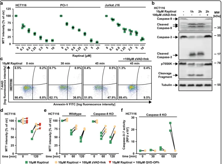
Exposure to Raptinal showed cytotoxic effects in various cancer cell lines and triggered rapid processing of caspase-9 (Fig.1a, b). Together with the observed cleavage of caspase-3 (a substrate of caspase-9) and p70S6K (a substrate of caspase-3), this indicated Raptinal-induced activation of the intrinsic apoptosis pathway14. Likewise, Raptinal-treated HCT116 cells stained positive for annexin-V and were rescued by the pan-caspase inhibitors zVAD-fmk and QVD-OPh (Fig. 1c, d). Deficiency of caspase-8, the initiator caspase of the extrinsic apoptotic pathway, had no protective effect and still allowed Raptinal-induced effector caspase activation (Fig. 1e, f).
Taken together, our results are in agreement with the original description of Raptinal as a rapid inducer of apoptotic cell death via the intrinsic pathway13.
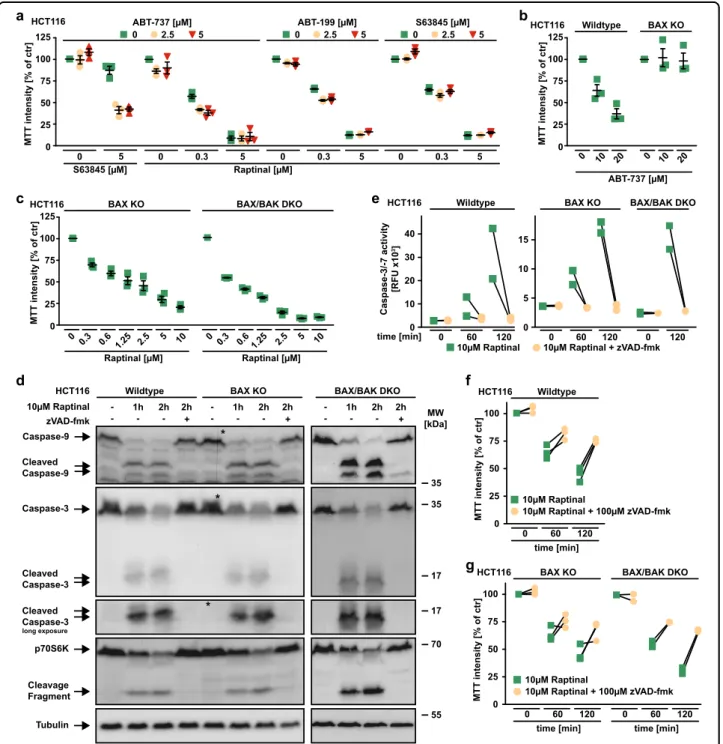
BAX and BAK are dispensable for Raptinal-induced apoptosis
Inhibition of antiapoptotic BCL-2 family proteins facil- itates activation of the pore-forming proteins BAX and/or BAK and thus primes for intrinsic apoptosis15. The BH3 mimetic ABT-737 (targeting BCL-2, BCL-XL, and BCL-W) expectedly primed HCT116 cells for death induced by the MCL-1 inhibitor S63845. Surprisingly, neither S63845 nor ABT-199 (targeting BCL-2) nor ABT-737 acted synergisti- cally with Raptinal in killing HCT116 cells (Fig.2a).
a
75 50 25 0 100
HCT116
0 ytisnetniTTM]rtcfo%[125
0.3 0.6 1.25 2.5 5 10 PCI-1
0 0.3 0.6 1.25 2.5 5 10
Caspase-9
35 Cleaved
Caspase-9
Tubulin 55 Caspase-3
Cleaved Caspase-3
55
17
p70S6K Cleavage Fragment
70 HCT116
10µM Raptinal
100µM zVAD-fmk - - - +
2h 2h
- 1h MW
[kDa]
b
0 30 60 120
Caspase-8 KO ytivitca7-/3-esapsaC [01xUFR]
0 10 20 30
time [min]
HCT116
f
75 50 25 0 tcfo%[ytisnetniTTM]r100
0 60 120 time [min]
Wildtype
0 60 120
Caspase-8 KO HCT116
e d
75 50 25 0 TM]rtcfo%[ytisnetniT100
0 120
time [min]
HCT116
0 0.3 0.6 1.25 2.5 5 10 Jurkat J16
c
0.5% 0.2%
8.8%
90.4%
0.7% 0.9%
36.8%
62.1%
0.4% 0.5%
47.9%
51.8%
1.3% 0.4%
9.5%
89.4%
10µM Raptinal 0 min 30 min 45 min 45 min
+100µM zVAD-fmk
Annexin-V FITC [log fluorescence intensity]
7-AAD [log fluorescence intensity]
Raptinal [µM]
10µM Raptinal + 100µM zVAD-fmk
10µM Raptinal 10µM Raptinal + 100µM QVD-OPh
HCT116
Fig. 1 Raptinal triggers intrinsic apoptosis in various cancer cells. aHCT116, PCI-1 and Jurkat J16 cells were challenged with the indicated concentrations of Raptinal for 18 h. Data points and mean ± SEM from three independent experiments are shown.bHCT116 cells were challenged with Raptinal (10 µM) for the indicated periods of time in the absence and presence of the pan-caspase inhibitor zVAD-fmk (100 µM). After washing and lysis, western blot analyses were performed with antibodies specific for the indicated proteins. Detection of tubulin served as a loading control.
cHCT116 cells were treated as inband subsequently analyzed byflow cytometry for 7-AAD- and annexin-V positivity. Forbandc, data shown are representative of two experiments performed.d,eHCT116 cells and caspase-8-deficient variants thereof were challenged with Raptinal (10 µM) for 60 min or 120 min in the presence and absence of the pan-caspase inhibitors zVAD-fmk (100 µM) or QVD-OPh (100 µM).fHCT116 Caspase-8 KO cells were treated with Raptinal (10 µM) for the indicated periods of time in the presence and absence of zVAD-fmk (100 µM). Caspase-3/-7 activity was assessed using the fluorogenic substrate (DEVD)2-R110. Ford–f, individual data points of at least two independent experiments are shown. RFU, relativefluorescence units
35 HCT116 Wildtype
Caspase-3
Cleaved Caspase-3 Cleaved Caspase-3
long exposure
Tubulin p70S6K
Cleavage Fragment
55 70 17 10µM Raptinal
zVAD-fmk - - - +
2h 2h 1h -
BAX KO - + - -
2h 2h 1h -
17 MW [kDa]
d
*
Cleaved Caspase-9 Caspase-9
35
*
*
ytivitca7-/3-esapsaC [01xUFR]
0 10 20 30 40
0 60 120
10µM Raptinal + zVAD-fmk 10µM Raptinal
HCT116
10 15 Wildtype
0 60 120
5
BAX KO
time [min] 0 120
BAX/BAK DKO
0 Raptinal [µM]
0 0.3 0.6 1.25 2.5 5 10 75
50 25 0 100 ]rtcfo%[ytisnetniTTM125
HCT116 BAX KO
c
75 50 25 0 100 HCT116 ]rtcfo%[ytisnetniTTM125
a
0 0.3 5
5 0 2.5 ABT-737 [µM]
0 0.3 5
S63845 [µM]
5 0 2.5
Raptinal [µM]
0 5
S63845 [µM]
0 0.3 5
ABT-199 [µM]
5
0 2.5
Raptinal [µM]
0 0.3 0.6 1.25 2.5 5 10 BAX/BAK DKO
BAX/BAK DKO - + - -
2h 2h 1h -
0 10 20
0 10 20
ABT-737 [µM]
Wildtype
75 50 25 0 100 cfo%[ytisnetniTTM]rt125
HCT116 BAX KO
b
e
f
75 50 25 0
fo%[ytisnetniTTM]rtc100 10µM Raptinal + 100µM zVAD-fmk
10µM Raptinal HCT116 Wildtype
time [min]
0 60 120
g
75 50 25 0 ]rtcfo%[ytisnetniTTM100
HCT116 BAX KO
time [min]
0 60 120
time [min]
0 60 120
BAX/BAK DKO
10µM Raptinal + 100µM zVAD-fmk 10µM Raptinal
Fig. 2 BAX and BAK are dispensable for Raptinal-induced caspase activation. aHCT116 cells were challenged for 18 h with the indicated concentrations of Raptinal and S63845 in the presence and absence of the indicated concentrations of ABT-199 (targeting BCL-2), ABT-737 (targeting BCL-2, BCL-XL and BCL-W), and S63845 (targeting MCL-1).b,cHCT116 cells and BAX- or BAX/BAK-deficient variants thereof were challenged with the indicated concentrations of ABT-737 and Raptinal for 18 h.dHCT116 cells and BAX- or BAX/BAK-deficient variants thereof were challenged with Raptinal (10 µM) for the indicated periods of time in the absence and presence of the pan-caspase inhibitor zVAD-fmk (100 µM). After washing and lysis, western blot analyses were performed with antibodies specific for the indicated proteins. Detection of tubulin served as a loading control. The asterisk (*) indicates a defect in the CCD sensor of the western blot imaging system. All samples were run on the same gel, no gels were sliced.
eHCT116, HCT116 BAX KO and BAX/BAK DKO cells were treated with Raptinal (10 µM) for the indicated periods of time in the presence and absence of zVAD-fmk (100 µM). Caspase-3/-7 activity was assessed using thefluorogenic substrate (DEVD)2-R110.f,gHCT116, HCT116 BAX KO and BAX/BAK DKO cells were challenged with Raptinal (10 µM) for the indicated periods of time in the presence and absence of zVAD-fmk (100 µM). Fora–cdata points and mean ± SEM from three independent experiments are shown; ford, data shown are representative of two experiments performed; for e–g, individual data points of at least two independent experiments are shown. RFU, relativefluorescence units
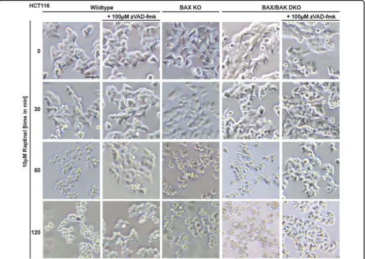
The loss of BAX was sufficient to abrogate cytotoxicity of ABT-737 in HCT116 cells, whereas cytotoxicity of Raptinal was not depending on BAX or BAK (Fig.2b, c). Proteolytic processing and activation of initiator and effector caspases was intact in Raptinal-treated BAX- and/or BAX/BAK- deficient HCT116 cells (Fig. 2d, e). Moreover, Raptinal rapidly caused‘membrane blebbing’, a morphological hall- mark of apoptotic cell death, irrespective of BAX/BAK (Fig. 3). Inhibition of caspase activity using zVAD-fmk expectedly abrogated Raptinal-induced apoptotic mor- phology (Fig. 3), reduced cytotoxicity (Fig. 2f, g) together with annexin-V/7-AAD-positivity in BAX/BAK-deficient and -proficient cells (Fig. 4a). Collectively, these results demonstrate that Raptinal triggers apoptotic cell death in the absence of the pore-forming proteins BAX/BAK.
Caspase-9 propagates Raptinal-induced apoptosis after BAX/BAK-independent cytochrome c release
The loss of BAX/BAK is known to severely impair activation of the mitochondria-controlled apoptotic cas- cade16,17. The efficient Raptinal-induced caspase activation
in BAX/BAK-deficient cells (Fig. 2d, e) could therefore either question an exclusive dependency of Raptinal on intrinsic apoptosis or point to an alternative, BAX/BAK- independent mechanism to initiate this pathway. Indeed, the latter seems the case as even in the absence of BAX/
BAK Raptinal treatment resulted in cytochrome c release from the mitochondria (Fig.4b, c). The loss of caspase-9 did expectedly not affect Raptinal-induced cytochrome c release (Fig. 4d), but conferred almost full-blown protec- tion to Jurkat cells challenged with Raptinal (Fig. 4e).
Importantly, we confirmed Raptinal-induced apoptosis in BAX/BAK-deficient DLD1 and SW48 cells to exclude cell line-specific effects (Fig. 5). In sum, our data support a model of fast Raptinal-induced activation of intrinsic apoptosis through a BAX/BAK-independent mechanism of cytochrome c release and subsequent caspase-9- dependent propagation of the death signal.
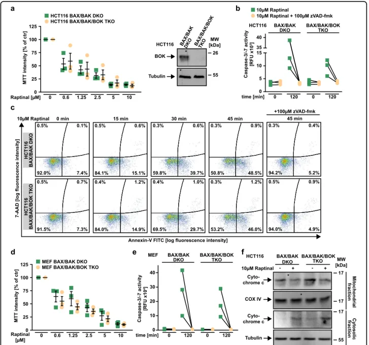
BOK is dispensable for Raptinal-induced MOMP
BOK is another protein capable to form pores in the outer mitochondrial membrane and has been reported to
Fig. 3 BAX/BAK-deficient cells display morphological signs of apoptosis upon Raptinal treatment.HCT116, HCT116 BAX KO, and BAX/BAK DKO cells were treated with Raptinal (10 µM) for the indicated periods of time in the presence and absence of zVAD-fmk (100 µM). Morphological changes were documented by brightfield microscopy. Scale bar: 50 µm. Data shown are representative of two experiments performed
HCT116 Wildtype BAX KO 10µM Raptinal - + - +
b
0.1% 0.0%
1.0%
98.9%
0.1% 0.0%
1.1%
98.8%
0.1% 0.1%
2.6%
97.4%
0.1% 0.0%
3.2%
96.8%
10µM Raptinal 0 min 15 min 30 min 45 min
Annexin-V FITC [log fluorescence intensity]
7-AAD [log fluorescence intensity]
d
10µM Raptinal 0 min 15 min 30 min 45 min 45 min
+100µM zVAD-fmk
a
7-AAD [log fluorescence intensity]
Annexin-V FITC [log fluorescence intensity]
c Jurkat J16 BAX/BAK DKO
- + - +
Mitochondrial fraction Cyto- chrome c
COX IV
17
17
Cytosolic fraction Cyto- chrome c
Tubulin
17
55 Mitochondrial fraction
Cyto- chrome c
COX IV
17
17 17
Cytosolic fraction 55 Cyto- chrome c
Tubulin
Jurkat Caspase-9 KO
- +
17
Cytosolic fraction 55 Cyto- chrome c
Tubulin 10µM Raptinal
10µM Raptinal
e
1.2% 0.4%
0.4%
96.6%
1.2% 0.4%
13.7%
85.2%
1.0% 0.7%
24.2%
74.5%
0.7% 0.5%
36.4%
62.8%
1.1% 1.0%
4.4%
93.9%
0.5% 0.8%
4.4%
94.3%
0.6% 1.4%
36.8%
61.6%
0.2% 2.2%
45.9%
52.4%
0.2% 3.1%
58.7%
38.6%
0.6% 2.9%
12.9%
83.7%
0.1% 0.2%
3.1%
97.0%
0.2% 0.7%
22.4%
77.4%
0.2% 7.0%
57.6%
36.3%
0.1% 13.1%
60.2%
28.6%
0.4% 1.8%
23.5%
75.9%
Jurkat Caspase-9 KO
0.6% 0.0%
2.4%
96.9%
0.1% 0.0%
28.5%
71.8%
0.2% 0.0%
29.9%
70.5%
0.1% 0.3%
60.1%
28.6%
0.2% 0.0%
5.9%
94.0%
Jurkat J16HCT116 BAX KOHCT116 BAX/BAK DKOJurkat BAX/BAK DKOHCT116 BAX KOHCT116 BAX/BAK DKOJurkat BAX/BAK DKO
Fig. 4(See legend on next page.)
induce cytochrome c release in the absence of BAX/
BAK18,19. To clarify whether BOK is involved in Raptinal- induced MOMP, we challenged HCT116 BAX/BAK/BOK triple knock-out (TKO) cells and BAX/BAK/BOK-defi- cient mouse embryonic fibroblasts with Raptinal. In the absence of BAX, BAK, and BOK, Raptinal still exerted cytotoxic effects (Fig. 6a, d), induced caspase-3 and -7 activation (Fig.6b, e), caused annexin-V/7-AAD positivity (Fig. 6c) and triggered cytochrome c release from the mitochondria (Fig. 6f). Collectively, these data argue against a dependency on BOK for Raptinal-induced intrinsic apoptosis.
Raptinal-induced loss of mitochondrial function exerts caspase-independent cytotoxic effects
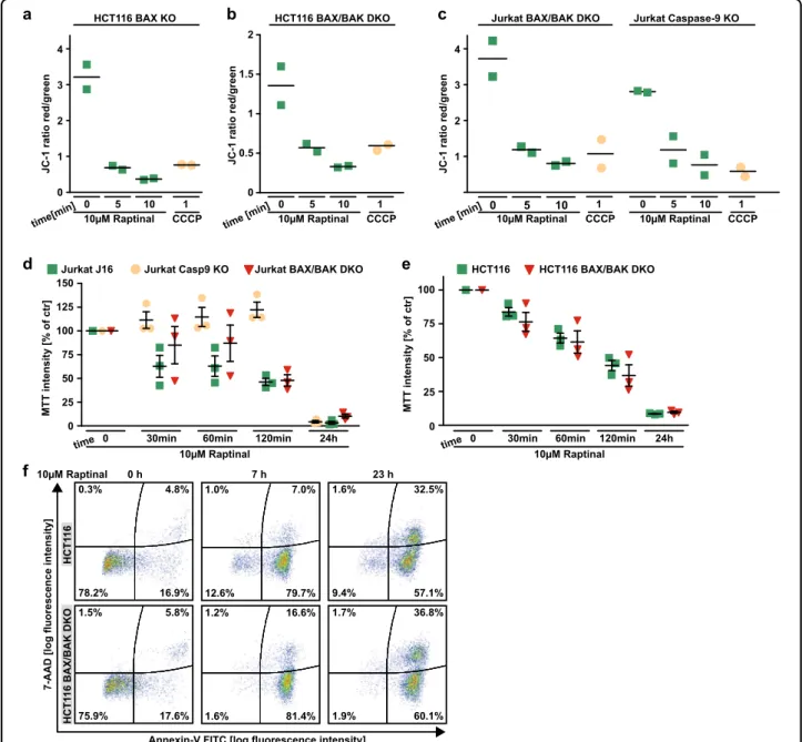
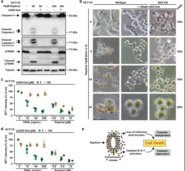
Apparently, Raptinal is capable to unleash the mitochondria-controlled death signal within minutes in a BAX/BAK/BOK-independent manner. Raptinal triggers release of cytochrome c from the mitochondria (Figs.4b, c,5f, and6f) and thereby disrupts the electron transport chain. In line with rapid MOMP induction, 5 min of exposure to Raptinal was sufficient to decrease the mitochondrial membrane potential in BAX-, BAX/BAK- and caspase-9-deficient cells (Fig. 7a–c). Caspase-9- deficient cells showed almost full-blown protection when exposed to Raptinal for up to 2 h (Fig. 7d). Over- night treatment, however, was highly toxic in caspase-9- and BAX/BAK-deficient cells (Fig. 7d–f). Caspase inhibition efficiently blocked Raptinal-triggered caspase-3 activation even after 24 h (Fig. 8a), abrogated apoptotic morphology (such as membrane blebbing) of Raptinal- treated cells (Fig. 8b) and was expectedly sufficient to protect against TRAIL-induced extrinsic apoptosis (Fig. 8c). However, blocking caspase activity in HCT116 cells only partially relieved cytotoxicity of short-term (2 h) Raptinal treatment (Fig.2f, g) and was even less protective upon long-term (24 h) exposure (Fig.8c, d). Thus, MOMP induction and subsequent loss of mitochondrial function additionally exert caspase-independent cytotoxic effects.
Collectively, our data support a dual mode of action for Raptinal to determine a cell’s fate following BAX/BAK/
BOK-independent MOMP (summarized in Fig. 8e): fast activation of the intrinsic apoptotic pathway (caspase-
dependent cell death) and the loss of mitochondrial function (caspase-independent cell death).
Discussion
Cancer cells differ widely in their threshold for activa- tion of the intrinsic apoptotic pathway and consequently display striking differences in their susceptibility to chemotherapy-induced apoptosis. A new class of anti- cancer drugs called ‘BH3 mimetics’ disturbs the sophis- ticated network of BAX/BAK-regulating BCL-2 family proteins and increases the readiness for mitochondrial cytochrome c release. Essentially, BH3 mimetics prime mitochondria for death and reduce the minimally required strength of death-promoting stimuli to unleash mitochondrial apoptosis10. As ‘mitochondria-priming drugs’, BH3 mimetics show limited efficacy as single agents in most cancer entities, but act synergistically with conventional chemotherapies15,20,21. However, the absence of the pore-forming proteins BAX and/or BAK renders cancer cells refractory to both, mitochondrial priming and inducers of intrinsic apoptosis10. From the latter, Raptinal is a notable exception as (1) the mito- chondrial priming of target cells does not enhance its cytotoxic activity (Fig. 2a) and (2) the cytochrome c release and MOMP occur in a BAX/BAK/BOK-indepen- dent manner (Figs. 4b, c, 5f, and 6f). Notably, Raptinal neither directly triggers cytochrome c release in isolated mitochondria nor via mitochondrial permeability transi- tion pore (MPTP) formation in the inner mitochondrial membrane13. Earlier studies already noted that even in the absence of MPTP, BAX and/or BAK are not always required for mitochondrial cytochrome c release22,23. Our data also argue against a decisive role for BOK in Raptinal-induced MOMP (Fig.6). Potentially, another yet unidentified mechanism for MOMP exists, which may involve specific lipids such as ceramide24,25.
Noteworthy, BAX/BAK/BOK-independent MOMP induction could also unlink mitochondrial priming from a cell’s readiness to activate the intrinsic apoptotic pathway.
When MOMP is not executed through BAX/BAK-medi- ated pore-formation in the outer mitochondrial membrane, disturbing BAX/BAK-regulatory BCL-2 family protein interaction (e.g., using BH3 mimetics) may have no effect
(seefigure on previous page)
Fig. 4 Raptinal causes cytochrome c release in the absence of the pore-forming proteins BAX/BAK. aHCT116 BAX KO, HCT116 BAX/BAK DKO, Jurkat J16 and Jurkat BAX/BAK DKO cells were challenged with Raptinal (10 µM) for the indicated periods of time in the presence and absence of the pan-caspase-inhibitor zVAD-fmk (100 µM). 7-AAD- and annexin-V positivity was analyzed byflow cytometry.b–dHCT116, Jurkat J16, Jurkat BAX/BAK DKO and Jurkat caspase-9 KO cells were challenged with Raptinal (10 µM) for 15 min (HCT116) or 30 min (Jurkat cells). After washing and lysis, western blot analyses were performed with whole cell lysates and mitochondria-containing fractions using antibodies specific for the indicated proteins. Detection of tubulin (whole cell lysate) and COX IV (mitochondria-containing fraction) served as loading control.eCaspase-9-deficient Jurkat cells were challenged with Raptinal (10 µM) for the indicated periods of time. 7-AAD- and annexin-V positivity was analyzed byflow cytometry.
Fora–e, data shown are representative of at least two experiments performed
on the apoptotic threshold. Raptinal could therefore be effective in cancer cells with no/low mitochondrial priming, which are considered as difficult-to-treat26. In addition, Raptinal could be unaffected by most mechan- isms that mediate primary or acquired resistance to BH3
mimetics. For example, the latter can loose their mitochondria-priming function when binding to BCL-2 proteins is reduced, expression levels of directly BAX- activating proteins (such as BIM) decrease or nontargeted BCL-2 pro-survival proteins are upregulated27–30. In stark
0.5% 0.3%
3.5%
95.8%
0.3% 0.2%
18.0%
81.6%
0.3% 0.2%
23.4%
76.5%
0.3% 0.6%
3.4%
95.9%
10µM Raptinal 0 min 30 min 45 min 45 min
+100µM zVAD-fmk
7-AAD [log fluorescence intensity] DLD1
1.2% 0.6%
4.8%
93.4%
0.7% 1.0%
9.5%
89.3%
0.6% 1.0%
28.6%
70.8%
0.6% 0.5%
4.8%
94.5%
DLD1 BAX/BAK DKO
60 40 20 0
10µM Raptinal
10µM Raptinal + 100µM zVAD-fmk
60 40 20 0 Caspase-3/-7 activity [RFU x10]
DLD1
DLD1 BAX/BAK DKO
d
1.0% 0.8%
4.0%
94.2%
1.3% 2.3%
24.0%
73.0%
0.9% 4.8%
46.3%
48.7%
1.7% 2.9%
4.1%
91.7%
Annexin-V FITC [log fluorescence intensity]
SW48
0.3% 0.3%
2.6%
96.8%
0.3% 2.0%
36.0%
62.2%
0.2% 2.2%
46.8%
51.4%
0.2% 0.9%
3.3-%
95.7%
SW48 BAX/BAK DKO
0
0
time [min] 0 60 120 SW48
SW48 BAX/BAK DKO
time [min] 0 30 60
time [min] 0 30 60
Caspase-3/-7 activity [RFU x10]Caspase-3/-7 activity [RFU x10]Caspase-3/-7 activity [RFU x10]
time [min] 0 60 120
b
Raptinal [µM]
0 0.3 0.6 1.25 2.5 5 10 75
50 25 0 100 snetni TTM]rtc fo %[ yti125
DLD1
Raptinal [µM]
0 0.3 0.6 1.25 2.5 5 10 DLD1 BAX/BAK DKO
Raptinal [µM]
0 0.3 0.6 1.25 2.5 5 10 75
50 25 0 100 ]rtc fo %[ ytisnetni TTM125
SW48
Raptinal [µM]
0 0.3 0.6 1.25 2.5 5 10 SW48 BAX/BAK DKO
Cytosolic fraction Cyto-
chrome c Tubulin
10µM Raptinal - + - + 17 DLD1 DLD1 BAX/
BAK DKO
55 MW [kDa]
Mitochondrial fraction Cyto-
chrome c COX IV
10µM Raptinal - + - + 17 DLD1 DLD1 BAX/
BAK DKO
17 MW [kDa]
*
10 20 30 4075 100
10 20 30 40 75 100
a
c
f e
Fig. 5(See legend on next page.)
contrast, Raptinal bypasses BAX/BAK (and also BOK) and is self-sufficient for MOMP induction. Reaching this point of no return irrevocably condemns a cell to death:
either via intrinsic apoptosis by caspase-9-dependent activation of downstream effector caspases or loss of mitochondrial function (Fig. 8e). Admittedly, further in vivo studies are needed to estimate the risk for clini- cally unacceptable side-effects of Raptinal. In combina- tion with novel drug delivery concepts (e.g., conjugation to target-directing antibodies), the tremendous death- inducing potential could perspectively be therapeutically exploitable.
In sum, we show that Raptinal bypasses coordination/
initiation of MOMP by pore-forming BCL-2 family pro- teins4. Raptinal exerts a dual cytotoxic effect by rapid activation of the intrinsic apoptotic pathway and simul- taneous shutdown of mitochondrial function.
Material and methods
Cell lines, antibodies, and reagents
HCT116 cells were obtained from the German Collec- tion of Microorganisms and Cell Culture (DSMZ, Braunschweig, Germany). HCT116 BAX/BAK DKO, BAX KO, and caspase-8 KO cells were kindly provided by Richard Youle (National Institutes of Health, Bethesda, USA), Bert Vogelstein (John Hopkins University, Balti- more, MA, USA) and Hamsa Puthalakath (La Trobe University, Bundoora, Australia), respectively31–33. BAX/
BAK/BOK-deficient HCT116 cells and MEFs were kindly provided by Thomas Kaufmann (Institute of Pharmacol- ogy, University of Bern, Bern, Switzerland). SW48 and DLD1 cells and BAX/BAK-deficient variants thereof were purchased from Sigma (Steinheim, Germany). PCI-1 cells were a gift from Richard Bauer (University of Regensburg, Germany). Jurkat J16 cells and caspase-9- or BAX/BAK- deficient variants thereof have been described before34. All cell lines were maintained in RPMI 1640 medium (PAN Biotech, Aidenbach, Germany) with 10% (v/v) fetal calf serum (Sigma). Medium of Jurkat cells was supple- mented with 100 U penicillin/mL and 0.1 mg streptomy- cin/mL (PAN Biotech). Antibodies: caspase-3 (#9662),
caspase-9 (#9502), COX IV (#4844), p70S6k (#2708): Cell Signaling (Beverly, MA, USA); tubulin (#MS-581):
Dunnlab (Asbach, Germany); cytochrome c (ab13575):
abcam (Cambridge, UK). Monoclonal rabbit anti-BOK (BOK-1-5) was a kind gift from Thomas Kaufmann (University of Bern, Bern, Switzerland)35. Chemicals:
Raptinal and MTT (3-[4,5-dimethylthiazol-2-yl]−2,5- diphenyl tetrazolium bromide): Biomol, (Hamburg, Ger- many); zVAD-fmk (carbobenzoxy-valyl-alanyl-aspartyl- (Omethyl)-fluoromethylketone): Bachem, (Bubendorf, Switzerland); zLEHD-fmk: BD Biosciences (Heidelberg, Germany); ABT-199, ABT-737, S63845 and QVD-OPh:
Hycultec (Beutelsbach, Germany); TRAIL: Apronex (Jesenice u Prahy, Czech Republic); cOmplete protease inhibitor cocktail: Roche (Mannheim, Germany).
MTT-based cell viability assay
Cells were seeded in 96-well plates (Jurkat: 2 × 10⁵cells/
well; all other cell lines: 2 × 10⁴cells/well) and challenged with the indicated concentrations of the indicated sub- stances in duplicates (technical replicates). Unless indi- cated otherwise, cell viability was determined 18 h after stimulation using MTT staining (2 h at 37 °C). Staining intensity was measured at 595 nm and the mean was calculated from the technical replicates of each experi- ment. The mean value for untreated controls was set to 100%. For any other condition, the MTT staining intensity is given relative to the corresponding untreated group (% of control). Data points shown are mean values (cal- culated from 2 technical replicates) of independent experiments (n≥2–3).
Western blot analysis
Cells were harvested by centrifugation and lysed in 4×
Laemmli sample buffer (8% (w/v) SDS, 0.1 M dithiothreitol, 40% (v/v) glycerol, 0.2 M Tris, pH 8.0) supplemented with phosphatase inhibitor cocktails-I and -II (Sigma). Samples were sonicated and boiled for 5 min at 96 °C before proteins were separated by SDS-PAGE and transferred to PVDF membranes. To block nonspecific binding sites, membranes
(seefigure on previous page)
Fig. 5 Raptinal induces intrinsic apoptosis in a variety of BAX/BAK-deficient cell lines. a,bDLD1 and SW48 cells and BAX/BAK-deficient variants thereof were challenged with the indicated concentrations of Raptinal for 18 h.cCells were treated with Raptinal (10 µM) for the indicated periods of time in the presence and absence of the pan-caspase-inhibitor zVAD-fmk (100 µM). 7-AAD- and annexin-V positivity was analyzed byflow cytometry.
dDLD1 and SW48 cells and BAX/BAK-deficient variants thereof were challenged with Raptinal (10 µM) in the presence and absence of the pan- caspase inhibitor zVAD-fmk (100 µM). Caspase-3/-7 activity was assessed using thefluorogenic substrate (DEVD)2-R110.e,fCells were challenged with Raptinal (10 µM) for 60 min. After washing and lysis, western blot analyses were performed with whole cell lysates and mitochondria-containing fractions using antibodies specific for the indicated proteins. Detection of tubulin (whole cell lysate) and COX IV (mitochondria-containing fraction) served as loading control. The asterisk (*) indicates a defect in the CCD sensor of the western blot imaging system. All samples were run on the same gel, no gels were sliced. Foraandb, data points and mean ± SEM from three independent experiments are shown. Forc,e, andf, data shown are representative of at least two experiments performed.dshows individual data points of at least two independent experiments. RFU, relative fluorescence units
were incubated in TBS containing 0.1% (v/v) Tween 20 and 5% (w/v) dry milk before primary antibodies of the speci- ficity of interest were added. Antigen-antibody complexes
were visualized using horseradish peroxidase-conjugated secondary antibodies (Dako, Hamburg, Germany) and ECL technology (Pierce, Rockford, IL, USA).
c
10µM Raptinal
Annexin-V FITC [log fluorescence intensity]
7-AAD [log fluorescence intensity] HCT116 BAX/BAK DKOHCT116 BAX/BAK/BOK TKO
0.5% 0.1%
7.4%
92.0%
0 min
0.5% 0.7%
7.3%
91.5%
0.5% 0.6%
15.1%
84.1%
15 min
0.4% 1.2%
14.9%
84.0%
0.3% 0.6%
39.7%
59.8%
30 min
0.4% 1.0%
29.7%
69.5%
0.3% 0.9%
48.5%
50.8%
45 min
0.3% 1.2%
46.0%
53.2%
0.3% 0.4%
5.2%
94.2%
45 min
0.5% 0.9%
4.9%
94.0%
+100µM zVAD-fmk
a b
d e
ytivitca7-/3-esapsaC [01xUFR]
time [min]
0 5 10 15 35 40
0 120 0 120
HCT116 BAX/BAK DKO
BAX/BAK/BOK TKO 10µM Raptinal + 100µM zVAD-fmk 10µM Raptinal
ytivitca7-/3-esapsaC [01xUFR]
0 10 20 30 40
time [min] 0 120 0 120
MEF BAX/BAK DKO
BAX/BAK/BOK TKO 75
50 25 100 125
]rtcfo%[ytisnetniTTM
0
0 0.6 1.25 2.5 5 10
Raptinal [µM]
HCT116 BAX/BAK/BOK TKO HCT116 BAX/BAK DKO
75 50 25 100 125
]rtcfo%[ytisnetniTTM
0
0 0.6 1.25 2.5 5 10
Raptinal [µM]
MEF BAX/BAK/BOK TKO MEF BAX/BAK DKO
f
10µM Raptinal - + - + BAX/BAK
DKO
BAX/BAK/BOK
TKO MW
[kDa]
Cytosolicfraction
Cyto- chrome c
Tubulin
17
55 HCT116
Mitochondrialfraction
Cyto- chrome c
COX IV
17
17
*
*
Tubulin * 55
BOK * 26
HCT116 BAX/BAKDKO BAX/BAK/BOKTKO MW [kDa]
Fig. 6 BOK is dispensable for Raptinal-induced MOMP. aLeft panel: HCT116 BAX/BAK DKO and BAX/BAK/BOK TKO cells were challenged with the indicated concentrations of Raptinal for 18 h. Right panel: BOK levels were analyzed in lysates generated from HCT116 BAX/BAK DKO and BAX/BAK/
BOK TKO cells by western blotting. The asterisk (*) indicates a defect in the CCD sensor of the western blot imaging system. All samples were run on the same gel, no gels were sliced.bHCT116 BAX/BAK DKO and BAX/BAK/BOK TKO cells were challenged with Raptinal (10 µM) for 120 min. Caspase- 3/-7 activity was assessed using thefluorogenic substrate (DEVD)2-R110.cCells were treated with Raptinal (10 µM) for the indicated periods of time in the presence and absence of the pan-caspase-inhibitor zVAD-fmk (100 µM). 7-AAD- and annexin-V positivity was analyzed byflow cytometry.dBAX/
BAK- and BAX/BAK/BOK-deficient mouse embryonicfibroblasts (MEFs) were challenged with the indicated concentrations of Raptinal for 18 h.eBAX/
BAK- and BAX/BAK/BOK-deficient MEFs were challenged with Raptinal (10 µM) for 120 min. Caspase-3/-7 activity was assessed using thefluorogenic substrate (DEVD)2-R110.fHCT116 BAX/BAK DKO and BAX/BAK/BOK TKO cells were challenged with Raptinal (10 µM) for 30 min. After washing and lysis, western blot analyses were performed with whole cell lysates and mitochondria-containing fractions using antibodies specific for the indicated proteins. Detection of tubulin (whole cell lysate) and COX IV (mitochondria-containing fraction) served as loading control. Foraandd, data points and mean ± SEM from three independent experiments are shown.b,eshow individual data points of three independent experiments. Forcand f, data shown are representative of at least two experiments performed
Cytochrome c release by immunoblot
Cytochrome c release by immunoblot was performed essentially as described previously13. In brief, 3 × 10⁶cells were treated with Raptinal for the indicated periods of time. Cells were harvested, centrifuged (1000 ×g, 2 min), washed with ice-cold PBS, resuspended in 200μL ice- cold digitonin permeabilization buffer (75 mM NaCl,
1 mM sodium phosphate monobasic, 8 mM sodium phosphate dibasic, 250 mM sucrose, 190μg/mL digito- nin, protease cocktail inhibitor, pH 7.5) and incubated on ice for 5 min. Following centrifugation (14,000 ×g, 5 min), 150μL of the supernatant (cytosolic fraction) was collected. The pellet (mitochondrial fraction) was washed in 200μL digitonin permeabilization buffer and lysed in a
d
75 50 25 0 100 ]rtcfo%[ytisnetniTTM 125
150
10µM Raptinal
0 30min 60min 120min 24h
time
Jurkat J16 Jurkat Casp9 KO Jurkat BAX/BAK DKO
0.3% 4.8%
16.9%
78.2%
1.0% 7.0%
79.7%
12.6%
1.6% 32.5%
57.1%
9.4%
10µM Raptinal 0 h 7 h 23 h
Annexin-V FITC [log fluorescence intensity]
7-AAD [log fluorescence intensity]
1.5% 5.8%
17.6%
75.9%
1.2% 16.6%
81.4%
1.6%
1.7% 36.8%
60.1%
1.9%
HCT116HCT116 BAX/BAK DKO JC-1 ratio red/green
0 0.5 1 1.5 2
CCCP 10µM Raptinal time [min]
0 5 10 1
HCT116 BAX/BAK DKO
CCCP 10µM Raptinal
HCT116 BAX KO
time[min]
JC-1 ratio red/green
0 1 2 3 4
0 5 10 1
75 50 25 0 ]rtcfo%[ytisnetniTTM 100
10µM Raptinal
0 30min 60min 120min 24h time
HCT116 HCT116 BAX/BAK DKO
Jurkat Caspase-9 KO
10µM Raptinal
0 5 10
10µM Raptinal
0 5 10
Jurkat BAX/BAK DKO
JC-1 ratio red/green
1 2 3 4
time [min] CCCP
1
CCCP 1
b c
e
f
Fig 7 Raptinal impairs mitochondrial function. a–cHCT116 BAX KO, HCT116 BAX/BAK DKO, Jurkat BAX/BAK DKO, and Jurkat caspase-9 KO were treated with Raptinal (10 µM) for the indicated periods of time. Membrane potential of mitochondria was assessed byflow cytometry after staining with JC-1. CCCP treatment served as a positive control. Individual data points together with mean of at least two independent experiments are shown.dJurkat J16, caspase-9 KO, and BAX/BAK DKO were challenged with Raptinal (10 µM) for the indicated periods of time. Cell viability was assessed by MTT staining.eHCT116 cells and BAX/BAK-deficient variants thereof were challenged with Raptinal (10 µM) for the indicated periods of time. Cell viability was assessed by MTT staining. Fordande, data points and mean ± SEM from three independent experiments are shown.fHCT116 and HCT116 BAX/BAK DKO cells were treated with Raptinal (10 µM) for the indicated periods of time. 7-AAD- and annexin-V positivity was analyzed byflow cytometry. Data shown are representative of two experiments performed
25μL RIPA lysis buffer (150 mM NaCl, 25 mM Tris, 1%
(v/v) Nonidet P-40, 1% (w/v) sodium deoxycholate, 0.1%
(w/v) SDS, pH 7.5). Forty micrograms of the cytosolic fraction and 50μg of the mitochondrial fraction were
resolved by SDS-PAGE. The cytosolic fraction was pro- bed for tubulin, and the mitochondrial fraction was probed for cytochrome c oxidase subunit IV (COX IV) to confirm equal loading.
17 kDa HCT116
55 kDa Tubulin
p70S6K
Cleaved p70S6K Cleaved Caspase-3 long exposure
73 kDa Caspase-3
Cleaved
Caspase-3 17 kDa
35 kDa 10µM Raptinal
zVAD-fmk - - +
- 8h 8h
- 24h 24h
- + -
a bHCT116 Wildtype
+ 100µM zVAD-fmk
Raptinal 10µM [time in h]
0
8
24
24
BAX KO
magn.
100x
100x
200x
c 100x
75 50 25 0 100 HCT116
]rtcfo%[ytisnetniTTM 125
0 100 zVAD-fmk [µM]
0 16 64 256 0.6 2.5 10
TRAIL [ng/mL] Raptinal [µM]
d
Raptinal
Cytochrome c release
Caspase-9/-3/-7 activation Loss of mitochon-
drial function
Caspase- independent
75 50 25 0 100 HCT116
]rtcfo%[ytisnetniTTM 125
0 16 64 256
0 100 zLEHD-fmk [µM]
0.6 2.5 10 TRAIL [ng/mL] Raptinal [µM]
e
Caspase- dependent
Cell Death
Fig. 8 The loss of mitochondrial function complements caspase-dependent cytotoxicity of Raptinal. aHCT116 cells were challenged with Raptinal (10 µM) for the indicated periods of time in the absence and presence of the pan-caspase inhibitor zVAD-fmk (100 µM). After washing and lysis, western blot analyses were performed with antibodies specific for the indicated proteins. Detection of tubulin served as a loading control.
bHCT116 and HCT116 BAX KO cells were challenged with Raptinal (10 µM) for the indicated periods of time in the presence and absence of zVAD- fmk (100 µM). Morphological changes were documented by brightfield microscopy. Scale bar: 50 µm. HCT116 cells were challenged with the indicated concentrations of KillerTRAIL and Raptinal in the presence and absence ofcthe pan-caspase inhibitor zVAD-fmk (100 µM) ordthe caspase- 9-specific inhibitor zLEHD-fmk (100 µM) for 18 h.eProposed model of Raptinal-induced cell death: Raptinal rapidly triggers cytochrome c release from the mitochondria in a BAX-, BAK- and BOK-independent manner. This determines a cell’s fate in two interdependent ways by (1) fast activation of caspases and subsequent apoptotic cell death or (2) the loss of mitochondrial function and caspase-independent cell death. Foraandb, data shown are representative of two experiments performed. Forcandd, data points and mean ± SEM from three independent experiments are shown