I
Biological Engineering and Characterization of an HIV-1 Envelope-based Genomic Library
D ISSERTATION ZUR E RLANGUNG
DES D OKTORGRADES DER N ATURWISSENSCHAFTEN (D R . RER . NAT .)
DER F AKULTÄT FÜR
B IOLOGIE UND V ORKLINISCHE M EDIZIN DER U NIVERSITÄT R EGENSBURG
Vorgelegt von
Julia Koop
aus
Karaganda, Kasachstan
im Jahr
2018
II
Biological Engineering and Characterization of an HIV-1 Envelope-based Genomic Library
D ISSERTATION ZUR E RLANGUNG
DES D OKTORGRADES DER N ATURWISSENSCHAFTEN (D R . RER . NAT .)
DER F AKULTÄT FÜR
B IOLOGIE UND V ORKLINISCHE M EDIZIN DER U NIVERSITÄT R EGENSBURG
Vorgelegt von
Julia Koop
aus
Karaganda, Kasachstan
im Jahr
2018
III Das Promotionsgesuch wurde eingereicht am:
26.02.2018
Die Arbeit wurde angeleitet von:
Prof. Dr. Ralf Wagner
Julia Koop
IV
Meinen Eltern
V Abstract ... XI
1 Introduction ... 1
1.1 Epidemiology of HIV ... 1
1.2 Origin and phylogeny of HIV ... 2
1.3 Genomic organization and structural biology ... 3
1.4 The HIV-1 life cycle ... 5
1.5 The envelope glycoprotein ... 6
1.5.1 Env synthesis and trafficking ... 6
1.5.2 Env structure... 7
1.5.3 Immune evasion mechanisms of Env ... 8
1.6 Humoral immune response to HIV infection ... 10
1.6.1 Ontogeny of the antibody response during HIV infection ... 10
1.6.2 Broadly neutralizing antibodies ... 11
1.7 HIV-1 vaccine development ... 12
1.8 Elicitation of broadly neutralizing antibodies ... 13
1.8.1 Engineering of envelope immunogens to induce cross-neutralizing antibody responses ... 13
1.8.2 Advantages of Env-based gene variant libraries ... 14
2 Objective ... 16
3 Materials and Methods ... 17
3.1 Molecular Biology ... 17
3.2 Next Generation Sequencing ... 18
3.2.1 Illumina Sequencing by Synthesis Technology ... 18
3.2.2 Sequencing library preparation ... 20
3.2.2.1 Generation of amplicon libraries ... 20
3.2.2.2 Extraction of genomic DNA ... 21
3.2.2.3 Generation of stable cell line samples for NGS ... 22
3.2.3 Purification of amplicon libraries ... 23
3.2.3.1 Agarose gel electrophoresis ... 23
3.2.3.2 Magnetic beads purification ... 24
3.2.4 Quantitation of amplicon libraries ... 24
3.2.4.1 Quantitation with the Agilent 2100 Bioanalyzer... 24
3.2.4.2 Generation of library pools and quantification by quantitative PCR ... 25
3.2.5 Denaturation and dilution of NGS libraries ... 26
VI
3.3.1 Cultivation of cell lines ... 27
3.3.2 Transient transfection of mammalian cells ... 28
3.3.2.1 Cationic-polymer-mediated transfection ... 28
3.3.2.2 Determination of optimal ratios between DNA and various transfection reagents ... 29
3.3.3 Expression of antibodies ... 30
3.3.4 Generation of stable cell lines ... 31
3.3.4.1 Cryopreservation and storage of stable cell lines ... 32
3.3.4.2 Thawing of stable cell lines ... 32
3.3.5 Flow cytometry of mammalian cells ... 32
3.3.6 Cell sorting ... 33
3.4 Protein Biochemistry ... 34
3.4.1 Purification of the HIV-1 specific human bnAbs VRC01 ... 34
3.4.2 Labeling of antibodies ... 35
3.4.3 SDS-PAGE ... 36
3.4.4 Envelope ELISA ... 36
4 Results ... 38
4.1 Overview of the sequential permutation library (SeqPer) ... 38
4.2 Generation of the stable cell line SeqPer library... 39
4.3 Quality control of the SeqPer library ... 41
4.3.1 Quality of plasmid DNA ... 41
4.3.1.1 Purity of plasmid DNA ... 41
4.3.1.2 Restriction enzyme assay ... 41
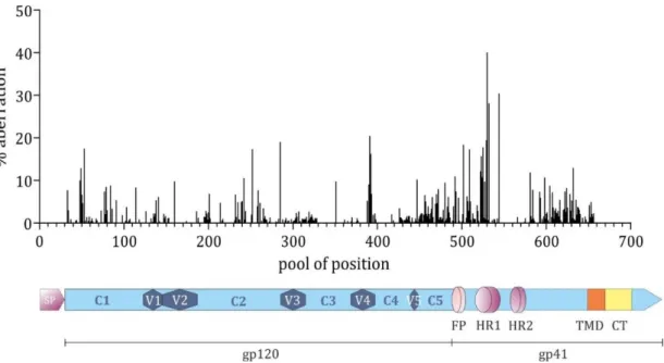
4.3.1.3 Densitometric analysis of aberrational phenotypes ... 43
4.3.1.4 Characterization of selected aberrational phenotypes ... 43
4.3.2 Establishment and validation of the Next Generation Sequencing library sample procedure ... 45
4.3.2.1 Determination of PCR conditions for NGS library preparation ... 45
4.3.2.2 Adjustment of the purification of amplicons ... 47
4.3.2.3 NGS background determination ... 48
4.3.3 Determination of the diversity of the SeqPer library on the example of the CD4 binding site ... 49
4.3.3.1 Diversity of the CD4 binding site on the level of plasmid DNA ... 50
4.3.3.2 Diversity of the CD4 binding site on the level of stable cell lines ... 52
4.3.3.3 Quantitative representation of amino acid diversity ... 54
VII
4.3.4.2 Impact of the number of integration events ... 58
4.3.4.3 Improvement of the transfection efficiency ... 60
4.4 High-throughput screening of a stable cell line library to identify improved HIV-1 antigen candidates ... 62
4.4.1 Overview of the mammalian cell-display-based screening technology ... 63
4.4.2 Purification and validation of the bnAb VRC01... 64
4.4.3 Identification of Env variants with increased or decreased binding affinity for the bnAb VRC01 ... 65
4.4.4 Optimization of the gating strategy ... 67
4.4.5 Validation of the detected GoB and LoB variants ... 69
5 Discussion ... 71
5.1 Evaluation of the SeqPer library ... 71
5.1.1 Advantages of the sequential permutation library ... 71
5.1.2 Quality of the pDNA and the possible implications ... 72
5.1.3 Impact of stable cell line quality on cell-display-based screening ... 72
5.2 Improvement of stable cell line generation ... 73
5.2.1 Identification of factors influencing SCL generation ... 73
5.2.2 Possible optimization approaches ... 75
5.3 Adaptation of NGS sample preparation for library applications ... 76
5.4 Analysis of a mammalian cell-display-based screening technology ... 78
5.4.1 Advantages of the mammalian cell-display technique ... 78
5.4.2 Evaluation of the screening technology ... 78
5.4.3 Structural analysis of envelope interactions with the bnAb VRC01 ... 80
6 Summary and conclusions ... 82
7 Perspective... 83
8 Appendix ... 84
8.1 List of Abbreviations ... 84
8.2 DNA constructs ... 85
8.2.1 Oligonucleotides ... 85
8.2.2 Plasmids ... 88
8.2.3 Cloning Constructs ... 89
8.3 Supplemental Material ... 90
8.4 References ... 109
Acknowledgements ... 119
VIII
IX
Zusammenfassung
Die zahlreichen immunologischen Ausweichstrategien, welche im HIV-1 envelope (Env) Glykoprotein verkörpert sind, stellen für die Entwicklung eines sicheren und effektiven Vakzins weiterhin ein enormes Hindernis dar. Zu den herausfordernsten Ausweichmechanismen zählt die unermessliche genetische Diversität, welche mit der Immundominanz hoch variabler Regionen von Env assoziiert wird. Die derzeitigen Impfstoffansätze zielen darauf hin, breitneutralisierende Antikörper (bnAK) hervorzurufen, von denen einige in der Lage sind mehr als 90% der kursierenden HIV-1 Stämme zu neutralisieren. Allerdings wird die Entwicklung von bnAK aufgrund der komplexen Koevolution von Virus und humoraler Immunantwort erheblich beeinträchtigt.
Daher bedarf es neuartiger Env Immunogene sowie innovativer Selektionstechnologien für deren Identifikation, um diesen Prozess zu begünstigen.
Der erste Abschnitt dieser Dissertation beschäftigte sich intensiv mit der biologischen Prozesstechnik einer auf Env basierten sequentiellen Permutationsbibliothek, mit besonderem Schwerpunkt auf Charakterisierung und Qualitätskontrolle der Bibliothek.
Jede Position des außenliegenden Env-Bereiches wurde durch 20 natürliche Aminosäuren ersetzt, wodurch eine Bibliothek bestehend aus 658 Unterbibliotheken und schätzungsweise 13.000 Varianten hervorgeht. Gleichzeitig wurden die jeweiligen stabilen Zelllinien durch stabile Transfektion jeder Unterbibliothek in Flp-In
TMT-Rex 293 Zellen hergestellt. Das Ziel bestand darin, diese Bibliothek einer Selektionstechnologie beruhend auf einer Zellsortierung zu unterziehen, um Env Varianten mit verbesserter Antigenität zu identifizieren. Sowohl die Plasmid-DNA- (pDNA), als auch die Zelllinien-Bibliothek wurden umfassend auf ihre Qualität kontrolliert. Die eingehende Analyse der pDNA offenbarte Deletionen verschiedenster Länge hauptsächlich in der Env-Region, welche etwa 48% der Bibliothek betreffen. Allerdings traten diese Deletionen in einem kleinen Bruchteil innerhalb der Unterbibliotheken auf, womit die tatsächliche Kontamination jeweils nur zwischen 6-18% lag. Mit dem Schwerpunkt auf der CD4-Bindestelle von Env, wurden Diversität und Verteilung der Aminosäuren der pDNA, sowie der stabilen Zelllinien mittels Next Generation Sequencing (NGS) ermittelt. Während die pDNA eine durchschnittliche Variabilität von 19 Aminosäuren und eine nahezu ideale Verteilung aufwies, zeigten die stabilen Zelllinien sowohl einen etwa 38%-igen Rückgang in der Diversität, als auch eine beträchtliche und zufallsbedingte Ungleichverteilung der Aminosäuren. Es wurde ersichtlich, dass die unzureichende Integration von Env bei der Herstellung der stabilen Zelllinien maßgeblich zu diesem Variabilitätsverlust beitrugen. Dementsprechend wurden einige vielversprechende Ansätze zur Optimierung der Herstellung stabiler Zelllinien eingeleitet, mit dem Ziel eine bessere Diversität und Aminosäureverteilung zu erlangen.
Das zweite Projekt beruhte auf der Identifikation von verbesserten Env-Kandidaten mit
vorteilhaftem Antigenitätsprofil. Zu diesem Zweck wurde eine Selektionstechnlogie
angewendet, die auf einer Zellsortierung beruht und folgende Vorteile in sich vereinigt: i)
Integration einer einzigen Env Variante in eine definierte FRT-Stelle pro Zelle, was eine
Kopplung zwischen Geno- und Phänotyp zur Folge hat, ii) induzierbare Env Expression, um
X Zytotoxizitätseffekten vorzubeugen, iii) translationale Verknüpfung von GFP und Env zur indirekten Normalisierung auf die induzierte Env Expression und iv) Expression der Env auf Hek293T Zellen, um native Faltung und Säugetierglykosylierung zu gewährleisten. In einem einzelnen Selektionszyklus wurden jeweils 12 Env Varianten mit erhöhter oder verminderter Affinität für den bnAK VRC01 aus der Zelllinien-Bibliothek angereichert.
Auffallend dabei war, dass keine der Varianten mit erhöhter, und nur drei Varianten mit
erniedrigter Bindungsfähigkeit mittels FACS-basierter Gleichgewichtstitration eindeutig
validiert werden konnten. Da die Selektionstechnologie zuvor an einer Bibliothek getestet
wurde, welche nur fünf Varianten umfasst, lag es der Vermutung nahe, dass die Methoden
für komplexere und größere Bibliotheken weiter ausgebaut und adaptiert werden müssen.
XI
Abstract
The numerous immune evasion strategies embodied in the HIV-1 envelope (Env) glycoprotein still represent a daunting challenge in the development of a safe and effective vaccine. Among the most defying of these evasive mechanisms is the tremendous genetic diversity associated with the immunodominance of highly variable regions of Env. Current vaccine design efforts aim to elicit broadly neutralizing antibodies (bnAb), some of which are able to neutralize more than 90% of circulating HIV-1 strains. However, a complex co- evolution of virus and humoral immune response considerably impairs the development of bnAbs. To facilitate this process, novel Env immunogens as well as innovative selection technologies for their identification are required.
The first part of this thesis concentrated on the biological engineering of an envelope-based sequential permutation library, specifically focusing on characterization and quality control of the library. Each residue in the external part of Env was substituted by 20 natural amino acids, thus creating a library of 658 sublibraries and approximately 13.000 variants.
Simultaneously, the respective stable cell lines (SCL) were generated by stably transfecting every sublibrary into Flp-In
TMT-Rex 293 cells with the goal to utilize the stable cell line library in a mammalian cell display- and cell sorting-based screening technology to identify Env variants with improved antigenicity. Comprehensive quality controls of plasmid DNA library and the respective stable cell line library were conducted to assess potential limitations. In-depth analysis of the pDNA revealed deletions of various lengths mainly in the Env region affecting about 48% of the library. However, these deletions occurred only in a small fraction within the sublibraries, thus the actual contaminations amounted to 6- 18%, respectively, deeming the library still eligible to work on. Focusing on the CD4 binding site (CD4bs) of Env, diversity and amino acid distribution of the pDNA- and the stable cell line-library was analyzed by Next Generation Sequencing (NGS). Whereas pDNA exhibited an average diversity of 19 amino acids in the sublibraries with a nearly ideal distribution, stable cell lines demonstrated a considerable decrease in diversity by approximately 38%, as well as a highly uneven and random distribution of amino acids. It became apparent that particularly insufficient integration of Env during the generation of stable cell lines contributed to this substantial loss of diversity. Accordingly, several promising approaches were tested to optimize the stable cell line generation aimed to improve the diversity and amino acid distribution.
The second project focused on the identification of improved Env candidates with favorable antigenicity from the stable cell line library. For this purpose, a mammalian cell display- and cell sorting-based technology was applied that combines the benefits of i) single integration of Env into a distinct FRT site resulting in the linkage of genotype and phenotype, ii) inducible Env expression to prevent cytotoxicity effects, iii) translational coupling of Env and GFP enabling an indirect normalization for induced Env expression and iv) display on Hek293T cells, thus ensuring native folding and mammalian glycosylation.
Using the CD4bs SCL library, twelve Env variants demonstrating increased (gain of binding,
GoB) and decreased (loss of binding, LoB) affinity for the bnAb VRC01, respectively, were
XII
selected in a single round of cell sorting procedure. Strikingly, none of the detected GoB
variants and merely three LoB candidates could be unequivocally validated by means of a
FACS-based equilibrium titration. As the selection technology was previously tested on a
five-variant library, there were grounds for supposition, that the methods require further
development and adaptation to be utilized for more complex and extensive libraries.
1
1 Introduction
1.1 Epidemiology of HIV
In June 1981, the U.S. Centers for Disease Control and Prevention (CDC) released a report describing cases of a rare lung infection called Pneumocystis carinii pneumonia (PCP) in five young, previously healthy gay men in Los Angeles
1. Concurrently, an increased incidence of an unusually aggressive cancer known as Kaposi’s Sarcoma was recognized in New York and California
2. At this point in time, no one established a connection between the two obviously different diseases. It was only two years later that scientists discovered a common thread of impaired cellular immunity that linked these malignancies and other opportunistic infections
3,4. Eventually, the human immunodeficiency virus (HIV) was identified as causative agent of the substantially increasing cases of severe immune deficiency worldwide. Due to symptoms and progression of an HIV infection, the term acquired immune deficiency syndrome (AIDS) was established by the CDC in 1982. Since then the virus spread globally, causing one of the most debilitating pandemics ever recorded in human history.
Approximately 80% of HIV infections occur during sexual intercourse with an infected partner through direct contact with semen and rectal or vaginal fluids
5. Blood-to-blood transmissions such as through sharing of needles or contaminated blood transfusions
6, as well as mother-to-child transmissions during pregnancy, childbirth or breastfeeding
7
, represent another 20% of all contracted HIV infections.
Natural progression of HIV infection encompasses three stages: an acute phase, followed by an early/clinically latent phase, and finally by the immune collapse/AIDS. The acute or primary phase lasts several months and is characterized by high level viral replication that is reflected in substantial concentrations of virus in plasma and lymphoid tissue.
After initial viral decline, concurrent with the appearance of virus-specific CD8
+cytotoxic T cells
8, the plasma viral load usually stabilizes at a steady state. This so-called
‘set-point’ is the consequence from the equilibrium between the HIV-1 replication and the corresponding immune responses and represents the beginning of the second stage, a long clinical latency. Ultimately, the regenerative CD4
+T cell population slowly diminishes below a crucial threshold rendering the immune system vulnerable to opportunistic infections, thus causing progression to AIDS.
According to the World Health Organization (WHO) more than 70 million people have
contracted HIV and an estimated 35 million people have died from AIDS-related illness
since the beginning of the pandemic. As of 2015, approximately 36.7 million [34.0–39.8
million] individuals were living with HIV, representing 0.8 % [0.7–0.9 %] of adults aged
15-49 years worldwide (figure 1). Although the prevalence of HIV varies considerably
among countries, 70 % of all accounted infections arise in Africa, specifically in the Sub-
Saharan regions. While there is currently no cure for HIV, the infection can be
2 suppressed by a combination of antiretroviral drugs, thus substantially reducing morbidity and mortality. At the moment, approximately 18.2 million [16.1-19.0 million]
HIV patients are receiving antiretroviral agents, termed combination antiretroviral treatment (cART). However, the low treatment rate, in addition to severe side effects from the medicaments, drug interactions and resistance demonstrate the importance of discovering a vaccine to finally conquer HIV infections globally.
Figure 1 - Global prevalence of HIV in 2016. The illustration shows that an estimated 0.8% [0.7-0.9%]
of adults aged 15-49 years worldwide are infected with HIV. Areas that are most severely affected, such as Sub-Saharan Africa, are indicated in dark red. Figure was adapted from WHO Global Health Observatory (GHO) data HIV/AIDS.
1.2 Origin and phylogeny of HIV
HIV appears to have its origin in the simian immunodeficiency virus (SIV) that infects
non-humate primates in West and Central Africa. Zoonotic transmission presumably
occurred as a consequence of hunting and butchering of primates and keeping of
monkeys as pets
9,10. Two distinct HIV types emerged from the transmissions, HIV type
1 (HIV-1) that descends from chimpanzees
9,11, and HIV type 2 (HIV-2) which is closely
related to the SIV of sooty mangabeys
12. Whereas HIV-2 is relatively uncommon and
majorly concentrated in West Africa, HIV-1 represents the predominant virus
worldwide.
3 HIV is characterized by tremendous genetic variability and rapid evolution. Several factors contribute to the extensive heterogeneity, such as the error-prone nature of the HIV-1 reverse transcriptase (RT)
13, host selective immune pressure
14,15, as well as genetic recombination events during replication
16. Due to this variability, the HIV-1 strains can be classified into four phylogenetic groups, which constitute the groups M (main), O (outlier), N (new or non-M, non-O) and P
17,18. Among these, group M viruses are globally the most prominent and can be further divided into nine genetically distinct subtypes or clades (A-D, F-H, J and K)
19. Furthermore, recombination events between strains and groups give rise to an increasing number of circulating recombinant forms (CRFs)
20,21(figure 2).
Figure 2 - Global distribution of HIV-1 subtypes and recombinants. Pie charts illustrate the percentage distribution of HIV-1 subtypes represented by different colors in each region. The distribution was calculated according to data gathered from 2004 to 2007. The figure was adapted from 22.
1.3 Genomic organization and structural biology
According to the International Committee on Taxonomy of Viruses (ICTV) the human immunodeficiency virus (HIV) is classified as a Retrovirus belonging to the genus Lentivirus. It features a roughly spherical morphology with a diameter of about 145 nm
23
. The approximately 10 kb genome is situated in the viral capsid as two non-covalently
4 linked positive stranded RNA molecules
24,25, and comprises nine open reading frames coding for 15 mature proteins (figure 3) which are divided into three classes
26,27: i) the major structural proteins, Gag, Pol and Env, ii) the regulatory proteins, Tat and Rev and iii) the accessory proteins, Vpu, Vpr, Vif, and Nef.
Figure 3 – Genomic organization and structure of HIV. (A) Structure of the RNA genome of HIV-1 that consists of roughly 10.000 nucleotides (nts). Open reading frames of nine genes are shown as rectangles that overlap in some cases. (B) The HIV genome encodes 15 proteins that are categorized into enzymes (PR – protease, RT – reverse transcriptase, IN – integrase) as well as structural (MA - matrix, CA – capsid, NC – nucleocapsid), auxiliary, surface (Env – envelope, gp – glycoprotein) and regulatory proteins. Colors of genes and their respective gene products are matched. (C) Schematic structure of a mature HIV-1 particle. The enveloped virus features one surface protein, the trimeric envelope glycoprotein (Env) comprising three gp120 and g41 subunits, respectively. Matrix proteins (MA, p17) line the host-derived membrane. The conical capsid (CA, p24) contains two copies of (+)-strand RNA molecules complexed with the nucleocapsid protein (NC, p7). The viral enzymes protease (PR, p10), reverse transcriptase (RT, p66/p51), integrase (IN, p32) are indicated in green, whereas the auxilliary (Vif, Vpr, Vpu) and regulatory proteins (Tat, Rev, Nef) are not shown.
Gag is synthesized as a 55 kDa (Pr55
Gag) precursor polyprotein on cytosolic ribosomes
and contains matrix (MA, p17), capsid (CA, p24), nucleocapsid (NC, p7), p6 domains, as
well as two spacer peptides SP1 & SP2, thus, comprising all of the viral elements required
5 for virus assembly
28–30. Every Gag protein (MA, CA, NC, p6) performs distinct functions during the viral assembly. The viral genome of HIV-1 is housed within a capsid that assembles into a conical outer shell
31,32. Matrix proteins are responsible for intracellular trafficking and binding of Gag to the plasma membrane
33,34, as well as directing the incorporation of the sole surface envelope glycoprotein (Env) into virions
33,35. NC serves as facilitator for viral replication
36and is a key component of RNA packaging, as well as Gag multimerization
31,37. Lastly, the p6 domain mediates budding and release of viral particles from the plasma membrane
38,39.
All essential enzymatic functions are provided by the three Gag-Pol proteins (Pr160
Gag-Pol
), protease (PR), reverse transcriptase (RT) and integrase (IN)
27,40.
HIV-1 entry into host cells is initiated by envelope glycoproteins by mediating virion attachment
41,42, as well as interaction with cellular CD4- and co-receptors
43,44(see below).
The proteins Tat and Rev assist in essential gene regulatory functions
45,46, while the four accessory proteins Vif, Vpr, Nef and Vpu contribute to infectivity and evasion of immune mechanisms
47–49.
1.4 The HIV-1 life cycle
HIV is able to infect cells which express CD4 molecules on their surface. Primarily, these
are macrophages and CD4
+T cells
50,51. In this context, the HIV-1 envelope (Env)
glycoprotein is crucial in the virus replication cycle by mediating the fusion between
viral and host cellular membranes during the entry process. After attachment of Env to
the cellular surface
41,42,52and subsequent binding to the CD4 receptor
53–55(figure 4), a
cascade of conformational changes in gp120 and gp41 occurs
56, augmenting its affinity
for a co-receptor
57. The relevant chemokine co-receptors for HIV-1 are CCR5 (R5) and
CXCR4 (X4)
58,59. Upon engagement of gp120 with the co-receptor, additional
conformational changes in gp41 trigger a membrane fusion reaction that delivers the
viral core into the host cell
60–62. Subsequently, the viral RNA genome is transcribed into
double-stranded DNA by the viral enzyme reverse transcriptase (RT)
63. Following
synthesis, viral DNA is translocated across a nuclear pore in the form of a nucleoprotein
complex (pre-integration complex, PIC) into the nucleus and integrated as a provirus
into the host cell genome
64, leading to a life-long reservoir of infected CD4
+T cells. The
virus-encoded integrase (IN) protein is a component of the PIC that mediates the
integration process
65. After transcription, viral RNAs are transported into the cytoplasm
where translation of the viral proteins occurs. At the plasma membrane, virion assembly
takes place, wherein newly synthesized proteins as well as two single-stranded copies
of viral RNA are packaged and bud from the cell as immature particles
66. Concomitant
with virion release, maturation takes place by proteolytic processing of Gag which leads
to a morphological rearrangement within the particle
66–68. The resulting virus is then
able to infect new cells.
6
Figure 4 - Schematic illustration of the main steps in the HIV-1 life cycle: (1) After attachment of the viral Env glycoprotein to cell surface proteins CD4 and a co-receptor (CCR5 or CXCR4), fusion of the viral and host cell membranes is mediated (2) enabling entry of the viral capsid into the cell. Once the capsid is uncoated and the viral RNA along with viral proteins are released into the cytoplasm, RNA is reverse transcribed to double stranded DNA (3) and translocated into the cell nucleus. Following successful integration (4), transcription of the provirus takes place resulting in viral RNAs, which are translated into proteins and transported (5) from the nucleus. Upon arrival on the cell surface, viral RNA and proteins are assembled into immature virions (6) that bud from the cell (7). Proteolytic processing of polyproteins initiates maturation (8), resulting in mature virions that are capable of infecting new cells. Many steps of the HIV life cycle can be inhibited by drugs which are displayed in the rectangles. With permission from69.
1.5 The envelope glycoprotein
The envelope glycoprotein (Env) is one of the most important proteins of HIV as it mediates host cell entry by binding to CD4 receptors. In addition, Env represents the sole target for the host’s humoral immune system, and therefore serves as target for HIV-1 neutralizing antibodies
58,70,71. Thus, Env is the major subject of investigation in respect to vaccine development which focusses particularly on the humoral immune response to the protein.
1.5.1 Env synthesis and trafficking
Env proteins are synthesized as heavily glycosylated gp160 polyprotein precursor
molecules from a singly spliced, bicistronic vpu/env mRNA on the rough endoplasmic
reticulum
72,73. After folding and oligomerizing
74,75, gp160 is transported to the Golgi
7 complex where it is subjected to various processing events, such as oligosaccharide modification and proteolytic cleavage
76. Proteolytic processing is mediated by cellular furin proteases within the trans-Golgi network (TGN) to yield the gp120 and the gp41 subunits that are required for viral infection of HIV-1
77–79. Three molecules each of gp120 and gp41 assemble into the final heterotrimeric Env spike, held together by meta- stable, non-covalent interactions
80,81. Following exit from the TGN, the glycoproteins traverse to the plasma membrane
74where Env either interacts with Gag and gets incorporated into viral particles, or alternatively is endocytosed again
82,83. In general, an average of 14 to 20 trimers are integrated into virions
84. Endocytosis of Env or disintegration of the trimeric structure into monomeric gp120 and gp41, also termed
‘shedding’, as a result of the non-covalent gp120-gp41 interactions can be attributed to the low incorporation events
85,86. Presumably, low surface spike density serves as an evasion mechanism against the host immune system
87.
1.5.2 Env structure
The envelope glycoprotein is a trimer of heterodimers comprising a complex of trimeric gp120 and gp41, respectively (figure 5C). The gp120 subunit is divided into discontinuous segments of constant and variable regions (figure 5A). Five variable domains (V1-V5) alternate with five relatively constant domains (C1-C5)
37,88–90. As already indicated by the name, variable regions feature a high degree of sequence and length diversity derived from recombination events, point mutations, insertions and deletions, with the V1V2 domain having the most variation in loop length (50-90 amino acids) and number of glycosylation sites. Typically, the variable regions are arranged in loops which are separated and delimited by disulfide bonds. 18 highly conserved cysteine residues, located throughout gp120 and gp41, form nine intramolecular disulfide bridges that are crucial in establishing the proper tertiary structure of Env
91,92. However, no disulfide bridge resides between the gp120 and g41 subunits.
Several N-linked glycans, with a small additional contribution of O-linked sugars, are located on the surface of gp120 (figure 5 D) comprising about 50% of its total mass.
Importantly these glycans have been shown to protect Env from host immune recognition, to influence Env conformation/oligomerization, as well as to affect viral entry, infectivity and antibody recognition
93.
The gp120 core consists of a highly conserved inner domain (figure 5B), facing the
trimer axis, and a heavily glycosylated outer domain, which is mostly exposed on the
surface of the trimer
94,95. One of the most relevant features of gp120 is represented by
the CD4 binding site (CD4bs) (figure 5C), which comprises the principal contact sites of
CD4. It is arranged in six discontinuous segments , consisting of residues that are highly
conserved
92,94,96,97. Considering the functional conservation among diverse HIV-1
8 isolates, the CD4 binding site is a favorable target for neutralizing antibodies, and thus also for vaccine design.
Anchored in the viral membrane, the gp41 subunit of Env comprises three major domains: an ectodomain, a transmembrane domain (TM), and a cytoplasmic tail (CT)
98(figure 5A). All major fusion determinants are located in the ectodomain, including an N-terminal fusion peptide (FP)
99,100, two hydrophobic heptad repeat regions (HR1 and HR2)
101,102(figure 5B) and a highly conserved tryptophane-rich domain referred to as the membrane-proximal external region (MPER)
103,104. The gp41 TM anchors Env in the lipid bilayer and is involved in fusion and modulation of immune responses during viral infection
61,105,106. Last but not least, the cytoplasmic tail mediates intracellular trafficking and incorporation of Env into virions
37,83.
Figure 5 – Structure of the HIV-1 envelope glycoprotein. Structures are based on the BG505 DS SOSIP trimer (PDB 5U1F) (A) Schematic representation of the HIV-1 gp160 envelope. The gp120 trimer comprises five constant regions (C1-C5) that are interspersed with five variable regions (V1-V5). The fusion peptide, heptad repeats 1 and 2, membrane proximal external regions, transmembrane domain (TM) and cytoplasmic tail (CT) are located in the gp41 trimer. Glycans are represented by tree-like symbols. (B) Structure of an Env protomer consisting of an outer and inner domain that are connected by the bridging sheet. The heptad repeats 1 and 2 (HR1, HR2) are located at the base of gp41, whereas fusion peptide is positioned at the interface of gp120. (C) Side and (D) top views of the Env trimer. Variable loops (V1-V5) and the CD4 binding site (CD4bs) are shown. Structures of the membrane proximal external region (MPER), transmembrane domain (TM) and cytoplasmic tail (CT) are not included in the illustration since they have not yet been determined. Glycans are shown in teal (only in one protomer). Figure was freely reproduced from 70.
1.5.3 Immune evasion mechanisms of Env
The virus features a multitude of evasion strategies to escape an efficient humoral
immune response, most of them embodied in structural properties of the envelope
protein. As previously mentioned, low density of viral spikes (14-20) on the surface of
HIV virions
107as well as shedding of Env represent effective evasion strategies
108. The
9 tremendous genetic diversity of Env which can exhibit up to 35% sequence variability between subtypes and 20% within a clade
108is particularly problematic for HIV vaccine design. This diversity is a result of the error-prone nature of the reverse transcriptase and high rates of viral replication
109,110. Many structures, especially the five variable loops (V1-V5), possess a high-level tolerance for point mutations
111,112, and even insertion and deletion of whole sequence stretches without loss of viral fitness
113,114. As a consequence, a multitude of escape variants can arise in fast succession, thus continuously evading the host’s hummoral immune responses. However, as Env is essential for cell entry, the variability is limited to non-conserved regions in order to maintain its functions.
In addition to the vast sequence diversity of the variable regions, the location and arrangement of structural features of Env lead to conformational masking. This phenomenon describes the capability of certain structures to conceal functionally essential regions of HIV from the immune system. For instance, Env trimer formation results in the burial of neutralizing epitopes within oligomeric interfaces
115,116. Furthermore, variable loops can successfully occlude conserved regions such as the CD4 binding site, thus restricting access for neutralizing antibodies
117,118. Extensive glycosylation covering the surface of Env is also able to shield exposed surfaces
119. In combination with the ability of repositioning of glycans in response to the selection pressure, this ‘glycan-shield’ (figure 6A) limits immunogenicity and obstructs binding of certain antibodies to Env.
Last but not least, unliganded Env was revealed to be intrinsically dynamic, transitioning between different conformations
120(figure 6C). During this so-called ‘breathing’
different non-essential epitopes are presented to the immune system resulting in the
generation of non-neutralizing antibodies
121.
10
Figure 6 – Evasion mechanisms of HIV-1 Env. (A) N-linked glycosylation and (B) sequence variability of Env (left) in comparison with influenza virus H3 haemagglutinin (HA) (middle) and RSV fusion glycoprotein subtype A (right). Conservation of glycans is represented in light green (conserved: > 90%conservation) or dark green (variable: < 90% conservation). Likewise, sequence variability is depicted in light or dark purple (B). Figure was adapted from 122 with minor modifications with permission from Nature Publishing Group. (C) Conformational states of Env. The pre-fusion trimer is assumed to be present in various reversible conformations that fluctuate between open and closed states which is referred to as
‘trimer breathing’. An antibody-bound state is also shown (right). The conformation remains in a more open state after binding the antibody (in this case b12). Figure was adapted from 123 with minor modifications with permission from Nature Publishing Group.
1.6 Humoral immune response to HIV infection
1.6.1 Ontogeny of the antibody response during HIV infection
Soon after HIV transmission, the B cell branch of the newly infected person’s immune
system becomes activated. The first antibody response to HIV-1 can be detected within
the first week of infection in the form of immunoglobulin (Ig) IgM and IgG antibodies
mainly targeting free-floating virions
124. However, the initial antibody responses do not
possess the ability to neutralize the virus. A few days later, circulating anti-gp41
antibodies are generated, followed by anti-gp120 antibodies that are primarily directed
against the V3 loop
125. Even though these antibodies have seemingly no effect on the
infecting viral strain, as they are directed mostly against dissociated gp120 and gp41
subunits or aberrantly folded proteins
126, the antibodies seem to be able to convey Fc-
mediated effector functions such as antibody-dependent cellular cytotoxicity (ADCC) or
antibody-dependent cellular phagocytosis (ADCP)
127. Several months post infection, the
first strain-specific autologous neutralizing antibodies occur which exert selective
pressure on the virus leading to the generation of escape mutants
124,128. After several
years of continuing co-evolution of escaping virus and the following adaptation of the
humoral immune response, antibodies with increased neutralization breadth and
potency can emerge in a small percentage of chronically infected individuals
129. Some
of these antibodies are able to neutralize more than 90% of circulating HIV strains
130.
Notably, the infected patients cannot benefit from the elicited bnAbs, as they acquired
escape mutants from said antibodies.
11 1.6.2 Broadly neutralizing antibodies
Despite the multitude of viral defense mechanisms, approximately 10-30% of the chronically infected individuals develop cross-reactive antibodies that are capable to neutralize various heterologous virus strains
131,132as a result of the co-evolution between HIV-1 escape variants and antibody affinity maturation. Furthermore, about 1% of the patients are described as ‘elite neutralizers’, pertaining to HIV-1-infected people with unusually potent cross-reactive neutralizing antibody response against a majority of HIV-1 subtypes
133. The monoclonal antibodies are referred to as broadly neutralizing antibodies (bnAbs) and target specific key sites of vulnerability on the envelope (figure 7): i) the CD4-binding site
134,135(e.g. bnAbs VRC01
136, NIH45-46
130), ii) the glycopeptide epitopes of the variable region 1 and 2
137(e.g. PG9 and PG16
136, PGT145
138), iii) the glycan-associated variable region 3
139(e.g. PGT121-134
140, iv) the membrane proximal external region (MPER) on gp41
141,142(e.g. 4E10
143, 10E8
144) and v) a gp120-gp41 spanning interface
145(e.g. PGT151
146, 35O22
145). In order to overcome the many viral defenses, bnAbs have acquired one or more unusual characteristics, such as extremely long or short heavy-chain complementarity-determining region 3 loops (HCDR3)
147,148, insertions and/or deletions
149and polyreactivity
150. In addition, many bnAbs undergo extensive somatic hypermutation (SHM)
151,152to achieve neutralization breadth and potency. To accumulate such degree of mutation can require a long time and might explain the unusual duration until cross-neutralizing antibodies occur in HIV- 1 infected individuals and why it proved to be challenging to elicit bnAbs so far.
Figure 7 – Location of bnAb epitopes on the HIV-1 Env trimer. So far, five sites of vulnerability were discovered that include the CD4 binding site (CDbs), the trimer apex (V1V2), the glycan-dependent V3 region, the gp120/gp41 interface and the membrane-proximal external region (MPER). Figure was adapted from 153.
12 1.7 HIV-1 vaccine development
After more than 30 years since the discovery of the HIV-1 pandemic, an effective vaccine for clinical use to prevent infection still remains elusive. Notwithstanding the significant efforts that have been undertaken toward developing an HIV remedy, from over 218 trials only seven vaccines advanced to clinical phase III trials
154–159. The first studies were performed in the late 1980s and early 1990s and involved the usage of recombinant gp120-based vaccines derived from the isolates MN and B (AIDSVAX B/B’) in the VAX004 trial and from clades B/E in the VAX003 trial. The efficacy for both vaccines was estimated at 0.1% and therefore failed to demonstrate protection against HIV-1 infection
160.
Thus, research was redirected toward reduction of viral load setpoints or delay of disease progression by eliciting cytotoxic T-lymphocyte (CTL) responses
161. Pursuing this strategy, the Step trial (HVTN502 in 2004) and Phambili trial (2007) were conducted by Merck and the HIV Vaccine Trials Network (HVTN), respectively. The vaccine candidate comprised a recombinant Adenovirus 5 (Ad5) vector expressing HIV- 1 Gag, Pol and Nef
161. Although the trials demonstrated CD8
+T-cell responses, both studies were stopped early on the basis of interim data proving futility and due to increased HIV incidence among vaccine recipients
154,162,163.
In September 2009, the first promising results were reported for the RV144 or Thai trial that was performed by the U.S. Military Research Program in collaboration with several Thai institutions. The vaccination strategy comprised a combination of a recombinant canarypox vector vaccine prime (ALVAC-HIV, Sanofi Pasteur) and the bivalent gp120 protein boost (AIDSVAX B/E) previously utilized in the VAX003 trial. Although only a mediocre efficacy of 31.2%
164was acknowledged, immune correlates could be identified suggesting that non-neutralizing antibodies directed against V1/V2 may have contributed to protection against HIV infection by elicitation of antibody-dependent cellular cytotoxicity (ADCC)
165.
The HVTN505 trial was designed to evaluate the regimen’s effect on viral load. In this respect, a prime/boost vaccination approach was applied which consisted of a DNA prime with Clade B gag/pol/nef and multiclade env followed by a boost with Ad5 vector from the Step study. However, the trial was stopped in 2013 due to futility
166.
The HVTN702 trial which started in November 2016 aims to provide greater and more
sustained protection than the Thai trial. To achieve this an improved vaccination
regimen was adapted to address HIV subtypes that predominate in southern Africa. In
this respect, an ALVAC vector containing a clade C Env insert followed by bivalent clade
C recombinant gp120 protein were applied in the vaccination regimen
167. This trial is
still ongoing.
13 1.8 Elicitation of broadly neutralizing antibodies
Results from the RV144 trial led to a general shift in the major focus of research toward an antibody-based HIV-1 vaccine. This concept was additionally substantiated by multiple in vivo challenge studies demonstrating that passive administration of bnAbs into humanized mice or non-human primates repeatedly conferred protection against HIV-1 infection
119,168–171. Thus, there is currently a well consolidated expectation that vaccines able to elicit bnAbs upon immunization would prevent HIV-1 infection.
1.8.1 Engineering of envelope immunogens to induce cross-neutralizing antibody responses
Significant efforts have been undertaken to develop an Env immunogen able to induce broadly neutralizing antibodies. Early strategies were based on the successful Hepatitis B vaccine design and thus involved monomeric gp120 subunits as immunogens.
Unfortunately, the antibody responses were relatively weak and were only able to
neutralize a very limited spectrum of sensitive viral strains
172,173. The many failures led
to a change in the general mindset of immunogen design and more attention has been
focused on candidates that simulate the natural Env structure. A strategy referred to as
structure-based reverse vaccinology was devised that first determined the
crystallographic structure of a complex between antibody and Env and then
reconstructed the respective epitopes
173,174. So far however, this approach produced
only little tangible progress in the elicitation of broadly neutralizing antibodies
(reviewed in
175–177). It is assumed that instability of Env was the major obstacle in the
induction of bnAbs
178, thus many efforts have been undertaken to prevent dissociation
of the glycoprotein. Approaches encompassed the generation of furin cleavage-deficient
Env proteins. This was achieved by mutating the primary recognition sites of the furin
protease that ocurr at KAKRRWQR
508EKR
511AVGIGALFLGFLGAAG between residues
508 and 509 or between 511 and 512
179,180. Accordingly, substitution of the motif REKR
with REKS or SEKS resulted in cleavage-incompetent Env proteins which prevented
dissociation of the protomers
181–183. Similarly, cleavage sites were replaced with a
glycine-serine peptide linker resulting in native flexibly linked (NFL) envelopes
184.
Another strategy to increase stability of Env was successfully accomplished by
introducing appropriately positioned cysteine residues in the ectodomains of Env which
created an engineered disulfide bond (SOS) between the two subunits
185,186.
Furthermore, this strategy was combined with the trimer stabilizing mutation I559P
185(IP), as well as with an improved 6xR furing cleavage site instead of REKR, leading to
fully cleaved and well-folded trimers that were referred to as BG505 SOSIP.664 Envs
185.
14 Many new discoveries corroborated the focus of vaccine research on natural, trimeric immunogens. First and foremost, it was revealed that many bnAb epitopes are strongly (trimer apex, gp120-g41 interface
187) or partially quaternary-dependent (CD4 binding site
188). Additionally, several CD4bs-specific bnAbs require contact to a second protomer within the trimer
188,189, thus constraining the presentation of epitopes.
Consequently, even though monomeric Env immunogens exhibit the same epitopes, they are not presented in the precise orientation of native spikes
190. This could potentially complicate the elicitation of bnAbs.
Recent approaches focused on the simulation of the co-evolution between Env and the humoral immune response. For this purpose, directional immunogens were designed to activate germline receptors on B cells that gradually guided the immune system towards the development of mature bnAbs over several intermediate stages. Some success was accomplished in several murine model systems by following this strategy. In a first approach, eight mutations were introduced into an engineered envelope variant (eOD- GT8) with the goal to impart affinity for VRC01 germline antibodies
191. Sequential immunization of transgenic mice with the germline-targeting constructs, as well as several intermediates and mature envelope (BG505 SOSIP N276D), finally resulted in the elicitation of VRC01-class neutralizing antibodies
192. Furthermore, the successful development of mature PGT121 bnAbs in mice was demonstrated after following a similar sequential immunization strategy
193. Ultimately, these approaches lead to the necessity of immunogens exhibiting high affinity towards germline, intermediate or mature antibodies, as well as appropriate methods to identify such improved Env variants.
1.8.2 Advantages of Env-based gene variant libraries
Despite continued advances in the field of protein structure and function, many aspects
still cannot be predicted. Therefore, protein engineering according to combinatorial
strategies is highly appealing. One fundamental component of such approaches
encompasses the construction of protein libraries which usually comprises a nucleic
acid library from which the protein library is then translated. This provides the benefit
that any protein can be directly identified by DNA sequencing. A wide variety of methods
have been devised to generate gene variant libraries. One approach focusses on
introducing sequence variation at random positions by physical (i.e. UV radiation) and
chemical mutagens (i.e. alkylating agents), mutator strains (i.e. XL1-Red strain) as well
as error-prone PCR
194. Recombination methods can rearrange already existing diverse
sequences into novel combinations. These techniques include DNA shuffling
195,196and
the staggered extension process
197. More controlled randomizations to specific
positions can be achieved with direct synthesis of mixtures of DNA molecules and their
15 subsequent incorporation into genes via PCR or cloning, as in the case of Gibson Assembly
198.
Gene mutant libraries offer beneficial possibilities to study properties, biological
functions and structure biology of many proteins simultaneously. As early as 1991, a 15-
mer random peptide libraries has been successfully utilized to map epitopes recognized
by the antibodies targeting the V3 loop of Env
199,200. Similarly, critical residues in the
epitope of the antibody 4E10 were revealed by screening of a 12-mer peptide library
201.
Another study demonstrated the identification of engineered soluble CD4-inhibitors
(sCD4) by combining structure-based design with sequential panning of a large mutant
library against different HIV-1 envelopes
201. The detected CD4-mutants displayed great
increase in affinity to gp120, neutralization of pseudoviruses and exhibited significant
inhibitory activities in clinical trials. Furthermore, Jardine et al (2016) were able to
demonstrate the detection of specific Env variants with improved binding affinity for
germline antibodies by screening of large mutant libraries
191. Overall, the positive
results from many previous studies consolidated that gene mutant libraries could
facilitate the search for Env variants with specific and favorable properties. Thus, a
multivariant genomic Env library provided the foundation of this PhD thesis.
16
2 Objective
The elicitation of broadly neutralizing antibodies still constitutes a major challenge.
However, the long-time search for immunogens able to induce bnAbs finally demonstrated the first successes. Recent studies suggested that the complex co- evolution between Env and the immune system can be mimicked by sequential immunization with germline-targeting, intermediate and finally mature variants of Env
192,193
. Such positive results emphasized the demand to develop methods for effective engineering, display and screening of envelope proteins with specific properties, such as improved binding affinities for bnAbs.
The focal point of this PhD thesis was the bioengineering and quality control of a
sequential permuation Env library, as well as the implementation of methods that
facilitate the work with complex and large libraries. The secondary objective focused on
identifying and characterizing trimeric Env immunogens with improved antibody
recognition from the library by using a mammalian cell display and cell sorting-based
panning approach.
17
3 Materials and Methods
3.1 Molecular Biology
Unless stated otherwise, all methods were performed in agreement with common protocols of molecular biology
202or of the respective manufacturers. A detailed list of all oligonucleotides and DNA constructs utilized in this work can be found in section 8.2.
Vector backbones for cloning purposes were generated by digestion with the appropriate restriction endonucleases and treatment with CIP to prevent re-ligation.
Subsequently, the desired DNA-fragment was isolated from a 0.8-1.0% agarose gel.
Inserts were created according to one of the following methods: 1) Amplification by PCR using eligible oligonucleotides and digestion with suitable restriction enzymes. PCR for analytical purposes was performed with GoTaq Green Master mix (Promega), whereas for preparative applications, Phusion DNA polymerase (NEB) was used. 2) Annealing of complementary oligonucleotides followed by phosphorylation of the 5‘-ends with T4- PNK. 3) Direct recovery from plasmids by restriction digestion and subsequent gel extraction.
Vector DNA was then mixed with a 3-fold molar excess of insert DNA and ligated using the Quick Ligation Kit (NEB). Subsequently, the ligation mixture was used for the transformation of chemically competent E.coli DH5α or DH10B according to standard protocols
203.
After cultivation of bacterial cultures in LB or TB medium containing the appropriate antibiotic, plasmid DNA was isolated by alkaline lysis
204or by usage of the GeneJET Plasmid Miniprep Kit/Plasmid Midi/Maxi Plus Kit. The concentration and purity of the purified DNA was determined spectrophotometrically by measuring absorbance at 260 nm and 280 nm or Agilent 2100 Bioanalyzer (Agilent) using the High Sensitivity DNA Kit (Agilent).
In order to confirm the correctness of the construct, restriction digestion and Sanger sequencing (Seqlab) were performed.
Oligonucleotides Biomers, Eurofins
Restriction endonucleases New England Biolabs, Fermentas, Thermo Fisher Scientific
Alkaline Phosphatase, Calf Intenstinal (CIP) New England Biolabs, M0290L QIAquick Gel Extraction Kit Qiagen, 28706
GoTaq® Green Master Mix Promega, M7123
Phusion ® High-Fidelity DNA Polymerase New England Biolabs, M0530 L
QuickLigation Kit New England Biolabs, M2200
T4-PNK New England Biolabs, M0201
GeneJET Plasmid Miniprep Kit Thermo Fisher Scientific, K0502
18
Plasmid Midi/Maxi Plus Kit Qiagen,12945, 12965
High Sensitivity DNA Kit Agilent, 5067-4626
E.coli DH5α F- supE44 dlacU169 (fi 80 lacZdM15) hsdR1recA1 endA1 gyrA96 thi-1 relA1 E.coli DH10B F– mcrA Δ(mrr-hsdRMS-mcrBC) Φ80lacZΔM15 ΔlacX74 recA1 endA1
araD139 Δ(ara leu) 7697 galU galK rpsL nupG λ–
Lysogeny broth (LB medium)
1% Bacto tryptone; 0.5% Bacto yeast extract; 1% NaCl; pH 7.5 Terrific broth medium
(TB medium)
1.2% Bacto tryptone; 2.4% Bacto yeast extract; 0.5 % glycerol; 0.17 M KH2PO4; 0.72 M K2HPO4
3.2 Next Generation Sequencing
3.2.1 Illumina Sequencing by Synthesis Technology
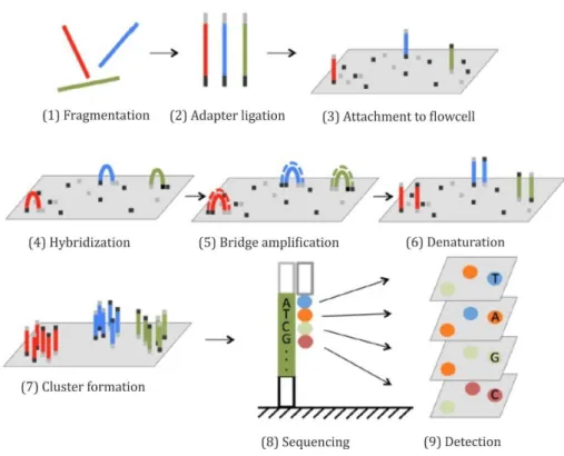
Next Generation Sequencing (NGS) represents a variety of sequencing methods which transcends the capacity of traditional DNA sequencing technologies in respect to cost, speed and data output, thus enabling an in-depth study of biological systems by rapid sequencing of whole genomes. One of the most prevalent NGS technologies was developed by Illumina and is referred to as ‘Sequencing-by-synthesis‘ (SBS)
205. This method supports massively parallel sequencing proving to be especially beneficial for questions that demand extensive information regarding highly diverse genomic libraries. The sequencing workflow is composed of four basic steps: i) sample preparation, ii) cluster generation, iii) sequencing and iv) data analysis.
In general, NGS sample preparation includes fragmentation of DNA sequences into suitable sizes (~ 300 bps) due to the limitation of reading length of the NGS device (figure 9, (1)) and the annealing of adapters (2) by PCR. The resulting product consists of a sequence of interest flanked 5‘ and 3‘ by the adapters P5 and P7 (figure 8), which allow attachment to the surface of a flow cell coated with a lawn composed of complementary adapter oligonucleotides (3). Additional motifs are also introduced during sample preparation, such as the NGS sequencing primer binding site and indices.
The indices or barcodes allow distinction among a multitude of samples.
Figure 8 – Schematic illustration of a ready-to-load index library eligible for NGS. The DNA fragment of interest is shown in grey. P5 (red) and P7 (green) indicate the adapters. Rd1 SP (yellow) and Rd2 SP (blue) designate the binding sites for NGS-specific sequencing primers. Barcodes or indices are represented in black.
19 After binding of samples to the flowcell, the DNA strand folds over and the adaptor region hybridizes to the second type of oligo on the flow cell (4). Polymerases create a complementary strand forming a double stranded bridge (5) which is subsequently denatured, resulting in two single stranded copies of the molecule (6). The process is repeated over and over so that millions of DNA ‘clusters’ are generated (7). Cluster densities have large impact on sequencing performance in terms of data quality and output.
Sequencing begins with the extension of the first sequencing primer to produce the first read (8). With each cycle, four fluorescently tagged reversible terminator bases compete for incorporation into the growing chain. Only one nucleotide is integrated, based on the sequence of the template while non-incorporated nucleotides are washed away.
Subsequently, clusters are excited by a light source and a characteristic fluorescent signal is emitted which is acquired as image by a camera (9). Emission wave length along with the signal intensity, determine the base call. The length of the read is determined by the number of cycles. The entire process generates millions of reads representing all fragments which then can be separated based on the unique indices introduced during the sample preparation. Forward and reverse reads are paired creating contiguous sequences that are aligned back to the reference genome for variant identification.
Sequencing coverage describes the average number of reads that align to known reference bases.
Figure 9 – Outline of Illumina NGS technology. After fragmentation of the samples (1), adapters are annealed to the ends of the sequence (2). Fragments attach to the flowcell (3) by hybridization to oligos complementary to the adapters (4). Subsequently, bridge amplification occurs to produce clusters of fragments (5-7). During each sequencing cycle, one fluorophore-attached nucleotide is added to the
20
growing strands (8). The fluorophores are then excited by a laser and signals from each fragment cluster are detected and recorded as images (9). Illustration was adapted from 206.3.2.2 Sequencing library preparation
The sequential permutation (SeqPer) library was the foundation for all steps involving Next Generation Sequencing. A key step in the NGS library sample preparation is generating the input for sequencing. In general, the library preparation was composed of four stages: i) DNA amplification, ii) attachment of oligonucleotide indices and adapters to the ends of the amplified fragments, iii) purification and iv) final library quantification and quality control.
3.2.2.1 Generation of amplicon libraries
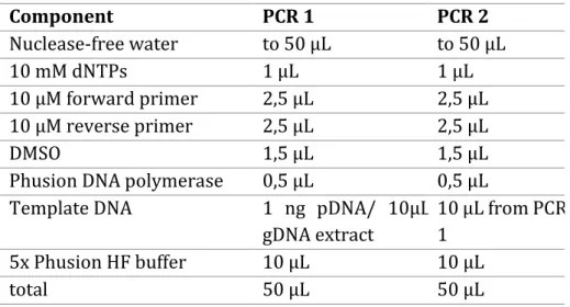
In the course of this PhD thesis, polymerase chain reaction was utilized to produce amplicons in the size of approximately 300 bps. These amplicon libraries were generated by two sequential PCRs using specific primers which carry the attachments required for NGS. In the first PCR, NGS-specific primer binding sites were introduced into the target DNA template. Fusion of indices and adapters was achieved by the second PCR (figure 8). The input DNA for the first PCR was derived from the plasmid DNA of the SeqPer library purchased from GeneArt or was isolated from the respective stable cell lines as genomic DNA (see 3.2.2.2).
For the amplification 20 ng plasmid DNA was applied in the first PCR. As the
concentration of isolated genomic DNA was always below the detection limit, 10 µL of
the extract was utilized for PCR1. Accordingly, 10 µL from the first amplification was
applied in the second PCR. The respective primers for PCR1 and PCR2 can be obtained
from section 8.2.1. Notably, only the reverse primers for PCR2 varied, whereas the
forward primer (ILLUMINAseq_fwd) remained the same. In general, a 50 µL reaction
was prepared containing 1 µL dNTPs (10 mM), 2.5 µL forward and reverse primer (10
µM), respectively, 1.5 µL DMSO, 0.5 µL Phusion DNA polymerase, 10 µL 5xHF Phusion
buffer, template DNA as mentioned above and nuclease-free water (see table 3). To
reduce accumulation of errors from the polymerase, 22 cycles were applied for both
amplification steps. The thermocycling conditions can be obtained from table 4. PCR
samples were stored temporarily (usually over night) at 4°C if it was not possible to
proceed with the experiment.
21
Table 3 – Composition of PCR 1 and 2 for generation of amplicon libraries.Component PCR 1 PCR 2
Nuclease-free water to 50 µL to 50 µL
10 mM dNTPs 1 µL 1 µL
10 µM forward primer 2,5 µL 2,5 µL
10 µM reverse primer 2,5 µL 2,5 µL
DMSO 1,5 µL 1,5 µL
Phusion DNA polymerase 0,5 µL 0,5 µL
Template DNA 1 ng pDNA/ 10µL
gDNA extract
10 µL from PCR 1
5x Phusion HF buffer 10 µL 10 µL
total 50 µL 50 µL
Table 4 – Thermocycling conditions for PCR 1 and 2.
PCR 1
98°C 1 min 98°C 10 sec
22 cycles 68°C 30 sec
72°C 6 sec 72°C 5 min
8°C ∞
3.2.2.2 Extraction of genomic DNA
Extraction of genomic DNA was performed by using the QIAamp DNA Mini Kit according to the protocol ‘DNA purification from blood and body fluids’. Unless stated otherwise, 2x10
5cells were utilized for gDNA isolation. If less than 10.000 genomic equivalents were present, 10 µg/mL carrier DNA (polyadenylic acid, poly dA) was added to the sample in order to enhance the recovery of DNA and to prevent the small amount of target nucleic acid from being irretrievably bound.
PCR 2
98°C 1 min 98°C 10 sec
22 cycles 64°C 30 sec
72°C 10 sec 72°C 5 min
8°C ∞
Deoxynucleotide (dNTP) Solution Mix New England Biolabs, N0447L Dimethyl Sulfoxide (DMSO) SigmaAldrich, D8418-100ML Phusion ® High-Fidelity DNA Polymerase New England Biolabs, M0530 L
![Figure 1 - Global prevalence of HIV in 2016. The illustration shows that an estimated 0.8% [0.7-0.9%]](https://thumb-eu.123doks.com/thumbv2/1library_info/3737594.1509104/14.892.132.799.303.707/figure-global-prevalence-hiv-illustration-shows-estimated.webp)