https://doi.org/10.1007/s15010-020-01503-7 ORIGINAL PAPER
A highly specific and sensitive serological assay detects SARS‑CoV‑2 antibody levels in COVID‑19 patients that correlate with neutralization
David Peterhoff1 · Vivian Glück2 · Matthias Vogel2 · Philipp Schuster1 · Anja Schütz1 · Philip Neubert2 ·
Veruschka Albert2 · Stefanie Frisch2 · Mara Kiessling2 · Philip Pervan2 · Maren Werner1 · Nicole Ritter2 · Leon Babl2 · Maria Deichner2 · Frank Hanses3,4 · Matthias Lubnow5 · Thomas Müller5 · Dirk Lunz6 · Florian Hitzenbichler3 · Franz Audebert7 · Viola Hähnel8 · Robert Offner8 · Martina Müller9 · Stephan Schmid9 · Ralph Burkhardt10 · Thomas Glück11 · Michael Koller12 · Hans Helmut Niller1 · Bernhard Graf6 · Bernd Salzberger3 · Jürgen J. Wenzel2 · Jonathan Jantsch1,2 · André Gessner1,2 · Barbara Schmidt1,2 · Ralf Wagner1,2
Received: 1 July 2020 / Accepted: 7 August 2020
© The Author(s) 2020
Abstract
Objective The severe acute respiratory syndrome coronavirus 2 (SARS-CoV-2) pandemic challenges national health systems and the global economy. Monitoring of infection rates and seroprevalence can guide public health measures to combat the pandemic. This depends on reliable tests on active and former infections. Here, we set out to develop and validate a specific and sensitive enzyme linked immunosorbent assay (ELISA) for detection of anti-SARS-CoV-2 antibody levels.
Methods In our ELISA, we used SARS-CoV-2 receptor-binding domain (RBD) and a stabilized version of the spike (S) ectodomain as antigens. We assessed sera from patients infected with seasonal coronaviruses, SARS-CoV-2 and controls.
We determined and monitored IgM-, IgA- and IgG-antibody responses towards these antigens. In addition, for a panel of 22 sera, virus neutralization and ELISA parameters were measured and correlated.
Results The RBD-based ELISA detected SARS-CoV-2-directed antibodies, did not cross-react with seasonal coronavirus antibodies and correlated with virus neutralization (R2 = 0.89). Seroconversion started at 5 days after symptom onset and led to robust antibody levels at 10 days after symptom onset. We demonstrate high specificity (99.3%; N = 1000) and sensitivity (92% for IgA, 96% for IgG and 98% for IgM; > 10 days after PCR-proven infection; N = 53) in serum.
Conclusions With the described RBD-based ELISA protocol, we provide a reliable test for seroepidemiological surveys.
Due to high specificity and strong correlation with virus neutralization, the RBD ELISA holds great potential to become a preferred tool to assess thresholds of protective immunity after infection and vaccination.
Keywords SARS-CoV-2 · COVID-19 · Antibody test · ELISA · Serology · Virus neutralization · Assay validation · Spike protein · S protein · Receptor binding domain
Introduction
Coronaviruses (CoV) are known to cause respiratory dis- eases in humans. The alphacoronaviruses HCoV-229E and HCoV-NL63 as well as the betacoronaviruses HCoV-OC43 and HCoV-HKU1 circulate seasonally in humans and cause common colds [1]. In contrast, the severe acute respiratory syndrome-related coronavirus 1 (SARS-CoV-1), the Mid- dle East respiratory syndrome-related coronavirus (MERS- CoV) and SARS-CoV-2 are zoonotic betacoronaviruses and can cause life-threatening severe respiratory distress syn- dromes and pandemics [2].
Barbara Schmidt and Ralf Wagner contributed equally.
Electronic supplementary material The online version of this article (https ://doi.org/10.1007/s1501 0-020-01503 -7) contains supplementary material, which is available to authorized users.
* David Peterhoff david.peterhoff@ur.de
* Barbara Schmidt barbara.schmidt@ukr.de
* Ralf Wagner ralf.wagner@ur.de
Extended author information available on the last page of the article
The immune response to the seasonal CoVs and SARS- CoV and MERS-CoV has been studied intensively [3, 4].
They are able to trigger a humoral immune response that correlates with disease severity [3]. Of note, mild infections resulted in short-lived and very low antibody titers near the detection limit. This loss of humoral immunity has been linked to the occurrence of (re-)infections with seasonal CoVs [3].
The current SARS-CoV-2-triggered COVID-19 pan- demic started in December 2019 in Wuhan, Hubei prov- ince of China and has led with August 2020 to > 18 million confirmed infections [5]. Diagnosis of COVID-19 relies on PCR testing of respiratory specimen [6]. However, to assess whether a patient had recovered from a previous infection serological analyses are required. Serological surveys for SARS-CoV-2 antibody responses are not only impor- tant to monitor how much of a given population has been infected, but also serology will be required to assess vaccine responses. Finally, serology will be key to identify the qual- ity of convalescent plasma that can be applied within clinical (compassionate) trials [7].
To allow for reliable serology, specific and sensitive assays are urgently needed. Here, we provide and validate a robust and simple ELISA protocol which is based on the SARS-CoV-2 receptor-binding domain (RBD). With this protocol, we performed a cross-sectional analysis of sera from PCR-proven COVID-19 patients. We demonstrate cor- relation of the ELISA test results with neutralizing antibody levels in a neutralization assay using a clinical isolate.
Materials and methods Patient samples
The SARS-CoV-2 ELISA was validated using pseu- donymized samples from patients aged older than 18 years from the diagnostic repository of the Institute of Clinical Microbiology and Hygiene, University Hospital Regens- burg, originating from summers of 2016–2019. Potential cross-reactivity of the ELISA protocol was analyzed using sera from patients with PCR-proven seasonal coronavirus infections and detectable antibody reactivity against sea- sonal coronavirus antigens. Sensitivity of the protocol was quantified with sera from patients with PCR-proven SARS- CoV-2 infection at different time points. This procedure was approved by the ethical commission of the Faculty for Medi- cine, University of Regensburg, Regensburg, Germany (ref.
no. 20-1854-101).
Design of recombinant proteins
S-protein sequences used for sequence analysis and gene synthesis are given in Table S1. Phylogenetic analysis, synthesis and cloning of the S protein variants used in this study (Fig. S1) are described in the supplementary materials and methods.
Protein structure analysis and visualization
Protein structures were visualized and analyzed by Pymol (LLC Schrodinger) using the structural data from the Pro- tein Data Bank (https ://www.rcsb.org) repository using entry codes 6vsb and 6m17.
Protein production and purification
Proteins were expressed in Expi293 cells (Thermo Fisher Scientific) in different scales using the commercial Expi- Fectamine™ system. Affinity purification of the proteins and quality controls are described in the supplementary materials and methods.
Line blot assay
CoV line blot assay (beta-version of recomLine Corona- virus IgG assay) was performed as described in the manu- facturer’s protocol. Line blots were evaluated by visual inspection and using recomScan software (Mikrogen Diag- nostik, Neuried, Germany).
Enzyme‑linked immunosorbent assay
A detailed ELISA protocol is provided in the supple- mentary materials and methods. Commercial Anti- SARS-CoV-2-ELISA IgG (EUROIMMUN, Lübeck, Ger- many) was performed according to the manufacturer’s recommendations.
Virus isolation and virus load quantification
SARS-CoV-2 isolation from respiratory specimen, deter- mination of the viral loads using quantitative SARS-CoV-2 real-time RT-PCR (RT-qPCR) and 50% tissue culture infective dose (TCID50) are described in the supplemen- tary materials and methods.
Virus neutralization assay
Virus neutralization assay using SARS-CoV-2 from respiratory specimen is described in the supplementary materials and methods.
Data evaluation and curve fitting
Experimental data were evaluated and plotted using GraphPad Prism (GraphPad Prism version 8.4.2 for Win- dows, GraphPad Software, San Diego, California USA).
Results
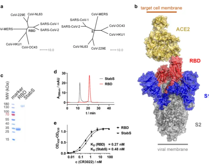
S-protein’s RBD has been shown to be SARS-CoV-1’s Achilles’ heel [8]. Due to the homology between the S-pro- teins as well as the RBDs of SARS-CoV-1 and SARS-CoV-2 (Fig. 1a), this may also hold true for the virus driving the current pandemic. This is why we decided to establish an ELISA protocol, which uses the SARS-CoV-2 RBD and stabilized ectodomain (StabS [9]) as antigens (Fig. 1b). To promote robust production of the two recombinant proteins, coding sequences were codon-optimized and the autologous S-protein signal peptide was replaced by a minimal version of the tissue plasminogen activator (tPA) signal peptide developed in our lab. RBD yielded 70 mg/l supernatant for
Fig. 1 Antigens used in SARS-CoV-2 ELISA. a Phylogenetic trees calculated for the RBD and complete S-protein of clinically relevant members of the coronavirus family. Trees are calculated on the basis of percent identity between each pair of sequences in the respective alignment. b Structure of the S-protein ectodomain in complex with its receptor, angiotensin-converting enzyme 2 (ACE2). c Reducing SDS-PAGE (linear gradient of 8–16% polyacrylamide) of the purified
RBD and StabS protein. d Size exclusion chromatography of the puri- fied RBD and StabS protein. e ELISA titrations of SARS-CoV-1 and SARS-CoV-2-binding antibody CR3022 against immobilized StabS and RBD [OD450–630: optical density (OD) at 450 nm after back- ground subtraction at 630 nm]. Resulting dissociation constants (KD) are given in the diagram
the RBD and 14 mg/l supernatant for the StabS protein.
Purity and homogeneity were verified by reducing SDS- PAGE and size exclusion chromatography (Fig. 1c, d).
A number of SARS-CoV-2 serology ELISA protocols have been published recently [10–15]. In our protocol, the antigens were directly absorbed to the plate’s plastic sur- face. To control protein integrity, we used our ELISA pro- tocol in combination with the structure-dependent mono- clonal anti-RBD antibody CR3022 [16–18]. High affinity binding as reflected by a KD of 0.27 nM for the RBD pro- tein and 0.48 nM for StabS as well as a strong absorption (OD450–630 > 1 at saturation after 4 min of development time) in both cases demonstrated sufficient amounts of well-folded protein (Fig. 1e). We used CR3022 at saturation concentra- tion to test the stability of RBD-coated plates, which had been coated up to 8 days earlier. No performance loss was detected during this period when plates were stored at room temperature in PBS-T (Fig. S2).
We used our ELISA to screen for anti-S and anti-RBD antibodies in sera from COVID-19 patients and controls.
In a first step, we quantified anti-RBD responses and anti-S responses in 22 sera, which displayed differential ELISA signals (Fig. S3).
We found that antibody responses against StabS corre- lated very well with antibody response to RBD as deter- mined by correlation of OD450–630 at 1:100 dilution, area under the curve (AUC), effective concentration at 50% sig- nal (EC50) and titer (Fig. S4). Moreover, comparable sig- nal strengths in these analyses suggest that RBD is a key immunogenic determinant of anti-S responses (Fig. S3).
Therefore, anti-RBD antibody levels represent a valuable surrogate for testing anti-S-directed antibody responses.
High-throughput titration of sera for the purpose of determining end point titers or calculating EC50 values—in particular in seroepidemiological surveillance studies on broader population scale—is a time- and material-consum- ing procedure. Thus, we tested whether OD450–630 at 1:100 dilution of sera correlate with standard serum characteristics such as area under the curve (AUC), effective concentration at 50% signal (EC50) and titer. This analysis revealed that 1:100 dilution of sera reflects total serum antibody responses (Fig. S5). Therefore, we used single-point measurements for all further assays.
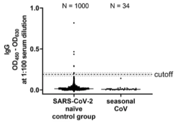
Next, we wanted to establish cutoff values for anti-RBD- directed IgG, IgA and IgM antibody responses following the requirements for validation of diagnostic assays in clinical virology [19]. For that purpose, we measured 190 SARS- CoV-2 naïve sera that had been collected before the current SARS-CoV-2 pandemic (Fig. S6). As recently proposed by Okba et al. [11], we used the mean of the background sig- nals plus six standard deviations (SD) to define the cutoff value. Using these parameters, we determined the specificity of our assay by measuring 1000 independent SARS-CoV-2
infection naïve sera. We obtained a false positive rate of 7 out of 1000 sera, corresponding to a specificity of 99.3%
(Fig. 2).
To further verify the specificity of our ELISA, we tested whether sera from patients, that suffered from infection with seasonal CoVs, cross-react with our assay. For that purpose, we retrieved in a total of 43 sera from patients that had PCR- proven seasonal corona virus infection from our diagnos- tic repository. In a commercial line blot assay, 34 sera out of 43 scored positive for seasonal CoV-specific antibodies (Table S2). We used these 34 sera to further validate our RBD ELISA. Of utmost importance, none of these seasonal coronavirus sera showed any cross-reactivity in our ELISA (Fig. 2).
To verify precision and reproducibility of our ELISA [19], we used a minimal panel of seasonal (N = 5), naïve (N = 15), weakly IgG reactive (N = 10) and strongly IgG reactive (N = 10) sera. For weakly IgG reactive sera, the rela- tive standard deviation (σrel) was 3.98% for IgG, 1.72% for IgM and 7.15% for IgA; for strongly reactive IgG positive sera σrel was determined to be 0.16% for IgG, 0.22% for IgM, and 2.73% for IgA. Combined inter-assay and inter-operator variabilities for weakly IgG reactive sera was determined as a σrel of 10.26% and for strong IgG reactive sera σrel was calculated to be 4.12%.
To determine the sensitivity of our assay, we analyzed sera from patients suffering from COVID-19 (N = 144), assuming that after symptomatic SARS-CoV-2 infection eventually all subjects develop antibodies. We quanti- fied the anti-RBD IgG responses, which correlated well (R2 = 0.8812, Spearmen’s ρ = 0.917, p value < 0.0001) with anti-SARS-CoV-2 responses measured using a commercial IgG ELISA (EUROIMMUN, Fig. S7) that has been vali- dated recently [20]. Next, we determined the IgM, IgG and
Fig. 2 Assay specificity of RBD ELISA. To define the specificity of the assay, 1000 sera isolated in the summers of 2016 and 2018 (SARS-CoV-2 naïve control group) and 34 sera of patients with sea- sonal corona virus infection (seasonal CoV) were measured (median is shown). The cutoff is set at six standard deviations above the mean of the reference panel (borderline ± 10% cutoff)
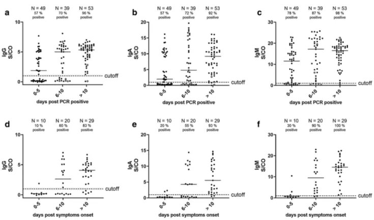
IgA levels at different time frames after the first detection of SARS-CoV-2 RNA by RT-qPCR in our cohort (proven infection; Fig. 3a–c). At > 10 days after proven infection, these assays displayed sensitivities of 92% for IgA, 96% for IgG and 98% for IgM. However, we found that the anti- body responses were already remarkably elevated at early time points after proven infection and, in concordance with previous findings, did not display a steady increase over time [21]. We speculated that infection preceded virus test- ing by several days. For 41% of the subjects (N = 59), the time point of symptom onset was available and we could calculate the average period between symptom onset and detection of SARS-CoV-2 RNA by RT-qPCR (5.7 days;
Fig. S8). This suggests that the majority of patients who were subjected to PCR testing had already suffered from COVID-19 for 5.7 days. Therefore, an analysis of anti-RBD responses in relation to days after symptom onset should result in lower anti-RBD responses at early time points and a steady increase of antibody levels over time. Indeed, in the subgroup of patients where the time point of symptom onset was available, we detected very low antibody responses early after symptom onset and a steady increase in all anti-RBD antibody isotypes (Fig. 3d–f).
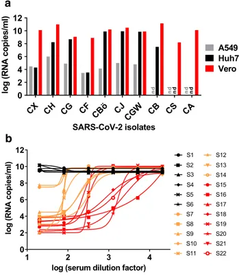
Neutralizing antibodies correlate with protection against several pathogens. Since we used the RBD of SARS-CoV-2, we expected the serum reactivity measured in our ELISA to correlate with virus neutralization due to sterical hindrance of RBD’s binding to its receptor ACE2 (Fig. 1b). In a first step, we isolated ten SARS-CoV-2 strains from respiratory specimen of COVID-19 patients. Of three different cell lines, kidney epithelial cells (Vero) supported virus propagation more efficiently than hepatocarcinoma-derived Huh-7 cells and lung carcinoma-derived A549 cells (Fig. 4a). For this reason, Vero cells were selected for neutralization experi- ments, using a highly replicative and cytopathic SARS- CoV-2 isolate (strain CA).
Prior to infection of Vero cells, the virus was incubated with serial dilutions of the 22 sera, which displayed dif- ferential ELISA titrations (Fig. S3). After removal of the inoculum at 12–24 h post-infection, viral loads were deter- mined in cell culture supernatants at 48 h post-infection using SARS-CoV-2 RT-qPCR. All sera which were negative in the ELISA (N = 6) did not inhibit virus entry and repli- cation (Fig. 4b). In contrast, sera with borderline or posi- tive ELISA values reduced SARS-CoV-2 viral loads by two (N = 2) to six log10 (N = 14). Notably, the reduction of viral
Fig. 3 Serological testing of COVID-19 patients using RBD ELISA.
a–c Serum reactivities of different immunoglobulin isotypes at 1:100 dilution of 144 sera of different time points after positive SARS- CoV-2 RT-qPCR, 1 per patient and time point (S/CO = signal/cut- off). Sampling of sera > 10 days post-PCR positive ranges from 11 to
29 days. Sera from patients with < 300 RNA copies/ml in respiratory specimen are shown as open circles, all others as closed circles. d–f Serum reactivities at 1:100 dilution (S/CO values) of 59 sera of dif- ferent time points after onset of symptoms (subset of values from a–c with known case history), one per patient and time point
loads showed a varying pattern, from a sudden to a more gradual inhibition of viral replication. Neutralization capac- ity was more pronounced in sera with high ELISA values.
Most importantly, neutralization titers (given as IC50 values) correlated strongly with anti-RBD (R2 = 0.8943, Spearmen’s ρ = 0.965, p value < 0.0001) and anti-StabS (R2 = 0.9057, Spearmen’s ρ = 0.964, p value < 0.0001) antibody levels (Fig. 5a, b and Fig. S9a–f).
Discussion
We established and validated a protocol for an easy-to-per- form, robust and sensitive ELISA for detection of S-directed SARS-CoV-2 antibody responses in sera that correlate with SARS-CoV-2 virus neutralization.
As reported for a commercial assay our ELISA is highly specific (99.3%) as well, and sensitivity is dependent on the time point after infection when the serum sample is taken [20]. In concordance with findings of others, we detected robust antibody levels at approximately 2 weeks after onset of symptoms [22, 23]. Moreover, our data suggest that sero- conversion for all Ig isotypes requires at least 6–10 days after onset of symptoms. In our cohort, IgM levels tend to appear first, followed by IgG conforming to earlier reports on SARS-CoV-2 Ig responses [21, 24–28]. Of note, we detected a late rise of the median IgA levels in our cohort. It is possible that this is an etiopathologic predictor of COVID- 19 [29]. Taken into account, that the cohort of our cross-sec- tional study contained only symptomatic COVID-19 cases, seroconversion rates in oligo- or asymptomatic patients, the persistence of antibody levels and the role of antibodies in mucosal fluids warrant further investigation. Longitudinal epidemiological surveillance studies to address these ques- tions are urgently needed. We provide an assay that can
CX CH CG CF CBö CJ
CGW CB CS CA 0
2 4 6 8 10 12
SARS-CoV-2 isolates
log(RNAcopies/ml)
Huh7A549 Vero
nd ndnd ndnd
a
b
1 2 3 4
0 2 4 6 8 10 12
log (serum dilution factor)
log(RNAcopies/ml) S1S2
S3 S4 S5 S6 S7 S8 S9 S10 S11
S12 S13 S14 S15 S16 S17 S18 S19 S20 S21 S22
Fig. 4 Virus isolation and neutralization assay. a Isolation of ten SARS-CoV-2 strains from respiratory specimen using three different cell lines (A549, derived from lung carcinoma; Huh7, hepatocyte- derived carcinoma; Vero, kidney epithelial cells from African green monkey; nd = not done). Viral loads in cell culture supernatants were determined using RT-qPCR. b After titration of the cytopathic SARS- CoV-2 strain CA, Vero cells were infected at a multiplicity of infec- tion (MOI) of 0.05. Prior to infection, the virus was incubated with dilutions of 22 serum samples (fourfold serial dilutions starting at 1:20). Of these, six (S1–S6, black), eight (S7–S14, orange), and eight (S15–S22, red) samples displayed negative, medium, and high SARS- CoV-2 IgG reactivities, respectively, in the RBD-based ELISA. Two days post-infection, viral loads were determined in cell culture super- natants using RT-qPCR
Fig. 5 Correlation of antibody levels with SARS-CoV-2 neutrali- zation. Correlation of OD from IgG ELISA measurements at 1:100 serum dilution with log IC50 values obtained from the neutralization experiments for a panel of 22 reference sera using RBD (a) and StabS
(b) as an antigen. Coefficients of determination (R2), Spearmen’s ρ, p value, linear regression line (solid line) and 95% confidence intervals (dashed line) are given in the diagrams
be easily implemented to quantify antibody responses in COVID-19.
The presence of RBD-directed antibodies, as determined by the herein described approach, correlate with SARS- CoV-2 neutralization. These findings strongly suggest that anti-RBD antibodies may confer protection and that the RBD as well as StabS proteins used in our study can be applied for sorting B cells and generating neutralizing mono- clonal antibodies. Surveillance of anti-RBD and anti-StabS antibody responses therefore represent, as far as we ana- lyzed, a reliable, simple, quick and high-throughput compat- ible alternative for any type of state of the art neutralization assay such as standard plaque reduction or lentiviral- and VSV-derived pseudotype assays. Such robust correlates will gain further importance once we start determining thresh- olds for protective immunity as a result of natural infection and, more importantly, following vaccination.
Acknowledgements Open Access funding provided by Projekt DEAL.
We acknowledge sponsoring of reagents by Mikrogen GmbH and finan- cial support through the pandemic responsiveness fund of The Bavarian Ministry of Science and Art.
Author contributions Conception and design of the study was accounted by DP, BSc, RW and AG. Samples were provided by FHa, ML, TM, DL, FA, VH, RO, MM, SS, TG, and BG. Data were collected by DP, BSc, AS, VG, MV, PN, VA, SF, MKi, PP, MW, NR, JJW, LB, and MD. DP, BSc, MV, and PS analysed and interpreted the data with valuable intellectual input by RW, HHN, JJ, JJW, MKo, RB, FHi, and BSa. Drafting the manuscript was done by DP and BSc. Manuscript was revised by RW and JJ. All authors approved the submitted version.
Compliance with ethical standards
Conflict of interest All authors declare no conflicts of interest.
Open Access This article is licensed under a Creative Commons Attri- bution 4.0 International License, which permits use, sharing, adapta- tion, distribution and reproduction in any medium or format, as long as you give appropriate credit to the original author(s) and the source, provide a link to the Creative Commons licence, and indicate if changes were made. The images or other third party material in this article are included in the article’s Creative Commons licence, unless indicated otherwise in a credit line to the material. If material is not included in the article’s Creative Commons licence and your intended use is not permitted by statutory regulation or exceeds the permitted use, you will need to obtain permission directly from the copyright holder. To view a copy of this licence, visit http://creat iveco mmons .org/licen ses/by/4.0/.
References
1. van der Hoek L. Human coronaviruses: what do they cause? Anti- vir Ther. 2007;12:651–8.
2. Fung TS, Liu DX. Human coronavirus: host–pathogen interaction.
Annu Rev Microbiol. 2019;73:529–57. https ://doi.org/10.1146/
annur ev-micro -02051 8-11575 9.
3. Kellam P, Barclay W. The dynamics of humoral immune responses following SARS-CoV-2 infection and the potential for reinfection.
J Gen Virol. 2020. https ://doi.org/10.1099/jgv.0.00143 9.
4. Dijkman R, Jebbink MF, El Idrissi NB, Pyrc K, Muller MA, Kui- jpers TW, et al. Human coronavirus NL63 and 229E seroconver- sion in children. J Clin Microbiol. 2008;46:2368–73. https ://doi.
org/10.1128/JCM.00533 -08.
5. Dong E, Du H, Gardner L. An interactive web-based dashboard to track COVID-19 in real time. Lancet Infect Dis. 2020;20:533–4.
https ://doi.org/10.1016/S1473 -3099(20)30120 -1.
6. WHO. Laboratory testing for 2019 novel coronavirus (2019- nCoV) in suspected human cases: interim guidance. 2020. www.
who.int.
7. Li L, Zhang W, Hu Y, Tong X, Zheng S, Yang J, et al. Effect of Convalescent plasma therapy on time to clinical improve- ment in patients with severe and life-threatening COVID-19: a randomized clinical trial. J Am Med Assoc. 2020. https ://doi.
org/10.1001/jama.2020.10044 .
8. Berry JD, Hay K, Rini JM, Yu M, Wang L, Plummer FA, et al.
Neutralizing epitopes of the SARS-CoV S-protein cluster inde- pendent of repertoire, antigen structure or mAb technology.
MAbs. 2010;2:53–66. https ://doi.org/10.4161/mabs.2.1.10788 . 9. Wrapp D, Wang N, Corbett KS, Goldsmith JA, Hsieh CL,
Abiona O, et al. Cryo-EM structure of the 2019-nCoV spike in the prefusion conformation. Science. 2020;367:1260–3. https ://
doi.org/10.1126/scien ce.abb25 07.
10. Stadlbauer D, Amanat F, Chromikova V, Jiang K, Strohmeier S, Arunkumar GA, et al. SARS-CoV-2 seroconversion in humans:
a detailed protocol for a serological assay, antigen production, and test setup. Curr Protoc Microbiol. 2020;57:e100. https ://doi.
org/10.1002/cpmc.100.
11. Okba NMA, Muller MA, Li W, Wang C, Geurtsvankessel CH, Corman VM, et al. Severe acute respiratory syndrome coro- navirus 2 - specific antibody responses in coronavirus disease patients. Emerg Infect Dis. 2019. https ://doi.org/10.3201/eid26 07.20084 1.
12. Sun B, Feng Y, Mo X, Zheng P, Wang Q, Li P, et al. Kinetics of SARS-CoV-2 specific IgM and IgG responses in COVID- 19 patients. Emerg Microbes Infect. 2020;9:940–8. https ://doi.
org/10.1080/22221 751.2020.17625 15.
13. Amanat F, Stadlbauer D, Strohmeier S, Nguyen THO, Chromik- ova V, McMahon M, et al. A serological assay to detect SARS- CoV-2 seroconversion in humans. Nat Med. 2020. https ://doi.
org/10.1038/s4159 1-020-0913-5.
14. Freeman B, Lester S, Mills L, Rasheed MAU, Moye S, Abiona O, et al. Validation of a SARS-CoV-2 spike protein ELISA for use in contact investigations and serosurveillance. bioRxiv. 2020. https ://doi.org/10.1101/2020.04.24.05732 3.
15. Guo L, Ren L, Yang S, Xiao M, Chang YF, et al. Profiling early humoral response to diagnose novel coronavirus disease (COVID- 19). Clin Infect Dis. 2020. https ://doi.org/10.1093/cid/ciaa3 10.
16. ter Meulen J, van den Brink EN, Poon LL, Marissen WE, Leung CS, Cox F, et al. Human monoclonal antibody combination against SARS coronavirus: synergy and coverage of escape mutants. PLoS Med. 2006;3:e237. https ://doi.org/10.1371/journ al.pmed.00302 37.
17. Tian X, Li C, Huang A, Xia S, Lu S, Shi Z, et al. Potent binding of 2019 novel coronavirus spike protein by a SARS coronavirus- specific human monoclonal antibody. Emerg Microbes Infect.
2020;9:382–5. https ://doi.org/10.1080/22221 751.2020.17290 69.
18. Yuan M, Wu NC, Zhu X, Lee CD, So RTY, Lv H, et al. A highly conserved cryptic epitope in the receptor binding domains of SARS-CoV-2 and SARS-CoV. Science. 2020;368:630–3. https ://doi.org/10.1126/scien ce.abb72 69.
19. Rabenau HF, Kessler HH, Kortenbusch M, Steinhorst A, Raggam RB, Berger A. Verification and validation of diagnostic laboratory
tests in clinical virology. J Clin Virol. 2007;40:93–8. https ://doi.
org/10.1016/j.jcv.2007.07.009.
20. Meyer B, Torriani G, Yerly S, Mazza L, Calame A, Arm-Vernez I, et al. Validation of a commercially available SARS-CoV-2 serological immunoassay. Clin Microbiol Infect. 2020. https ://
doi.org/10.1016/j.cmi.2020.06.024.
21. Wolfel R, Corman VM, Guggemos W, Seilmaier M, Zange S, Muller MA, et al. Virological assessment of hospitalized patients with COVID-2019. Nature. 2020;581:465–9. https ://doi.
org/10.1038/s4158 6-020-2196-x.
22. Long QX, Liu BZ, Deng HJ, Wu GC, Deng K, Chen YK, et al.
Antibody responses to SARS-CoV-2 in patients with COVID- 19. Nat Med. 2020;26:845–8. https ://doi.org/10.1038/s4159 1-020-0897-1.
23. Caruana G, Croxatto A, Coste AT, Opota O, Lamoth F, Jaton K, et al. Diagnostic strategies for SARS-CoV-2 infection and inter- pretation of microbiological results. Clin Microbiol Infect. 2020.
https ://doi.org/10.1016/j.cmi.2020.06.019.
24. Zhao J, Yuan Q, Wang H, Liu W, Liao X, Su Y, et al. Antibody responses to SARS-CoV-2 in patients of novel coronavirus disease 2019. Clin Infect Dis. 2020. https ://doi.org/10.1093/cid/ciaa3 44.
25. Whitman JD, Hiatt J, Mowery CT, Shy BR, Yu R, Yamamoto TN, et al. Test performance evaluation of SARS-CoV-2 serological assays. medRxiv. 2020. https ://doi.org/10.1101/2020.04.25.20074 26. To KK, Tsang OT, Leung WS, Tam AR, Wu TC, Lung DC, 856.
et al. Temporal profiles of viral load in posterior oropharyngeal saliva samples and serum antibody responses during infection by SARS-CoV-2: an observational cohort study. Lancet Infect Dis.
2020;20:565–74. https ://doi.org/10.1016/S1473 -3099(20)30196 27. Tan W, Lu Y, Zhang J, Wang J, Dan Y, Tan Z, et al. Viral kinetics -1.
and antibody responses in patients with COVID-19. medRxiv.
2020. https ://doi.org/10.1101/2020.03.24.20042 382.
28. Lassaunière R, Frische A, Harboe ZB, Nielsen AC, Fomsgaard A, Krogfelt KA, et al. Evaluation of nine commercial SARS-CoV-2 immunoassays. medRxiv. 2020. https ://doi.
org/10.1101/2020.04.09.20056 325.
29. Cervia C, Nilsson J, Zurbuchen Y, Valaperti A, Schreiner J, Wolfensberger A, et al. Systemic and mucosal antibody secretion specific to SARS-CoV-2 during mild versus severe COVID-19.
bioRxiv. 2020. https ://doi.org/10.1101/2020.05.21.10830 8.
Affiliations
David Peterhoff1 · Vivian Glück2 · Matthias Vogel2 · Philipp Schuster1 · Anja Schütz1 · Philip Neubert2 ·
Veruschka Albert2 · Stefanie Frisch2 · Mara Kiessling2 · Philip Pervan2 · Maren Werner1 · Nicole Ritter2 · Leon Babl2 · Maria Deichner2 · Frank Hanses3,4 · Matthias Lubnow5 · Thomas Müller5 · Dirk Lunz6 · Florian Hitzenbichler3 · Franz Audebert7 · Viola Hähnel8 · Robert Offner8 · Martina Müller9 · Stephan Schmid9 · Ralph Burkhardt10 · Thomas Glück11 · Michael Koller12 · Hans Helmut Niller1 · Bernhard Graf6 · Bernd Salzberger3 · Jürgen J. Wenzel2 · Jonathan Jantsch1,2 · André Gessner1,2 · Barbara Schmidt1,2 · Ralf Wagner1,2
1 Institute for Medical Microbiology and Hygiene, University of Regensburg, Regensburg, Germany
2 Institute for Clinical Microbiology and Hygiene, University Hospital Regensburg, Regensburg, Germany
3 Department for Infection Control and Infectious Diseases, University Hospital Regensburg, Regensburg, Germany
4 Emergency Department, University Hospital Regensburg, Regensburg, Germany
5 Department of Internal Medicine II, University Hospital Regensburg, Regensburg, Germany
6 Department of Anesthesiology, University Hospital Regensburg, Regensburg, Germany
7 Praxiszentrum Alte Mälzerei, Regensburg, Germany
8 Institute of Clinical Chemistry and Laboratory Medicine, Transfusion Medicine, University Hospital Regensburg, Regensburg, Germany
9 Department of Internal Medicine I, University Hospital Regensburg, Regensburg, Germany
10 Institute of Clinical Chemistry and Laboratory Medicine, University Hospital Regensburg, Regensburg, Germany
11 Kreisklinik Trostberg, Trostberg, Germany
12 Center for Clinical Studies, University Hospital Regensburg, Regensburg, Germany