https://doi.org/10.1007/s10930-020-09929-6
RNA and DNA Binding Epitopes of the Cold Shock Protein TmCsp from the Hyperthermophile Thermotoga maritima
Konstanze von König
1· Norman Kachel
1· Hans Robert Kalbitzer
1· Werner Kremer
1Accepted: 8 October 2020 / Published online: 22 October 2020
© The Author(s) 2020
Abstract
Prokaryotic cold shock proteins (CSPs) are considered to play an important role in the transcriptional and translational regulation of gene expression, possibly by acting as transcription anti-terminators and “RNA chaperones”. They bind with high affinity to single-stranded nucleic acids. Here we report the binding epitope of TmCsp from Thermotoga maritima for both single-stranded DNA and RNA, using heteronuclear 2D NMR spectroscopy. At “physiological” growth temperatures of TmCsp (≥ 343 K), all oligonucleotides studied have dissociation constants between 1.6 ((dT)
7) and 25.2 ((dA)
7) μM as determined by tryptophan fluorescence quenching. Reduction of the temperature to 303 K leads to a pronounced increase of affinity for thymidylate (dT)
7and uridylate (rU)
7heptamers with dissociation constants of 4.0 and 10.8 nM, respectively, whereas the weak binding of TmCsp to cytidylate, adenylate, and guanylate heptamers (dC)
7, (dA)
7, and (dT)
7is almost unaffected by temperature. The change of affinities of TmCsp for (dT)
7and (rU)
7by approximately 3 orders of magnitude shows that it represents a cold chock sensor that switches on the cold shock reaction of the cell. A temperature dependent conformational switch of the protein is required for this action. The binding epitope on TmCsp for the ssDNA and RNA heptamers is very similar and comprises β-strands 1 and 2, the loop β1–β2 as well as the loops connecting β3 with β4 and β4 with β5. Besides the loop regions, surprisingly, mainly the RNA-binding motif RNP1 is involved in ssDNA and RNA bind- ing, while only two amino acids, H28 and W29, of the postulated RNA-binding motif RNP2 interact with the uridylate and thymidylate homonucleotides, although a high affinity in the nanomolar range is achieved. This is in contrast to the binding properties of other CSPs or cold shock domains, where RNP1 as well as RNP2 are involved in binding. TmCsp takes up a unique position since it is the only one which possesses a tryptophan residue instead of a usually highly conserved phenyla- lanine or tyrosine residue at the end of RNP2. NMR titrations suggest that neither (dT)
7nor (rU)
7represent the full binding motif and that non-optimal intercalation of W29 into these oligonucleotides blocks the access of the RNP2 site to the DNA or RNA. NMR-experiments with (dA)
7suggest an interaction of W29 with the adenine ring. Full binding seems to require at least one single purine base well-positioned within a thymine- or uracil-rich stretch of nucleic acids.
Keywords Cold-shock protein · TmCsp · Thermotoga maritima · Single-stranded DNA · Single-stranded RNA · Protein/
ssDNA complex · Protein/RNA complex · NMR spectroscopy · Temperature sensing
1 Introduction
When microorganisms experience a sudden decrease in temperature, they suffer from a cold shock. Its most strik- ing characteristic is the impairment of all growth–related
processes within the cell. However, after an acclimatization period, a physiological cold shock response sets in, which allows for growth to be resumed and is therefore consid- ered essential for the survival of unicellular organisms (for a review, see [1, 2]). This effect is based on the transient appearance of cold-induced proteins (CIPs). In particular, a subgroup comprising the so-called cold shock proteins (CSPs) is induced to an extreme extent.
CSPs are small, acidic proteins found in a wide variety of Gram-negative and Gram-positive species and are classi- fied together as one family since they possess high sequence homology among each other (> 45%). They seem to exert
*
Hans Robert Kalbitzer
hans-robert.kalbitzer@biologie.uni-regensburg.de
*
Werner Kremer
werner.kremer@biologie.uni-regensburg.de
1
Institut für Biophysik und Physikalische Biochemie,
Universität Regensburg, 93040 Regensburg, Germany
their biological function by binding to single-stranded nucleic acids, especially RNA. In this way, secondary struc- tures such as hair pins, which are increasingly formed in DNA and RNA at low temperatures, may be destabilized, thus enabling both transcription and translation to continue [3]. Due to their binding to mRNA with low sequence speci- ficity, cold shock proteins are also called ‘RNA’ chaperones [3]. Because of their homology to the cold shock domains of the eukaryotic Y-box transcription factors, CSPs were origi- nally thought to bind to the Y-box-sequence ATTGG. How- ever, for B. subtilis CspB (BsCspB) no apparent sequence specificity was found [4, 5]. More recent experiments have revealed that BsCspB possesses a strong preference for polypyrimidine ssDNA [5]. Its affinity is highest for thy- midylate-rich stretches of DNA and directly correlates with the number of T-bases present [6]. In this respect, (dT)
7is sufficient for obtaining nanomolar dissociation constants [7].
The tertiary structures of a number of cold shock proteins have been solved, i.e., of Csp from hyperthermophilic Ther- motoga maritima [8], of thermophilic Bacillus caldolyticus [9], or of CspB from mesophilic Bacillus subtilis [10, 11], and of CspA from mesophilic Escherichia coli [12, 13].
They belong to the OB (oligonucleotide/oligosaccharide binding) fold family, which typically is found in Greek-key β-barrel proteins. In their primary structures, CSPs contain the sequence motifs RNP1 and RNP2 commonly found in RNA-binding proteins [14, 15]. Taken together, these struc- tural features further support their role as nucleic acid bind- ing proteins.
The crystal structures of CspB from the mesophilic Bacil- lus subtilis (BsCspB) complexed with the hexanucleotide 5′-UUU UUU -3′ and the pentanucleotide 5′-GUC UUU A-3′
single stranded RNA (ssRNA) were published recently [16].
Here, one CSP is complexed with one ssRNA-molecule. In contrast, the ssDNA 5′-TTT TTT -3′ (dT)
6is bridging to two BsCspB molecules in the crystal structure [17]. A solution NMR structure of BsCsp in complex with the ssDNA dT
7shows that in solution the binding mode of ssDNA is prob- ably different to that observed in X-ray crystallography [18].
Crystal structures of CSP-nucleotide complexes of hyper- thermophilic bacteria have not yet been published.
In this paper we aim to characterize the nucleic acid interactions of the cold shock protein from the hyperther- mophilic bacterium T. maritima (TmCsp) with both ssRNA and ssDNA oligonucleotides. A thorough analysis was considered necessary because it had been shown that the binding specificity among the different CSPs varies slightly [19]. Cold chock proteins have two main functions after cold shock application, (1) an inhibition of the bulk protein expression and (2) an increased expression of CSPs them- selves and other cold shock induced, specific proteins (for a review see e.g. [2]). At the millimolar concentrations of CSPs obtained at cold shock conditions, transcription as well
as translation is downregulated completely as studies in cell free systems show [20]. For TmCsp 50% inhibition of the two processes is obtained at a concentration of 140 μM. Inhi- bition can completely be abolished by addition of hepta-2′- desoxy-thymidylate (dT)
7that competitively binds to TmCsp with high affinity [20]. Specific activation of translation by high affinity binding of EcCspA to the 5′-untranslated region of the CspA mRNA can be observed in cell-free systems at cold shock temperatures largely independent of the cod- ing sequence used [21]. For TmCsp detailed data about the unspecific bulk effects and the sequence specific activation effects are still missing, especially with regard to the ques- tion if binding to RNA or DNA involves different amino acid motifs of the protein. Preliminary studies had indicated that TmCsp would be an ideal candidate for such studies since its binding affinity for DNA seemed to be significantly higher than those of other CSPs. Furthermore, it could be deduced that TmCsp appeared to require oligomers comprising seven nucleotides as a minimum binding site.
According to the findings that ssDNA binding by BsCsp predominantly depends on the base composition of the tem- plate and not on the specific base sequence [5], simplified oligonucleotide sequences were used for the experiments presented in the following, which consisted of only one type of nucleotide each.
2 Results
2.1 Nucleotide Binding Properties of TmCsp Monitored by Electrophoretic Mobility Shift Assay (EMSA)
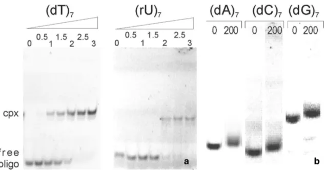
To investigate which types of nucleotides are recognized as TmCsp substrates, binding studies were performed with a non-radioactive electrophoretic mobility shift assay. Con- stant amounts of 5′-fluorescein-labeled oligonucleotides were incubated with varying concentrations of TmCsp. In the case of the pyrimidine oligonucleotides (dT)
7or (rU)
7, increasing concentrations of TmCsp resulted in a gradual decrease of the intensity of the band corresponding to the free oligonucleotide, while an additional double band recip- rocally appeared in the gel with a smaller electrophoretic mobility (Fig. 1a). Silver-staining of the gels showed that both bands contain protein. From this it was concluded that the shifted bands at high oligonucleotide concentrations correspond to the nucleoprotein complex (data not shown).
Complete retardation of the free oligonucleotides occurred at a molar ratio of TmCsp: (dT)
7or (rU)
7of 2:1–2.5:1.
In contrast, incubation of the cold shock protein with the
pyrimidine oligonucleotide (dC)
7as well as the purine oli-
gonucleotides (dA)
7or (dG)
7at a ratio as high as 200:1 (cor-
responding to 100 μM TmCsp) did not lead to a band shift
characteristic for a stable nucleoprotein complex at the con- centrations of oligonucleotides (0.5 μM) used in this assays (Fig. 1b). The purine oligonucleotides were re-tested at 300 μM of TmCsp but no band shift at lower migration height was observed that contained protein as determined by Silver staining (data not shown).
2.2 One Dimensional NMR Studies of the ssDNA Binding Interactions
Moreover, binding interactions were analyzed by NMR spec- troscopy to define the regions of the protein involved in com- plex formation. An aqueous solution of TmCsp (0.5 mM) in sodium phosphate buffer was separately titrated with (dA)
7, (dC)
7,or (dT)
7in the range of 0 to 0.625 mM by adding equal amounts of oligonucleotide in each step and recording a 1D
1H NMR spectrum of each of these samples at 303 K.
We initially intended to use the oligonucleotides (dA)
7and (dC)
7merely as controls since they do not strongly interact with TmCsp in the shift assay (see above). However, one has to note that in the NMR-experiments the protein and nucle- otide concentrations are higher by almost three orders of magnitude compared to the shift assays. It can be expected that under these conditions relatively weak interactions are also visible as, indeed, we will demonstrate in the follow- ing. Without DNA, the protein spectrum was well resolved and displayed sharp signal lines (Fig. 2a). Depending on the type of oligonucleotide added, the protein underwent distinct
changes in each titration series, which was clearly reflected in the 1D spectra.
In the presence of (dT)
7, the resonances broadened all over the spectrum (including also the signals of non- exchangeable protons). The resonance lines of the ε1-side chain protons of the tryptophan residues W7 and W29, of the main chain amide proton of V25, and of the high-field shifted H
δof I17 (Fig. 2b) at 10.45, 10.35, 9.77, and − 0.146 ppm, respectively, are of special interest since they encoun- tered significant changes upon nucleotide binding which could be easily monitored due to their isolated position.
In the first titration step, these signals broadened severely (Fig. 2b). Increased (dT)
7concentrations first led to a split- ting of the signal of V25 and I17; at high nucleotide con- centrations only one downfield shifted signal remains as it is typical for a slow exchange process. A similar process is observed for the tryptophan side chain resonances. At increasing (dT)
7-concentrations a new downfield shifted resonance at 9.457 ppm and a high field shifted resonance at 10.536 ppm appear and the intensity of the lines correspond- ing to nucleotide-free Csp simultaneously get weaker. The chemical shifts of these signals are virtually independent of the oligonucleotide concentration as to be expected for slow exchange processes. In slow exchange, the assignment of new resonances is not straightforward without additional information. When during a titration a new peak in close vicinity of the original peak is created and the total integral of the two peaks is constant, one can assume that they cor- respond to the same residue in the free and complexed form.
Fig. 1
Relative binding affinities as observed by electrophoretic mobility shift assays. a Gels run after the incubation of 0.5 μM (dT)
7or (rU)
7with TmCsp in different concentrations at room temperature.
The molar ratio of protein : oligonucleotide is indicated above the gel.
In both cases, good binding interactions were observed. Cpx: Com-
plex.
b Incubation of 0.5 μM (dA)7, (dC)
7or (dG)
7without TmCsp
and with TmCsp at a ratio of DNA:protein = 1:200. Note that the ver-
tical scale was increased by a factor of 2 for visualizing smaller shift
changes with addition of Csp. No band could be observed characteris-
tic for complex formation
With this argument, the downfield shifted signal at 9.81 ppm and the high-field shifted resonance at − 0.17 ppm, respec- tively, can be assigned to the amide proton of V25 and the methyl protons of I17 in the complex. In the BsCspB-dT
7complex the side chain resonance of W8 (corresponding to W7 in TmCsp) is shifted upfield by approximately 1 ppm. By analogy, it is likely that the signal at 9.457 ppm corresponds to W7 in the TmCsp nucleotide complex and the signal at 10.536 ppm to W29.
Above a molar ratio of approximately 1:2 of (dT)
7:TmCsp, the signals of free TmCsp are completely abolished and the integrals corresponding to complexed protein do not change anymore. Consistently, even at the endpoint of the titration with a (dT)
7concentration of 1.25 mM, the resonance line of W29 is broadened by a factor of 1.25 compared to the oligonucleotide free sample. Note that doubling the molecu- lar mass by dimerization would lead to an increase of the rotational correlation time and thus of the approximate line width by a factor of 1.26.
Titration of TmCsp with (dA)
7leads again to significant spectral changes proving that at the high concentrations used for NMR the oligonucleotide interacts with the pro- tein. When again considering the 4 resonances that were used for monitoring the binding of (dT)
7, spectral changes are also observed but now typical for fast exchange processes (Fig. 2c). Addition of (dA)
7leads to a very small continuous upfield shift of the resonance line of W7 accompanied by a significant line broadening by a factor of 1.8. Similar effects are observed for V25 and Ile17. Both of them are upfield
shifted with increasing nucleotide concentration to positions close to those observed for TmCsp ligated with (dT)
7.In contrast to the titration with (dT)
7, the signal of W29 is con- tinuously shifted upfield and severely broadens. At a ratio of approximately 1:0.375 of TmCsp:(dA)
7, it is broadened almost beyond detection. In fact, under fast to moderately fast exchange conditions a stronger exchange broadening effect is to be expected for lines with a larger chemical shift difference in the two states involved (nucleotide-free and nucleotide-bound). At oligonucleotide concentrations higher than 0.5 mM a saturation effect is observed for all consid- ered resonances. However, the signal of W29 could not be identified at saturation conditions suggesting an additional heterogeneity of the interaction mode at high oligonucleo- tide concentrations.
As in case of (dA)
7spectra, continuous chemical shift changes are observed when the oligonucleotide (dC)
7was added, indicating again a protein-nucleotide interaction, fast on the NMR time scale (Fig. 2d). As with (dA)
7W7, V25, and I17 are shifting downfield. However, W29 is downfield shifted as observed in the case of (dT)
7. No larger exchange broadening is observed, but at the highest DNA concentra- tion used (0.625 mM), the line widths are increased by a factor of 1.3. Again a saturation behavior is observed for all of these amino acids. (dG)
7did not yield well-resolved spectra and was therefore omitted from analysis.
In conclusion, these spectral changes indicate interac- tions of the cold shock protein with all ssDNA fragments studied. It is clear that the extent of the line broadening and
Fig. 2
1D
1H NMR spectra of TmCsp in the absence and presence of ssDNAs. a Overview spectrum of 0.5 mM TmCsp in 50 mM sodium phosphate, pH 6.5, 20 mM NaCl, 0.2 mM EDTA, and 0.1 mM DSS at 303 K. b to d 1 mM TmCsp in 50 mM sodium phosphate, pH 6.5, 20
mM NaCl, 0.2 mM EDTA, 0.1 mM DSS, and varying nucleotide con-
centrations as indicated in the plots. Temperature 303 K. b Spectral
changes induced by the addition of (dT)
7, c of (dA)
7, and d of (dC)
7in varying concentrations as indicated
the changes in chemical shift of the protein resonances upon binding are different. The main difference between the gel shift assays and the NMR spectra shown here are the con- centrations used. At the high concentrations used in NMR (1 mM protein and ≥ 0.625 mM nucleotide) and the lack of shear force as exerted in EMSA by the electric field, low- affinity interactions with all oligonucleotides were detectable which could not be detected by the electrophoretic mobility shift assay shown above with oligonucleotide concentrations of 0.5 μM. Surprisingly, a single oligonucleotide molecule binds two Csp-molecules.
2.3 Identification of (dT)
7and (rU)
7Binding Epitopes by Two‑Dimensional NMR
The complex formation was monitored on a residue-by-resi- due basis by recording a series of
1H–
15N HSQC fingerprint spectra of
15N-enriched TmCsp samples in the presence of increasing amounts of the oligonucleotides (dT)
7or (rU)
7at 303 K. Resonance assignments of free TmCsp were per- formed on the basis of the assignments published by Kremer et al. [8]. The spectral changes due to the addition of oligo- nucleotide were tracked by comparison of the successively recorded [
1H,
15N] HSQC spectra (Fig. 3).
Upon complex formation with (dT)
7, the backbone and side chain amide resonances of the protein in the [
1H,
15N]
HSQC spectra shifted to different extents. In addition, many peaks became undetectable. Such an effect was observed for the amide resonances of W7, D9, S10, K12, G13, Y14, F16, D24, H28, S30, E33, F37, K38, T39, K41, Q51, K54, K55, K63, and V65 (Fig. 4). It could be the effect of strong line broadening by intermediate exchange or it could correspond to the slow exchange effects observed for the H
ε1of W7 where the signal in the bound state shifts considerably in the 1D-spectra but cannot be identified unequivocally in the 2D-spectra. The combined chemical shift changes ∆δ
combincluding the backbone amide nitrogen and proton reso- nances as well as some side chain resonances are depicted in Fig. 4. Amino acids whose resonances shifted after the interaction with the oligonucleotides (dT)
7by more than σ
0and 2 σ
0were considered moderately and very probable interacting sites, respectively (Fig. 4). The [
1H,
15N]-HSQC spectrum of the complex with (rU)
7, is much better defined than that with (dT)
7. As already observed for (dT)
7upon complex formation with (rU)
7, the backbone and side chain amide resonances of the protein in the [
1H,
15N] HSQC spec- tra shifted to different extents. As in the case of (dT)
7many peaks became undetectable or shifted considerably. Dur- ing the titration with (rU)
7also significant chemical shift changes ∆δ
combwere observed. In most cases they were associated with the same residues as already described for the interaction with (dT)
7(Fig. 4). A consensus interacting surface was derived by using only residues that are most
likely interacting in at least one of the complexes and in the other structure have at least an intermediate interaction probability (Fig. 4). They are labeled in the consensus plot Co in Fig. 4 in bold letters. These residues are plotted on the NMR structure of TmCsp determined at 303 K (Fig. 5).
In the complex of Csp with (rU)
7many resonances remained either unaffected or shifted continuously with increasing oligonucleotide concentration, indicating that fast or moderately fast exchange conditions prevailed. How- ever, in the presence of (dT)
7, the chemical exchange rates of many of these resonances of TmCsp behaved in a way which is typical for intermediate exchange or slow exchange on the NMR time scale (Figs. 2, 3, 4). This result reflects most probably that a smaller exchange correlation time of the complex leads to slow exchange conditions. It is also supported by the fact that the overall dissociation constant of TmCsp for (dT)
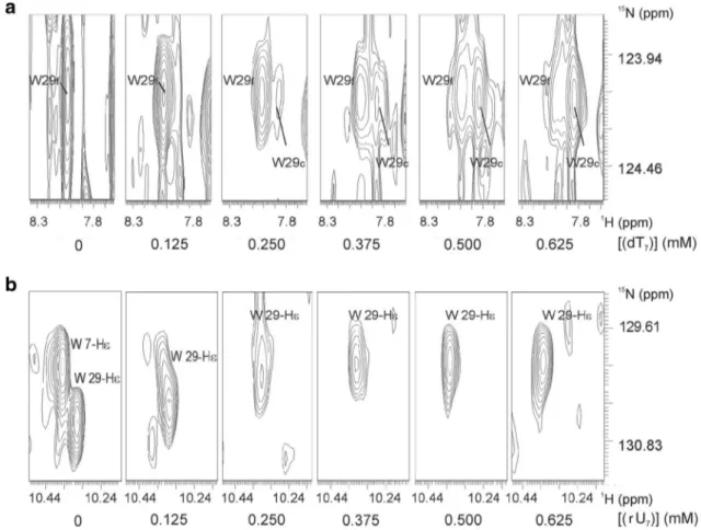
7is 2.7-fold lower than the one for (rU)
7. An example for slow exchange is shown in Fig. 6a, where the backbone H
N-resonance signal of W29 in free TmCsp becomes weaker and broader while simultaneously a new resonance signal of W29 in complex with the oligonucleo- tide appears and becomes stronger.
In contrast, many resonances in the complex TmCsp- (rU)
7show typical fast exchange behavior with continuously shifting cross peaks during the titration, probably since the exchange correlation time is smaller. This is illustrated in Fig. 6b for W29 again, where the H
ε1-resonance of W29 shifts with increasing oligonucleotide concentrations. The H
ε1-resonance of W7 is strongly broadened at a concen- tration of 0.125 mM (rU)
7already and then disappears completely.
2.4 Determination of the Dissociation Constants of the TmCsp‑RNA/ssDNA‑Complexes by Trp Fluorescence Quenching
The K
Dvalues for the binding of (dT)
7and (rU)
7by TmCsp were determined by fluorescence quenching titra- tion experiments. Tryptophan residues contribute most to the fluorescence emission spectrum and were used to monitor the binding process. In order to determine the affinities, the protein fluorescence intensity was meas- ured as a function of the oligonucleotide concentration at three different temperatures. The dissociation constants were then calculated from the experimental binding iso- therms using a stoichiometry of 2:1. This stoichiometry initially derived from the NMR-data has been confirmed by the fluorescence quenching titration experiments.
Titration curves were all monophasic, indicating that the
ligands ((dA)
7, (dC)
7, (dG)
7, (dT)
7or (rU)
7) bound to the
two Csp molecules with equal affinity. Hence identical
binding sites were assumed in the fit model (Table 1).In
line with that, the ligand binding of either (dA)
7, (dC)
7,
Fig. 3
Changes in the [
1H-
15N]- HSQC spectrum of
15N-TmCsp induced by the oligonucleotides (dT)
7and (rU)
7. The samples contained 1 mM uniformly
15
N-enriched TmCsp in 50 mM
sodium phosphate, pH 6.5, 20
mM NaCl, 0.2 mM EDTA,
and 0.1 mM DSS, temperature
303 K. The last step of the
titration with the RNA (upper
spectrum) and the DNA (lower
spectrum) is shown at a ratio
of TmCsp:oligonucleotide =
1:0.625. (top) TmCsp only,
(middle) TmCsp with (rU)
7,
(bottom) TmCsp with (dT)
7. (*),
side chain resonances
(dG)
7, (dT)
7or (rU)
7, does not show any protein NMR peaks characteristic for non-equivalent positions in the two Csp molecules in the complex wit the oligonucleo- tide. Strikingly, the K
Dvalues for the oligonucleotides shown to be strongly bound in the gel shift assays ((dT)
7and (rU)
7)) were in the nanomolar range at 303 K. For all other oligonucleotides the K
Dvalues lay in the micromo- lar range (Table 1). In addition, the effect of temperature upon binding is very different for different oligonucle- otides. At 30 °C, the affinities of the protein for (dT)
7were approximately 400 fold higher than at 70 °C, and about 1700 fold higher for (rU)
7, which is in excellent agreement with the idea that one physiological function of TmCsp is temperature sensing. For (dA)
7, (dC)
7and (dG)
7, the affinities increased only about 1.5–5 fold when the temperature was reduced.
2.5 Stoichiometry of Binding Derived from NMR Spectroscopy
In principle, NMR spectroscopy allows the quantifi- cation of oligonucleotide-protein interactions and the evaluation of different binding models, e.g., cooperative binding or independent binding to several binding sites can be distinguished. Since the K
Dvalues for (dT)
7and (rU)
7binding as determined by the fluorescence quench- ing studies are in the nanomolar range and the protein and oligonucleotide concentrations used in the NMR experiments were in the millimolar range, the K
Dcould be neglected for the quantitative analysis of the chemical shift changes observed during the oligonucleotide titra- tions (see Eqs. 2, 3 and 4 in Materials and Methods).
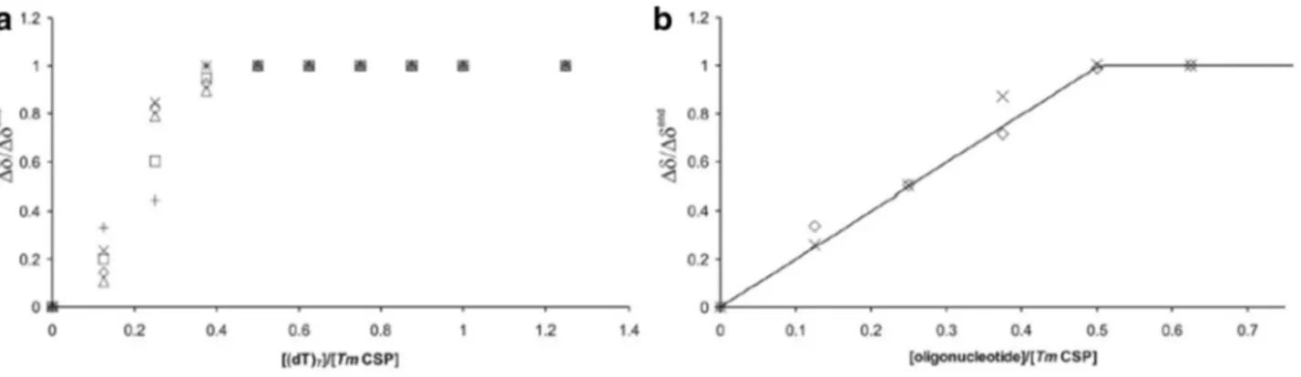
Figure 7a shows the normalized chemical shift changes
Fig. 4
Residue specific chemi- cal shift perturbations by the oligonucleotides (dT)
7and (rU)
7. The residue and amino acid specific combined chemi- cal shift perturbations ∆δ
comb[37] for the (rU)
7RNA (top) and (dT)
7ssDNA (bottom) were calculated from the 1D and 2D spectra in the absence of oligonucleotides and in the presence at molar ratios of TmCsp:oligonucleotide = 1:0.625 (dark grey bars). The horizontal lines depict standard deviations σ
0and 2 σ
0. The residues whose unperturbed resonances disappeared in the presence of oligonucleotides and could not be identified in the spectra are presented as light grey bars. Their value was set to 2 σ
0. The missing entry in posi- tion 57 represents a proline resi- due (no amide proton). (Middle) In addition, residues are marked as interaction partners in the
TmCsp sequence with moderateprobability (cursive letters, σ
0≥ ∆δ
comb< 2 σ
0) and with high probability (bold letters,
∆δ
comb≥ 2 σ
0). (Co) indicates
residues that have high interac-
tion probability for at least one
oligonucleotide and moderate to
high probability for the second
nucleotide. Boxes indicate the
limits of β-strands in TmCsp
for isolated protein signals in 1D
1H NMR spectra taken from a (dT)
7titration experiment with a molar ratio of (dT)
7:TmCsp up to 1.25:1. The data were fitted by using the K
D-values obtained from fluorescence quenching. The saturation effect of the chemical shift change at a ratio of 0.5 indicates a stoichiometry of two molecules TmCsp per (dT)
7. These chemical shift changes are in perfect agreement with the intensity changes of residues involved in slow exchange described above. We further averaged the normalized
1H and
15N chemical shift changes of clearly observable resonances in fast exchange obtained from the HSQC titration experiments. The data were fit- ted using the K
D-values from fluorescence quenching for (dT)
7and (rU)
7and eqs. 2 and 3. We got values of 1.99
± 0.05 ((dT)
7) and 2.21 ± 0.04 ((rU)
7), respectively, for the number of independent binding sites N. The average maximal chemical shift changes Δδ
end, led to Δδ/Δδ
endof 0.93±0.12 ((dT)
7) and 0.94±0.10 ((rU)
7), respectively (see Fig. 7a and b). So, the stoichiometry is close to two molecules TmCsp per heptanucleotide.
3 Discussion
3.1 Stoichiometry of Oligonucleotide Binding and Size of the Oligonucleotide Binding Site The titration experiments consistently prove that under cold shock conditions (303 K) two molecules of TmCsp are bound per uridylate or thymidylate heptamer (Figs. 1, 2 and 7). The same also holds for (dA)
7and (dC)
7(Fig. 2).
The concentrations of Csp and the oligonucleotides were carefully checked (see Materials and Methods) and could be confirmed directly by NMR spectroscopy.
No hints for a stronger interaction between the two Csp molecules are visible in the NMR spectra. We conclude that their mutual interaction must be very weak or that it must have an internal symmetry leading to equivalent chemi- cal shifts of a given nucleus in the two proteins. As a con- sequence the length of the oligonucleotide binding site is only 3 nucleotides per Csp (or, alternatively, for a dimer binding to 6 of the 7 nucleotides). In fact, dimerization in the absence of oligonucleotides was observed for BsCspB in crystals [13] and in solution [22] via strand β4, for StCspE from Salmonella typhimurium [23]. Most interesting is the interaction of a mutant of BcCspB from Bacillus caldolyti- cus [24] through its nucleic acid-binding surface in a point symmetrical arrangement. The latter structures form a sym- metric dimer with antiparallel orientation that would lead to the same chemical shifts of nuclei in equivalent positions in the two Csps in the dimer.
When compared with the binding properties of the cold shock proteins from other species, these findings let us suggest that Csp from the hyperthermophilic organism T.
maritima takes up a unique position because of its unu- sual origin. It represents a Csp homologue, which previous results cannot be transferred to. For example, chemical shift mapping with CspA from E. coli [12] and CspB from B. subtilis [18] demonstrated that both the RNP1 and the RNP2 motifs strongly contribute to DNA binding. Analy- sis of the binding stoichiometries showed that the binding sites for EcCspA and BsCspB on homogenous templates consist of 6–7 nucleotides per Csp molecule [5, 16].
3.2 Identification of Interaction Sites
Chemical shift mapping as well as mapping of inten- sity changes due to slow and intermediate exchange effects show that the location of the binding epitopes for the pyrimidine based ssDNA or RNA oligomers (dT)
7and (rU)
7on TmCsp do not differ significantly (Figs. 4 and 5). The epitope mainly comprises the last amino acids of β-strand 1 (W7, D9) and the first amino acids of β-strand
Fig. 5
Mapping of the chemical shift perturbations caused by oligo-
nucleotide binding. Residues that probably are involved (red) or not
involved (blue) by oligonucleotide binding (see Fig. 4) were mapped
onto the 3D model of pure TmCsp [8]. These representations were
generated using the program MOLMOL [35]
and are scattered along the long loop connecting β3 and β4 (S30, A31, E33, M34, F37, K38, T39, K41), and the loop between β4 and β5 (G53, K55). In strand β5 K63 and V65 see the oligonucleotide binding. Surprisingly, these bind- ing sites only partly involve the classical RNA-binding sites, which had been predicted from the sequence align- ment of 23 cold shock domains known from other CSPs and which consists of the motifs RNP1 (residues 13–20) and RNP2 (residues 25–29) [4, 12, 25, 26]. In RNP1 G13, Y14, and F16 are concerned, in RNP2 only the aromatic amino acids H28 and W29. Nevertheless, a high binding affinity in the nanomolar range is achieved at cold shock conditions.
Judging from the K
Dvalues and the gel shift assays, the other oligonucleotides interact significantly weaker with TmCsp. Nevertheless at least for the two oligonucleotides (dA)
7and (dC)
7interactions with TmCsp can be detected at the high concentrations used in the 1D NMR experiments. A detailed analysis of their binding by 2D-NMR spectroscopy
Fig. 6
Exchange time scales in the complexes TmCsp-(dT)
7and
TmCsp-(rU)7. 1 mM uniformly
15N-enriched
TmCsp in 50 mMsodium phosphate, pH 6.5, 20 mM NaCl, 0.2 mM EDTA, 0.1 mM DSS, and varying nucleotide concentrations. a Slow exchange of the side chain amide resonance of W29 in the complex with (dT)
7: By the addition of (dT)
7, the signal of the main chain amide proton of W29
in free TmCsp (W29
f) disappears, whereas the signal of W29 in com- plexed TmCsp (W29
c) appears simultaneously. b Fast exchange of the main chain amide resonance of W29 in the complex with (rU)
7: Inter- action with (rU)
7leads to a continuous shift of the signals as seen e.g.
for the side chain amide proton of W29
Table 1
Dissociation constants of TmCsp complexed with various nucleotide heptamers at different temperatures
The dissociation constants were determined by quenching of the tryp- tophan fluorescence (see Materials and Methods). For data evalua- tion, two binding sites for TmCsp per heptamer were assumed. K
Dis the dissociation constant for one binding site assuming independent binding. 343 K (70 °C): physiological temperature of T. maritima.
323 K (50 °C): temperature range in which the cold shock sets in. 303 K (30 °C): cold shock. n.d. not determined
Oligonu-
cleotide
KD(303 K) [µM]
KD(323 K)
[µM]
KD(343 K) [µM]
(dA)
75.0 ± 0.2 19.2 ± 0.5 25.2 ± 0.6
(dC)
72.8 ± 0.1 n.d. 11.0 ± 0.1
(dG)
73.4 ± 0.1 n.d. 5.6 ± 0.1
(dT)
7(4.0 ± 0.2)·10
−30.44 ± 0.02 1.6 ± 0.04 (rU)
7(10.8 ± 0.8)·10
−31.52 ± 0.01 17.4 ± 0.1
2 (G13, Y14, F16) and the loop region in between the
two strands (S10, K11, K12). Further interaction points
include H28 and W29, the last amino acids of strand β3,
has not been performed. Concerning the side chains of the two tryptophan residues qualitatively the same behavior is observed, both nucleotides influence the shifts of the H
ε1-resonance of W7 and W29, but size and direction of the shifts vary considerably. A moderate downfield shift of the H
ε1-resonance of W29 is induced by (dT)
7and a small upfield shift by (dC)
7but a large upfield shift is caused by binding of (dA)
7(Fig. 2). For all three nucleotides upfield shifts of the H
ε1-resonances of W7 are observed, a very large shift for (dT)
7, a moderate shift for (dC)
7, and a very weak shift for (dA)
7. This suggests that the two tryptophan residues take part in the recognition of the thymine–base.
If the shifts of the tryptophan H
ε1-resonances are mainly ring-current shifts by the bases, it indicates that thymine is optimally oriented relative to the tryptophan residues and that also the adenine ring has to come close to W29 but with an orientation different from the pyrimidine bases.
Under our conditions the binding site of a TmCsp mol- ecule for (rU)
7or (dT)
7may be only 3 nucleotides long.
In consequence, the question arises whether this is true for selected heptapeptides with different nucleotide sequences.
Csp from T. maritima is the only one which possesses a tryp- tophan residue (W29) instead of a highly conserved pheny- lalanine or tyrosine residue at the end of RNP2 [8]. During the 1D NMR titration experiments, the chemical shift of this residue (W29) changed to different extents according to the kind of oligonucleotide added. The two pyrimidine nucleotides lead to a downfield shift of the H
ε1-resonance of W29 with a larger shift for (dT)
7. However, the high-field shift induced (dA)
7,is much larger and may indicate that in TmCsp this tryptophan favors an interaction with an adenine residue.
3.3 TmCsp as an Important Sensor Switching to the Cold Shock Response
When discussing the TmCsp-oligonucleotide interaction, it is important to have in mind that Csp itself is part of a tempera- ture sensing system or even may be the main temperature sen- sor for switching on and off the cold shock response. Under cold shock conditions, cold shock proteins have two different cellular functions, namely (1) the unspecific inhibition of the expression of the majority of proteins at high Csp concen- trations and (2) the specific activation of cold chock related proteins (for a review see e. g. [2]). At physiological growing conditions, these regulatory functions should be switched off.
At the millimolar concentrations of Csps present at cold shock conditions transcription as well as translation is down- regulated completely as studies in cell free systems show. For TmCsp 50% inhibition of the two processes is obtained at a concentration of 140 μM. Inhibition can completely be abol- ished by addition of the hepta-2′-desoxy-thymidylate (dT)
7that competitively binds to TmCsp with high affinity [20]. This is in line with our binding data since all studied purine and pyrimi- dine heptanucleotides bind with sufficient micromolar affinity to single stranded RNA and DNA under cold shock conditions (Table 1). Especially, Csp seems to be able to form larger, prob- ably more stable clusters on the nucleotides inhibiting transcrip- tion as well as translation. This is in agreement with our study that shows that at cold shock temperatures TmCsp forms dimers with heptanucleotides. It is most likely that longer nucleotides would also lead to larger nucleoprotein complexes.
The second mechanism, initializing and propagating expression of heat shock related proteins would require rec- ognition of specific nucleotide sequences with high affin- ity. For this aim the recognition of a sequence motif larger than three nucleotides would be required when only one Csp molecule would bind. However, the problem could be
Fig. 7
Stoichiometries of nucleic acid binding from NMR data. a Relative chemical shift change for isolated protein signals in 1D
1
H NMR spectra caused by addition of (dT)
7(see Fig.
2) indicat-ing a stoichiometry of two TmCsp molecules per (dT)
7. The chemi- cal shifts were normalized by setting the chemical shift at the high-
est (dT)
7.concentration to 1. b Mean values of the relative chemical
shift change taken from HSQC titration experiments (see Fig.
3)with (dT)
7(◊) and (rU)
7(X). The solid line symbolizes the theo-
retical binding curve as defined by (Eqs.
2 and 4) with N = 2 andΔδ/Δδ
end= 1
solved even with binding of a short sequence of three nucle- otides recognized by a single Csp-molecule when more than one Csp would bind cooperatively to a longer nucleotide sequence as observed experimentally in this study.
Generally, a preference for pyrimidine based oligonucleo- tides was detected for TmCsp as it had been earlier described for BsCsp [5, 6]. This specificity is rather small at optimal growing conditions where also the affinity of Csp for all nucle- otides studied here is quite low (Table 1). However, at cold shock conditions (303 K) the protein showed a nanomolar affinity for thymidylate or uridylate heptamers but binds with three orders of magnitude lower affinities to purine nucleo- tides and to the pyridine heptamer (dC)
7. Under cold shock conditions for the affinity to the ssDNA or RNA templates (dT)
7and (rU)
7no major differences were observed in line with the dual function of Csp in transcription and translation.
We obtained most of our information concerning the oli- gonucleotide-Csp interaction at cold shock conditions. The gel shift assays were performed at room temperature with nucleotide concentrations in the nanomolar range (0.5 μM) and maximum Csp concentrations of 100 μM ((dA)
7, (dC)
7, (dG)
7) or 1.5 μM (((dT)
7, (rU)
7). No clear binding activity was observed with any of the purine based substrates, while (dT)
7and (rU)
7show strong binding. The NMR titrations were performed at a Csp-concentration of 1 mM at 303 K.
Because of this high concentration, they are not suited for a quantitative determination of dissociation constants but for the determination of the stoichiometry of binding (see above). Quantitative binding constants were obtained by fluorescence quenching (Table 1). At 303 K and Csp con- centrations between 5 nM and 1 μM, the dissociation con- stants of (dT)
7and (rU)
7are very small with 4 and 10.8 nM, respectively. For (dA)
7, (dC)
7, and (dG)
7they are approxi- mately 3 orders of magnitude higher with 5.0, 2.8, and 3.4 μM, respectively. As a consequence a preferential interaction of Csp with nucleotides containing uracil or thymine is to be expected, although at the typical total concentrations of Csp of 100 μM during cold shock [2] also unspecific interac- tions should occur. At an almost physiological growing tem- perature for T. maritima of 343 K, the affinity of Csp to the model single stranded is significantly lower. It is very similar for all nucleotides studied here (Table 1) that is the specific recognition of nucleotides is decreased much. Together with the reduced Csp concentration at physiological temperatures, it means that in the cellular environment most of the nucleo- tide interactions are switched off as observed.
The temperature switch can now be understood from the structural perspective since at high non-cold shock tempera- tures the interaction with residues interacting specifically with uracil or thymine bases at low temperatures have to be per- turbed. Since NMR structures of TmCsp at 303 K [8] and 343 K [36] are available, we have a structural basis for estimating differences of the cold shock and the physiological state with
respect to nucleotide binding. From the chemical shift per- turbation β-strands 1 (W7, D9) and 2 (G13, Y14, F16) and the loop region in between the two strands (S10, K11, K12) appear to be involved in the nucleotide interaction. At cold shock temperatures, the H
ε1-resonance of W7 shows very large upfield shift after binding of (rU)
7or (dT)
7that is not observed for the other oligonucleotides studied here. This suggests that a specific interaction of these two bases with the tryptophan ring exists causing an upfield ring current. At physiological high temperature this region is destabilized, especially a partial melting of β-strand 1 is observed including amino acids K6 and W7 that characteristically change their orientation in the structure [36]. A second characteristic temperature induced large conformational change was observed for the large loop (E52 to P57) between β-strand 4 and β-strand 5. At 303 K and 343 K, it adopts a well-defined orientation that differs signifi- cantly, at 328 K an equilibrium between these orientations is observed. Again, significant chemical shift perturbation after binding are observed here (G53, K55).
Directly after switching to low temperature, the Csp concentration is still rather small. Here, even higher affin- ity and specificity may be required. This suggests that more elaborated nucleic acid sequences may be recognized. It is likely that they contain at least a short sequence of rU or dT (probably three units/Csp according to our data). If a longer sequence would be required, the presence of at least one dif- ferent base in a well-positioned place within the thymidylate- rich nucleic acid sequence would make sense. A cytidine is not a likely candidate because of the relative weak response in the NMR-spectra of the Csp (dC)
7complex (Fig. 2). One the other hand, the (dA)
7has a very strong shift effect on the H
ε1resonance of W29 suggesting a stacking interaction of its base with the tryptophan side chain. One can speculate what the recognition site of TmCsp may look like. When we assume that (1) the binding of two TmCsps is preferen- tial, that (2) RNP1 also is a physiological interaction site requiring the pyrimidine nucleotides dT or rU in the TmCsp- complex, and that (3) a direct interaction with the purine nucleotide (dA)
7and W29 at the end of RNP2 is proposed from the NMR-data, then a palindromic sequence like AUU UUU UA and UUU AUU U (or ATT TTT TA and TTT ATT T in case of DNA binding) would satisfy these conditions.
Since we did not test the oligonucleotide (dG)
7by NMR, we cannot exclude that here the guanine base is even more spe- cific. These findings support the notion that W29 plays a major role in the binding process. It is conceivable that in the case of (dT)
7or (rU)
7some steric hindrance occurs because of the non-optimal intercalation of W29 into these oligonucleotides which blocks the access of the RNP2 site to the DNA or RNA.
Future experiments will include an in vitro selection
approach (SELEX) [27] to determine the optimal nucleotide
binding sequence for TmCsp. Additionally, a point mutation
experiment will be performed, in which W29 will be mutated
to a phenylalanine or tyrosine residue, which are highly con- served in the sequences of cold shock proteins of most species.
In this way we will be able to distinguish whether the dimeri- zation of TmCsp was caused by the use of non-optimal tem- plates, or whether such a short nucleotide sequence represents its true binding site and therefore a new binding mode. The excellent binding affinity found for the pyrimidine templates would argue for the latter possibility.
4 Conclusion
At 343 K, close to the physiological temperatures for optimal growth of T. maritima [2] of 353 K, the affinities of all DNA and RNA homopolymers studied is relatively low, indicating that specific cold shock response by TmCsp is switched off.
The obtained affinities towards the homopolymers is close to 3.4 μM (at 353 K) estimated earlier in an in-vitro transcription- translation system for arbitrary genes [20]. Under cold shock conditions the affinities of (dT)
7and (rU)
7increase by three orders of magnitude, whereas the other oligonucleotides do not change their affinity much. This would perfectly fit to a picture where as specific cold shock response Csp would rec- ognize nucleotide sequences containing poly-U or poly-T, but where the affinity of Csp after its millimolar concentration has been reached is sufficient to inhibit general protein expres- sion. Here also multimerization of the bound Csp molecules could strengthen the inhibitory effect. The specific recognition sequences required for activation of the expression of cold shock induced proteins are not known yet but probably are rich in rU or dT. From the side of Csp, the unspecific inhibitory effect on protein transcription and translation does not require specific, temperature dependent conformational changes, since even at physiological temperatures its affinity to a variety of sequences is sufficient. However, the specific increase of affin- ity for poly-U or poly-T sequences under cold shock condi- tions requires specific conformational changes in the protein that switch on the cold shock response by a strongly increased affinity to the specific recognition sequences, analogous to the dramatic increase of the affinity for (rU)
7or (dT)
7by three orders of magnitude we observed here.
5 Materials and Methods 5.1 Protein Purification
For the expression of unlabeled protein, E. coli Rosetta (DE3) pLysS cells transformed with the plasmid coding for TmCsp were grown in Luria Bertani medium in the presence of 50 µg/ml carbenicillin and 68 µg/ml chloramphenicol to an OD
600of 1, or, for the expression of
15N isotopically enriched protein, in New Minimal Medium [28] containing 1 g/ L
15NH
4Cl and 2 g/ L glucose at 37°C to an OD
600of
0.8, respectively. Expression was induced by adding 1 mM isopropyl β-D-thiogalactopyranoside, and bacterial growth was continued for 3 h. Purification of the cold shock pro- tein was performed as described [29]: to remove the bulk of the E. coli proteins without significant co-precipitation of TmCsp, the cell extract was diluted fourfold with buffer and then heated to 70°C for 20 minutes. Pure TmCsp was obtained by hydrophobic interaction chromatography at pH 8 and size exclusion chromatography. Its concentration was determined photometrically using an extinction coefficient ε
280of 12,660 M
−1cm
−1.
5.2 Oligonucleotides
All unlabeled and 5′-fluorescein-labeled ssDNA and RNA oligonucleotides were purchased in HPSF-quality (Highly Purified Salt Free) from MWG Biotech (Ebersberg, Ger- many) except for the unlabeled RNA, which was purchased from BioSpring (Frankfurt/Main, Germany). The concentra- tions of the unlabeled oligonucleotides were calculated from their absorbance at 260 nm. The extinction coefficients ε
260used for the individual oligomers were 15,400 M
−1cm
−1for (dA)
7,7300 M
−1cm
−1for (dC)
7, 11,700 M
−1cm
−1for (dG)
7, and 8400 M
−1cm
−1for (dT)
7and (rU)
7[30].
5.3 NMR Spectroscopy
NMR samples of unlabeled and uniformly
15N-labeled TmCsp contained 0.5 mM and 1 mM protein, respectively, in 50 mM NaH
2PO
4(pH 6.5), 20 mM NaCl, 0.2 mM EDTA (sodium salt), 0.1 mM DSS in
1H
2O/
2H
2O (92%/8%), and oligonucleotides in varying concentrations. The NMR exper- iments were carried out on a Bruker DRX-600 spectrometer (
1H resonance frequency 600 MHz) at 303 K.
1H–
15N het- eronuclear single-quantum coherence (HSQC) spectra [31]
were recorded with an echo/anti-echo-gradient selection in ω
1[32], the digital resolution was 4.75 Hz per point for
15N and 5.86 Hz per point for
1H.
The proton chemical shifts were referenced to sodium- 2,2-dimethylsilapentane-5-sulfonic acid (DSS) used as an internal reference.
15N chemical shifts were referenced indi- rectly to DSS using an X-value of 0.101329118 [33]. Spectral analysis and peak picking were performed using the program AUREMOL (available at http://www.aurem ol.de) [34].
5.4 NMR Data Evaluation
For the identification of the interaction sites, amino acid and
atom specific combined chemical shifts [37] were calculated
using the tool implemented in the program AUREMOL
(www. auremol.de). The Euclidian norm was used. Note
that the implementation in AUREMOL in its version from November 2019 now contains weighting factors for all types of atoms in the 20 amino acids (not only the backbone atoms as described in [37]). In our case not only the backbone N and H shifts were used but we could also include the effects on the tryptophan N
ε1and H
ε1, as well as the amino groups of the glutamine side chains. The combined chemical shift perturbation can be applied for fast and slow exchange as long as the chemical shifts in the absence and presence of the ligands under saturation conditions are known. If in the slow or intermediate exchange region the chemical shifts in the complex could not be identified, they were treated separately.
A significant interaction has been assumed
The probability P
ABthat a molecule TmCsp is bound to an oligonucleotide is given by (Eq. 1), where c
Aand c
Bare the total concentrations of TmCsp and the heptanucleotide in solu- tion, N is the number of independent binding sites on the hep- tanucleotide and K
Dis the dissociation constant. Equation 1 assumes that there are N independent and equal binding sites.
With K
Din the nanomolar and c
Aand c
Bin the millimolar range, Eq. 1 can be simplified to Eq. 2, neglecting K
D.
For signals under fast chemical exchange regime, the chemical shift δ is given by (Eq. 3). δ
Aand δ
ABare the chem- ical shifts of free and bound protein. The relative change of the chemical shifts is given by Eq. 4 with Δδ = δ − δ
Aand Δδ
end= δ
AB− δ
A.
5.5 Electrophoretic Mobility Shift Assay
The binding reaction was performed by incubating 7.5 pmol of 5′-fluorescein-labeled ssDNA or RNA with differ- ent amounts of protein in NMR buffer (50 mM NaH
2PO
4, pH 6.5, 20 mM NaCl, and 0.2 mM EDTA) at room tem- perature for 1h (total volume 15 µl). Prior to gel elec- trophoresis, 5 µl of dye solution (20% glycerol, 0.034%
bromphenol blue) were added to the samples. Native gel electrophoresis was performed at 4 °C in 1xTBE buffer (5xTBE buffer: 54 g Tris, 27.5 g boric acid, 20 ml EDTA 0.5 M) through a 20% mini-polyacrylamide gel at 80 V until the dye had reached the bottom of the gel (ca. 7 h).
(1) P
AB= 1
2 c
A(
c
A+ Nc
B+ K
D−
√
( c
A+ Nc
B+ K
D)
2− 4 Nc
Ac
B)
(2) P
AB= 1
2 c
A( c
A+ Nc
B− |
| c
A− Nc
B|
| )
(3) 𝛿 = P
ABδ
AB+ ( 1 − P
AB)δ
AΔ𝛿 (4)
Δ𝛿
end= P
ABThe fluorescent oligonucleotides were detected by expos- ing the gels to UV light (302 nm). Then the gels were silver-stained to localize the position of the protein.
5.6 Fluorescence Spectroscopy
The dissociation constants of TmCsp complexed with vari- ous nucleotide heptamers (Table 1) were determined by quenching of the intrinsic tryptophan fluorescence of the nucleoprotein complex as described by Zeeb and Balbach [26] assuming a stoichiometry of two protein molecules bound to one oligonucleotide. Titration experiments at 303 K were carried out with protein concentrations of 5 nM ((dT)
7), 25 nm ((rU)
7) or 1 µM ((dC)
7, (dA)
7, (dG)
7). For the determination of the K
Dvalues at 343 K TmCsp con- centrations of 1 µM ((dT)
7, (rU)
7) or 5 µM ((dC)
7, (dA)
7, (dG)
7) were used. The successive addition of oligonucleo- tides was performed until the protein was fully saturated with the complex partner and no further quenching of the Trp fluorescence was detectable.
Acknowledgements
We are grateful to the DFG (Kr1407/4-1) to sup- port this study. We thank Claudia Munte for valuable help in the final stages of the project.
Funding
Open Access funding enabled and organized by Projekt DEAL.
Open Access
This article is licensed under a Creative Commons Attri- bution 4.0 International License, which permits use, sharing, adapta- tion, distribution and reproduction in any medium or format, as long as you give appropriate credit to the original author(s) and the source, provide a link to the Creative Commons licence, and indicate if changes were made. The images or other third party material in this article are included in the article’s Creative Commons licence, unless indicated otherwise in a credit line to the material. If material is not included in the article’s Creative Commons licence and your intended use is not permitted by statutory regulation or exceeds the permitted use, you will need to obtain permission directly from the copyright holder. To view a copy of this licence, visit http://creat iveco mmons .org/licen ses/by/4.0/.
References
1. Graumann PL, Marahiel MA (1998) A superfamily of pro- teins that contain the cold-shock domain. Trends Biochem Sci 23:286–290
2. Horn G, Hofweber R, Kremer W, Kalbitzer HR (2007) Structure and function of bacterial cold shock proteins. Cell Mol Life Sci 64:1457–1470
3. Jiang W, Hou Y, Inouye M (1997) CspA, the major cold-shock protein of escherichia coli, is an RNA chaperone. J Biol Chem 272:196–202
4. Schroder K, Graumann P, Schnuchel A, Holak TA, Marahiel
MA (1995) Mutational analysis of the putative nucleic acid-
binding surface of the cold-shock domain, Cspb, revealed an
essential role of aromatic and basic residues in binding of
single-stranded-DNA containing the Y-Box motif. Mol Micro- biol 16:699–708
5. Lopez MM, Yutani K, Makhatadze GI (1999) Interactions of the major cold shock protein of bacillus subtilis CspB with single- stranded DNA templates of different base composition. J Biol Chem 274:33601–33608
6. Lopez MM, Yutani K, Makhatadze GI (2001) Interactions of the cold shock protein CspB from bacillus subtilis with single- stranded DNA. Importance of the T base content and position within the template. J Biol Chem 276:15511–15518
7. Martin A, Sieber V, Schmid FX (2001) In-vitro selection of highly stabilized protein variants with optimized surface. J Mol Biol 309:717–726
8. Kremer W, Schuler B, Harrieder S, Geyer M, Gronwald W, Wel- ker C, Jaenicke R, Kalbitzer HR (2001) Solution NMR structure of the cold-shock protein from the hyperthermophilic bacterium Thermotoga maritima. Eur J Biochem 268:2527–2539 9. Mueller U, Perl D, Schmid FX, Heinemann U (2000) Thermal
stability and atomic-resolution crystal structure of the Bacillus caldolyticus cold shock protein. J Mol Biol 297:975–988 10. Schindelin H, Marahiel MA, Heinemann U (1993) Universal
nucleic acid-binding domain revealed by crystal-structure of the bacillus-subtilis major cold-shock protein. Nature 364:164–168 11. Schnuchel A, Wiltscheck R, Czisch M, Herrler M, Willimsky G,
Graumann P, Marahiel MA, Holak TA (1993) Structure in solu- tion of the major cold-shock protein from bacillus-subtilis. Nature 364:169–171
12. Newkirk K, Feng W, Jiang W, Tejero R, Emerson SD, Iouye M, Montelione GT (1994) Solution NMR structure of the major cold shock protein (CspA) from escherichia coli: Identification of a binding epitope for DNA. Proc Natl Acad Sci USA 91:5114–5118 13. Schindelin H, Jiang W, Inouye M, Heinemann U (1994) Crystal
structure of CspA, the major cold shock protein of escherichia coli. Proc Natl Acad Sci USA 91:5119–5123
14. Bandziulis RJ, Swanson MS, Dreyfuss G (1989) RNA-binding proteins as developmental regulators. Genes Dev 3:431–437 15. Burd CG, Dreyfuss G (1994) Conserved structures and diversity
of functions of rna-binding proteins. Science 265:615–621 16. Sachs R, Max KEA, Heinemann U, Balbach J (2012) RNA single
strands bind to a conserved surface of the major cold shock protein in crystals and solution. RNA 18:65–76
17. Max KE, Zeeb M, Bienert R, Balbach J, Heinemann U (2006) T-rich DNA single strands bind to a preformed site on the bacte- rial cold shock protein Bs-CspB. J Mol Biol 360:702–714 18. Zeeb M, Max KEA, Weininger U, Löw C, Sticht H, Balbach
J (2006) Recognition of T-rich single-stranded DNA by the cold shock protein Bs-CspB in solution. Nucleic Acids Res 34:4561–4571
19. Lopez MM, Makhatadze GI (2000) Major cold shock proteins, CspA from Escherichia coli and CspB from Bacillus subtilis, interact differently with single-stranded DNA templates. Biochim Biophys Acta - Protein Struct Mol Enzym 1479:196–202 20. Hofweber R, Horn G, Langmann T, Balbach J, Kremer W, Schmitz
G, Kalbitzer HR (2005) The influence of cold shock proteins on transcription and translation studied in cell-free model systems.
FEBS J. 272:4691–4702
21. Freischmidt A, Hiltl J, Kalbitzer HR, Horn-Katting G (2013) Enhanced in vitro translation at reduced temperatures using a cold-chock RNA motif. Biotech. Lett. 35:389–395
22. Makhatadze GI, Marahiel MA (1994) Effect of pH and phosphate ions on self-association properties of the major cold-shock protein from Bacillus subtilis. Protein Sci 3:2144–2147
23. Morgan HP, Wear MA, McNae I, Gallagher MP, Walkinshaw MD (2009) Crystallization and X-ray structure of cold-shock protein E from Salmonella typhimurium. A Crystallogr Sect F Struct Biol Cryst Commun 65:1240–1245
24. Carvajal AI, Vallejos G, Komives EA, Castro-Fernandez V, Leon- ardo DA, Garratt RC, Ramırez-Sarmiento CA, Babul J (2017) Unusual dimerization of a BcCsp mutant leads to reduced con- formational dynamics. FEBS J 284:1882–1896
25. Kloks CPAM, Spronk CAEM, Lasonder E, Hoffmann A, Vuister GW, Grzesiek S, Hilbers CW (2002) The solution structure and DNA-binding properties of the cold-shock domain of the human Y-box protein YB-1. J Mol Biol 316:317–326
26. Zeeb M, Balbach J (2003) Single-stranded DNA binding of the cold-shock protein CspB from Bacillus subtilis: NMR mapping and mutational characterization. Prot Sci 12:112–123
27. Tuerk C, Gold L (1990) Systematic evolution of ligands by expo- nential enrichment - RNA ligands to bacteriophage-T4 Dna-pol- ymerase. Science 249:505–510
28. Budisa N, Steipe B, Demange P, Eckerskorn C, Kellermann J, Huber R (1995) High-level biosynthetic substitution of methio- nine in proteins by its analogs 2-aminohexanoic acid, selenom- ethionine, telluromethionine and ethionine in escherichia-coli. Eur J Biochem 230:788–796
29. Welker C, Bohm G, Schurig H, Jaenicke R (1999) Cloning, over- expression, purification, and physicochemical characterization of a cold shock protein homolog from the hyperthermophilic bacte- rium Thermotoga maritima. Prot Sci 8:394–403
30. Wallace RB, Miyada CG (1987) Oligonucleotide probes for the screening of recombinant DNA libraries. Meth Enzym 152:432–442
31. Palmer AG, Cavanagh J, Wright PE, Rance M (1991) Sensitiv- ity improvement in proton-detected 2-dimensional heteronuclear correlation NMR-spectroscopy. J Magn Reson 93:151–170 32. Schleucher J, Schwendinger M, Sattler M, Schmidt P, Sched-
letzky O, Glaser SJ, Sorensen OW, Griesinger C (1994) A gen- eral enhancement scheme in heteronuclear multidimensional Nmr employing pulsed-field gradients. J Biomol NMR 4:301–306 33. Markley JL, Bax A, Arata Y, Hilbers CW, Kaptein R, Sykes BD,
Wright PE, Wuthrich K (1998) Recommendations for the presen- tation of NMR structures of proteins and nucleic acids - IUPAC- IUBMB-IUPAB inter-union task group on the standardization of data bases of protein and nucleic acid structures determined by NMR spectroscopy. Eur J Biochem 256:1–15
34. Gronwald W, Kalbitzer HR (2004) Automated structure deter- mination of proteins by NMR spectroscopy. Progr NMR Spectr 44:33–96
35. Koradi R, Billeter M, Wuthrich K (1996) MOLMOL: a program for display and analysis of macromolecular structures. J Mol Graph 14:51–52
36. Jung A, Bamann C, Kremer W, Kalbitzer HR, Brunner E (2004) High-temperature solution NMR structure of TmCsp. Prot Sci 13:342–350
37. Schumann FH, Riepl H, Maurer T, Gronwald W, Neidig K-P, Kalbitzer HR (2007) Combined chemical shift changes and amino acid specific chemical shift mapping of protein-protein interac- tions. J Biomol NMR 39:275–289
Publisher’s Note

![Fig. 3 Changes in the [ 1 H- 15 N]- N]-HSQC spectrum of 15 N-TmCsp induced by the oligonucleotides (dT) 7 and (rU) 7](https://thumb-eu.123doks.com/thumbv2/1library_info/3731281.1508626/6.892.307.812.84.1042/fig-changes-h-hsqc-spectrum-tmcsp-induced-oligonucleotides.webp)