Molecular mechanisms of mitochondrial DNA replication
Inaugural-Dissertation
zur
Erlangung des Doktorgrades
der Mathematisch-Naturwissenschaftlichen Fakultät
der Universität zu Köln
vorgelegt von
STANKA MATIĆ
aus Ljubovija
2 Berichterstatter: Prof. Dr. Nils-Göran Larsson
Prof. Dr. Aleksandra Trifunovic
Tag der mündlichen Prüfung: 16/01/2017
Cover page and title
3
Molecular mechanisms of mitochondrial DNA replication
PhD Thesis
Submitted by Stanka Matic Supervisor: Nils-Göran Larsson
Graduate School for Biological Sciences, University of Cologne
GRADUATE SCHOOL
FOR BIOLOGICAL SCIENCES
UNIVERSITY OF COLOGNE
“The nitrogen in our DNA, the calcium in our teeth, the iron in our blood, the carbon in our apple pies were made in the interiors of collapsing stars. We are made of starstuff.”
Carl Sagan, Cosmos
“Scientific research is one of the most exciting and rewarding of occupations. It is like a voyage of discovery into unknown lands, seeking not for new territory but for new knowledge. It should appeal to those with a good sense of adventure.”
Frederick Sanger
5
Abstract
Mitochondria are cellular organelles responsible for energy conversion to form the cell’s energy currency, adenosine triphosphate (ATP), through the process of oxidative phosphorylation. In addition, mitochondria play a vital role in diverse cellular processes including apoptosis, calcium homeostasis and intracellular signalling. As a consequence, mitochondrial dysfunction can lead to numerous disorders that display variability in clinical presentation and tissue specificity. Mitochondria contain their own genome, known as mitochondrial DNA (mtDNA), and have distinct enzymes involved in mtDNA expression and maintenance. Even though the core components of the machinery necessary for mtDNA replication have been identified and reconstituted in vitro, its underlying regulatory mechanisms are largely unknown. The displacement (D) loop, a triple stranded structure that is formed by premature replication termination generating the 7S DNA, likely plays an important role in the control of mammalian mtDNA replication in response to cellular bioenergetics demands.
The work described in this thesis aimed to study the effects of the mitochondrial replicative helicase TWINKLE and the mitochondrial nuclease MGME1 on mtDNA replication regulation. Both of these factors were previously described to impact 7S DNA levels.
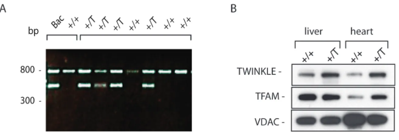
The effects of TWINKLE on mtDNA levels were studied by generating Twinkle bacterial artificial chromosome (BAC) transgenic mice. This TWINKLE overexpressor model showed that TWINKLE upregulation leads to an increased replication and augmented mtDNA copy number.
To study the in vivo role of the MGME1 nuclease in mtDNA replication regulation, Mgme1 knockout mice were generated and analyzed. Our results show that MGME1 is not essential for mouse embryonic development and survival. This MGME1 knockout model showed mtDNA depletion, and accumulation of an 11-kb linear mtDNA fragment that spans the entire major arc of the mtDNA and is present in different mouse tissues. Interestingly, a similar linear fragment was also present in
6 mice carrying an exonuclease deficient DNA polymerase (mtDNA mutator mice). These mice show a progeroid phenotype that is likely not driven by linear deletions as Mgme1 knockout mice do not display a premature ageing phenotype. Finally, we dissected the role of MGME1 in mtDNA replication and transcription. The lack of abortive replication events and diminished H-strand transcription termination in Mgme1 knockout mice suggests a possible role of MGME1 in the regulation of those processes at the end of the D-loop region.
7
Zusammenfassung
Mitochondrien sind essentielle Zellorganellen, die durch den Prozess der oxidativen Phosphorylierung für die Energiegewinnung der Zelle in Form von Adenosintriphosphat (ATP), zuständig sind. Zusätzlich spielen Mitochondrien eine wesentliche Rolle in verschiedenen zellulären Prozessen, einschließlich Apoptose, Kalziumhomöostase und intrazellulärer Signalwege. Daraus folgt, dass mitochondriale Dysfunktion zu einer Vielzahl von Funktionsstörungen führen kann, die eine ganze Bandbreite von klinischen Befunden und gewebsspezifischen Auswirkungen widerspiegeln. Mitochondrien besitzen ihr eigenes Genom, bekannt als mitochondriale DNA (mtDNA), und haben eigene, spezielle Enzyme, die bei der mtDNA Expression und Replikation involviert sind. Obwohl die Hauptkomponenten, die zur Replikation der mtDNA benötigt werden, bekannt sind und in vitro rekonstruiert wurden, sind die zugrunde liegenden regulatorischen Mechanismen weitestgehend unbekannt. Der Displacement (D) Loop, eine dreisträngige Struktur, die durch einen verfrühten Replikationsabbruch entsteht und aus der die 7S DNA hervorgeht, spielt vermutlich in Säugetieren eine wichtige Rolle bei der Kontrolle der mtDNA Replikation als Antwort auf bioenergetische Bedürfnisse.
Die hier vorliegende Arbeit befasst sich mit der Untersuchung der Effekte der mitochondrialen replikativen Helikase TWINKLE und der mitochondrialen Nuklease MGME1 auf die Regulation der Replikation von mtDNA. In der Vergangenheit wurde bereits für beide Faktoren gezeigt, dass sie die Quantität von 7S DNA beeinflussen.
Die Auswirkungen von TWINKLE auf die mtDNA Level wurden anhand von transgenen Mäusen, die Twinkle in einem künstlichen Bakterienchromosom (BAC) tragen und überexprimieren, untersucht. Anhand dieses Modells konnte gezeigt werden, dass die Hochregulierung von TWINKLE zu einer vermehrten Replikation und erhöhten Kopienzahl von mtDNA führt.
Um die in vivo Rolle der Nuklease MGME1 während der Regulation der mtDNA Replikation zu untersuchen, wurden Mgme1 knockout Mäuse generiert und analysiert.
Unsere Ergebnisse zeigen, dass MGME1 für die Entwicklung und Vitalität von
8 Mausembryonen nicht essentiell ist. Die Mäuse des MGME1 Knockout Modells zeigten eine Depletion der mtDNA und eine Akkumulierung eines linearen 11-kb-Fragments von mtDNA, welches den gesamten major arc der mtDNA umfasst und in verschiedenen Mausgeweben präsent ist. Interessanterweise gibt es ein vergleichbares lineares Fragment in Mäusen, die eine mutierte DNA Polymerase mit defizienter Exonuklease besitzen (mtDNA Mutator Mäuse). Diese Mäuse zeigen einen vorzeitigen Alterungsphänotyp, der vermutlich nicht von linearen Deletionen verursacht wird, da dies bei Mgme1 knockout Mäuse nicht der Fall ist. Abschließend untersuchten wir die Rolle von MGME1 sowohl bei der mitochondrialen Replikation als auch bei der Transkription. Das Ausbleiben verfrühter Replikationsabbrüche sowie verminderter Termination der H-Strang Transkription in Mgme1 knockout Mäusen deutet auf eine mögliche Rolle von MGME1 bei der Regulation dieser wichtigen Prozesse am Ende der D-Loop Region hin.
9
Contents
Cover page and title ... 2
Erklärung ... 98
Abstract ... 5
Zusammenfassung ... 7
Contents ... 9
Table of figures ... 12
Table of abbreviations ... 14
1. INTRODUCTION ... 17
1.1 Mitochondrial origin ... 17
1.2 Mitochondrial function and form ... 18
1.3 Organization of mtDNA ... 20
1.4 Replication of mtDNA ... 22
1.4.1 The strand-displacement model ... 23
1.4.2 The strand-coupled model ... 24
1.4.3 RITOLS model ... 24
1.4.4 Mitochondrial non-coding region (D-loop region) ... 25
1.4.5 Mitochondrial DNA polymerase POLγ ... 26
1.4.6 Mitochondrial replicative helicase - TWINKLE ... 27
1.4.7 Mitochondrial single-stranded DNA binding protein - mtSSB ... 29
1.4.8 Additional proteins involved in mitochondrial DNA replication ... 30
1.4.9 Mitochondrial genome maintenance exonuclease 1 – MGME1 ... 31
1.5 Transcription of mtDNA ... 32
1.5.1 The basic components of the mitochondrial transcription machinery ... 32
1.5.2 Regulation of transcription termination ... 34
1.6 Mitochondrial DNA repair ... 35
1.7 Mitochondrial genetics and diseases ... 37
10
1.7.1 Mitochondrial disorders caused by mtDNA mutations ... 39
1.7.2 Mitochondrial disorders caused by nDNA mutations ... 39
2. AIMS ... 41
3. RESULTS ... 42
3.1 Twinkle overexpression leads to elevated mtDNA levels ... 42
3.2 Generation and verification of Mgme1 knockout mice ... 47
3.2.1 Mgme1 knockout mice display multiple mtDNA deletions and depletion of mtDNA in various tissues ... 49
3.2.2 Mgme1 knockout mice have an increase in steady-state levels of 7S DNA but severely diminished de novo synthesis of 7S DNA ... 52
3.2.3 MGME1 deficiency results in accumulation of 7S DNA with extended 5 ends ... 55
3.2.4 Lack of MGME1 affects mitochondrial transcription differentially ... 56
3.2.6. Mgme1 deficiency causes stalled mtDNA replication close to OL in liver and accumulation of replication intermediates ... 60
3.2.7. Sequence coverage for Mgme1 knockout mice from different tissues ... 63
3.2.8 Steady-state protein levels in Mgme1 knockout mice ... 66
4. DISSCUSSION ... 68
5. METHODS ... 76
Generation of MGME1 knockout mice ... 76
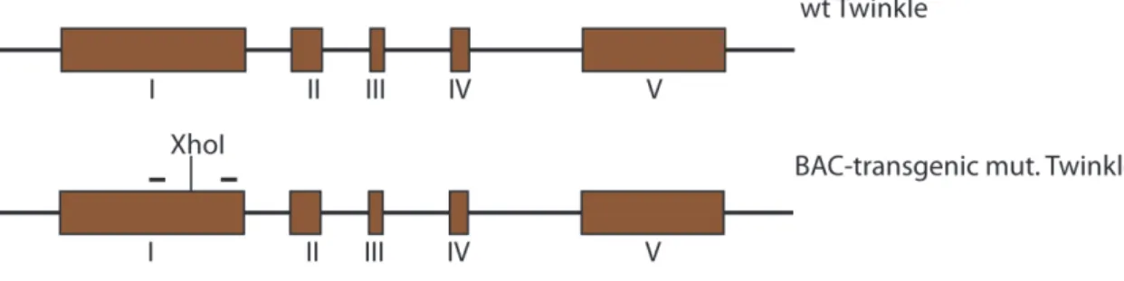
Generation of BAC and BAC FLAG TWINKLE transgenic mice ... 76
Southern blot analysis ... 77
De novo DNA synthesis ... 77
Northern blot analysis ... 78
De novo Transcription Assay ... 78
Quantitative PCR ... 78
Western Blot analysis ... 79
Long-extension PCR ... 79
Phenol chloroform extraction ... 79
DNA extraction with Puregene® Core Kit A (Qiagen) ... 80
RNA isolation with ToTALLY RNA TM kit (Ambion) ... 80
DNA/RNA quantification with Qubit® 1.0 fluorometer (Invitrogen) ... 80
DNA agarose gelelectrophoresis ... 81
11
Reverse transcription ... 81
Isolation of mitochondria from mouse tissue ... 81
Blue Native PAGE ... 82
Probe labelling with [α−32P] dCTP using Prime-It® II Random Primer Labeling Kit (Agilent) ... 82
Oligonucleotide labelling with [γ−32P] ATP using T4-polynucleotide kinase ... 82
ACR transcript labeling using Riboprobe System T7 Kit (Promega) ... 83
Preparation of purified mitochondria from different tissue and mitochondrial DNA83 Northern Blotting using biotinylated probes ... 84
Sequencing library preparation and pair-end DNA sequencing ... 84
Two-Dimensional (2D) Agarose Gel Electrophoresis ... 85
Cell culture and ddC treatment ... 85
Material ... 85
6. REFERENCES ... 88
Acknowledgments ... 98
Curriculum Vitae ... 102
12
Table of figures
Figure 1: Structure of mitochondria and biogenesis of the oxidative phosphorylation
system. ... 18
Figure 2: Schematic illustration of the structure and function the oxidative phosphorylation system.. ... 19
Figure 3: Organization of mammalian mtDNA ... 21
Figure 4: Components of the basic mtDNA replication machinery. ... 23
Figure 5: Mitochondrial D-loop region with regulatory elements. ... 25
Figure 6: Schematic representation of the MGME1 protein and localization of the investigated homozygous mutations causing disease.. ... 31
Figure 7: Common clinical manifestations of mitochondrial disorders ... 38
Figure 3.1: BAC transgenic strategy. ... 42
Figure 3.2: TWINKLE BAC screening and protein levels ... 43
Figure 3.3: mtDNA levels in TWINKLE overexpressor mice. ... 44
Figure 3.4: Transcript levels and levels of respiratory chain complex subunits in TWINKLE overexpressor mice.. ... 45
Figure 3.5: Western blot analysis in TFAM knockout and overexpressor mice. ... 46
Figure 3.6: Mgme1 knockout mice. ... 47
Figure 3.7: MGME1 protein levels in Mgme1 knockout mice.. ... 48
Figure 3.8: mtDNA levels in Mgme1 homozygous and tissue-specific knockout mice.. . 50
Figure 3.9: mtDNA levels in Mgme1 homozygous knockout mice in different tissues.. . 51
Figure 3.10: 7S DNA synthesis and steady-state level Mgme1 knockout mice. ... 53
Figure 3.11: mtDNA and 7S DNA quantification in Mgme1 knockout mice. ... 54
Figure 3.12: Mapping 7S DNA ends in Mgme1 knockout mice.. ... 55
Figure 3.13: Steady-state transcript levels in Mgme1 knockout mice.. ... 57
Figure 3.14: De novo and relative transcript levels in Mgme1 knockout mice. ... 59
Figure 3.16: Replication in heart of Mgme1 knockout mice. ... 61
Figure 3.17: Replication in liver of Mgme1 knockout mice ... 62
Figure 3.18: Heart mtDNA pair-end sequencing from PolγAmut and Mgme1 knockout mice.. ... 64
13 Figure 3.19: Brain and liver mtDNA pair-end sequencing from Mgme1 knockout mice.
... 65
Figure 3.20: Protein levels in Mgme1 knockout mice. ... 67
Figure 4.1: Origin of the linear deletion.. ... 70
Figure 4.2: The termination complex at the end of D-loop region. ... 74
14
Table of abbreviations
Abbreviation Meaning
AMP adenosine monophosphate
ATP adenosine triphosphate
ACR anti-control region
adPEO autosomal dominant progressive external
ophthalmoplegia
BAC bacterial artificial chromosome
BER base excision repair
BioID biotin identification
BirA bifunctional ligase/repressor A
BN-PAGE blue native polyacrylamide gel electrophoresis
bp base-pair
CoA acetyl -coenzyme A
CoQ Coenzyme Q
ChIP chromatin immunoprecipitation
CSB conserved sequence block
CytB cytochrome b
D-Loop displacement-loop
ddC 2′-3′-dideoxycytidine
DNA deoxyribonucleic acid
DNA2 DNA replication ATP-dependent helicase/nuclease 2
dsDNA double-stranded DNA
E. 8.5 embryonic day 8.5
ExoG endo/exonuclease (5'-3'), endonuclease G-like
ETAS Extended termination-associated sequence
EM electron microscopy
EMSA electrophoretic mobility shift assay
H2O2 hydrogen peroxide
hTFAM human mitochondrial transcription factor A
HSP heavy-strand promoter
HMG high mobility group
15
IM inner membrane
IMS intermembrane space
kb kilobase
KO knockout
KSS Kearns-Sayre syndrome
LHON Leber's hereditary optic neuropathy
LM-PCR ligation-mediated polymerase chain reaction
LSP light-strand promoter
LP-BER long-patch base excision repair
LRPPRC leucine-rich PPR motif-containing protein
MGME1 mitochondrial genome maintenance exonuclease 1
MMR mismatch repair
mRNA messenger RNA
MUTYH mutY DNA glycosylase
MEFs mouse embryonic fibroblasts
mt mitochondrial
MTS mitochondrial targeting sequence
mtSSB mitochondrial single-stranded DNA-binding protein
MTERF1 mitochondrial transcription termination factor 1
NCR non-coding region
NEIL neil DNA glycosylase
nt nucleotide
Ni-NTA nickel-nitrilotriacetic acid
OH origin of heavy-strand replication
OL origin of light-strand replication
O2 – • superoxide anion
OH• hydroxyl radical
OXPHOS oxidative phosphorylation
PBS phosphate buffered saline
PCR polymerase chain reaction
POLγ DNA polymerase gamma
POLRMT mitochondrial RNA polymerase
16
PD-(D/E)XK the PD-(D/E)XK nuclease superfamily
RNA ribonucleic acid
ROS reactive oxygen species
RNS reactive nitogen species
RITOLS ribonucleotide incorporation throughout the lagging strand
RNase H1 ribonuclease H1
rRNA ribosomal RNA
SDM strand-displacement model
SDS-PAGE sodium dodecyl sulfate polyacrylamide gel electrophoresis
ssDNA single-stranded DNA
STED stimulated emission depletion microscopy
si-RNA small interfering RNA
TAS termination-associated sequence
TFB1M rRNA dimethyladenosine transferase (previously
transcription factor B1, mitochondrial)
TFB2M transcription factor B2, mitochondrial
TTF-1 transcription termination factor
TOP1mt mitochondrial topoisomerase 1
TCA tricarboxylic acid cycle
tRNA transfer RNA
WT wild type
17
1. INTRODUCTION
1.1 Mitochondrial origin
Mitochondria are eukaryotic organelles that originated in an endosymbiotic event about 2 billion years ago1. According to the most prevalent hypothesis, this event involved a facultative anaerobic α-protobacterium and a hydrogen-dependent archaeal host able to respire in the presence of oxygen2. In this symbiotic relationship the endosymbiont provided the host cell with ATP in exchange for carbohydrates3.
Additional evidence consequence of the endosymbiotic origin of mitochondria is the presence of mitochondrial genome (mtDNA). During the course of evolution some of the ancient genes from the endosymbiont were transferred to the nuclear genome (nDNA), others were lost, while yet others were replaced. Interestingly many genes involved in mtDNA replication and transcription are similar to bacteriophage enzymes and have probably been acquired by specific horizontal gene transfer events4.
Another interesting question in mitochondrial evolution is why mitochondria have retained their genome. One theory suggests that there is a need for a minimal mitochondrial genome to encode highly hydrophobic proteins that are difficult to import into the organelle. When encoded by mtDNA, such hydrophobic proteins can be synthesized in situ and any import difficulties are avoided. In support of this theory, the mitochondrial highly conserved Cox1 and Cytb genes encode some of the most hydrophobic proteins present in mitochondria5. A second theory for the retention of mtDNA is based on the variations in codon usage between the nucleus and the mitochondria, which may have prevented any further gene transfer from the mitochondrion to the nucleus6. A third theory is the co-location for redox regulation theory, according to which the organelle has retained the genome in order to directly regulate the expression of key components of the respiratory electron transport chain depending on the intramitochondrial redox balance7,8. In support of this latter theory, hydrogenosomes (degenerate mitochondria without ETC) have not retained their genome.
18 1.2 Mitochondrial function and form
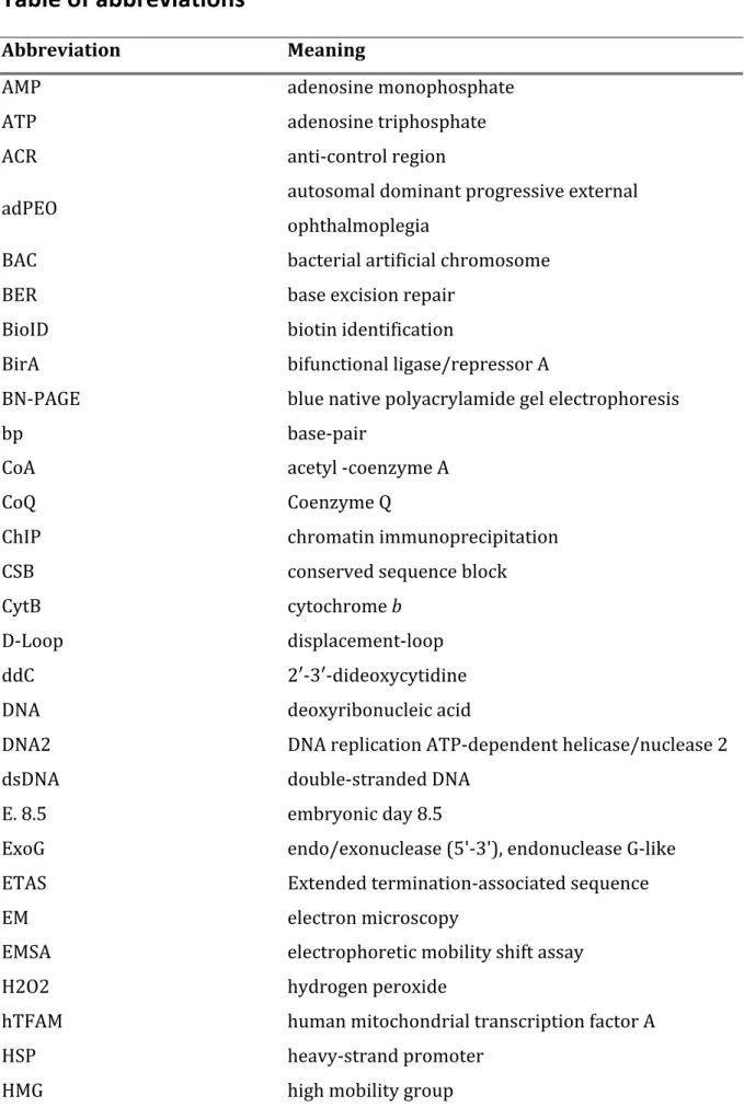
Mitochondria are enclosed by double membrane that separate two aqueous compartments: the mitochondrial matrix and the intermembrane space9 (Figure 1).
The outer membrane is permeable to all molecules of 5000 daltons or less. The inner mitochondrial membrane contains an extremely high protein content and is folded into invaginations, creating characteristic cristae10. The five complexes of the mitochondrial oxidative phosphorylation system (OXPHOS) are located in the inner membrane.
Figure 1: Structure of mitochondria and biogenesis of the oxidative phosphorylation system (OXPHOS). Mitochondrial compartments: IM (inner membrane), OM (outer membrane), IMS (intermembrane space) and matrix. The majority of the OXPHOS subunits and other proteins involved in mitochondrial metabolism or mtDNA maintenance are nuclear encoded, translated on the cytosolic ribosomes and imported into mitochondria through specialized import machineries.
19 The matrix contains a mixture of enzymes that metabolize pyruvate and fatty acids, and catalyze the citric acid cycle. Moreover, the matrix contains copies of mitochondrial DNA, proteins required for mtDNA replication and expression, and mito- ribosomes11.
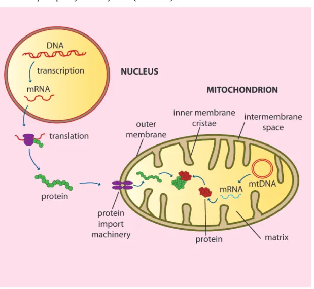
Mitochondria form a dynamic intracellular network and have an essential role in living systems by performing energy conversion to produce adenosine triphosphate (ATP) through the process of oxidative phosphorylation12. The OXPHOS system is composed of five complexes (I-V) and consists of approximately 90 different subunits.
Mitochondria can use both pyruvate and fatty acids as a fuel. Oxidation of carbohydrates in the citric acid cycle and lipids via β-oxidation generates the electron carriers NADH and FADH2 which donates electrons to the ETC13. The ETC consists of four protein complexes, Complex I (NADH: ubiquinone oxidoreductase), complex II (succinate: ubiquinone reductase), complex III (ubiquinol-cytochrome c reductase), complex IV (cytochrome c oxidase). All of the complexes are under dual genetic control (mtDNA and nDNA) except complex II that is exclusively nuclear encoded14 (Figure 2).
Figure 2: Schematic illustration of the structure and function the oxidative phosphorylation system. The OXPHOS system consists of five different enzyme complexes (Complexes I-V), and mobile carriers, coenzyme Q (CoQ) and cytochrome C. NADH and succinate are oxidized by complex I and II, the electrons are transferred to coenzyme Q, complex III, cytochrome C, complex IV and finally to molecular oxygen O2 which is reduced to water (H20). Protons are pumped out from mitochondrial matrix by complexes I, III and IV forming a proton gradient across the inner membrane.
The protons return to the matrix trough ATP synthase and the energy of the proton gradient is used to drive ATP synthesis.
The electrons received by complex I and II are passed further through the ETC in a controlled series of redox reactions resulting in reduction of oxygen to water15. The electron transfer via ETC is coupled to the transfer of the protons from the matrix side, transferring them to the intermembrane space side thus creating an electrochemical proton gradient across the inner membrane. This electrochemical
20 gradient is fully utilized by the fifth component of the OXPHOS system, the ATP synthase or complex V, which by rotary catalysis combine ADP and phosphate into ATP, the main energy currency of the cell16,17.
Besides cell energy conversion, mitochondria are also crucial for other biologically important functions including the regulation of cellular metabolism, fatty acid β-oxidation, the citric acid cycle, formation of reactive oxygen species (ROS), apoptosis, assembly of iron-sulphur clusters, and intracellular signalling18. As a consequence of their critical roles, mitochondrial dysfunction is implicated in many rare human genetic disorders and diseases such as Parkinson’s and Alzheimer’s disease, diabetes, as well as the ageing process18.
1.3 Organization of mtDNA
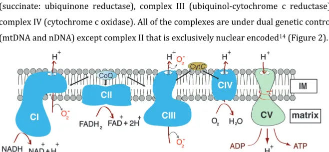
As a result of their endosymbiotic origin, mitochondria have retained their genome, and eukaryotic cells contain ~103 to ~105 copies of mtDNA. Mammalian mtDNA is a circular, double-stranded molecule of ~16.5kb. The mtDNA contains 37 genes and encodes 2 ribosomal RNAs (rRNAs), 22 transfer RNAs (tRNAs), and 11 messenger RNAs (mRNAs) (Figure 3). The individual strands of the mtDNA molecules are referred to as the heavy (H) strand and the light (L) strand due to their different densities in alkaline cesium chloride gradients. Its only longer non-coding region is the so-called control region (non-coding region or displacement loop (D-loop) region, which harbors regulatory elements for transcription and replication (the promoters for the transcription of the L and H strands (LSP and HSP) and the origin of replication of the H-strand (OH) (Figure 3). However, all of the proteins required for mtDNA maintenance and expression are encoded by the nuclear genome and have to be imported into mitochondria to fulfill their function20.
The mtDNA is maintained in compact protein-coated structures known as nucleoids, with a mean diameter of ~100nm21. In addition to its role in transcription, TFAM (mitochondrial transcription factor A) also plays a key role in mtDNA packaging into nucleoids22. Electron microscopy (EM) of biochemically in vitro reconstituted nucleoids combined with stimulated emission depletion (STED) microscopy of fixed cells showed that TFAM packages single mtDNA molecules in nucleoids that have a
21 slightly ellipsoid shape23. TFAM is present at around 1000 copies per mtDNA molecule and has structural properties consistent with an important role in DNA compaction24. Among the other suggested nucleoid associated proteins, some assist in processes such as DNA replication and transcription25. Many different proteins involved in mitochondrial biogenesis and metabolism are suggested to be recruited to the nucleoids. This may allow mtDNA packaging, nucleoid division, and inheritance to be coupled to these processes26.
Figure 3: Organization of mtDNA. Non-coding regions harbor regulatory elements for transcription and replication. Transcription promoters: light-strand promoter (LSP) and heavy-strand promoter (HSP). Origins of replication: OH, origin of H-strand DNA replication and OL, origin of L-strand DNA replication. The tRNA genes encoded on each of the two strands are labeled with orange. mRNA and rRNA abbreviations: COI, cytochrome c oxidase subunit I; COII, cytochrome c oxidase subunit II; COIII, cytochrome c oxidase subunit III; Cytb, cytochrome b; ND1, NADH dehydrogenase subunit 1; ND2, NADH dehydrogenase subunit 2; ND4, NADH dehydrogenase subunit 4; ND5, NADH dehydrogenase subunit 5, ATP6, ATP Synthase 6, ATP8, ATP Synthase 8, ND6, NADH dehydrogenase subunit 6; 16S rRNA gene, 12S rRNA gene.
22 1.4 Replication of mtDNA
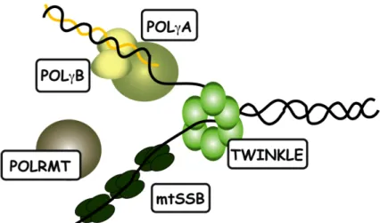
Replication of mtDNA is independent of the cell cycle and nDNA replication, and mtDNA can be replicated several times or not at all during mitosis. Replication of mtDNA occurs also in post-mitotic cells, as mitochondrial turnover continues in order to support their post-mitotic metabolic requirements. Complex machineries that are required for faithful mtDNA replication are present in the mitochondria and their basic components have been reconstituted in vitro27. Cooperation of the mitochondrial heterotrimeric DNA polymerase (POLγ), the hexameric helicase TWINKLE, the tetrameric mitochondrial single-stranded binding protein (mtSSB) and the mitochondrial RNA polymerase (POLRMT) was shown to be necessary and sufficient for mtDNA replication in a recombinant in vitro reconstituted system27,28 (Figure 4).
Numerous other factors may also play important roles in mtDNA replication.
Mitochondrial isoforms of the nucleases RNaseH1, FEN1 and DNA2 are implicated in replication primer removal and/or processing of the DNA flap that is formed when the replication apparatus has circled the entire mtDNA molecule. After processing of the DNA flap is complete, the mitochondrial DNA ligase (DNA ligase III) ligates the 5´and 3´nascent DNA ends to form a completed circle21.
The exact mechanism of mtDNA replication is still a matter of controversy and three different models have been suggested to date: a) the strand displacement model29, b) strand-coupled model30, c) mtDNA replication via ribonucleotide incorporation throughout the lagging strand (RITOLS) model31,32.
23
Figure 4: Components of the basic mtDNA replication machinery.
1.4.1 The strand-displacement model
Electron microscopy and atomic force microscopy analyses of replication intermediates greatly contributed to the generally accepted strand-displacement model (SDM) for mtDNA replication. This model proposes a unidirectional, asymmetrical synthesis of the leading H-strand until the region containing the origin of replication of the lagging L-strand is unwound initiating L-strand replication.
Furthermore, in support of the SDM, ChIP analysis of mtSSB distribution in vivo has elucidated the additional role of mitochondrial single-stranded protein (mtSSB) in covering the displaced parental H-strand and blocking random primer synthesis on the displaced strand. It was shown that this wrapping with mtSSB restricted POLRMT activity and initiation of light-strand mtDNA synthesis. The finding that it is possible to initiate replication from OL in vitro33-35 and that is impossible to mutate some nucleotides in OL in vivo lends support to the SDM as this is the only model in which OL
has a proposed function as a origin of the L-strand replication34.
The origin for the L-strand replication is located in a non-coding region of ~30 nt and is flanked by five tRNA genes36. OL is activated when the growing daughter H- strand synthesis reaches about two-thirds of its total length, displacing the parental H- strand as a single strand. After strand displacement, OL adopts a stem-loop structure
24 and RNA priming starts at the T-rich region. Recent work suggests that L-strand mitochondrial DNA replication is primed by mitochondrial RNA polymerase (POLRMT)37. The biochemical analysis presented by Wanrooij and colleagues38 demonstrated that POLRMT has two distinct modes of action. The enzyme efficiently transcribes long regions of dsDNA, but becomes much less processive on ssDNA, producing only short RNA molecules of 25-75 nt. The short RNA primers can be used by the mitochondrial DNA polymerase POLG to initiate DNA synthesis in vitro and this reaction is stimulated by the mitochondrial ssDNA binding protein (mtSSB). It was further demonstrated that, when combined, POLRMT, DNA polymerase POLγ, TWINKLE and mtSSB are capable of simultaneous leading and lagging-strand DNA synthesis in vitro. Following initiation at OL, the lagging strand synthesis proceeds over the whole length of the mtDNA molecule 36.
1.4.2 The strand-coupled model
In addition to the SDM of replication, Holt and coworkers proposed an alternative mode of mammalian mtDNA replication, the so-called strand coupled model39. This model for mtDNA replication proposes simultaneous leading and lagging strand mtDNA synthesis and it is based on observation of replication intermediates by two-dimensional agarose gel electrophoresis (2D-AGE)39. According to the strand- displacement model, mtDNA replication starts bidirectionally from multiple origins across a region including the CytB and ND5 and ND6 genes. When the replication process reaches OH, the replication fork is arrested and replication is restricted to one direction only40.
1.4.3 RITOLS model
An additional model of replication called RITOLS mode (ribonucleotide incorporation throughout the lagging strand) was proposed32. This model differs from the strand displacement model by suggesting that the lagging strand is first synthetized as an RNA molecule that is later replaced by DNA. The authors speculate that the RNA may help to protect and stabilize the displaced ssDNA, or alternatively act to block transcription machineries that could interfere with the replication process31.
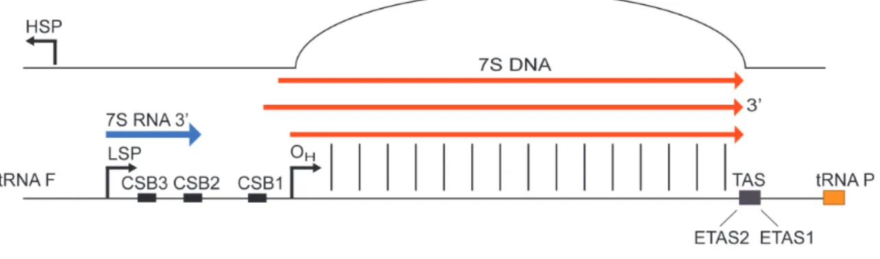
25 1.4.4 Mitochondrial non-coding region (D-loop region)
The mitochondrial D-loop or non-coding region (NCR) ranges between 880 and 1400 bp in length, and in its 5' domain it contains the main regulatory elements of the mitochondrial genome: the two promoters (HSP and LSP) and the origin of replication of the H strand.
Short conserved sequence blocks called CSB1, CSB2 and CSB341 are present at the NCR 5’ end . Those conserved sequences blocks are possibly involved in the RNA:DNA transitions and formation of the RNA primers for H-strand replication.
Mitochondrial replication is coupled with transcription in mitochondria because transcription from LSP terminates prematurely at the conserved sequence block 2 (CSB2)42,43. This termination event generates an RNA replication primer of ∼100 nt (Figure 5).
A remarkable feature of mtDNA replication is the premature termination of the replication of the newly synthesized H-strands at the end of the mtDNA control region that results in the formation of a ~650 bp long structure known as 7S DNA or D-loop44 (Figure 5). Nearly 95% of the H-strand initiation events are terminated in this manner.29 The D-loop structure is triple stranded since the newly replicated DNA molecule remains bound and displaces the non-replicated strand45. The half-life of the D-loop in cell culture is around 70 min, which shows that it is rapidly turned over29.
Figure 5: Mitochondrial D-loop region with regulatory elements.
Important roles for the 7S DNA have been proposed in mtDNA replication regulation, maintenance of DNA topology, recombination, dNTP metabolism and nucleoid
26 segregation, but its exact function and the mechanisms underlying its formation are still not understood46.
In support for a regulatory role for the D-loop, the termination associated sequences (TAS sequences) composed of ~15-bp long repetitive conserved sequences are present at the end of the D-loop region45 (Figure 5). Furthermore two new conserved blocks of about 60 bp were mapped at the 3’ end of D-loop region called ETAS1 and ETAS2 (extended TAS sequences). Mapping such a region of conserved sequences suggested their possible role in multiple terminations signals functioning between mitochondrial transcription and replication. Potentially, protein factor(s) that are binding to the conserved TAS sequences are leading to premature DNA synthesis and 7S DNA formation. In vitro, in organello, and in vivo footprinting approaches in different mammalian species identified protein-binding sites at the 3’ region of the control region of mitochondrial DNA. Proteins with contrahelicase activity have been described in prokaryotic and eukaryotic systems47,48. In sea urchin, the mitochondrial D-loop binding protein (mtDBP) has been shown to bind to the 3’ end of the D-loop region, to display contra-helicase activity, and to negatively regulate DNA synthesis49. In Escherichia coli, the replication termination factor TUS arrests the replication fork via protein-protein interaction with the replicative helicase50. Moreover a 48 kDa protein of unknown function has been reported to bind to ETAS sequences and promote 7S DNA formation in bovine mitochondria47. However no TAS-binding protein in mammals has yet been identified.
1.4.5 Mitochondrial DNA polymerase POLγ
There are 16 specialized polymerases present in mammalian cells required for nuclear and mitochondrial genome maintenance. POLγ is a highly efficient and processive polymerase located in mitochondria, and is responsible for mtDNA replication and repair. Disruption of the POLγ in mice causes an early developmental arrest between E7.5 and E8.5 associated with severe mtDNA depletion, confirming its essential role in mtDNA maintenance51. POLγ is a member of a family A class of DNA polymerases which also includes the E. coli DNA polymerase I and the T7 DNA polymerase52. The mammalian mtDNA polymerase holoenzyme is a heterotrimer with one catalytic POLγA subunit and two accessory POLγB subunits.
27 The POLγA subunit is a 140 kDa peptide with a 3’-5’ exonuclease domain that is connected with the 5’-3’ polymerase domain via a linker region. In humans, the linker region is a 482 amino acids long region and mutations in this region have been linked to several mitochondrial disorders. The exonuclease domain has proofreading activity during replication and it can remove inaccurately incorporated nucleotides53. Recently it has been reported that the exonuclease activity of POLγ is required for successful DNA ligation in the final step of mtDNA replication at OH54. Moreover the coordination between POLγ and newly described mitochondrial nuclease MGME1 has been shown to be sufficient for flap removal and ligation to complete mtDNA replication55. Mice that have an exonuclease-deficient version of POLγ have increased levels of point mutations, linear mtDNA fragments, and develop symptoms of premature ageing56.
The accessory subunit POLγB has a molecular mass of 55 kDa. POLγB was demonstrated to stimulate the catalytic activity and processivity of POLγA, by enhancing DNA binding and increasing the polymerase rate57. POLγB has sequence and structural similarities with the anticodon-binding pocket of the prokaryotic aminoacyl- tRNA synthetases, a group of enzymes that catalyze the attachment of each amino acid to its corresponding tRNA. The accessory subunit of DNA polymerase is present in Homo sapiens, Mus musculus, and Drosophila melanogaster, but is absent from Saccharomyces cervisiae58. The protein has dsDNA binding activity and is required to ensure that the polymerase stays bound to the template behind the moving TWINKLE helicase59.
In conclusion, the role of POLγ is vital for mitochondrial function, and defects in this enzyme are the most common cause of mtDNA stability disorders causing conditions such as progressive external ophthalmoplegia (PEO), Alpers syndrome, adult onset ataxia, and infertility.
1.4.6 Mitochondrial replicative helicase - TWINKLE
Helicases are universally present enzymes in prokaryotes, eukaryotes and viruses with key functions in DNA and RNA metabolic processes, including replication, recombination, DNA repair, transcription and translation60. During DNA replication, helicases play an essential role in unwinding the double-stranded template in front of
28 the DNA polymerase and the resulting ssDNA strands are used as a templates for the synthesis of the complementary DNA strands23. The mitochondrial replicative helicase TWINKLE unwinds double-stranded DNA in a 5’ -> 3’ direction using NTP hydrolysis as an energy source. This protein is homologous to the bacteriophage T7 gene 4 primase/helicase (T7gp4 protein), especially in its helicase domain. Interestingly, the TWINKLE protein has not retained the primase role in mitochondrial replication, hence POLRMT is responsible for priming DNA synthesis at both strands 61,37.
The Twinkle gene was originally discovered just over a decade ago, in an screen for mutations linked to autosomal dominant PEO (adPEO)61. Patients with this human disorder present exercise intolerance, muscle weakness, peripheral neuropathy, deafness, ataxia, hypogonadism, and cataracts62. adPEO patients accumulate various mtDNA deletions across different postmitotic tissues62. The TWINKLE protein is comprised of a carboxy-terminal helicase domain, a middle linker domain, important for subunit interaction and multimerisation, and an amino terminal domain of unknown function.61
Over 30 different TWINKLE mutations are known to cause accumulation of deletions or depletion of mtDNA, thereby inducing mitochondrial energy defects which result in neuro-muscular symptoms63,64.
The consequences of various TWINKLE mutations have been studied in vitro by using recombinant proteins and expressing mutant protein versions in human and insect cell lines65,66. In particular, the effects of adPEO-causing mutations in the linker region have been studied as those show abolished protein hexamerisation and DNA helicase activity, which causes replication stalling. Overexpression of the mutated Twinkle in human cells showed mtDNA depletion and accumulation of replication intermediates and also induced changes in nucleoid structure 64,66,67.
Additionally, the consequences of dominant Twinkle-PEO mutations have been investigated in vivo using a transgenic mouse strategy. To this end, a so called Deletor mouse, which contains a transgene carrying a 13 amino acid-long duplication in the TWINKLE linker region, was generated68. In this model, older mice expressing the PEO linked mutations accumulate mtDNA deletions in muscle and brain, and show a cellular phenotype similar to PEO patients with present aberrant mitochondrial structures.
These mice also show mtDNA depletion in brain, but not in muscle and heart68.
29 Severe phenotypes in patients with PEO mutations correlate with the amount of residual helicase activity in Twinkle mutants, suggesting that proper helicase activity is required for TWINKLE to function64. Additionally to dominant PEO mutations, the recessive mutation Y508C has been described in the helicase domain of TWINKLE. This recessive mutation is known to cause IOSCA a form of infantile-onset spinocerebellar ataxia69. From the analysis of patient cells and a homozygous IOSCA knockin mouse model, it has be shown that IOSCA is associated with tissue-specific mtDNA depletion in the liver and cerebellum, and decreased concentrations of all dNTPs. Furthermore, IOSCA mice eventually developed mitochondrial epileptic encephalopathy, mimicking the IOSCA phenotype seen in human patients70.
Several mutant forms of TWINKLE have been expressed in Drosophila melanogaster, where they caused severe depletion of mtDNA and lethality65. Mitochondrial impairment caused by these mutations promotes apoptosis65. This observation is in line with the finding that in vitro depletion of TWINKLE in Schneider cells is decreasing mtDNA copy number71.
Finally, the in vivo function of TWINKLE has been investigated using a mouse model 72. We created and analyzed conditional Twinkle knockout (KO) mice and proved that TWINKLE is the only mitochondrial replicative helicase since it is essential for the mouse embryonic development72. Tissue-specific TWINKLE KO in the heart and skeletal muscle of mice resulted in severe and rapid mtDNA depletion and consequently decreased mitochondrial encoded transcripts and protein steady-state levels. Southern blot analysis showed that TWINKLE is required for the unwinding of the entire mtDNA molecule as well as for the abortive replication at the D-loop region72.
1.4.7 Mitochondrial single-stranded DNA binding protein - mtSSB
A single-stranded DNA binding protein, mtSSB is present in mitochondria to maintain integrity of long single-stranded DNA regions during replication. mtSSB is a tetramer that consists of four 16 kDa subunits and shares similarities with the E. coli SSB protein. Besides DNA stabilization, mtSSB has a stimulatory effect on the rate of DNA unwinding by TWINKLE27,73, probably due to a direct protein-protein interaction74
30 1.4.8 Additional proteins involved in mitochondrial DNA replication
The list of the proteins involved in mitochondrial DNA replication has been expanded during recent years. Just like any other DNA, mtDNA requires the activity of various enzymes with nuclease, ligase, and topoisomerase activities.
Topoisomerases are enzymes that adjust DNA topology for replication and transcription processes75. Mitochondrial topoisomerase I (TOP1mt) transiently breaks one DNA strand at a time and thereby belongs to the type I topoisomerases. In TOP1mt knockout mice, an increased negative supercoiling of mtDNA indicates that mitochondria contain an efficient topoisomerase activity that relaxes positive supercoiling of DNA76. Apart from TOP1mt, two additional topoisomerases (Top2α and Top2β) were shown to localize to mitochondria in mouse and human models. Top2α and Top2β are type IIA topoisomerases - in mitochondria they are potentially critical for supercoil relaxation, enabling transcription and replication77, and may also have roles in decatenating locked mtDNA circles formed during replication76.
Ribonuclease H1 (RNase H1) is localized in both the nucleus and mitochondria.
Analysis of mtDNA from knockout mouse models and human patient studies strongly indicates that RNase H1 removes the RNA primers at the origins of the light and heavy strand replication. Therefore, RNase H1 is essential for proper mtDNA replication in humans, and pathogenic RNaseH1 mutations cause mtDNA replication stalling, mtDNA depletion and deletions78.
DNA ligase III activity is present in mitochondria, and human cells with impaired DNA ligase expression have reduced mtDNA copy number and multiple single-stranded nicks79. Additionally, deletion of ligase III in mice causes embryonic lethality at E8.5, similar to the other mouse knockouts with germline disruption of genes essential for maintenance or expression of mtDNA80.
31 1.4.9 Mitochondrial genome maintenance exonuclease 1 – MGME1
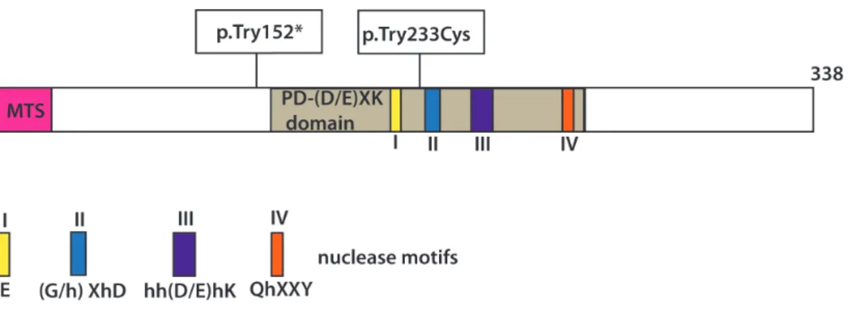
Recently, two different laboratories reported an exclusively mitochondrial protein called mitochondrial genome maintenance exonuclease 1 (MGME1 a.k.a. Ddk1) to be involved in processing of mitochondrial DNA during the replication81,82. MGME1 belongs to the RecB subclass of PD-(D/E)XK endonucleases (Figure 6).
Figure 6: Schematic representation of the MGME1 protein and localization of the investigated homozygous mutations causing disease. The motifs of the PD−(D/E)XK nuclease superfamily are indicated with Roman numerals. MTS indicates mitochondrial targeting signal.
Loss of function mutations in the gene encoding MGME1 cause a severe multisystemic mitochondrial disorder82. The mtDNA from muscle of affected patients shows various defects, including deletions, depletions, rearrangements, and 7S DNA accumulation83. In vitro analysis of recombinant MGME1 showed that the protein exhibits 5’-3’ nuclease activity with a specificity for single-stranded DNA81,82. It shows no activity on RNA, even when hybridized to DNA. In vitro nuclease-ligation assays have confirmed that MGME1 cleaves longer flaps efficiently but imprecisely around the flap base. The MGME1 activity requires the 3’-5’ exonuclease activity of POLγ for proper flap removal and nick ligation at OH55. Precisely processing the long primer formed at OH in the mitochondrial NCR may require a teamwork of different nucleases.
Initiation of mtDNA synthesis includes the RNA-DNA transition immediately after CSB2, but the 5’ ends of the nascent DNA are moved around 100 bp downstream of the transition site by presumably a long-flap pathway84. In support of this, 7S DNA from MGME1 deficient patient cells contains 5’ extended ends suggesting that Mgme1
32 activity is necessary for processing of the single-stranded flap intermediate83,84. MGME1 patient cells also contain an 11 kb truncated linear mtDNA fragment, the ends of which map near OH and OL suggesting that MGME1 is involved in primer flap processing at OH83. Interestingly nucleases like FEN1 and DNA2 may also have roles in mtDNA base excision repair and flap processing in mitochondria85,86. Therefore it has been hypothesized that additionally to its exonuclease role, which is necessary for proper replication and flap processing in mitochondria, MGME1 might have a role in mtDNA repair.
1.5 Transcription of mtDNA
RNA synthesis in mammalian mitochondria is initiated from two different promoters, both located in the D-loop; LSP and HSP. Transcription of the L-strand is initiated at the LSP, resulting in a primary transcript encoding mRNA-ND6 and eight tRNAs. Transcription of the H-strand produces a polycistronic transcript covering almost the entire H-strand87,88. Almost all the mRNA and rRNA genes are flanked by one or more tRNA gene. Excision of tRNA molecules is required to produce mature mRNA and rRNA molecules from the polycistronic transcripts according to so-called tRNA punctuation model89.
1.5.1 The basic components of the mitochondrial transcription machinery
The basal transcription initiation machinery consists of three proteins:
mitochondrial RNA polymerase (POLRMT), mitochondrial transcription factor A (TFAM), and mitochondrial transcription factor B2 (TFB2M)90-92. Recently, a transcription elongation factor (TEFM) was reported to enhance POLRMT processivity to produce large polycistronic transcripts covering almost the entire length of each strand93,94.
POLRMT is a large single subunit enzyme of 140 kDa that shows a high sequence similarity with both the T3 and T7 bacteriophage polymerases. It is the only RNA polymerase operating in mammalian mitochondria. Loss of POLRMT leads to embryonic lethality at E8.5, as seen in other mouse models with germline disruption of genes essential for replication or expression of mtDNA37. It has been confirmed that
33 POLRMT is essential for primer synthesis to initiate mtDNA replication also in vivo37.
Although POLRMT can specifically recognize mitochondrial promoters, it cannot initiate transcription without assistance from two additional transcription factors, TFAM and TFB2M90.
TFAM belongs to the high-mobility (HMG)-box family of proteins, and can bind, unwind and bend DNA95. The vital importance of TFAM for mtDNA maintenance was demonstrated in studies of homozygous TFAM knockout mice, which showed embryonic lethality and severe depletion of mtDNA96. As one of the major protein components of nucleoids, TFAM is likely to fully coat mtDNA21,97. TFAM binds sequence-specifically to promoter elements immediately upstream of the HSP and LSP transcription start sites. Structural studies showed that TFAM-binding introduces sharp U-turns of mtDNA, which may facilitate promoter melting98. The DNA bending is clearly important for transcription activation and previous studies indicate that the C- terminal tail of TFAM plays an essential role in transcription activation99,100..
TFB1M and TFB2M can assist TFAM and POLRMT during the initiation of the transcription process. TFB2M is more active in stimulating specific transcription as is TFB1M in vitro. Both factors are closely related to the family of bacterial rRNA dimethyltransferases and our results suggest that TFB2M is a transcription factor whereas TFB1M has no role in transcription but rather functions a 12S rRNA methyltranserase{Metodiev:2009dz}.
TFB2M is essential for complex formation during mitochondrial transcription initiation101, and crosslink experiments reveal a direct interaction of TFB2M with the promoter starting site, where it is involved in promoter melting to facilitate transcription initiation by POLRMT101. Additionally increased levels of TFB2M activates mitochondrial transcription and causes elevated steady-state levels of mitochondrial transcripts90,102 suggesting a direct and essential role in mitochondrial transcription.
TFB1M was shown to methylate two adenine residues in a conserved stem loop region of the 12S rRNA, which is an important post-transcriptional modification in the process of mitochondrial ribosomal biogenesis103,104. Therefore, TFB1M is thought to have its main function in ribosomal maturation rather than in transcriptional initiation104.
Recently, the transcription elongation factor (TEFM) was reported to enhance
34 POLRMT processivity by promoting the production of large polycistronic transcripts covering almost the entire length of each strand93,94. Mitochondrial transcription elongation factor (TEFM) interacts with the catalytic domain of POLRMT and its depletion leads to a reduced transcription elongation and decreased promoter-distal mitochondrial transcripts. Therefore, TEFM aids the transcription of longer RNA products and the circumvention of highly structured regions. Furthermore it has been shown that interaction of the transcription elongation factor (TEFM) with the mitochondrial RNA polymerase (POLRMT) and the nascent transcript prevents the generation of replication primers, increases transcription processivity and thereby serves as a molecular switch between replication and transcription93,105.
1.5.2 Regulation of transcription termination
Mechanisms regarding mtDNA transcription termination in mammalian organisms are still under debate. Previously it was reported that transcription of the heavy strand is initiated from two different sites: HSP1 and HSP2106. Transcripts from HSP1 were proposed to terminate immediately after the 16S site, while transcription from HSP2 promoter continues past the termination site, along almost the entire length of the H-strand. This premature termination justified why HSP1 transcripts resulted in a 50-times higher abundance of rRNAs than mRNAs produced downstream of the termination site107. This termination event was suggested to be mediated by specific binding of mitochondrial termination factor 1 (MTERF1). However, in vivo studies in MTERF1 knockout mouse reported that MTERF1 blocks L-strand transcription to prevent transcription interference at the light-strand promoter, while not having an effect on H-strand transcription108. Additional roles of MTERF1 have been investigated in vitro, where overexpression of MTERF1 causes replication pausing. These findings have been confirmed after a recent study proposed a model where the MTERF1 binding site is a pausing site for the replication machinery while POLRMT passes the rRNA region. After the halt, the transcription machinery can remove the MTERF1 block and replication can continue. Hence MTERF1 acts as contrahelicase postponing the progression of DNA replication and avoiding collisions between the replication and transcription machineries in the rDNA region109. The MTERF protein family has an
35 important role in the regulation of mitochondrial transcription at the level of termination in different species. In addition to its transcriptional termination activity, mtDBP, the MTERF1 homologue in sea urchin, also plays a role in mtDNA replication acting as a contra-helicase, which negatively regulates mtDNA synthesis49. The L- strand transcription is frequently terminated at the mitochondrial non-coding region, at the CSB1 and CSB2 sequences, when a G-quadruplex structure is formed between the nascent RNA and the non-template DNA strand. Interestingly, this structure covers the 3’ end of the RNA primer formed by premature transcription termination at the CSB2 site, and the primer appears inaccessible to the DNA replication machinery.
There are possible additional proteins involved in resolving this hybrid structure and enabling utilization of the RNA primer for initiation of mtDNA replication at OriH 110.
H-strand transcription is initiated from only one promoter and encompasses nearly the entire mitochondrial genome with termination occurring in a region immediately upstream of the tRNAPhe gene, at the termination associated sequences in the mitochondrial control region108. Recent findings suggest that termination of H- strand transcription and premature mtDNA termination at core-TAS are functionally linked111. Further studies are necessary to elucidate the exact regulatory molecular mechanisms of mitochondrial transcription and replication on both mtDNA strands.
1.6 Mitochondrial DNA repair
The mitochondrial respiratory chain is a site for ROS (reactive oxygen species) and RNS (reactive nitrogen species) production, since molecular oxygen can differently react with RC electrons112. ROS are generated at several locations in the cell, but 90% is produced by the ETC in the mitochondria113. Imbalance between ROS and/or RNS production and antioxidant defense mechanisms can lead to “oxidative stress”, which is highly harmful for cell functionality. Contrary to this detrimental role or ROS, fluctuations in the steady-state levels of ROS have appeared important for cell signaling, cell cycle progression, and apoptosis114. ROS consist of a variety of highly reactive molecules and free radicals formed from reactions including molecular oxygen. One of the most common ROS species is the superoxide anion (O2•–), which can produce hydrogen peroxide (H2O2) in a reaction catalyzed by superoxide dismutases or via spontaneous reactions. H2O2 can be further reduced to water or via a Fenton
36 reaction converted to the highly reactive hydroxyl radical (OH•). In mitochondria, the majority of O2•– production is through complex I and complex III115. The well-known mitochondrial free radical theory of ageing poses that ROS produced in mitochondria are a primary source of cellular free radicals contributing to the ageing process116. Furthermore a “vicious cycle” has been suggested whereby subsequent ROS induced mutations in mtDNA would accumulate and lead to dysfunctional OXPHOS components that would produce more ROS and mtDNA mutations, finally resulting in cell damage117. Therefore the main assertions for vulnerability of mtDNA and source of mtDNA mutations are: a) proximity of mtDNA to OXPHOS system and exposure to superoxide radicals b) lack of mtDNA repair mechanisms; and c) replication errors during normal DNA synthesis in mitochondria. Although mtDNA was initially thought to lack repair systems, several repair pathways have recently been reported.
The main repair pathway found in mitochondria is base excision repair (BER), which includes single-nucleotide BER (SN-BER) and long-patch BER (LP-BER)118,119. Initial damage recognition and cleavage is performed by the specific glycosylases, and mitochondria contain six damage-specific glycosylases. UDG-uracil DNA-glycosylase I and OGG1-8-oxo-G DNA-glycosylase/AP lyase are expressed as both nuclear and mitochondrial isoforms. Additionally MUTYH and NTH (thymine glycol glycosylase) and two endonuclease VIII-like proteins (NEIL1 and NEIL 2) are reported to be present in mitochondria. After DNA-glycosylase cleavage, the DNA phosphate backbone is cleaved by AP endonuclease. During thesingle-nucleotide SN-BER, POLγ is filling single nucleotide gap prior to ligation. In the case of long patch LP-BER, during the gap filling POLγ is displacing the 5’DNA strand thus creating a 5’-flap structure. FEN1, DNA2 and EXOG enzymes were implied to play a role in 5’-flap removal prior to ligation84. Long patch repair is mainly responsible for repairing common lesions caused by oxidative damage to sugar-phosphate backbone120.
Nucleotide excision repair (NER) has not been reported to operate in mitochondria, even though many chemical carcinogens enter in mitochondria and interact with mtDNA, possibly causing large lesions53.
Another repair mechanism required for removal of base mismatches and small insertions called mismatch repair (MMR) has been reported to operate in S. serevisiae and S. pombe121. Regarding the MMR in higher eukaryotes, a low level MMR repair
37 activity was identified in rat liver mitochondrial lysate122 but the main MMR enzymes active in nucleus have not be identified. Many recent studies that involve knockout mouse models of repair enzymes (MUTYH, OGG1, NEIL1) develop tumors123, metabolic syndrome124 and obesity125. Additionally disruption of OGG1/MYH genes in mice did not result in a higher mtDNA mutation rate126. In the future, a mitochondrial specific knockout of these repair enzymes can contribute to the investigation of their specific role only in mitochondria. Finally many of the proteins playing a role in mtDNA repair are also implicated in mtDNA replication showing tight coordination of those pathways in keeping the integrity of mtDNA.
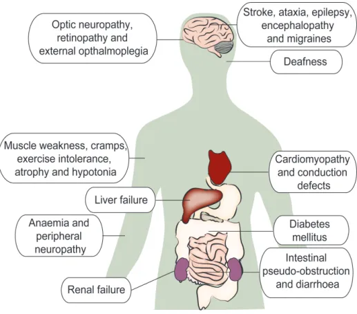
1.7 Mitochondrial genetics and diseases
Mitochondrial diseases are the most common causes of inherited metabolic disease in humans with a frequency of about 1 in 5000127. Human mitochondrial disorders encompass a genetically heterogeneous group of different diseases, caused by mutations in mitochondrial and/or nuclear DNA and display both clinical heterogeneity and tissue specificity128, with the brain and muscle being the most commonly affected tissues129. The inheritance pattern of mtDNA-related diseases can vary from case to case. Some patients appear to be sporadic cases, while others are clearly inherited. Clinical heterogeneity can be explained by the specific features of mtDNA genetics; since mtDNA is maternally inherited, mutations in mtDNA are not transmitted according to Mendelian principles. In normal cells, most mtDNA copies are assumed to be identical, a condition know as homoplasmy. Pathogenic mutations of mtDNA are usually present only in a subset of all mtDNA molecules. This mixture of mutated and wild-type mtDNA molecules is called heteroplasmy130.
38
Figure 7: Common clinical manifestations of mitochondrial disorders
Very often the proportion of mutant and normal mtDNA in a tissue determines the clinical outcome of the mutation. This means that a disease or biochemical defect will be caused only when a pathogenic mtDNA mutation is above a certain threshold level131. This threshold level in turn depends on the mutation type and tissue energy demand. Furthermore, mtDNA populations undergo a bottleneck effect which means that only a small proportion of the total number of mitochondrial genomes are passed on from mother to offspring132. Therefore through this bottleneck effect, pathogenic mutations can segregate to very different heteroplasmy levels in the offspring.
Consequently, a mother carrying a pathogenic mutation can give birth to both severely sick and healthy children130.
39 1.7.1 Mitochondrial disorders caused by mtDNA mutations
There are two major types of mtDNA defects that may occur: large rearrangements (including deletions and duplications) and point mutations. The defects can be present in the germline and be transmitted maternally, or appear randomly in somatic cells133.
Point mutations are the most predominant cause of disease in humans and there are several mitochondrial disorders know to be induced by them. Mitochondrial encephalomyopathy, lactic acidosis and stroke-like episodes (MELAS) syndrome is a multisystem disorder with impaired motor ability, vision and deficient cognitive activity133. MELAS is caused by a few point mutations, the most prevalent of which is an A>G transition at m.3243 in tRNALeu(UUR), but also in the ND5 gene. Another neuromuscular disorder known as myoclonus epilepsy with ragged red fibers (MERRF) syndrome is a consequence of point mutation m8344 A>G in tRNALys. The clinical severity of MERRF correlates with heteroplasmy levels, and the biochemical manifestations of MELAS and MERRF are seen as a defect in complex I and/or IV. One of the most common mtDNA-mutation disorders is Leber’s hereditary optic neuropathy (LHON), characterized by subacute bilateral visual failure in young adults134. Usually patients carry homoplasmic point mutations m.3460 G>A, m.11778 G>A , m14484 T>C in genes coding for complex I subunits of the RC.
Another type of mitochondrial DNA mutations are mtDNA rearrangements that are sporadic. Single large-scale deletions have been associated with progressive external ophthalmoplegia (PEO), Kearn-Sayre syndrome (KSS) and Pearson’s syndrome18.
1.7.2 Mitochondrial disorders caused by nDNA mutations
Several diseases have been shown to be due to mutations in nuclear genes encoding mitochondrial proteins. During the last decade, researchers have defined a list of genes linked to human mtDNA maintenance disorders mostly affecting mtDNA replication. Numerous pathogenic mutations leading to mtDNA instability are reported in the genes encoding basic replication components such as POLGA, POLGB, TWINKLE135, and replication/repair related factors including DNA2, RNAseH1, and the recently reported MGME1 nuclease82,136,137. Mutations in these genes are causing