LOV domain signaling:
A study of LOV–LOV interactions
Dissertation
zur Erlangung des Doktorgrades der Naturwissenschaften (Dr. rer. nat.)
der Fakultät für Chemie und Pharmazie der Universität Regensburg
vorgelegt von Kathrin Magerl
München aus
im Jahr 2017
Prüfungsausschuss:
Vorsitzender: Prof. Dr. Arno Pfitzner 1. Gutachter: Prof. Dr. Bernhard Dick 2. Gutachter: PD Dr. Tilman Kottke
weiterer Prüfer: Prof. Dr. Frank-Michael Matysik
List of abbreviations
The following list contains all abbreviations repeatedly used in this thesis except SI units. The standard one or three letter code was used for amino acids and DNA bases.
A. thaliana Arbabidopsis thaliana (At)
BLUF blue light sensor using flavin adenine dinucleotide bZIP basic region leucine zipper
CCD charge-coupled device
C. reinhardtii Chlamydomonas reinhardtii (Cr)
CRY cryptochrome
C-terminal carboxy-terminal
cw continuous wave
DADS decay associated difference spectrum DNA deoxyribonucleic acid
E. coli Escherichia coli
EDTA ethylenediaminetetraacetic acid EPR electron paramagnetic resonance
eT electron transfer
FAD flavin adenine dinucleotide FbFP flavin-binding fluorescent protein FDHW full duration at half width
FMN flavin mononucleotide
FP fluorescent protein
FRET Förster resonance energy transfer FWHM full width at half maximum GFP green fluorescent protein
HTH helix-turn-helix
I
IR infrared
LC lumichrome
LED light emitting diode
LOV light-, oxygen-, voltage-sensitive
NHS N-hydroxysuccinimide
N-terminal amino-terminal
NWML nominal molecular weight limit MALS multiangle light scattering
MD molecular dynamics
PAS PER-ARNT-SIM
PB phosphate buffer
PCET proton coupled electron transfer
Phot phototropin
PMT photomultiplier tube
pT proton transfer
PtAureo1a Aureochrome 1a Phaeodactylum tricornutum
RF riboflavin
R. sphaeroides Rhodobacter sphaeroides (Rs) SAXS small angle X-ray scattering SEC size exclusion chromatography STK serine/threonine kinase
SVD singular value decomposition
TA transient absorption
TCSPC time-correlated single photon counting
TEV Tobacco etch virus
UPLC ultra performance liquid chromatography
UV ultraviolet
UVR-8 UV resistance locus 8
VfAureo1 Aureochrome-1 Vaucheria frigida
Vis visible
WCC white collar complex
III
wt wild type
ZTL ZEITLUPE
Contents
1 Introduction 1
1.1 LOV domains . . . . 2
1.2 Aim of this work . . . . 5
2 Electron transfer in mutated LOV domains of C.reinhardtii 7 2.1 Introduction . . . . 7
2.2 Results . . . . 9
2.2.1 Photoreaction of CrLOV1-F41Y . . . . 9
2.2.2 Additional mutation of the reactive Cys57 . . . 12
2.2.3 pH influence . . . 15
2.2.4 Decay of the S 1 state suggests electron transfer via the excited singlet state . . . 17
2.2.5 Molecular Dynamics Simulations . . . 19
2.3 Discussion . . . 22
2.4 Conclusions . . . 25
3 Reengineering the flavin photochemistry in iLOV 27 3.1 Introduction . . . 27
3.2 Results . . . 28
3.2.1 Target protein selection and design rationale . . . 28
3.2.2 Illumination of iLOV-Q489D results in effective formation of the neutral FMN semiquinone radical under aerobic conditions. 29 3.2.3 UV / Vis spectra of iLOV-Q489D along with computational prediction suggest that the newly introduced Asp is proto- nated at neutral pH values . . . 36
3.2.4 Transient absorption spectroscopy hints at the formation of a stable FMN:protein radical pair in iLOV-Q489D . . . 38
3.2.5 FMNH formation occurs from the excited triplet state in iLOV-Q489D . . . 41
3.2.6 Electron paramagnetic resonance (EPR) spectroscopy reveals the presence of a stable radical pair in iLOV-Q489D . . . 42
3.2.7 Mutational analysis of the electron transfer pathway in iLOV- Q489D suggests the involvement of multiple Tyr/Trp residues 43 3.3 Discussion . . . 47
3.3.1 Efficient proton transfer is essential for stabilizing FMNH in parental iLOV . . . 48
V
3.3.2 The electron transfer pathway present in iLOV is conserved
in all LOV proteins . . . 49
3.4 Conclusions . . . 51
4 Investigation of the intermolecular interactions of LOV domains 53 4.1 Intermolecular interactions and biological function of LOV domains . 53 4.2 Förster Resonance Energy Transfer (FRET) . . . 56
4.2.1 Principles of FRET . . . 56
4.2.2 Determination of the FRET efficiency . . . 58
4.2.3 FRET measurement setup . . . 59
4.2.4 Labeling with Cy3 and Cy5 . . . 60
4.3 Rhodobacter sphaeroides LOV . . . 62
4.3.1 RsLOV wt . . . 62
4.3.2 RsLOV-Cy3 and RsLOV-Cy5 . . . 64
4.3.3 Discussion . . . 73
4.4 The LOV1 domain of C.reinhardtii phototropin . . . 77
4.4.1 CrLOV1 wt . . . 77
4.4.2 CrLOV1-Cy3 and CrLOV1-Cy5 . . . 79
4.4.3 Emission measurements of CrLOV1-Cy3/Cy5 . . . 83
4.4.4 CrLOV1 A16C C32S C83S . . . 86
4.4.5 Discussion . . . 92
4.5 Conclusions . . . 96
5 Experimental 97 5.1 Microbiological methods . . . 97
5.1.1 Bacterial strains and plasmids . . . 97
5.1.2 Preparation of chemical-competent cells . . . 98
5.1.3 Heat shock transformation . . . 98
5.1.4 Bacteria growth . . . 98
5.2 Molecular biological methods . . . 99
5.2.1 Polymerase chain reaction (PCR) . . . 99
5.2.2 Oligonucleotides . . . 101
5.2.3 Gene synthesis . . . 102
5.2.4 Isolation of plasmid DNA . . . 102
5.2.5 Agarose gel electrophoresis . . . 102
5.3 Proteinbiochemical methods . . . 103
5.3.1 Cell lysis and protein extraction . . . 103
5.3.2 Protein purification via immobilized metal ion affinity chro- matography (IMAC) . . . 103
5.3.3 Buffers . . . 104
5.3.4 His-tag cleavage . . . 105
5.3.5 Sodium dodecyl sulfate polyacrylamide gel electrophoresis (SDS- PAGE) . . . 105
5.3.6 Size exclusion chromatography (SEC) . . . 106
Contents VII
5.3.7 Labeling with the cyanine dyes Cy3 and Cy5 . . . 107
5.4 Chemicals . . . 108
5.5 Spectroscopy . . . 109
5.5.1 UV/Vis absorption spectroscopy . . . 109
5.5.2 Transient absorption (TA) spectroscopy . . . 109
5.5.3 Time-correlated single photon counting (TCSPC) . . . 111
5.5.4 Determination of fluorescence quantum yields, Φ F . . . 112
5.5.5 Emission measurements . . . 112
5.5.6 Stopped-flow measurements . . . 112
5.6 Theoretical methods . . . 113
5.6.1 Modeling . . . 113
5.6.2 Molecular dynamics (MD) simulations . . . 113
6 Summary 115
A Appendix 119
Bibliography 123
1 Introduction
Sunlight is prerequisite for life on earth. All life processes depend directly or in- directly on this ever-present energy source. The ability to adapt and respond to environmental light is an evolutionary selection factor and has developed across all kingdoms of life. Light reception in nature is realized by photosensory pro- teins, which respond to the environmental stimulus light, providing the regulation of metabolic reactions and physiological functions.
The solar radiation reaching the earth’s surface peaks in the blue-green range of the spectrum. Due to the optical properties of water, blue light between 420 nm and 500 nm penetrates deepest of all spectral ranges into the oceans, enabling organisms to make use of the light energy also below the sea level [1]. Hence, it is not surprising that many physiological responses in nature are induced only by blue and near ultraviolet (UV) light. First reports of the influence of blue light on plants were given as early as in the 19th century [2]. Amongst others, Julius von Sachs investigated plant growth under different light conditions. He used either colored glass or colored chemical solutions to provide red and blue light and observed differences in the biological plant response depending on the light color he used [3]. A few years later in 1880, Charles Darwin described experiments on phototropism and postulated that it cannot be induced by red light [4]. Over the next 100 years many studies on blue light-induced effects regarding plant growth were performed and a debate about the chromophore molecule responsible for the light absorption between carotenoids and flavins flared up [5]. Since no photoreceptor for blue light had been identified by then, the question could not be answered unambiguously. A milestone in the history of the investigation of blue light reception was the molecular identification of the photoreceptor cryptochrome cry1 from Arabidopsis thaliana in 1993 [6]. Only four years later, phototropin was identified in Arabidopsis thaliana and was shown to be responsible for phototropism [7, 8]. Since then great process has been made and a lot more blue light photoreceptors have been identified across almost all taxonomic groups.
A great number of physiological functions in nature are triggered by blue light, e.g.
photosynthesis [9] and processes related to it like phototropism [7], stomata opening [10], chloroplast relocation [11] and solar tracking of leafs [10], but also phototaxis [12], gametogenesis [13], entrainment of circadian rhythms [14] and many more.
The blue light photoreceptors responsible for these processes can be classified into the families of xanthopsins [15], BLUF (blue light using flavin adenin dinucleotide) domains [16], cryptochromes [6], LOV (light-, oxygen-, voltage-sensitive) protein
1
families [17], phototropins [7] and ZEITLUPEs (ZTL) [18]. Besides this, there are the UV-sensitive UVR-8 (UV resistance locus 8) receptors [19], the red light-sensing phytochromes [20] and rhodopsins [21]. Taken together, the eight photoreceptor families cover almost the entire UV, visible and far-red region of the electromagnetic spectrum.
In general, photoreceptors consist of two components: a chromophore molecule to absorb the light and a protein core to host the chromophore and propagate the signal downstream to an effector domain or a signaling partner. With the exception of xan- thopsins, all blue light photoreceptors non-covalently bind flavin as a chromophore, either as flavin mononucleotide (FMN) or flavin adenine dinucleotide (FAD). The excitation of flavin by light represents the key element of the photoreception mech- anism.
Phototropins, ZEITLUPEs and LOV domain proteins can be summarized as light-, oxygen-, voltage-sensitive domains. LOV domains, BLUF domains and crypto- chromes all share a flavin chromophore, but the photochemistry upon blue light excitation is different. While BLUF domains and cryptochromes perform electron transfer reactions, LOV domains form a covalent bond between flavin and a nearby cysteine - the so-called adduct state - via a triplet state reaction of FMN. In the following the LOV domains which are the main aspect of this work will be described in more detail.
1.1 LOV domains
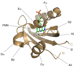
LOV domains form a subset of the PER-ARNT-SIM (PAS) superfamily and have preserved their structure [22–25]. The LOV core is composed of a central β -scaffold with five antiparallel β -strands (A β , B β , G β , H β , I β ) and four α -helices (C α , D α , E α , F α ) as shown in Figure 1.1.1. E α and F α pack against the β -sheet and form a pocket to non-covalently bind the flavin chromophore. The reactive cysteine participating in adduct formation is located on E α within the highly conserved se- quence GXNCRFL(Q). The LOV core is flanked by variable N- and/or C-terminal extensions, which are often helical. These flanking regions are thought to act as con- nective elements that propagate light-induced signals from the LOV core to effector domains or interacting partner proteins.
Most LOV domains non-covalently bind FMN as a chromophor, in some cases they
harbor FAD. FMN and FAD are the biologically active forms of riboflavin. They ex-
hibit a very rich photochemistry and can act as one- or two-electron redox reagents
[26]. The chemical versatility is linked to the fact that flavins can switch between
three different oxidation states: the fully oxidized form (ox), the one-electron re-
duced, semiquinone form (sq) and the fully reduced, hydroquinone (hq) form (see
Figure 1.1.2A). Nature makes use of these attributes in the form of various flavoen-
1.1 LOV domains 3
Figure 1.1.1 PAS-fold of LOV domains exemplified by the crystal structure of the LOV1 domain from C.
reinhardtii phototropin (PDB 1N9L). The central anti-parallel β-scaffold is linked by a helical connector, together forming the binding pocket of FMN. The LOV core is shown in cartoon representation in brown, the FMN chromophore in stick representation in green. The nitrogen atoms of FMN are colored blue, the oxygens red and phosphorus orange.
zymes, which are involved in the four major energy metabolism systems: photosyn- thesis, aerobic respiration, denitrification and sulfur respiration [27].
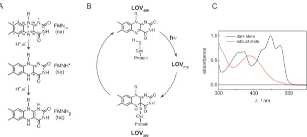
In the dark state the flavin chromophore of LOV domains in the fully oxidized form strongly absorbs in the blue region of the spectrum. The absorption bands are cen- tered around 450 nm (S 0 → S 1 transition), 360 nm (S 0 → S 2 ) and 280 nm (S 0 → S 3 ).
The flavin chromophore is non-covalently bound in the LOV binding pocket via hy- drogen bonds leading to a characteristic vibrational fine-structure of the absorption bands (Figure 1.1.2C) [17]. Blue light excitation induces covalent bond formation of FMN-C4a with the thiol group of a nearby, conserved cysteine. This so-called adduct state represents the active, signaling state and is strictly conserved among all currently characterized LOV domains. Adduct formation is accompanied by a change of the absorption spectrum resulting in a peak centered at 390 nm as can be seen in Figure 1.1.2C. Adduct formation is reversible in the dark. The flavin- cysteine bond breaks on the timescale of seconds to hours [28, 29]. The photocycle of LOV domains has been extensively investigated, but still some details remain unclear. A consensus exists, however, that upon blue light absorption FMN gets ex- cited into the singlet state and subsequently undergoes intersystem crossing to the triplet state, LOV 715 (numbers indicate the absorption maximum of the particular intermediate), within ns to µ s. From there, the covalent bond between FMN-C4a and the sulfur group of a nearby cysteine is formed resulting in the adduct state (LOV 390 ) [30]. A scheme of the LOV photocycle is shown in Figure 1.1.2B.
Several mechanistic proposals for the adduct formation have been debated in the
literature, including an ionic mechanism, a concerted mechansim and a radical pair
FMNox (ox)
FMNH (sq)
FMNH2 (hq)
88
77 66 5a5a 9a 99 9a
N55 4a4a
10a
N10a 10 10
4422NH33 N11 R
O O
NH N
NH N R
O O
NH N
NH N R
O O H+,e-
NH N
NH N R
O SO Cys Protein N N
NH N R
O
SO Cys Protein H
LOV
390LOV
450LOV
715hν
A B
H H+,e-
C
Figure 1.1.2 A: Structures of the fully oxidized state of FMN (ox), the one-electron reduced neutral semiquinone radical FMNH (sq) and the fully reduced FMNH
2(hq). B: Scheme of the LOV photocycle.
Upon absorption of blue light, LOV
450gets excited in the singlet state and subsequently undergoes intersystem crossing to the triplet state (LOV
715) from which the adduct state is formed ( LOV
390).
LOV
390reverts back to LOV
450in the dark. C: Steady-state UV/Vis spectra of the dark state (black line) and the adduct state (red line) of LOV domains in the range of 300 nm to 550 nm. Panel A and B are taken from [31].
mechanism [32–34]. The latter one seems to be most likely. According to this, LOV 715 initiates either concerted or sequential electron and proton transfer from the reactive cysteine resulting in the formation of the biradical pair FMNH SCys which subsequently combines to yield the adduct [35, 36]. However, although indications of the neutral semiquinone radical of FMN could be detected in a LOV domain of C. reinhardtii, there is so far no unequivocal proof that these are intermediates in the photocycle [37].
Adduct formation is just the primary step in LOV domain signaling. Protonation of FMN-N5 due to adduct formation induces the flip of the amide side chain of a nearby, conserved glutamine located on I β to adjust hydrogen-bonding interactions [38–42]. This in turn leads to further changes in the hydrogen-bonding network within the LOV core, transducing the light trigger into a physiological signal [43, 44]. The signal is propagated from the flavin chromophore to the β -scaffold, from where it can be further transferred to adjacent N- and C-terminal elements like linkers or flanking helices. Alteration of the dynamics and the structure of these elements influences the oligomeric state and the activity of output modules [42, 45].
Recently, a bioinformatics study on 6700 LOV domains identified 119 clusters of
associated domains including so far unidentified effectors such as regulators for G
protein signaling and lipases [46]. This emphasizes the high modularity of LOV
domains which allows them to control many processes in nature but also makes
them suitable for applications in the field of optogenetics, fluorescence microscopy
or singlet oxygen production.
1.2 Aim of this work 5
1.2 Aim of this work
LOV domains have been investigated in great detail for more than 15 years. Much knowledge has been attained regarding the structure, function and signaling of LOV domains. The LOV pardigm which is strictly conserved across all kingdoms of life, offers already today various application possibilities. But nevertheless, still open questions remain to be answered, e.g. the mechanism of adduct formation and the details of signal transduction and the communication with interaction partners.
This thesis addresses some of these questions. It has two main objectives: at first, engineering the flavin photochemistry in a LOV domain towards efficient electron and proton transfer and, secondly, the investigation of intermolecular interactions of LOV domains in dependence of light activation. The questions addressed were, how can the flavin photochemistry be tuned towards efficient electron and proton transfer instead of adduct formation? Which factors influence the flavin reactivity and what is the underlying mechanism? Can we follow LOV-LOV interactions via Förster resonance energy transfer? Which role do intermolecular interactions of LOV domains play in signaling?
Taken together, primary and secondary signaling events of LOV domains were in- vestigated to further extend the knowledge about these photosensory domains. The results are presented in the following chapters, which consist of publications already submitted and under review process and of yet unpublished data.
In chapter 2 the LOV1 domain of C. reinhardtii was used as a template to mu- tationally induce efficient electron transfer towards flavin instead of the naturally occurring adduct formation. The photocycle of CrLOV1 wild type has already been investigated previously so that the impact of the mutations can be analyzed in great detail. A single point mutation in the LOV core, F41Y, was sufficient to switch from adduct formation towards electron transfer in CrLOV1 under aerobic conditions, al- though the reactive Cys was present. This allows for deductions regarding the wild type adduct mechanism, which is still not fully understood. Furthermore, the knowl- edge how to switch the flavin reactivity inside the LOV core may come in useful for LOV-based applications.
The flavin photochemistry of LOV domains is also subject of chapter 3 . By using a mutational approach the naturally non-photoreducible and photo-inactive fluo- rescent reporter protein iLOV has been engineered towards efficient photoreduction under aerobic conditions. The introduction of aspartate as a proton donor into the LOV core does not only allow for efficient photoreduction of FMN but also has impli- cations for the presence of natural electron transfer pathways in LOV domains. The ability to tune the flavin photochemistry inside a LOV core could also contribute to the development and design of novel photoreceptor-based tools for optogenetics or imaging technologies.
It becomes more and more apparent that blue light-dependent LOV-LOV interac-
tions play a major role in LOV protein signaling. Oligomeric state changes of the
LOV domain from R. sphaeroides and of the LOV1 domain from C. reinhardtii were investigated by size exclusion chromatography and Förster resonance energy trans- fer. The results are presented in chapter 4 . Using fluorescence spectroscopy offers the great advantage that the interactions could be followed to the nM range, but labeling of the LOV domains required for FRET on the other hand entails some experimental difficulties. The results on RsLOV extend the knowledge about the oligomeric state changes revealed so far for this short LOV domain, while the re- sults on CrLOV further confirm this LOV domains’ divergence compared to LOV1 domains from higher plants.
The experimental methods used in this thesis are described in detail in chapter 5.
And completing this thesis, the results are summarized in chapter 6.
2 Electron transfer in mutated LOV domains of C.reinhardtii
2.1 Introduction
LOV domains undergo a photocycle upon blue light excitation. The FMN is excited in the singlet state and subsequently undergoes intersystem crossing to the excited triplet state. On a timescale of 100 ns up to 5 µ s a covalent bond is formed between the C4(a) atom of FMN and a nearby Cys [30]. This flavin-cysteinyl adduct repre- sents the active, signaling state of the protein. The signaling chain continues with conformational changes of the protein, presumably a flip of a Gln in the binding pocket [41], inducing downstream signaling in the effector domain of Phot. The adduct fully reverts in the dark on the timescale of seconds to hours depending on the particular LOV domain [28, 29]. Several mechanistic proposals for this adduct formation have been debated in the literature [32–34]. Of these, a radical pair mech- anism seems to be most likely: the triplet state of FMN initiates electron and proton transfer, or a sequence of electron and proton transfer, from the reactive Cys result- ing in the biradical pair FMNH SCys which subsequently combines to yield the adduct [35, 36]. However, although the neutral semiquinone radical of FMN could be detected in a LOV domain of C. reinhardtii, there is so far no unequivocal proof that these are intermediates in the photocycle [37, 47].
In contrast, proton coupled electron transfer (PCET) [48] from a conserved Tyr to the excited singlet state of FAD is the primary photochemical reaction in BLUF domains [49, 50]. It is believed that formation of the signaling state is completed by a rearrangement of the hydrogen bond network. There is still some debate as to which specific amino acids are involved, e.g. rotation of a Gln or a Trp [51–54].
cryptochromes also perform a photoreduction of the FAD cofactor via an electron transfer after light activation. In this case a conserved Trp triad is involved as electron donor. Some details, especially the oxidation state of the FAD in the dark state, are still under debate [45].
LOV domains, BLUF domains and CRY exhibit only a low similarity at the se- quence level. Regarding their different primary activation mechanisms the question arises whether the different reactions of flavin in these domains are determined by individual amino acids or governed by the whole protein environment.
7
Some work has been published already to address this topic. Mutational studies on BLUF domains unraveled that replacing the electron donor Tyr by the inert amino acids Phe or Ala abolishes the photoinduced electron transfer completely, and instead formation of the excited triplet state of FAD was observed [55]. After substitution of the Tyr with Trp the electron transfer remains active, forming a radical pair with Trp instead of Tyr [56]. Additionaly, it has been shown recently that the redox potential of FAD and Tyr as well as the pK a value of Tyr can be tuned by using fluorinated Tyr, which in turn has a large influence on the reaction kinetics of electron and proton transfer [57, 58]. A different approach was adopted by Suzuki and co-workers. By sequence alignment of two crystal structures they identified the position in a BLUF domain that corresponds to that of the Cys in LOV domains regarding the distance and orientation with respect to the flavin chromophore. They placed a Cys into this position in BLUF and inactivated the electron donor Tyr via a mutation to Phe. In this mutant the UV/Vis and IR spectra taken after photoexcitation strongly resemble those of the LOV adduct state [59].
In contrast, the formation of stable and long-lived flavin radicals was observed under aerobic and anaerobic conditions in the presence and absence of external electron donors in LOV domains when replacing the reactive Cys by Ser, Gly or Ala [36, 47, 60]. However, so far LOV domains could not be made to follow an alternative reaction type other than adduct formation as long as the reactive Cys is present, albeit there is some tolerance about the position of this Cys as shown in a mutational study on the LOV2 domain of A. sativa [29]. These authors presented several alternative locations of the reactive Cys within the binding pocket of the LOV domain which were still photocycling.
Very recently, Yee et al. demonstrated that the LOV domain VVD and the artifi- cial construct YF1 are capable of transducing the light-induced signal downstream within the protein although the reactive Cys is lacking, identifying the neutral semiquinone radical as the signaling state [61]. Analogously, some BLUF domains do not require the formation of a radical intermediate to retain their biological func- tion [62]. These findings lead to the hypothesis that the primary reaction of these photoreceptors is not decisive for the initial activation of the proteins but rather modulates second order events of the signaling process. Gaining more knowledge how to manipulate the photoreactivity of a flavin chromophore inside a protein core and hence being able to control the timescales of activation via switching between different mechanisms within the same domain structure could therefore contribute substantially to the design and engineering of optogenetic tools and reporter proteins in living cells.
In the following chapter, we succeeded in changing the reaction type of FMN in the
LOV1 domain of C. reinhardtii via substitution of Phe41 with the electron donor
amino acid Tyr. Upon blue-light excitation, the F41Y mutant does not form a flavin-
cysteinyl adduct characteristic for LOV domains but performs an electron transfer
with the introduced Tyr even though the reactive Cys57 is present. When addition-
ally mutating this Cys to Ala or Ser the quantum yield and protonation state of the
2.2 Results 9 intermediate flavin radicals can be tuned. We present spectroscopic data in combi- nation with MD simulations to unravel the primary FMN photochemistry, identify radical species participating in the reaction and discuss structural changes, hydrogen bond rearrangements and solvent accessibility in the proteins. These results allow us to propose key parameters important for an electron transfer to occur in LOV domains.
2.2 Results
2.2.1 Photoreaction of Cr LOV1-F41Y
Phe41 was substituted isosterically with Tyr in CrLOV1 in order to investigate the influence of an electron donor amino acid in the protein core on the photoreactivity of the chromophore FMN. The F41Y mutant was expressed and purified like the wild type (wt) as a soluble, green protein in solution.
The steady-state UV/Vis spectrum of Cr LOV1-F41Y (Figure 2.2.1) exhibits the typical fine structured absorption bands with maxima at 473 nm, 445 nm, 368 nm and 352 nm, blue-shifted by 3 nm compared to the wt. The absorption band in the UVA is a good indicator for the hydrogen bonding network and the polarity in the FMN binding pocket [63, 64]. The high spectral similarity between CrLOV1- F41Y and CrLOV1 wt indicates that the mutation did not lead to major structural changes within the protein. In contrast to Cr LOV1 wt, the steady-state UV/Vis spectrum of Cr LOV1-F41Y does not change upon blue light illumination, meaning no adduct is formed in this mutant.
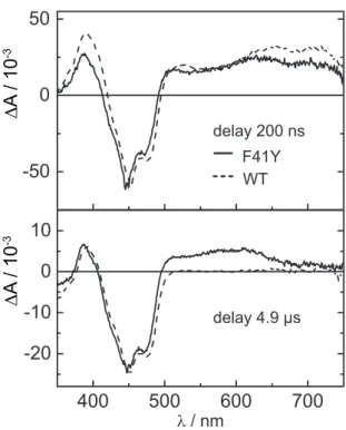
To further investigate the behavior of CrLOV1-F41Y upon blue light excitation, transient absorption (TA) spectroscopy in the µ s timerange was applied. Figure 2.2.2 shows difference absorption spectra of Cr LOV1-F41Y at 200 ns and 4.9 µ s after
norm. Absorbance
Figure 2.2.1 Comparison of the steady-state UV/Vis spectra of CrLOV1 wt in the dark (black line) and
in the adduct state (dotted line) and of CrLOV1-F41Y (red line). The steady-state UV/Vis spectrum
of Cr LOV1-F41Y does not change upon blue light illumination.
delay 200 ns F41Y WT
delay 4.9 µs
∆ A / 10
-3∆ A / 10
-3Figure 2.2.2 Difference absorption spectra extracted from a TA measurement recorded with a 10 µs time window of CrLOV1-F41Y (solid line) 200 ns and 4.9 µs after laser excitation. Spectra of CrLOV1 wt (dotted line) are shown for comparison. The noise level of the data was reduced by singular value decomposition (SVD) and all spectra were scaled to the same minimum at 447 nm.
laser excitation. For comparison, previously published spectra of Cr LOV1 wt are included [47]. All spectra are scaled to the same minimum at 447 nm. The negative absorption band at around 450 nm corresponds to the ground state bleach of the oxidized FMN, FMNox. Positive absorption signals are detected for CrLOV1-F41Y from 493–745 nm and below 415 nm in Figure 2.2.2 after 200 ns. The wt exhibits a similar spectrum blue-shifted by 5 nm. After 4.9 µ s, a positive band can be detected at 374–407 nm and above 493 nm up to 660 nm in the case of Cr LOV1-F41Y. The positive absorption band above 660 nm almost decayed to zero at this time delay.
The difference spectrum of Cr LOV1 wt in Figure 2.2.2 after 4.9 µ s is characterized by the positive absorption of the adduct state at 376-409 nm and the ground state bleach. Above 500 nm, no absorption signals contribute to the spectrum.
We performed global lifetime analysis of the TA data (see chapter 5, subsection 5.5.2), two lifetime components were sufficient for a good fit. Figure 2.2.3 shows the two decay associated difference spectra (DADS) corresponding to decay time constants of τ 1 = 1.3 µ s and τ 2 > 10 µ s. The first DADS (D 1 ) exhibits positive signals above 490 nm and below 420 nm next to the negative ground state bleach of FMNox.
The broad absorption band with the typical structure in the range 650 to 700 nm
can be associated with the excited triplet state of FMN [65]. The triplet decay
time of CrLOV1-F41Y is almost the same as in CrLOV1 wt, which was reported
to be 1.16 µ s [47]. The second DADS (D 2 ) is non-decaying within the observed
2.2 Results 11
∆ A / 10
-3Figure 2.2.3 DADS obtained from global lifetime analysis of CrLOV1-F41Y. The first DADS (solid line) decays with a time constant of 1.3 µs, while the second DADS (dashed line) is non-decaying within the measured time window of 10 µs.
streak window of 10 µ s. This D 2 has positive absorption in the range of 375 nm to 403 nm and from 495 nm to 670 nm, resembling the spectrum of the neutral semiquinone radical FMNH [65, 66]. The spectrum of this radical in the LOV environment is known from photochemical reduction experiments of CrLOV1-C57G with EDTA or β -mercaptoethanol [60, 67–69]. Comparison of these spectra with D 2
of Cr LOV1-F41Y revealed a significant difference in the range 500-530 nm, indicat-
ing an additional species contributing to D 2 . FMNH is formed by electron transfer
to FMN, leading to the radical anion FMN – which is subsequently protonated. In
the photoreduction experiment with Cr LOV1-C57G the electron was donated by
β -mercaptoethanol. The cation of this donor or its decay products do not absorb in
the visible spectral range and protonation of FMN – is complete in this steady state
experiment. In the case of Cr LOV1-F41Y, however, the electron is most probably
donated by Tyr41. The initially produced tyrosine radical cation usually deproto-
nates quickly to the neutral radical. On the other hand, protonation of the radical
anion FMN – can be a slow process on the µ s timescale. Hence we considered the
following species for a possible contribution to D 2 : FMNH , FMN – and TyrO .
We then tried to fit the observed D 2 (black line, Figure 2.2.4) to a linear combi-
nation of reference spectra of FMNox, FMNH , FMN – minus FMNox and TyrO
(for description how these reference spectra were generated see chapter 5, subsub-
section 5.5.2.1). A fit to the reference spectrum of FMNH minus FMNox (figure
2.2.4, green line) did not lead to reasonable agreement, as did a fit including also
the reference spectrum of FMN – minus FMNox (blue line, figure 2.2.4). However,
the linear combination of FMNox, FMNH and FMN – minus FMNox yields a good
description of D 2 in the range λ > 500 nm (magenta line, figure 2.2.4). The difference
between this linear combination and D 2 < 500 nm corresponds to the red spectrum
shown in figure 2.2.4, inset. The peak with maxima at 397 nm and 413 nm resem-
bles the spectrum of TyrO minus TyrOH. The tyrosyl radical spectrum in aqueous
solution shows a narrow absorption peak at 408 nm with a shoulder at 391 nm [70].
∆ A / 10
-3D2 - LK
Figure 2.2.4 D
2of CrLOV1-F41Y (black line) compared to the linear combination (LK) of the reference difference spectra (purple line) for the species FMNH minus FMNox (green line) and FMN
–minus FMNox (blue line). Inset: The reference spectrum of the TyrO radical (black line) compared to the difference between D
2and the sum of the flavin radical species, i.e. the black and the purple lines from the main plot (red line). The TyrO reference spectrum (black line), generated with FMN and Tyr, was red-shifted by 9 nm for better comparison.
It is shown as the black spectrum in the inset in figure 2.2.4, red-shifted by 6 nm for better comparison. It is evident from the spectra that the peak at 397 nm of the red spectrum is in good agreement with the reference spectrum of TyrO . The different shape of the second peak at 413 nm can be explained by the fact that the reference spectrum of FMN – minus FMNox was generated with CrLOV1/C57S which has a spectral shift of the ground state bleach compared to CrLOV1/F41Y. We conclude that electron transfer in CrLOV1-F41Y occurs from Tyr41 to FMN, and that the flavin radical anion is still not completely protonated after 10 µ s.
2.2.2 Additional mutation of the reactive Cys57
No evidence for adduct formation was found in Cr LOV1-F41Y although the reactive Cys is still present. To elucidate, whether this Cys57 is participating in the observed electron transfer reaction of Cr LOV1-F41Y, we additionally replaced the reactive Cys with the polar amino acid serine and the unpolar amino acids alanine and glycine. The UV/Vis spectra (Figure 2.2.5) are in good agreement with the UV/Vis spectrum of CrLOV1 wt indicating an intact hydrogen bonding network around the FMN.
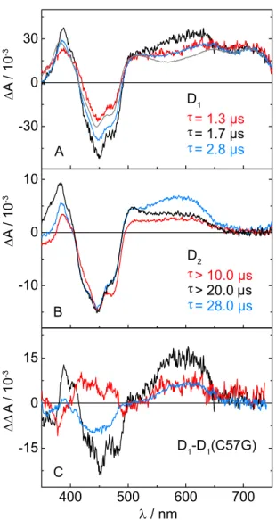
We performed TA measurements at pH 8.0 on a 20 µ s time window with Cr LOV1- F41Y/C57A and on a 50 µ s time window with Cr LOV1-F41Y/C57S. The data were analyzed by global lifetime analysis using two exponential functions for each data set.
The D 1 displayed in Figure 2.2.6A decay with time constants of 1.7 µ s for Cr LOV1-
F41Y/C57A and 2.8 µ s for Cr LOV1-F41Y/C57S, i.e. they are associated with the
decay of the excited triplet state of FMN. D 2 of Cr LOV1-F41Y/C57S decays within
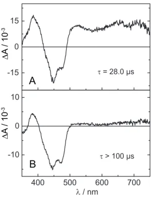
28.0 µ s, while the C57A mutant is non-decaying within the measured time window
2.2 Results 13
WT F41Y F41Y/C57A F41Y/C57S F41Y/C57G
norm. Absorbance
Figure 2.2.5 UV/Vis spectra of the Cr LOV1-F41Y mutants measured in the range of 300-600 nm.
(Figure 2.2.6B). Both DADS in Figure 2.2.6B exhibit similar spectral features of FMNH like Cr LOV1-F41Y. Interestingly, the spectral signatures of the radicals appear not only in the D 2 , but also in the D 1 exemplified by subtraction of the D 1
of the C57G mutant which is entirely due to triplet absorption (see Figure 2.2.6C).
The FMNH absorption varies significantly between the different mutants in the range of 540 nm to 655 nm. The highest amplitude can be
found in CrLOV1-F41Y/C57S, followed by Cr LOV1-F41Y/C57A and CrLOV1- F41Y. Furthermore, the positive absorption below 405 nm differs between CrLOV1- F41Y/C57S and CrLOV1-F41Y/C57A, being more intense in the latter one with a shoulder at 398 nm. In combination with the pronounced peak around 500 nm in Cr LOV1-F41Y/C57A, these spectral features are characteristic for the FMN radical anion, FMN – .
A fit of the linear combination of the aforementioned reference spectra revealed con- tributions of FMNH , FMNox, FMN – and TyrO to the D 2 of CrLOV1-F41Y/C57S.
In contrast, D 2 of Cr LOV1-F41Y/C57A could be well described using a linear com- bination of FMNH , FMNox and FMN – . Including the TyrO spectrum did not improve the fit. The results from spectral fitting indicate that the introduced Tyr41 is quite likely the terminal amino acid of the electron transfer chain and therefore the electron donor, since the spectrum of TyrO needs to be taken into account to fit the D 2 of Cr LOV1-F41Y and Cr LOV1-F41Y/C57S. Only in the case of CrLOV1- F41Y/C57A we could not establish the contribution of TyrO via spectral fitting of D 2 . But in this mutant, the FMN – features are more pronounced compared to the other F41Y mutants, which has a strong absorption in the same spectral region as the TyrO radical.
The photophysical behavior of CrLOV1-F41Y/C57G is different compared to the
other F41Y mutants investigated. Analysis of a 100 µ s TA dataset results in two
DADS (Figure 2.2.7). D 1 exhibits the typical spectral features of the FMN triplet
state and decays with τ = 28.0 µ s, prolonged by a factor of 10 compared to the
corresponding DADS of the other F41Y mutants. D 2 is non-decaying within the
measured streak window and the spectrum exhibits high similarity with D 1 . This
τ = 1.3 µs τ = 1.7 µs τ = 2.8 µs D
1τ > 10.0 µs τ > 20.0 µs τ = 28.0 µs D
2D
1-D
1(C57G) C
B A
λ / nm
∆ A / 10
-3∆ A / 10
-3∆∆ A / 10
-3Figure 2.2.6 DADS obtained from the global lifetime analysis for Cr LOV1-F41Y/C57S (blue line) and CrLOV1-F41Y/C57A (black line). The DADS in A decay with a time constant of 2.8 µs and 1.7 µs.
The DADS of Cr LOV1-F41Y (red line) and of CrLOV1-C57G (gray line) are shown for comparison. The
spectrum of Cr LOV1-C57G corresponds to the pure triplet transient absorption. B: The second DADS
of Cr LOV1-F41Y/C57S decays with a time constant of 28.0 µs, while the one of CrLOV1-F41Y/C57A
is non-decaying within the measured streak window of 20 µs. The DADS were scaled to the same
minimum at 447 nm. C: The D
1of panel A after subtraction of the D
1of Cr LOV1-C57G.
2.2 Results 15
∆ A / 10
-3∆ A / 10
-3A
B
Figure 2.2.7 DADS of Cr LOV1 F41Y C57G obtained by global lifetime analysis. A: D
1decaying with a rate constant of 28.0 µs B: D
2non-decaying within 100.0 µs.
component has an amplitude of approx. 10 % of the faster decaying component. In Cr LOV1-F41Y/C57G no spectral evidence for FMN radicals can be observed. These findings suggest a biphasic triplet decay like already observed in the case of CrLOV1- C57G [47] indicating the presence of an additional quenching channel. The same has been reported for Pp2FbFP L30M, a LOV domain based singlet oxygen sensitizer [71]. It has been suggested that two distinct triplet states are formed in those proteins which can be divergently quenched by oxygen due to varying accessibility of molecular oxygen into the binding pocket. Glycine is the least sterically demanding natural amino acid. This allows for more space in the binding pocket and therefore increased flexibility of the chromophore.
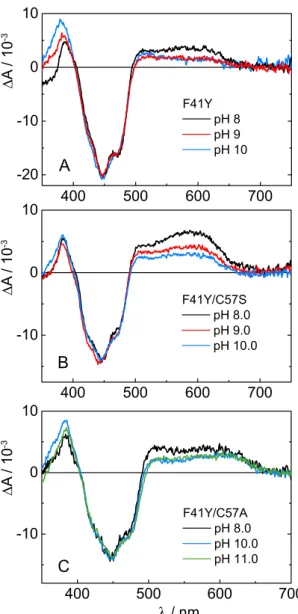
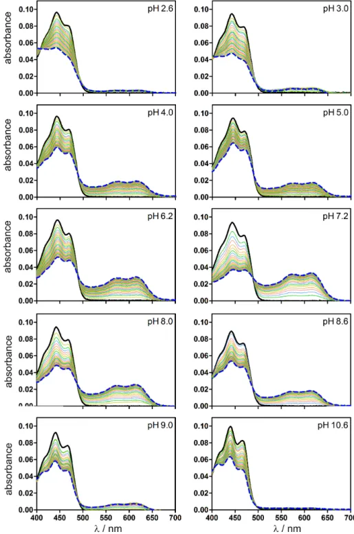
2.2.3 pH influence
Our TA data show, that mainly the protonated FMNH can be observed upon blue light excitation of the Cr LOV1-F41Y mutants. In order to identify the proton donor in this reaction we increased the pH of the buffer solutions up to 11.0 and repeated the TA measurements. Figure 2.2.8 compares the D 2 at different pH values of the respective Cr LOV1-F41Y mutants resulting from analysis of the TA data.
All three F41Y mutants form the neutral radical FMNH even at strongly basic pH,
albeit with a reduced yield. The strongest decrease of approx. 50 % can be observed
A
B
C
∆ A / 10
-3∆ A / 10
-3∆ A / 10
-3Figure 2.2.8 Comparison of the D
2of Cr LOV1-F41Y (A), F41Y/C57S (B) and F41Y/C57A (C) at pH 8.0, 9.0, 10.0 and 11.0, respectively.
in the case of Cr LOV1-F41Y/C57S (Figure 2.2.8B). In the D 2 of Cr LOV1-F41Y and CrLOV1-F41Y/C57A (Figure 2.2.8A and C) the spectral features of FMN – become more pronounced with increasing pH including the absorption bands around 500 nm and below 400 nm. The fact that FMNH can still be observed at strongly basic pH indicates that the proton donor must be protonated as well up to a pH of 10.0-11.0.
Therefore, protonation by the solvent is not possible but must be performed by an
amino acid of the LOV core. According to the crystal structure of Cr LOV1 (pdb
1N9L) as well as our MD simulations, the only proton donor in the binding pocket
of the CrLOV1-F41Y mutants is Tyr41. In the case of CrLOV1-F41Y also Cys57 is
possible. The pK a value of free Cys is 8.18 and the one of Tyr is 10.07. However, the
pK a values of amino acids can be significantly shifted by the protein environment
2.2 Results 17 [72]. Hence, we calculated the pK a value of Tyr41 in the Cr LOV1-F41Y mutants using the H ++ program v3.2 [73] resulting in pK a values of 11.45 (F41Y/C57S) and > 12 (F41Y, F41Y/C57A). Cys57 in CrLOV1-F41Y has a calculated pK a of 10.9 and is thus also strongly shifted towards a more basic value. We conclude that Tyr41 is the proton donor in Cr LOV1-F41Y/C57A and C57S. In the case of Cr LOV1-F41Y, however, we can not exclude Cys57 as a possible proton donor. It should be noted that the accessibility of the solvent molecules into the binding pocket should be considered within the context of protonation. This will be discussed in subsection 2.2.5.
2.2.4 Decay of the S 1 state suggests electron transfer via the excited singlet state
The TA data of the CrLOV1-F41Y mutants provide evidence for the occurrence of an electron transfer reaction. Next to the observation of FMN and Tyr radical species, the triplet decay times obtained are as short as in CrLOV1 wt indicating a quenching of the excited FMN triplet state. The only exception is CrLOV1- F41Y/C57G in which no radical species could be detected in the µ s timerange and the triplet decay is prolonged compared to the other F41Y mutants. Determination of the S 1 decay times (Table 2.1), however, yields a biexponential decay behavior for all CrLOV1-F41Y mutants, including Cr LOV1-F41Y/C57G. In all F41Y mutants one decay time corresponds to the expected value for Cr LOV1 (i.e. 3 ns for wt and 4.5-5 ns for photo-inactive Cr LOV1 mutants) while the second S 1 decay time is a component that is faster than the time resolution of our apparatus (ca. 1 ns). This indicates, that the CrLOV1-F41Y mutants can adopt at least two conformations to account for the biexponential decay behavior.
Furthermore, the fluorescence quantum yields, Φ F , of the Cr LOV1-F41Y mutants were determined (Table 2.1). The fluorescence of the photoinactive mutants C57S, C57A and C57G are strongly quenched from Φ F = 0.30, 0.30 and 0.25 to Φ F = 0.18 (F41Y/C57S), 0.15 (F41Y/C57A), and 0.10 (F41Y/C57G) when additionally intro- ducing Tyr41. The wt and the F41Y mutant have similar values of 0.16 and 0.14, respectively. We conclude from our measurements of Φ F and the S 1 decay times that both, the photoactive C57 and the extra tyrosine, provide quenching channels that do not exist in the photoinactive mutants. This could be enhanced ISC to the triplet state, but might also indicate that some fraction of the electron transfer occurs from the singlet state.
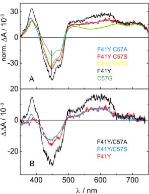
To check this, we analyzed the sum of all DADS of each particular F41Y mutant.
The sum of all DADS corresponds to the initial situation before any µ s-processes
occur and will be named hereafter t 0 spectra. They are shown in Figure 2.2.9A. All
spectra were scaled to the same value at 715 nm. At this wavelength exclusively the
FMN triplet state absorbs and no other species contribute to the spectrum. As a
reference, the t 0 spectrum of Cr LOV1 C57G is included. It is known from a previous
A
B
∆ ∆ A / 10
-3∆ A / 10
-3norm.
Figure 2.2.9 A: Sum for all DADS of CrLOV1-F41Y, CrLOV1-F41Y/C57S, CrLOV1-F41Y/C57G and CrLOV1-F41Y/C57A referring to the spectra at t
0before any µs processes occur. The corresponding spectrum of CrLOV1-C57G representing the pure triplet spectrum of FMN in the CrLOV1 core is shown for comparison. B: Difference spectra obtained by subtracting the reference spectrum of CrLOV1-C57G from the t
0spectrum of each F41Y mutant in A. The difference spectra show characteristic features of FMN radical species.
study, that this spectrum refers to the pure triplet spectrum of FMN in the CrLOV1 core [47]. When comparing this reference spectrum with the t 0 spectra of Cr LOV1- F41Y, F41Y/C57S and F41Y/C57A significant differences become apparent in the range of 500 nm to 660 nm and below 410 nm (see Figure 2.2.9A). Subtraction of the C57G reference spectrum from the t 0 spectra of Cr LOV1-F41Y, F41Y/C57S and F41Y/C57A yields the spectra in Figure 2.2.9B, which show characteristic fea- tures of the flavin radical spectra described above. Since these spectra can already be observed at t 0 , the radical species in CrLOV1-F41Y, Cr LOV1-F41Y/C57S and CrLOV1-F41Y/C57A can not be formed exclusively via a triplet state reaction.
Thus, the electron transfer in these mutants must occur to some extent in the ex-
cited singlet state of FMN. The t 0 spectrum of CrLOV1-F41Y/C57G on the other
hand is very similar to that of Cr LOV1-C57G except for the spectral range of the
ground state bleach. This means, that if CrLOV1-F41Y/C57G performs an electron
transfer via the excited singlet state, as suggested by the fluorescence data, the rad-
ical is not stabilized and probably recombines on a faster timescale than formation
of the triplet state occurs.
2.2 Results 19
Table 2.1 Fluorescence quantum yields, Φ
F, and S
1decay times of all F41Y mutants. The fluorescence lifetimes were determined using time correlated single photon counting with excitation at λ = 376 nm and detection at λ = 510 nm. Values for CrLOV1 wt and CrLOV1-C57S are taken from Holzer et al.
[74].
τ 1 /ns τ 2 /ns Φ f
F41Y 3.0 ( ∼ 50 %) 0.9 ( ∼ 50 %) 0.14
WT a 2.9 - 0.16
F41Y/C57A 3.4 ( ∼ 50 %) 1.2 ( ∼ 50 %) 0.15
C57A 4.5 - 0.30
F41Y/C57S 3.6 ( ∼ 65 %) 1.0 ( ∼ 35 %) 0.18
C57S a 4.6 - 0.30
F41Y/C57G 4.3 ( ∼ 80 %) 0.9 ( ∼ 20 %) 0.10
C57G 4.5 - 0.25
a values shown for comparison.
2.2.5 Molecular Dynamics Simulations
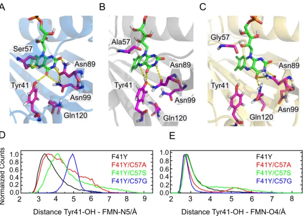
The microenvironment surrounding the FMN chromophore and its interactions with the protein is of crucial importance. The photoreactivity of riboflavin is strongly affected by its hydrogen bond network [63, 75]. Ivan Stambolic kindly performed 20 ns MD simulations of Cr LOV1-F41Y using the dark state crystal structure of Cr LOV1 wt (pdb 1N9L) as a starting structure [76]. The MD data reveal that substitution of Phe41 with Tyr has only minor influence on the overall structure of Cr LOV1-F41Y. Small perturbations occur around Tyr41 but the hydrogen bonding network built around FMN C2(=O), N3(H) and C4(=O) including Asn89 and Asn99 remains intact. Tyr41 can form a hydrogen bond with FMN C4(=O) as well as with FMN N5 instead of Gln120, which is rotated away from FMN. A close-up view of the binding pocket of the 20 ns simulation structure of Cr LOV1-F41Y in comparison with the dark state crystal structure of the wild type is shown in Figure 2.2.10.
Figure 2.2.11A and B shows the structures of Cr LOV1-F41Y/C57S and CrLOV1- F41Y/C57A. In Cr LOV1-F41Y/C57S the hydrogen bond network between Asn89, Asn99 and FMN C2(=O), N3(H) and C4(=O) remains intact. In this case, the distance between Tyr41 and FMN C4(=O) is small enough to form a hydrogen bond, while direct interaction with FMN N5 like in CrLOV1-F41Y is quite unlikely.
Instead, FMN N5 can form a hydrogen bond with the polar side chain of Ser57.
In the case of CrLOV1-F41Y/C57A (Figure 2.2.11B) the hydrogen bond network
on the polar side of FMN is changed and does not include Asn99. Asn89 in turn,
can form a hydrogen bond with N3(H) and C4(=O) of FMN. The introduced Tyr41
is very close to the FMN and well stabilized in this mutant, with possible hydrogen
bonds to FMN N5, C4(=O) and Asn89. Since Ala57 cannot form hydrogen bonds,
Tyr41 is the only amino acid in the close proximity of FMN which can
Cys57
Asn89
Asn99 Gln120
Phe41/
Tyr41
Figure 2.2.10 Superposition of the 20 ns MD simulation structure of CrLOV1-F41Y (gray) with the dark state crystal structure of Cr LOV1 wt (green) visualized with PyMOL [77]. The mutation F41Y does not cause significant perturbations of the Cr LOV1 structure except for Gln120, which is rotated away from FMN.
make a connection with FMN N5. In both mutants, Cr LOV1-F41Y/C57S and CrLOV1-F41Y/C57A, Gln120 is rotated away from FMN, not interacting with the chromophore.
In contrast, FMN N5 does not participate in hydrogen bond interactions in Cr LOV1- F41Y/C57G (Figure 2.2.11C). The distance between Tyr41 and FMN C4(=O) and FMN N5 is approx. 5 Å and therefore larger than in the other F41Y mutants.
Furthermore, the hydrogen bond network between FMN and Asn89 and Asn99 is changed compared to Cr LOV1 wt. Asn89 can interact with FMN N3(H) and C4(=O), but due to a rotation of Asn99 no interaction between this asparagine and FMN is possible. Gln120 is interacting with C4(=O) instead.
Figure 2.2.11D and E compares the distances between Tyr41-OH and either FMN N5 (D) or FMN O4 (E) of the distinct F41Y mutants. The distances between Tyr41 and FMN N5 vary strongly between the different mutants from around 3.0 Å for CrLOV1-F41Y to 5.0 Å for Cr LOV1-F41Y/C57G. The distances of Cr LOV1- F41Y/C57A and C57S are broadly distributed with a maximum at 3.0-4.0 Å. The distances between Tyr41 OH and FMN O4 on the other hand are almost equal in all F41Y mutants between 2.7 and 3.0 Å (Figure 2.2.11E).
Furthermore, the dihedral angle distributions between Tyr41-CE2, FMN - (C5a-C4a)
and Tyr41 - CE1 of all CrLOV1 F41Y mutants were determined from the 20 ns MD
trajectories (figure ). The distribution curves are broad in all cases with varying
2.2 Results 21
A B C
Ser57
Gln120 Asn99
Asn89
Tyr41 Tyr41 Tyr41
Asn99
Asn99
Asn89 Asn89
Gln120 Gln120
Ala57 Gly57
D E
Distance Tyr41-OH - FMN-N5/Å Distance Tyr41-OH - FMN-O4/Å
2 3 4 5 6 7 8
2 3 4 5 6 7 8 9
1.0 0.8 0.6 0.4 0.2 0.0 1.0 0.8
0.6 0.4 0.2 0.0
Normalized Counts
F41Y F41Y/C57A F41Y/C57S F41Y/C57G
F41Y F41Y/C57A F41Y/C57S F41Y/C57G
Figure 2.2.11 20 ns simulation structures of A: CrLOV1-F41Y/C57S, B: CrLOV1-F41Y/C57A and C: CrLOV1-F41Y/C57G. Possible hydrogen bonds are indicated by dashed yellow lines. D and E:
Distance distribution plots of F41Y mutants between Tyr41-OH and FMN-N5 and O4, respectively.
maxima from 18 ◦ (F41Y C57S) to 22 . 5 ◦ (F41Y). CrLOV1 F41Y C57A additionally has a second minor peak. The dihedral angle distribution indicate a considerable flexibility between Tyr41 and FMN possibly leading to different geometry-dependent reaction pathways. The structural differences of the particular CrLOV1-F41Y are accompanied by varying accessibilities for solvent molecules into the binding pocket.
In Cr LOV1-F41Y, a slow water exchange was observed, with always one water molecule being present in the binding pocket. A very fast water exchange occurs in Cr LOV1-F41Y/C57G with constantly three to four water molecules present.
Cr LOV1-F41Y/C57A also shows fast water exchange. The water molecule is lo-
cated around FMN-O4, but the pathway for the HOH entry is different. While
in the other CrLOV1-F41Y mutants the water enters via the phosphate backbone
of FMN, in CrLOV1-F41Y/C57A a new water channel opens on the polar side of
FMN. Solely in Cr LOV1-F41Y/C57S, no water exchange was observed. During
the whole simulation time one non-exchanging water molecule is present, located
between Ser57 and FMN oriented towards N10.
F41Y F41Y/C57A F41Y/C57S F41Y/C57G
Normalized Counts
Dihedral angle/degree
Tyr41-CE2 - FMN-(C5a-C4a) - Tyr-CE1
0 5 10 15 20 25 30
0.0 0.5 1.0
Figure 2.2.12 Dihedral angle distributions between Tyr41-CE2, FMN - (C5a-C4a) and Tyr41 - CE1 of all CrLOV1 F41Y mutants.
2.3 Discussion
Adduct formation as the initial step of activation is a well known attribute of LOV domains. While the reaction mechanisms of other flavin based blue light sensors, e.g. BLUF and CRY, implicate electron transfer reactions, no other light induced photoreaction than adduct formation has been observed so far in the case of LOV domains if the reactive Cys was present. In this study, we present spectroscopic and simulation data on CrLOV1 mutants, in which a Tyr was introduced as a potential electron donor in close proximity to the flavin chromophore. The substitution of Phe41 by Tyr leads to a change in the primary photoreaction of Cr LOV1. The naturally preferred adduct formation reaction is inhibited and instead an electron transfer can be observed.
The mutation did not significantly alter the electronic properties of FMN since the ground state absorption spectrum shows only minor changes in comparison to CrLOV1 wt. Moreover, MD simulations of both, Cr LOV1 wt and CrLOV1 F41Y, reveal that both structures are highly similar with mainly preserved hydrogen bond interactions between FMN and the surrounding amino acids. Only minor perturba- tions around the introduced Tyr occur.
TA measurements in the µ s-time range showed that Cr LOV1-F41Y populates the
excited FMN triplet state like observed for the wt which decays also with a simi-
lar rate constant. But instead of the characteristic flavin-cysteinyl-adduct, radical
species of FMN evolved from the triplet state. Spectral fitting of the data demon-
strated the presence of FMNH , FMN – and TyrO . Likewise, when additionally sub-
stituting the reactive Cys57 with Ala or Ser, the formation of flavin radical species
can be observed. CrLOV1-F41Y/C57A and C57S as well populate the FMN triplet
state that decays with comparably short lifetimes like observed for CrLOV1-F41Y
and wt. However, in these cases the deactivation channel of the triplet is electron
transfer. Other photo-inactive CrLOV1 mutants, in which electron transfer occurs
only in the presence of an external electron donor like EDTA, usually have a triplet
2.3 Discussion 23 decay time by at least a factor of ten longer [34, 47]. Presumably, Tyr41 is the initial electron donor in the F41Y mutants although the amino acid sequence of Cr LOV1 wt contains four Tyr and one Trp as natural candidates. According to the crystal structure, they are located quite distant from the acceptor FMN, with Tyr47 being closest with a distance of 11.0 Å between Tyr47-OH and FMN-O4. Tyr41 on the other hand, is located very close with an average distance of 2.75 Å to FMN-O4 in each F41Y mutant providing a favored initial situation since electron transfer reactions are strongly distance dependent processes. Furthermore, the described electron transfer reactions are only observed, when Tyr41 is present.
On the other hand, our findings on the excited singlet state deactivation, including a reduced fluorescence quantum yield and a biexponential fluorescence decay, lead to the hypothesis that a second pathway for electron transfer via the excited singlet state exists in the Cr LOV1-F41Y mutants. This is supported by the fact that the radical species are already formed immediately after the exciting laser pulse (ca 10 ns) in the TA measurements. A reasonable explanation for the two reaction pathways would be the existence of more than one conformation of each F41Y mutant most likely regarding Tyr41. It has been reported recently, that already subtle conformational changes of the terminal electron donor Trp in the A. thaliana cryptochrome triade can inhibit the electron transfer to the next Trp due to a drastic change in the electronic coupling between donor and acceptor [78]. And in fact, the dihedral angle distribution including FMN and Tyr41 is rather broad for all F41Y mutants indicating a variation in the donor to acceptor orientation.
Since Cr LOV1-F41Y, F41Y/C57S and F41Y/C57A feature the same photo-chemical characteristics upon blue-light excitation we propose that these mutants follow the same mechanism, meaning that the reactive Cys in CrLOV1-F41Y apparently be- comes unreactive. Although the orientation and distance of Cys57 and FMN seem to be favorable for adduct formation according to our MD simulations, we do not observe any matching signals in our spectroscopic data. The triplet decay time of Cr LOV1-F41Y is 1.3 µ s and therefore in the same timerange as in the wt. Conse- quently, the first step leading to the radical pair in the mutants containing Tyr41 is about as fast as the first step leading to the adduct in the wt. In the F41Y mutants this first step should be electron transfer from Tyr to 3 FMN, for the wt the first step most likely is also electron transfer, this time from Cys57 to 3 FMN. Accordingly, in F41Y we should expect both, adduct and FMN radicals, with similar yield.
In a previous study of the Cr LOV1 wt photocycle we could not detect any interme-
diate between the FMN triplet state and the adduct [47]. We concluded that the
reaction is either concerted or it occurs as a sequence of electron transfer, proton
transfer and radical recombination, with electron transfer being the rate limiting
step. If adduct formation is a concerted reaction, it should compete with electron
transfer from Tyr in the F41Y mutant, and both products should be observed. If
however, the reaction occurs in several steps, competition between the two reactions
might occur only in the second step, meaning after electron transfer from Cys the
formed cysteine radical cation reacts faster with the Tyr than with the FMN radical
k
Tk
C3
FMN + Cys + Tyr
2
FMN .
-+ Cys .
++ Tyr
2