1222
Strain
etal
reply:We found the comments of Dr Bowsher on our article very informative, and a suitable starting point for a discussion on the diag- nostic value of heat pain sensitivity testing.
His main concern was that the mean heat pain thresbold in the dermatome ipsilateral to the chronic nerve root compression was 48· l •c, which he found unaccountably hig:h.
We wish to point out that our controls' value was 47·3°C in this study and that we had obtained a value of 46·5°C at the same sites in a much younger group of healthy sub- jects.' We therefore think the question is why are our values consistently hig:hcr than those reportcd by BoWllhcr, which arc in the range of 41°C to 43°C.
Even with contact thermodes, the heat pain threshold cannot be considered as a physiological constant given in •c. lt is clearly dependent on the physical character- istics of the thermodc and the mcasuremcnt procedures. Wc will givc some examples of our experience. We recently changcd from Marstock type thermodes (used also in the study under discussion) to a more advanced model with the same surface area (6 cm").
Because of thc dift'crent charactcristics of thcsc thennodes that is, the isolation layer between the Pelticr elements and the ther- modc surfacc) an avcrage lowering of the
Maturs Arising
thresholds of about 1·5°C occurrcd.
2The hcat pain thresholds of
threeage groups measured at thc lateral dorsum pedis wcre 44·9(1 ·5)°C (17-29 years), 44·8(1-9)°C (30-44 years) and 45·7(1 ·2)°C (45-63 ycars). These valucs arc still considerably hig:her
thanthose given by Bowsher. We think that therc may be two reasons.
First, threshold estimates
inthe early trials wcre lower and more variable
thanin the later oncs. For example, the difference between thc first and the eig:hth trial with measure- ments on thc foot was found tobe -1·3°C.' Wc thereforc disregard thc first three trials
inevaluation. Second, with thc traditional Mar- stock proccdures thc tcmpcrature increascs start from tcmpcratures around 30°C and may lcad to what we would call "prematute pain rcsponses" at temperatures weil below 40°C. This can
againbe clcarly seen in a vcry reccnt study by Jensen
et al. 4To avoid this, wc set the base temperatures to 38°C or 40°C, which arc levcls that have not becn feit painfully by any of our patients or control subjects.
Considering these factors, wc
thinkthat the pain threshold values wc reponed, althoug:h different from those of Bowsher, should be considercd valid and that it
isvery unlikely that thc difference betwecn thc pain thresholds measured ipsi-and contralaterally to the nerve root comprcssion is the con- sequcncc of an ipsilateral threshold increase rathcr than a contralateral thrcshold dccrcasc. (As a rcmindcr, the contralatcral valuc was 45·8°C and significantly smallcr than thc valuc ofthe hcalth controls).
Wc rcad thc findings of Bowsher on heat
pain and warmth scnsitivity
indifferent
paticnt groups with intcrest and have no
difficulty agreeing with them. However,
hypalgcsic phenomena
indiabetic, posthcr-
patic and post-stroke patients do not excludc
thc possibility of hyperalgcsic phenomcna
inMaiurs Arising
paticnts with chronic lumbosacral disc dis- eases as descnbed in our study. We think thcre are important diffcrences in pathophy- siolo&Y. So far the testing of neuropathic conditions with heat pain stimulation has rarely resulted in strong evidence for hyper- algesic or hyperpathic changes.
Ina very recent publication,' however, Wall gave a great number of examples of hyperalgesic changes produced by different kinds of neu- ropathies and he also pointed to the fact that a non-selective blockade of peripheral affer- ent impulses may lead to a "partial disinhibi- tion" and, in consequence, to hyperalgesia.
This is what seemed to have happened in our patients with chronic lumbosacral disc dis- ease. That such an event might produce effects at the contralateral side appears not too speculative when the results of con- tralateral TENS-effects cited in our paper are considered. Thken together, we still believe that the conclusions
drawnfrom the pilot study described are justified.
Finally, we want to answer the questions raised
byBowsher. 1\vo patients were affec- ted at the L5 root affection and 7 at the S l root. We measured the thresholds at the medial (L5) and lateral (Sl) side of the dorsum pedis, where the peripheral denna- tomes are to be found, and verified the location of the dennatomes in the preceding neurogical examination. As Bowsher expec- ted, the dennatones do normally not differ in wannth and pain sensitivity.
S l.AUTENBACHER Max Planck Institut far
l'Jychiatrie,
Kraepelinsrr I 0, J)..8000 Munich, Germany Strian F, Lautenbacher S, Galfe G, Hölzl R.Diurnal variations in pain per<:eption and thennal sensitivity.
Poin
1989;J6:125-31.2 Lautenbacher S, Strian F. Similarities in age differences in heat pain perception and ther-
IDll senaitivity.
Funct N6UrrJI
1991:6:125-31.3 Galfe G, Lautenbacher S, H61zl R, Strian F.
Diaan011is of small fibre neuropatby: com- putcr-usisted methods of combined pain and therlDll semitivity determination.
Ht1$JJitrudi-
c11 1990:8:38-48.
4 Jensen TS,
BachFW, Kastrup J,
DejpardA,
BrennumJ.
Vibrstory and thermaltbresholds
indiabetics
with and without clinical neurop- athy. Acta Nnnol Scand 1991 :84:326-33.5 Wall PD Neuropathie pain and injured nerve:
central mechanisms. Br Med Bull 1991:
47:631-43.
1223
1222
Disturbances ofC-fibre·mediated 1emi- bllity fn lumbosacral dfac cUsease 1 was interested to read the communication by F Strian
etal.' There is difficulty, however, in accepting that heat pain threshold is lowered
inthe foot contralaural to the sciatic root compression, rather
thanraised in the foot
ipsilateralto the lesion (as is the warmth threshold).
With JA Campbell, AW Chan, G Leijon and T Nurmikko, thresholds to most somato- sensory modalities have been measured, by the method of limits, at five body sites on each side in a !arge number of volunteers, as weil as in patients with neurogenic pain conditions. In the case of the foot, a
Mar-stock Peltier thermode measuring 25·0 x 50·0 mm (12·5 cm2)
wasapplied just below the medial malleolus, where the skin is neither thickened nor hairy. Our results for warmth and heat pain
in(1) the normal foot;
(2) the diabetic foot, (3) postherpetic neu- ralgia, and (4) central post- stroke pain ("thalamic syndrome") are shown
intable 1.
In
cases 3 and 4, comparison is made {paired
ttest) with the una1fected mirror-image area on the other side of the body, while
in(2) age-matched normal subjectS were used (unpaired
ttest). Our results suggest that while there is a !arge rise
inthe warmth threshold (average about 6°C), or twice this for the warm-cold limen, on the affected side, there is only a very small (averagc about 2°C), but significant, rise
inthe heat pain threshold, also on the affected side.
Strian et al used a thermode measuring 6 cm•. With J Giewald, we have performed experiments using both !arge (12·5 cm") and small (1·3 cm") thermodes, and found that while warmth thresholds
varyaccording to the surface area of the thermode, heat pain thresholds do not. We find the ipsilateral mean (SD) heat pain threshold of 48· l (l ·6)°C' unaccountably high compared with our normal thresholds (see table 2) and those found by others, and would suggcst that the ipsilateraJ heat pain threshold
inthe patients of Strian et al may be raised.
lt would be helpful to know how many of their 9 patients bad L5 root lesions and how many S 1; where on the foot they measured heat pain thresholds for the two roots; and whether there
is anydifference according to
Mattm Arising
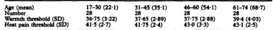
Table 1 Mean wamrth and heat pain thresholds ("C) in normal feet Ace
(mean)Number
Wannth threshold (SO) Heat pain threshold (SD)
17-30 (22-1) 28 36·75 (3-22) 41-5 (2-7)
31-45 (35-1) 37-65 (2·89) 28 41-75 (2-4)
46-60 (54-1) 28 37-75 (2·88) 43·0 (3·3)
61-74 (68·7) 28 39·4 (4·03) 43-1 (2-5)
Table 2 Mean (SD) warm thresholds, warm-cold limen, and heat pain thresholds ("C) in neurogmü:
pain conditions
Palnfbl dlabedc aeuropatbr 64 Patienu:
Median aae: 54 28 Controls:
Warm-cold Limen (SD): 19·0 (8·8) Heat pain threahold 46·6 (2'6) unpaired r test: p < 0·000001 p < 0·00001
Warm-cold Limen (SD): 6· l (2'65) Heat Paln Thresbold: 43·0 (3-3) Postherpetlc
neuralsi•
39 Patienu: Warm Tbreshold (SD): 40·8 (0·9) Heat Pain Threshold (SD): 45·8 (0·5) Median age: 69·5 paired t test:
p <
0·000001p_
< 0·0000339 Conrroh: Wann Tbreshold (SD): 35·4 (0·3) Heat Pain Tbreshold (SD): 43-1 (0·5) Central post-•trolic
paln
38 Parienu: Wann Tbreshold (SO): 40·5 (0·8) Heat Paln Threshold (SD): 43·8 (0·4) Median aae: 64
38 Controls: paired t test: p < 0·000001 p
=
0·02Warm Tbreshold (SO): 33-5 (0·4) Heat Paln Threshold (SD): 42·7 (0·45) Palnfbl cllabetlc neuropatby
64 Parienu: Wann-cold
Diffcrcncc
(SO): 19·0 (8·8) Heat Pain Thresbold: 46·4 (2·6) Median age: 54Z8 Controls: unpaired t test: p < 0·000001 p < 0·00001
Warm-cold Difference (SO): 6·1 (2·65) Heat PainThresbold: 43·0 (3-3) Poetherpetlc
neunJPa
39 Parienu: Warm Threshold (SD): 40·8 (0·9) Heat Pain Thresbold (SD): 45·8 (0·5) Median age: 69·5 paired r test: p < 0·000001 p < 0·00003
39 Controls: WannTbreshold (SO): 35·4 (0·3) Heat PainThresbold (SD): 43·1 (0·5) Central post-stroke paln
39 Patienu:
Median age: 64 38 Conrrols:
Wann Threshold (SDJ: 40·5 (0·8) Heat Pain Threshold (SD): 43·8 (0·4) paired r test: p < 0·000001 p
=
0·02Wann Threshold (SD): 33•5 (0·4) Heat Pain Thresbold (SD): 42·7 (0·45)
site, in both patient population and control subjects-although it must be admitted that our own results on the unaffected side of postheipetic neuralgia patients suggest that site makes little difference.
DAVID BOWSHER Pain Reuarch Insrifuu Wfdron Hospital Liwrpool
L9 lAE
1 Scrain F, uurenbacher S, Karlbauer G, Galfe G. Disturbancesof
C-fibre-mediated sensi- bility in lumbrosacral disc disease.J Neurol
N1Urosurg P1ychialry 1991;54:1013-14.Strain et al
reply:We found the comments of Dr Bowsher on our article very informative, and a suitable starting point for a discussion on the diag- nostic value of heat pain sensitivity testing.
His
mainconcern was that the mean heat pain threshold in the dermatome ipsilateral to the chronic nerve root compression was 48· l 0C, which he found unaccountably hiah.
We wish to point out that our controls' value was 47·3°C in this study and that we bad obtained a value of 46·5°C at the same sites in a much younger group of healthy sub- jects.' We therefore think the question is why are our values consistently higher
thanthose reponed by Bowsher, which are
inthe range of41°C to 43°C.
Even with contact thermodes, the heat pain threshold cannot be considered as a physiological constant givcn in •c. lt is clearly dependent on the physical character- istics of the thermode and the measurement procedures. We will give some examples of our experience. We recently changed from Marstock type thermodes (used also in the study under discussion) to a more advanced model with the same surface area (6 cm").
Bccause of the different characteristics of these thermodes that is, the isolation layer between the Peltier elements and the ther- mode surface) an average lowering of the
thresholds of about l ·5°C occutred.2 The heat pain thresholds of three
agcgroups measured at the lateral dorsum pedis wcre 44·9(1 ·5)°C (17-29 years), 44·8(1 ·9)°C (3o-44 years) and 45·7(1 ·2)°C (45-63 years). These values are still considerably higher than those
givenby Bowsher. We think that there may be two reasons.
First, threshold estimates
inthe early trials werc lower and more variable than in thc later ones. For example, the difference between the first and the eighth trial with measure- ments on the foot was found tobe - l ·3°C. • We therefore disregard the first three trials
inevaluation. Second, with the traditional Mar- stock proccdures the temperature increases start from temperatures around 30°C and may lead to what wc would call "prematute pain responses" at temperatures well below 40°C. This can again be clearly seen
ina very recent study by Jensen et al.
4To avoid this, we set the base temperatures to 38°C or 40°C, which are levels that have not been feit painfully by any of our patients or control subjects.
Considering these factors, we think that the pain threshold values we reponed, although different from those of Bowsher, should be considered valid and that it
isvery unlikely that the difference between the pain thresholds measured ipsi-and contralaterally to the nerve root compression is the con- sequence of an ipsilateral threshold increase rather than a contralateral threshold dccrease.
(Asa reminder, the contralateral value was 45·8°C and significantly smaller
thanthe value of the health controls).
We read the findings of Bowsher on heat
pain and warmth sensitivity
indifferent
patient groups with interest and have no
difficulty agreeing with them. However,
hypalgesic phenomena in diabetic, posther-
patic and post-stroke patients do not exclude
the possibility of hyperalgesic phenomena in
Mattm Arising
paticnts with chronic lumbosacral disc dis- eases as descnbed in our study. We tbink there arc important ditferences in pathophy- siology. So far the testing of neuropathic conditions with heat pain stimulation has rarely resulted in strong evidence for hyper- algesic or hyperpathic changes. In a very recent publication, • however, Wall gave a great number of examples of hyperalgesic changes produced by different kinds of neu- ropathies and he also pointed
tothe fact that a non-sclective blockade of pcripheral affer- ent impulses may lead to a "partial disinhibi- tion" and, in conscquence, to hyperalgesia.
This is what seemed to have happcned in our patients with chronic lumbosacral disc dis- ease. That such an event might produce
effectsat the contralateral side appears not too speculative whcn the results of con- tralateral TENS-etfects cited in our paper are considered. Thkcn togethcr, we still believe that the conclusions
drawnfrom the pilot study described are justified.
Finally, we want to answer the questions raised
byBowsher. 1\vo patients were affec- ted at the L5 root affection and 7 at the S 1 root. We measured the thresholds at the medial (L5) and lateral (SI) side of the dorsum pedis, where the pcripheral denna- tomes are to be found, and verified the location of the dermatomes in the preceding neurogical examination. As Bowsher expec- ted, the dermatones do normally not differ in warmth and pain scnsitivity.
S LAUTENBACHER Max Planclc ln1titut far l'sychiatrie, Kraepe/in1tr 10, D-8000 Munkh, Gmnany 1 Strian F, Lautenbacher S, Galfe G, Hölzl R.
Diurnal variations in pain perception and thennal sensitivity. Pain 1989;36: 125-31.
2 Lautenbacher S, Strian F. Similarities in age differences in heat pain perception and ther-
mal aemitivity.
Funa Neurol
1991:6:125-31.3 Ga1fe G, Lautenbacher S, H61zl R, Strian F.
Diaplosis of small fibre neuropathy: com- puter-usisted methods of combined pain and thermal sensitivlty determination. HtnpilrNdi-
ca
1990:8:38-48.4 Jensen TS, Bach FW, Kastrup J, Dejpard A, Brennum
J.
Vibratory and thermal thresholds in diabetics with and without clinical neurop- athy. Acta Nwroi &4lld 1991:84:326--33.5 Wall PD Neuropathie pain and injured nerve:
central mechanisms. Br Med Bull 1991:
47:631-43.
1223