Eur J Clin Chem Clin Biochem 1996; 34:237-244 © 1996 by Walter de Gruyter · Berlin · New York
A Simple and Sensitive Assay for Determination
of Human Anti-idiotypic Anti-B72.3 Antibodies, which Is not Affected by the Presence of Tumour-Associated Glycoprotein 72
Jochen Reinsberg, Jan Schmolling and Dietlind Ackermann
Zentrum fur Frauenheilkunde und Geburtshilfe, Universit t Bonn, Bonn, Germany
Summary: An immunoradiometric assay is described for the determination of human anti-idiotypic anti-B72.3 IgG.
The latter is formed in ovarian cancer patients after treatment with the murine monoclonal antibody B72.3, which is directed against the tumour-associated glycoprotein 72 (TAG-72). A gel coupled with Fc-specific anti-human IgG antibodies is used as a solid phase for the extraction of serum IgG. The anti-B72.3 IgG is then specifically detected by incubation with radiolabelled B72.3 detector antibodies. Calibration standards were prepared from serum obtained from a patient repeatedly treated with B72.3 antibodies. The concentration of anti-idiotypic anti- B72.3 antibodies was expressed as TAG-72-like arb.units/1. The assay performed with two 60-minute incubation steps is characterized by a high sensitivity (detection limit: 3 Χ 103 arb.units/1) and precision (coefficients of variation: intra-assay = 6.4% and 5.8% at 80 X 103 arb. units/1 and 217 Χ 103 arb.units/1, inter-assay = 8.7% and 7.1% at 91 X 103 arb.units/1 and 212 Χ 103 arb.units/1) and a good linearity of dilution (recovery after dilution between 99% and 107%). The assay is more specific than previously described methods; no interference was observed by TAG-72 up to 3.3 Χ 107 arb.units/1. Also, non-specific human anti-mouse antibodies did not cross- react up to 34.8 mg/1. The test may be modified for detection of anti-idiotypic antibodies, which are formed after treatment with other monoclonal antibodies.
Introduction
A novel therapeutic approach to the treatment of ma- lignant neoplasms is the application of unconjugated monoclonal antibodies directed against a tumour-asso- ciated antigen expressed on the tumour cells. An indi- rect mechanism is thought to be involved in the bene- ficial effect observed in cancer patients after treatment with monoclonal antibodies (1). According to the idio- typic network hypothesis proposed by Jerne (2), injec- tion of a monoclonal antibody specific for a tumour- associated antigen induces the formation of anti-idio- typic antibodies which subsequently may induce the formation of anti^anti-idiotypic antibodies, which in return recognize the same epitope on the tumour-asso- ciated antigen as on the original antibody. These anti- anti-idiotypic antibodies as well as nti-idiotype re- active T-cells (3) may kill the tumour-cells bearing the tumour-associated antigen by a direct mechanism.
Induction of such a humoral idiotypic cascade has been shown after the in vivo application of various monoclonal antibodies (4—7), and the formation of anti-idiotypic antibodies has been favorably correlated with the response of the patients (1, 6, 8, 9).
In most studies, anti-idiotypic antibodies have been measured with an "indirect" assay, in which the anti- idiotypic antibodies are captured by the immobilized
original antibody, then detected by incubation with la- belled anti-human immunoglobulin G (IgG) or immuno- globulin M antibodies (4, 6, 10). Alternatively, in the direct "sandwich"-assay, the original antibody is used as both capture and detector antibody (9—12). In both types of assay the original antibody bound to a solid phase is incubated with whole serum. Thus, for serum samples containing the original tumour antigen, interfer- ence is to be expected, leading to falsely low or falsely high assay results (7, 10, 13, 14).
Recently, in a clinical trial evaluating the effect of repeated infusion of low doses of the murine monoclo- nal antibody B72.3 in ovarian cancer patients, the formation of anti-idiotypic anti-B72.3 antibodies was shown (9). The B72.3 antibody defines the tumour- associated glycoprotein 72 (TAG-72), a surface antigen expressed in the majority of human epithelial cancers (15, 16), which is also released into the serum (17, 18). Thus, the anti-idiotypic antibody concentration could be assessed only in samples with low TAG-72 concentrations because the TAG-72 present in patient serum cross-reacted with the "sandwich" assay used in this study (19). To allow the determination of hu- man anti-idiotypic anti-B72.3 antibodies, even in the presence of TAG-72, we developed an immunoradio-
metric assay which is not affected by TAG-72. This assay uses a gel coupled with Fc-specific anti-human IgG antibodies to extract the serum IgG, before spe- cific detection of the anti-idiotypic anti-B72.3 antibod- ies by radiolabelled B72.3 detector antibodies.
Materials and Methods Patients and serum samples
Serum samples were obtained from ovarian cancer patients (stage III-IV FIGO) who had received one or more infusions of 1 mg of the B72.3 antibody (Oncoscint; Eurocetus, Frankfurt, Germany).
Samples drawn before antibody treatment served as control. Addi- tional control samples were obtained from four patients during the in vitro fertilization cycle. Samples with elevated non-specific hu- man anti-mouse antibody concentrations were obtained from four patients treated with radiolabelled F(abf>2 fragments of the murine antibody OC125 (IMACIS-2; Isotopen Diagnostik CIS, Dreieich, Germany) for the purpose of radioimmunodetection (8), and from one patient treated with the murine antibody B43.13 (Biomira, Ed- monton, Canada) for the purpose of photodynamic therapy (20).
All samples were aliquoted and stored at -20 °C until analysis.
The procedures followed in this study were in accordance with the standards of the ethical committee of our faculty.
Coupling of anti-human IgG antibodies to hydrazide gel
Fc-specific anti-human IgG antibodies were coupled to hydrazide gel (Avidchrom Hydrazide Gel F; Sysmex, Hamburg, Germany) according to the procedure described by Radparvar et al. (21). For buffer exchange 2 ml of a Fc-specific goat anti-human IgG anti- body solution (2.4 g/1; Sigma, Deisenhofen, Germany) were con- centrated with a Centricon-30 concentrator (Amicon, Witten, Ger- many) to an end volume of 0.04 ml and refilled with 2 ml of cou- pling buffer (0.05 mol/1 sodium acetate, pH 5.0). Then 0.2 ml of a sodium /weta-periodate solution (0.1 mol/1) was added. After incu- bation for 20 minutes at room temperature, the oxidized antibody sample was applied to a 2 ml hydrazide gel column equilibrated with 16ml coupling buffer. After a 15-minute incubation at room temperature, the column was washed with 16ml coupling buffer and 16 ml phosphate-buffered saline (0.01 mol/1 sodium phosphate, 0.0027 mol/1 potassium chloride, 0.137 mol/1 sodium chloride, pH 7.4) to elute the uncoupled antibodies. Then the gel was removed from the column and suspended in phosphate-buffered saline (1 ml gel in 9 ml buffer).
The maximum binding capacity of the gel was determined by incu- bating 0.1 ml gel with 0.5 ml of human IgG (reagent grade, Sigma, Deisenhofen, Germany; 0.2 g/1) dissolved in assay buffer (1 ml/1 Tween-20 in phosphate-buffered saline) for one hour at room tem- perature with continuous shaking. At the end of the incubation, the gel was sedimented and washed three times with 3 ml assay buffer.
The bound human IgG was then eluted by incubation with 0.3 ml glycine buffer (0.1 mol/1 glycine-HCl, pH 2.7). After centrifugation the amount of IgG recovered in the supernatant was quantitated by measuring the absorbance at 280 nm.
Standard material
Serum drawn from an ovarian cancer patient two weeks after the fourth B72.3 infusion was used as standard material. A nominal value of 6.1 X 10⁶ arbitrary units/1 (arb.units/1) of anti-idiotypic anti-B72.3 antibodies was assigned to this standard material, ac- cording to the value measured with the "sandwich assay" described below. Serum drawn from 5 healthy women was pooled and diluted 1/200 with assay buffer to give the zero standard. Dilutions of the standard material were made in zero standard after 1/200 predilu- tion of the standard material with assay buffer to give a range of standards from 0 to 3.05 103 arb.units/1. The standards were aliquoted and stored at -20 °C until used.
of human anti-idiotypic anti-B72.3 IgG
Standard assay protocol: 0.4 ml of the anti-human IgG gel suspen- sion prepared as described above was pipetted into conical polysty- rene test tubes and centrifuged at 3500 g for 10 minutes. After aspiration of the supernatant, 0.2 ml of standards prepared as de- scribed above or 0.2 ml of test serum diluted 1/200 in assay buffer were added and incubated at room temperature for one hour with continuous shaking. At the end of the incubation the gel was sedi- mented by centrifugation and washed three times with 3 ml assay buffer. Then 0.2 ml of a solution containing I25l-labelled B72.3 detector antibodies (provided as tracer in the B72.3- M-K-S kit;
Sorin Biomedica, Düsseldorf, Germany) diluted 2-fold with assay buffer were added to each tube. To block non-specific binding of the B72.3 detector antibodies to human anti-iso/allotypic anti- B72.3 antibodies, the solution was supplemented with non-specific murine IgG: 0.008 ml of murine IgG (reagent grade; Sigma, Deisenhofen, Germany; 20 g/1) were added to 0.232 ml of antibody solution. After incubation for one hour at room temperature with continuous shaking the gel again was sedimented and washed three times with 3 ml assay buffer. The radioactivity bound to the gel was measured.
To evaluate the effect of increasing the amount of gel, in some experiments the test volume in the first incubation step was raised from 0.12 ml (0.1 ml sample + 0.02 ml gel) to 1.2 ml (1.0 ml sample + 0.2 ml gel), followed by a second incubation with 0.5 ml labelled B72.3 detector antibodies.
"Sandwich assay" for determination of anti-idiotypic anti-B72.3 antibodies
For comparison, anti-idiotypic anti-B72.3 antibodies were mined with a homologous "sandwich assay" using the B72.3-M^
K-S kit (Sorin Biomedica, Düsseldorf, Germany) with slight modi- fications. In this commercially available test for TAG-72, antl·
B72.3 antibodies can cause a falsely high TAG-72-like assay re- sponse. In samples with low real TAG-72 concentrations the results of the B72.3-M-K-S reflect the concentration of the anti-B72.3 an- tibodies (19). To block anti-iso/allotypic anti-B72.3 antibodies, 0.008 ml of a solution of non-specific murine IgG (20 g/1) was added to 0.232 ml of sample. The concentration of the anti-idio- typic anti-B72.3 antibodies was expressed as TAG-72-like arbi- trary units/1.
Determination of real tumour-associated glycoprotein 72
The real TAG-72 concentrations of the serum samples were mea- sured with the ELSA CA 72-4 (Isotopen Diagnostik CIS, Dreieich, Germany) as described elsewhere (19). To eliminate interference by anti-B72.3 antibodies, samples were heated before the TAG-72 determination as described previously, to extract interfering immu- noglobulins (19).
Determination of human anti-mouse antibodies
Non-specific human (anti-iso/allotypic) anti-mouse antibodies (HAMAs) were determined with the HAMA-ELISA-medäc (Medac, Hamburg, Germany) using mouse IgG as both capture and detector antigen. In patients not treated with murine antibodies the concentration of human anti-mouse antibodies ranged from 0-0.2 mg/1.
Determination of human serum immunoglobulin G Human serum IgG was determined by radial immunodififusion on LC-Partigen IgG plates (Behring, Marburg, Germany).
Evaluation of analytical performance
The within-assay precision was calculated from replicate determin- ations of a sample in a single assay. Between-assay precision was calculated from the values measured for a sample in consecutive
Reinsberg et al.: Determination of anti-idiotypic anti-B72.3 antibodies in the presence of tumour-associated glycoprotein 72 239
I £>y— .
-f
i 1 1
14- 12- 10- 8- 6 - 4 - 2 - 0 -
--—-— "
I 1 1 1 1- n
50 100 150
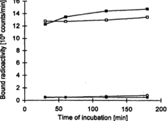
Time of incubation [min] 200 Fig. 1 Variation of incubation time. When the contact time of the zero standard (o) and the 1.53 X 10³ arb.units/1 standard (D) with the solid phase-bound anti-human IgG antibodies was increased from 30 to 180 minutes (first incubation step), this was followed by 60 minutes incubation with the B72.3 detector antibodies. When the contact time with the B72.3 detector antibodies (second incuba- tion step) was increased from 30 to 180 minutes, the zero standard (o) and the 1.53 X JO3 arb.units/1 standard (O) were preincubated with the solid phase bound anti-human IgG antibodies for 60 min- utes. Except for variation of the incubation time, the IRMA was carried out according to the standard protocol.
assays. The detection limit was assessed by calculating the con- centration which corresponds in the standard curve to the mean signal of 10 replicates of the zero standard + 3 standard devia- tions. For recovery studies, samples were spiked with a standard pool and the resulting concentration of anti-idiotypic antibodies was measured. For assessment of the linearity of dilution, the anti-idiotypic antibody concentrations measured in samples seri- ally diluted with zero standard (after 1/200 predilution with assay buffer) were compared with the expected values. To examine the effect of elevated concentrations of serum IgG and TAG-72 on assay results, a standard solution with a known concentration of anti-idiotypic antibodies was measured after addition of increas- ing amounts of human serum IgG and TAG-72, respectively. As a source of TAG-72 we used a mucin prepared from bovine submaxillary glands (Sigma, Deisenhofen, Germany). For com- parison of methods, linear regression analysis was performed by the method of Bablok et al. (22).
Results
Development of an immunoradiometric assay (IRMA) for determination of human anti-idio- typic anti-B72.3 IgG
As shown in figure 1, for the zero standard as well as the 1.53 Χ 103 arb.units/1 standard, only small differences in the resulting test signal were observed when the incuba- tion time of the two incubation steps of the IRMA was increased from 60 to 180 minutes. Thus, for the standard assay protocol we used an incubation time of one hour for each incubation.
The binding capacity for human IgG of two different gel preparations coupled with anti-human IgG antibodies was 37 and 42 μg per 0.04 ml gel. Because of the high IgG concentration in the serum samples (3 to 25 g/1), we diluted the standard material and serum samples 1/200 with assay buffer before applying the standard as- say protocol, to guarantee that serum IgG is completely bound to the solid phase. When the volume of gel and
o.oo 0.05 0.10 0.15 Gel volume [ml] 0.20
Fig. 2 Variation of gel volume. The signal (D; radioactivity bound in the presence of the 0.76 Χ 103 arb.units/1 standard), the noise (o; radioactivity bound in the presence of the zero standard) and the ratio between signal and noise (^) were determined when the test volume in the first incubation step was raised from 0.12 ml (0.1 ml standard + 0.02 ml gel) to 1.2 ml (1.0 ml standard + 0.2 ml gel), followed by a second incubation with 0.5 ml labelled B72.3 detector antibodies. Except for variation of the incubation volumes the IRMA was carried out according to the standard proto- col.
sample in the first incubation step was increased (from 0.02 ml gel and 0.1 ml sample up to 0.2 ml gel and 1.0 ml sample) both the signal (bound radioactivity due to the presence of anti-idiotypic antibodies) of the 0.76 Χ 103 arb.units/1 standard and the noise (bound radioac- tivity in the absence of anti-idiotypic antibodies) of the zero standard continued to increase. However, the sig- nal-to-noise ratio also continuously increased (fig. 2).
Thus, subsequent assays were performed with 0.04 ml gel and 0.2 ml of standards or serum samples prediluted
1/200 with assay buffer.
We used serum from an ovarian cancer patient treated repeatedly with B72.3 antibodies as standard material.
Whereas the heterologous TAG-72 assay detected no real TAG-72 in this standard material, the homologous
"sandwich assay" showed apparent TAG-72 value of 6.1 Χ 106 arb.units/1, obviously due to the activity of human anti-idiotypic anti-B72.3 antibodies present in the se- rum. Thus, an arbitrary anti-idiotypic anti-B72.3 anti-
o.o 1.0 2.0 3.0 4.0 Anti-idiotypic antibodies [10s a rb. u nits/I]
Fig. 3 Dilution curve of standard material. The standard pool was diluted with zero standard after 1/200 predilution with assay buffer.
The IRMA was carried out according to the standard protocol.
body concentration of 6.1 X 10⁶ TAG-72-like units/1 (arb.units/1) was assigned to the standard material. No human anti-mouse antibodies and anti-idiotypic anti- B72.3 antibodies were detected in the serum pool used as zero standard. Standards ranging from 0 to 3.05 X 10³ arb.units/1 were prepared from the standard mate- rial by dilution with zero standard. As shown in figure 3 the standard curve measured with the IRMA was lin- ear up to a concentration of 3.05 Χ 103 arb.units/1.
Analytical performance
The minimal detectable concentration assessed on the basis of 10 replicate measurements of the zero calibrator
to detection limit in the undiluted serum of 3 X 10³
arb.units/1. Within-assay precision, tested with two sam- ples with mean anti-idiotypic antibody concentrations of 80 and 217 Χ 103 arb.units/1, revealed coefficients of variation of 6.4% and 5.8% (n = 20)· The between-as- say precision, calculated from values measured in 20 consecutive runs for two samples with mean anti-idio- typic antibody concentrations of 91 and 212 X 103
arb.units/1, revealed coefficients of variation of 8.7%
and 7.1%. The linearity of dilution was checked for three serum samples with elevated anti-idiotypic anti- body concentrations (153, 277, 290 Χ 103 arb.units/1) Tab. 1 Assay performance of the IRMA standard protocol.
a) Linearity of dilution. Serum samples were serially diluted with zero standard after 1/200 predilution with assay buffer.
Sample identification Dilution Anti-idiotypic anti-B72.3 antibodies
(103 arb.units/1) Recovery (%)a
10117
10618
8107
1/11/2 1/4 1/11/2 1/4 1/11/2 1/4
Measured 15376
39 277144 74 290148 76
Expected 7738
13969
14573
10299
104107
102105
b) Recovery. 0.01 ml of the standard pool diluted 1/200 with assay
buffer was added to 0.29 ml serum prediluted 1/200 with assay buffer and the resulting anti-idiotypic antibody concentrations were measured.
Sample identification
930b
931b
932b 933b 600C
11225d
Anti-idiotypic anti-B72.3 antibodies Endogenous
144
<3
<3
<3
<3
Added 193193 193193 193193
(103 arb.units/1) Expected 207197 193193 193193
Recovered 208192
159187 188183
Recovery (%)a
10198 9782 9795
c) Interferences by non-specific human anti-mouse antibodies (HAMA). Serum samples with elevated human anti-mouse anti- body concentration were measured directly (after 1/200 predilution
with assay buffer) or after addition of 0.01 ml of the standard pool (diluted 1/200 with assay buffer) to 0.29 ml of the prediluted se- rum.
Sample identification
8027e
22788°
7754e
6437e
11055f
HAMA(mg/1)
2.15.1 34.88.3 12.3
Anti-idiotypic anti-B72.3 antibodies (103 arb.units/1) Endogenous Added
<3 86
<3
<3
203203 203—
Expected 211209 203
Recovered _ 219210 201
Recovery (%)a
_ 100104
99
a Recovery = (anti-idiotypic antibody concentration measured/ex- pected) X 100
b Obtained from four in vitro fertilization patients
c TAG-72 concentration = 709 Χ 103 arb.units/1
d TAG-72 concentration = 6940 Χ 103 arb.units/1
e Obtained from four ovarian cancer patients treated with OC125 fragments
f Obtained from an ovarian cancer patient treated with B43.13 an- tibodies · ί
Reinsberg et al.: Determination of anti-idiotypic anti-B72.3 antibodies in the presence of tumour-associated glycoprotein 72 241
1 -- 0.8-- 0.6 -- 0.4 -- 0.2--
0 - - -4-
100 200 300 Human IgG [mg/l]
400
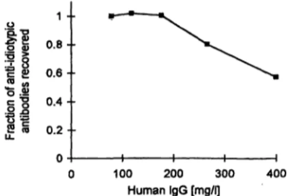
Fig. 4 Recovery of anti-idiotypic anti-B72.3 antibodies in the presence of increasing serum IgG concentration. A 0.76 Χ 103 arb.
units/I standard solution prediluted 1/200 with assay buffer was supplemented with increasing amounts of human IgG and mea- sured with the IRMA according to the standard protocol.
obtained from three patients. The concentrations mea- sured after 2- and 4-fold dilutions of the 1/200 predi- luted serum samples were only slightly different from the expected values (tab. la).
Interferences
As shown in figure 4 the results of the IRMA for a 0.76 X 10³ arb.units/1 standard solution were not affected by addition of increasing amounts of human serum IgG up to an IgG concentration of 175 mg/l (corresponding to a IgG concentration of 35 g/1 in the undiluted serum sample). However, in the presence of higher IgG con- centrations the assay results were considerably reduced.
Furthermore, six serum samples from different patients with serum IgG concentrations from 11 to 18 g/1, showed almost complete recovery of added anti-idio- typic antibodies (tab. Ib), confirming that the different IgG concentrations of the samples have no effect on the assay results.
As shown in table Ib, the two samples with elevated TAG-72 concentrations (sample identification: 600 and 11225) showed almost complete recovery of anti-idio- typic antibodies, indicating that the TAG-72 antigen has no effect upon the results of the IRMA. In the same way, increasing the TAG-72 concentration up to 3.3 Χ ΙΟ**
arb.units/1, by addition of bovine mucin containing TAG-72-like immunoreactive material, has virtually Ho effect on the results measured in a 1.15 Χ 103 arb.units/1 standard solution (apparent concentration of the standard in the presence of 3.3 Χ 107 arb.units/1 TAG-72 = 1.19
Χ 103 arb.units/1).
The apparent anti-idiotypic anti-B72.3 antibody concen- tration measured in five serum samples from patients treated with the murine antibodies OC125 (four patients) and B43.13 (one patient), with non-specific human anti- mouse antibody concentrations ranging from 2.1 to 34.8 mg/l, did not exceed 8 Χ 103 arb.units/1. Also anti-idio- typic antibodies added to three of the samples were reco- vered almost completely (tab. Ic).
Clinical evaluation
As shown in table 2, in the samples obtained from 16 ovarian cancer patients before treatment with B72.3 an- tibodies, the concentration of anti-idiotypic anti-B72.3 antibodies did not exceed 12 Χ 103 arb.units/1. This is in accordance with the results measured in the control samples (tab. Ib). However, after infusion of B72.3 anti- bodies, 9 patients showed grossly elevated anti-idiotypic antibody concentrations ranging from 204 to 2164 Χ 103 arb.units/1; in 4 patients values were slightly ele- vated ranging from 27 to 66 Χ 103 arb.units/1; anti-idio- typic antibodies were undetectable in only 3 patients.
The human anti-mouse antibodies concentration of the samples drawn after antibody treatment ranged from
<0.1 to 4.2 mg/l.
Figure 5 shows the time course of the concentration of the anti-idiotypic anti-B72.3 antibodies measured with the IRMA and with the "sandwich" assay, respectively, in serum samples drawn from an ovarian cancer patient treated three times with B72.3 antibodies. Three weeks after the third B72.3 infusion, the IRMA detected an increase of the anti-idiotypic antibody concentration up to 587 Χ 103 arb.units/1 followed by a decrease to 20xl03 arb.units/1 five months later. Also the values measured with the "sandwich" assay rose three weeks after the third infusion to 443 Χ 103 arb.units/1 followed by an initial decrease to 187 Χ 103 arb.units/1 ten weeks later. However, the values then again rose up to 260 X 103 arb.units/1, obviously due to the continous increase of the TAG-72 concentration measured in the same period.
Tab. 2 Anti-idiotypic anti-B72.3 antibody concentration in pa- tients treated with the B72.3 antibody. Serum samples drawn from 16 patients before and after B72.3 infusion were measured with the IRMA according to the standard protocol.
Patients
EHKE RCWU HIVM MUBB AMSR BIBM FRMH TLSE
Number of infusions
22 63 31 34 31 43 63 31
Anti-idiotypic anti-B72.3 antibodies (103 arb.units/1) before
treatment
<34
<3
<3
<3
<3
<3
<3
<3
<3
<3
<3
<3
<312 6
after last infusion
<3
<3
<3 2734 4366 420204 470526 587793 1572815 2164
50 150 250 350 Time after first B72.3 infusion [d]
Fig. 5 Typical time course of the formation of anti-idiotypic anti-B72.3 antibodies measured with the IRMA (Π) and the
"sandwich" assay (o), respectively, during repeated treatment with Β72.3 antibodies in an ovarian cancer patient with increas- ing TAG-72 serum concentration (+)· Arrows indicate the time of OC125 infusions.
0.1 1 10 100 TAG-72-like activity (IRMA) [105 arb.units/l]
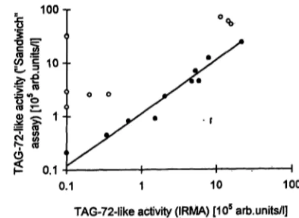
Fig. 6 Comparison of the results measured with the IRMA and the "sandwich" assay, in serum samples obtained from patients after treatment with B72.3 antibodies with low (O) and elevated (o) TAG-72 concentrations. The regression line was calculated for the values measured in the samples with low TAG-72 con- centration.
In figure 6 the values measured with the IRMA are compared with the results of the "sandwich" assay.
For 10 samples with low TAG-72 concentrations there was a good correlation between both rests (r = 0.981;
regression line y = 1.1 χ + 7.6). For 9 samples with TAG-72 concentrations ranging from 189 to 5490 Χ 103 arb.units/l the values measured with the "sand- wich" assay were considerably higher than those from the IRMA.
Discussion
The advantage of the IRMA described here is that the human serum IgG is first selectively extracted by ad- sorption to solid phase coupled anti-human IgG antibod- ies. Thus, serum components which may react with the B72.3 detector antibodies can be removed by a washing step and possible interference can be excluded. The data also confirm that very high TAG-72 concentrations have no effect upon the results of the IRMA, whereas the
"sandwich" assay gave falsely high values, due to cross- reaction with TAG-72.
In most assays for detection of anti-idiotypic antibod- ies described in various papers, the murine antibody bearing the original idiotype is coupled to a solid phase and incubated with the serum samples (4, 6, 9-12, 14). Thus, interfering serum components in- cluding circulating antigen can react with the immobi- lized antibody leading to falsely low or falsely high assay results (7, 10, 13, 14). In patients treated with the anti-TAG-72 antibody B72.3 or the anti-CA-125 antibody OC125, which are directed against antigens which can be present in serum in very high concentra- tions, interference by elevated antigen concentrations in the patient serum can be a problem.
In order to eliminate interference by circulating anti- gen in the determination of anti-idiotypic anti-B72.3
antibodies, Ferroni et al. (14) proposed that the TAG- 72 be removed from patient serum by adsorption to the solid phase coupled anti-TAG-72 antibody CC49 which recognizes a separate epitope on the TAG-72 antigen (23). This is a time-consuming procedure and an additional antibody is required which specifically binds the antigen but does not react with the anti- idiotypic antibodies. A more simple method has been described by Moseley et al. (13). For determination of human antibodies formed against the anti-CA-125 antibody OC125 they used microtitre wells coated with protein A to extract the immunoglobulins from serum. The anti-idiotypic anti-OC125 antibodies bound to protein A are subsequently detected by incubation with radiolabelled F(ab)2 fragments of the OC125. This may be a simple assay, but F(ab)2 fragments of the original antibody must be available to avoid binding of detector antibodies to protein A on the solid phase.
Further problems arise from the high concentration of non-specific IgG present in human serum samples.
Because of the limited binding capacity of the protein A on the solid phase (10 μg per well), serum samples must be prediluted 1/100 for correct determination, leading to a decline of sensitivity (13). In the IRMA described here the sensitivity is also limited by the binding capacity of the solid phase for human serum IgG. However, this problem can be compensated partly by increasing the amount of gel used in the first incubation step. On the other hand, the density of antibodies coupled to the hydrazide gel can be sub- stantially enhanced by increasing the amount of anti- body reacted (21). This may be a further way of increasing the binding capacity of the'solid phase in order to achieve an improved analytical sensitivity.
A farther advantage of the IRMA is that, because of the specificity of the capture antibodies used, only anti- idiotypic antibodies of the IgG class are detected. The
Reinsberg et aL: Determination of anti-idiotypic anti-B72.3 antibodies in the presence of tumour-associated glycoprotein 72 243 assay may be adapted for the determination of anti-idio-
typic antibodies of the IgM class by using a gel coupled with anti-human-IgM capture antibodies.
The validity of the IRMA is confirmed by the data ob- tained for serum samples from patients treated with B72.3 antibodies. In samples drawn before antibody treatment, the IRMA detected no or only marginally ele- vated anti-idiotypic anti-B72.3 IgG, thereby indicating that the assay is highly specific. Obviously, non-specific interference, as observed with the "indirect" assay for- mat (10), does not affect the IRMA. Also, no false posi- tive values were found in samples with high concentra- tions of non-specific human anti-mouse antibodies up to 34.8 mg/1. Thus, the amount of murine IgG added to the detector antibodies of the IRMA is sufficient to block the human anti-mouse antibodies activity of the samples drawn after B72.3 treatment which did not exceed 4.2 mg/1.
In 81% of the posttreatment samples we found ele- vated anti-idiotypic anti-B72.3 IgG concentrations, in- dicating that the assay is highly sensitive. The time course of the appearance of anti-idiotypic antibodies during antibody treatment shows a good correlation between the time of antibody infusion and the appear-
ance of anti-idiotypic antibodies. The validity of the results is further confirmed by the coincidence with the values measured with the "sandwich" assay for samples with low TAG-72 concentration. However, because the "sandwich" assay detects all classes of anti-idiotypic antibodies, the good agreement between both assays suggests that in the samples compared, the concentration of the anti-idiotypic IgG antibodies is considerably higher than that of the anti-idiotypic antibodies of the other immunoglobulin classes. This may be due to the fact that only one of these samples was drawn within the first weeks following to the first antibody infusion. As reported by Courtenay-Luck et al. (4), the immune response to repeated antibody infusions is mainly of the IgG class.
Because the only specific reagent required for the IRMA is the labelled original antibody, the test may be modi- fied to detect anti-idiotypic antibodies, which were formed after treatment with other monoclonal antibod- ies. At present we have performed preliminary experi- ments which suggest that this assay design also can be used for determination of human anti-idiotypic antibod- ies directed against the anti-CA-125 antibodies B43.13 and OC125.
References
1. Koprowsky H, Herlyn D, Lübeck M, DeFreitas E, Sears HF.
Human anti-idiotypic antibodies in cancer patients: is the mod- ulation of the immune response beneficial for the patients?
Proc Natl Acad Sei USA 1984; 81:216-9.
2. Jerne NK. Towards a network theory of the immune system.
Ann Immunol 1974; 125:373-89.
3. Fagerberg J, Frödin JE, Ragnhammar P, Steinitz M, Wigzell H, Mellstedt H. Induction of an immune network cascade in cancer patients treated with monoclonal antibodies (abi). II. Is induction of anti-idiotype reactive T cells (T3) of importance for tumor response to mAB therapy? Cancer Immunol Immu- nother 1994; 38:149-59.
4. Coutenay-Luck NS, Epenetos AA, Sivolapenko BG, Lärche M, Barkans JR, Ritter MA. Development of anti-idiotypic anti- bodies against tumor antigens and autoantigens in ovarian can- cer patients treated intraperitoneally with mouse monoclonal antibodies. Lancet 1988; 15:894-7.
5. Wettendorf M, Iliopoulos D, Tempero M, Kay D, DeFreitas E, Kpprowsky H, Herlyn D. Idiotypic cascades in cancer patients treated with monoclonal antibody CO 17-1 A. Proc Natl Acad Sei USA 1989; 86:3787-91.
6. Frödin JE, Faxas ME, Hagström B, Lefvert AK, Masucci G, Nilsson B, et al. Induction of anti-idiotypic (ab2) and anti-anti- idiotypic (ab3) antibodies in patients treated with the mouse monoclonal antibody 17-1A (ab^. Relation to the clinical out- come - an important antitumoral effector function? Hybri- doma 1991; 10:421-6.
7. Reinsberg J. Idiotypic cascades after MAb OC125 application.
Hybridoma 1993; 12:577-82.
8. Wagner U, Oehr P, Reinsberg J, Schmidt S, Schlebusch H, Werner A, Krebs D. Immunotherapy of advanced ovarian car- cinomas by the activation of the idiotypic network. Biotech Therapeutics 1992; 3:81^9.
9. Schmolling J, Reinsberg J; Wagner Ü, Krebs D. Antiidiotypic antibodies in ovarian cancer patients treated with the monoclo- nal antibody B72.3. Hybridoma 1995; 14:183-6.
10. Herlyn DM, Lübeck M, Sears H, Koprowski H. Specific detec- tion of anti-idiotypic immune responses in cancer patients
treated with murine monoclonal antibody. J Immunol Methods 1985; 85:27-38.
11. Wagner U, Reinsberg J, Oehr P, Briele B, Schmidt S, Werner A, et al. Clinical courses of patients with ovarian carcinomas after induction of anti-idiotypic antibodies against a tumor-as- sociated antigen. Tumordiagn Ther 1990; 11:1—4.
12. Mäher VE, Drukman SJ, Kinders RJ, Hunter RE, Jennings J, Brigham C, et al. Human antibody response to the intravenous and intraperitoneal administration of the F(ab')2 fragment of the OC125 murine monoclonal antibody. J Immunotherapy
1992; 11:56-66.
13. Moseley KR, Knapp RC, Haisma HJ. An assay for detection of human anti-murine immunoglobulins in the presence of CA 125 antigen. J Immunol Methods 1988; 106:1-6.
14. Ferroni P, Milenic DE, Schlom J, Colcher D. Assay for detec- tion of anti-idiotypic antibodies to monoclonal antibody B72.3.
J Clin Lab Anal 1990; 4:465-73.
15. Thor A, Ohuchi N, Szpak CA, Johnston WW, Schlom J. Distri- bution of oncofetal antigen tumor-associated glycoprotein-72 defined by monoclonal antibody B72.3. Cancer Res 1986;
46:3118-24.
16. Thor A, Gorstein F, Ohuchi N, Szpak CA, Johnston WW, Schlom J. Tumor associated glycoprotein (TAG-72) in ovarian carcinomas defined by monoclonal antibody B72.3. J Natl Cancer Inst 1986; 76:995-1006.
17. Guadagni F, Roselli M, Amato T, Cosimelli M, Pern P, Casale V, et al. CA-72-4 measurement of tumor-associated glycopro- tein-72 (TAG-72) as a serum marker in the management of gastric carcinoma. Cancer Res 1992; 52:1222-7.
18. Esposito G, Panza N, Mansi L, De Matteis A, D'Aiuto G, La- bonia V, et al. Evaluation of TAG 72 as a serum marker in ovarian and breast carcinoma. J Nucl Med Allied Sei 1990;
34:88-93.
19. Reinsberg J, Gast B, Schmolling J, Wagner U, Krebs D. Spe- cific interferences with the determination of the Tumor Associ- ated Glycoprotein 72 (TAG-72) by human antiidiorypic anti- bodies formed after treatment with the anti-TAG-72 antibody B72.3. Eur J Clin Chem Clin Biochem 1994; 32:691-6.
doma 1993; 12:539-41. - sociated glycoprotein 72 antigen. Cancer Res 1988;
21. Radparvar S, Fung G, Ngo TT. Simple, quick and efficient site- 48:4588-96.
directed antibody immobilization in a cartrige. Biotechniques
^ I9??;i9u/32n""5'· n D ,ι ο ο ι Μ η Δ ι * . Received October 4/December 4, 1995 22. Bublok W, Passing H, Bender R, Schneider B. A general re-
gression procedure for method transformation. J Clin Chem Corresponding author: Dr. J. Reinsberg, Zentrum f r
Clin Biochcm 1988; 26:783-90. Frauenheilkunde und Geburtshilfe, Universit t Bonn, Sigmund- 23. Muraro R, Kuroki M, Wunderlich D, Poole DJ, Colcher D, Freud-Stra e 25, D-53127 Bonn, Germany
Thor A, et al. Generation and characterization of B72.3 second