van Straalen et al.: Prostate-specific antigen after prostatectomy 53
Eur. J. Clin. Chem. Clin. Biochem.
Vol. 32, 1994, pp. 53-55
© 1994 Walter de Gruyter & Co.
Berlin · New York
Biological Half-Life of Prostate-Specific Antigen after Radical Prostatectomy
By J. P. van Straalen !, M M F. Bossens 2, T. M de Reijke 2 and G. T. B. Sanders l
1 Department ofdinical Chemistry, Academical Medical Centre, Amsterdam, The Netherlands
2 Department ofUrology, Academical Medical Centre, Amsterdam, The Netherlands
(Received July 12/October 15, 1993)
Summary: The disappearance pattern of prostate-specific antigen frorn serum after a Standard radical prostatectomy was studied in eight patients with cancer confined to the prostate. The results were used to plot an elimination curve and calculate the best fit. A biphasic pattern was found with an average biological half-life of l .63 hours in the -phase, and 4.63 days in the ß-phase. Based on these results it is concluded that determination of prostate- specific antigen concentrations less than one month after a Standard radical prostato-vesiculectomy has no value for the detection or exclusion of residual malignant processes.
Introduction
In 1971, a gamma seminoprotein was detected (1) in seminal plasma, and subsequently isolated and charac- terized by Li (2) and Sensabaugh (3). In 1979 Wang (4) purified this protein from natural prostate tissue and named it prostate-specific antigen. After the introduction of commercial kits for the determination of prostate- specific antigen, prostatic acid phosphatase was replaced by prostate-specific antigen äs a tumour marker for pros- tate cancer (5, 6). After radical prostatectomy äs a treat- ment for localized cancer, the absence of residual tu- mour (pT2-3N0M0) is indicated by a decrease of serum prostate-specific antigen until it is undetectable by the current generation of immunoassays for prostate-specific antigen (6). In view of differences in the öbserved bio- logical washout patterns and disappearance time of pros- tate-specific antigen after a Standard radical prostatec- tomy, we initiated the present study to determine the biological half-life of prostate-specific antigen.
Methods and Patients
Prostate-specific antigen was measured by a solid-phase two-site immunoenzymatic assay employing two monoclonal antibodies.
We used a commercial kit, Tandem R-E, from Hybritech (Hybri-
tech Ine, San Diego CA, USA), according to the instructions of the manufacturer. The colorimetric measurements were performed on a Photon II spectrophotometer.
Prostate-specific antigen excretion pattems were followed in eight patients ranging from 59 to 75 years, suffering from prostatic can- cer. The prostates were removed surgically, together with the semi- nal vesicles. In all eight patients the cancer was confined to the prostate; the bone scan was negative, and there was no lymph node involvement at lymphadenectomy (pT2-3NoM0). The final patho- logical stage and grade was pT2G2 (n = 2), pT3G2 (n = 4) and pT3G3 (n = 2), respectively. During follow-up the prostate-specific antigen concentration remained for at least one year after surgical Intervention within the female ränge; during this time there was no evidence ofdinical recurrence, based on digital rectal examination, trans rectal ultra sonography of the prostate, bone-scan and ehest X-ray. Venous blood was collected according to the following scheine: before the Intervention two samples were taken at 9 a. m.
with a time interval of one day. The actual moment of removal of the prostate was ealled T0. During and after the Intervention, blood was collected every hour until two hours after T0, followed by collection at 3, 6, 9 and 12 hours after T0. During the following ten days one blood sample was taken each day, always at 9 a. m.
Until analysis all the serum samples were stored at —20 °C. Pros- tate-specific antigen was determined when all the samples from one patient were available.
For the statistical evaluation of the data we used a Computer pro- gram ealled PCNONLIN NONUNEAR ESTIMATION from Stat- istical Consulting Ine (Lexington, Kentucky, USA), which is able to choose the right form of elimination through best fit of the elim- ination curves of the individual patients (one, two or three compart- ment model).
Eur. J. Clin. Chem. Clin. Biochem. / Vol. 32,1994 / No. 2
54 van Straaien et al.: Prostate-specific antigen afler prostatectomy
Results and Discussion
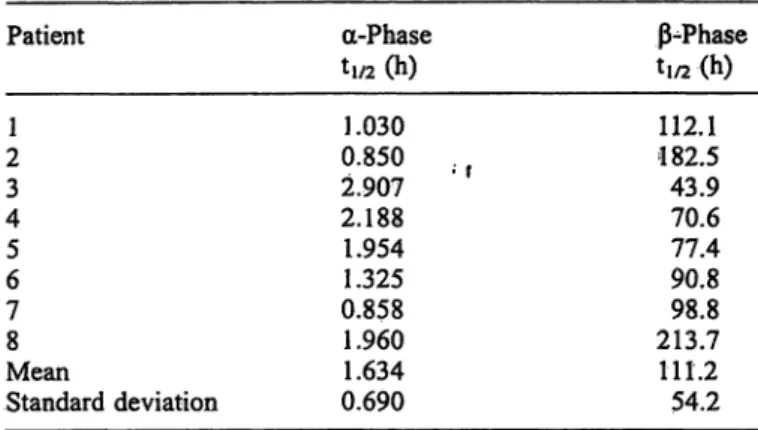
During the surgical extirpation of the prostate, prostate- specific antigen concentrations were increased to be- tween three and ten times their starting levels. It was noted that a biphasic disappearance curve resulted after total removal of the prostate. The mean biological half- life in the first phase ( -phase) was 1.63 hours (SD 0.69). After the first twenty-four hours the prostate- specific antigen disappearance was more gradual. In this phase (ß-phase) the biological half-life was 4.63 days (SD 2.26). The graphic fit of the disappearance is shown in figure l and the separate data of the individüal patients are shown in table l.
The correctness of fit is very high (0.997). Data are ex- pressed äs percentage of T0 values. In the literature sev- eral other studies have been reported but they show no general agreement äs to the disappearance rate. Our ob- servations contradict those of Oesterling et al. (7), who found a single phase elimination with a half-life of 3.15 days (SD 0.09.) determined with a log-linear regression model. They stated that prostate-specific antigen con- centrations were undetectable after 5 to 6 times the bio- logical half-life, meaning that 16 days after the radical prostatectomy the prostate-specific antigen concen- trations were in the female ränge. Stamey et al. (6) found a two-phase elimination: an -phase of 12.6 hours (SD 19.7) and a ß-phase of 2.2 days (SD 0.8). Their data were calculated from the natural log of the prostate- specific antigen concentration plotted against time.
These data fitted a two compartment model of first order elimination kinetics. They also claimed that prostate- specific antigen concentrations were no longer detect- able after fourteen days. The fast decline in the first phase, äs seen in our results, is probably based on com-
Tab. l Disappearance rates, expressed äs tj/2 of prostate-specific antigen in the 8 individüal patients.
Patient
21 43 56 78
MeanStandard deviation
-Phase
tl/2 (h)
1.030 0.850 2.907 2.188 1.954 1.325 0.858 1.960 1.634 0.690
ß-Phase ti/2(h) 112.1 182.5 43.970.6 90.877.4 213.798.8 111.2 54.2
plex formation between liberated prostate-specific anti- gen and mainly arantichymotrypsiii (10), and possibly other acute phase proteins such äs a2-macroglobulin.
Examples of these complexes are deseribed by Chris-?
tensson (11).
There is some doubt äs to whether the Tandem- pros- tate-specific antigen test method determines these com- plexes. a2-Macroglobulin is known to encapsulate the target protein at complex formation (11). This häppens in such a way that no antiserum will detect prostate- specific antigen bound to this acute phase protein. But since the concentration of these coinplexes is low (12) it is not to be expected that their presence will inflüence our results to a great extent. The performance character- istics of the Tandem- prostate-specific antigen ässay are similar to those of the Tandem^R assay (13), and it has been reported (10) that the latter kit also detects prostate-specific antigen complexed to a^antichymo- trypsin. Therefore, it may be concluded that the Hybri-
§ 1.00
ce
0.80
0.60
g 0.40 0.20
Fig. l Typical disappearance curve of prostate-specific antigen tion of initial concentration versus tirne. Values given are means concentrations frorn serum after prostatectomy, expressed äs frac- ± l SD (n = 8).
Eur. J. Clin. Chem. Clin. Biochem. / Vol. 32, 1994 / No. 2
van Straalen et al.: Prostate-specific antigen after prostatectomy 55
tech Tandem- , äs used by us, measures free prostate- specific antigen, äs well äs prostate-specific antigen bound to -antichymotrypsin. In the second phase the slow decrease of prostate-specific antigen might be due to a slow release of prostate-specific antigen from the complexes and/or their gradual disappearance from plasma. This issue will require further study.
Conclusion
In conclusion it can be stated that prostate-specific anti- gen disappears after radical prostatectomy in accordance with a biphasic excretion model, consisting of an a-
phase with a half-life of l .63 hours and a ß-phase with a half-life of 4.63 days.
Based on our results and the assumption that 6 times the half-life should elapse before a steady level has been reached, we advise that determination of prostate- specific antigen levels less than one month after a Stan- dard radical prostato-vesiculectomy has no value for the detection or exclusion of residual malignant processes.
Acknowledgement
We wish to express our thanks to the department of Clinical Phar- macology (head: Prof. dr. C. J. van Boxtet) for the statistical pro- cessing of the data.
References
1. Hara, M., Koyanagi, Y., Inoue, T. & Fukuyama, T. (1971) Some physicochemical characteristics of gamma-semenpro- tein, an antigenic component specific for human seminal plasma. Jpn. J. Legal Med. 25 322-324.
2. Li, T. S. & Beling, C. G. (1973) Isolation and characterization of two specific antigens of human seminal plasma. Fertil.
Steril. 24, 134-144.
3. Sensabaugh, G. F. (1978) Isolation and characterization of a semen-specific protein from seminal plasma: A potential new marker for semen identification. J. Forensic Sei. 23, 106—115.
4. Wang, M. C., Valenzuela, L. A., Murphy, G. P. & Chu, T. M.
(1979) Purification of a human prostate specific antigen. In- vest. Urol. 77, 159-163.
5. Killian, C. S., Emrich, L. J. & Vargas, F. P. (1986) Relative reliability of five serially measured markers for prognosis of progression in prostate cancer. J. Natl. Cancer Inst 76, 176—
185.
6. Stamey, T. A., Yang, N., Hay, A. R., McNeal, J. E., Freiha, F.
S. & Redwine, E. (1987) Prostate-speeific antigen äs a serum marker for adenocarcinoma of the prostate. N. Eng. J. Med.
377, 909-916.
7. Oesterling, J. E., Chan, D. W, Epstein, J. L, Kimball, A. W., Bruzek, D. J., Rock, R. C., Brendler, C. B. & Walsh, P. C.
(1988) Prostate specific antigen in the preoperative and pqstop- erative evaluation of localized prostatic cancer treated with radical prostatectomy. J. Urology 139, 766-772.
8. Mannini, D., Maver, R, Aiello, E., Corrado, G., Vecchi, F., Bellänova, B. & Marengo, M. (1988) Spontaneous circadian
fluctuations of prostate specific antigen and prostatic acid phosphatase serum activities in patients with prostatic cancer.
Urol. Res. 76,9-12.
9. Chan, D. W. 81988) PS A äs a marker for prostatic cancer. Lab.
Management 26, 135-139.
10. Lilja, H., Christensson, A., Dahlen, U., Matikainen, M. T., Nilsspn, O., Pettersson, K. & Lövgren, T. (1991) Prostate- specific antigen in serum occurs predominantly in complex with -antichymotrypsin. Clin. Chem. 37, 1618-1625.
11. Christensson, A., Laurell, C. B. & Lilja, H. (1990) Enzymatic activity of prostate-specific antigen and its reaction with extra- cellular serine proteinase inhibitors. Eur. J. Biochem. 194, 755-763.
12. Stenman, U-H., Leinonen, J., Alfthan, H., Rannikko, S., Tuhkanen, K. & Alfthan, O. (1991) A complex between pros- tate-specific antigen and -antichymotrypsin is the major form of prostate-specific antigen in serum of patients with prostatic cancer: Assay of the complex improves clinical sensi- tivity for cancer. Cancer Res. 57, 222-226.
13. Oesterling, J. E. (1991) Prostate specific antigen: A critical assessment of the most useful tumor marker for adenocarcin- oma of the prostate. J. Urology 145, 907-923.
Jan P. van Straalen Academic Medical Centre Meibergdreef 9
NL-1105 AZ Amsterdam The Netherlands
Eur. J. Clin. Chem. Clin, Biochem. / Vol. 32,1994 /No. 2