138
Analysis of Placental Structures by Ultrasound in Normal andf
Complicated Pregnancies
P. KOZLOWSKI, R. TERINDE, H. SCHMIDT, J. HERBERGER, H.6. BENDER
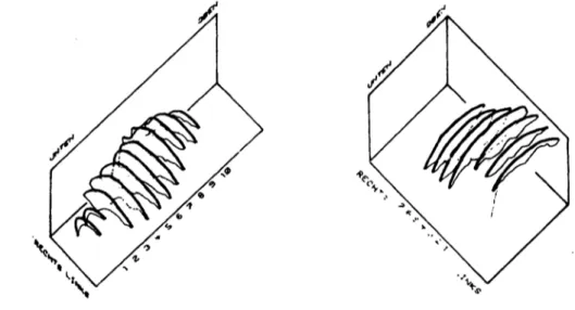
Grey scale echography allows to study the intrauterine dimensions of the placenta äs it is possible to separate the chorionic plate, marginal areas and the basal plate from the surrounding structures. Subject of the present study were the progress of placental -growth in the last third of pregnancy and the changes in placental tissue structure. 267 placentas of 157 gra- vidas were examined at various stages of gestation. 4372 longitudinal and transversal section scans were done - on an average 15 scans of one pla- centa. The anterior wall placenta of a patient was scanned in steps of 2 cms in longitudinal just äs in transverse direction ( Fig. l ).
Abb. 1: Model of transverse (left) and longitudinal (right) section scans of a placenta
The scans were stored on a video tape and measured by means of a X-Y-di- gitizer, outlining the placenta on each scan. The Implantation area, total surface and the placental volume were calculated.
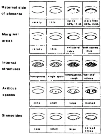
The sonographical tissue structure of each single section scan was dif- ferentiated by a scored scheme of five different signs in a four-step graduation ( Fig. 2 ). The degree of appeärance of each criterion in a section scan was brought in relation to its part of the whole placental volume.
Results
Fig. 3 demonstrates the increase of placental volume during pregnancy by means of a mathematically fitted growth curve including the Standard de- viation. The gestational age was calculated not only by the term of the last menstruation, but also by the progress of the fetal biparietal dia- meter. There is no growth of placental volume beyond the 34th w.o.g.
The placental volumes of pregnancies complicated by diabetes B, C or .D according to the WHITE scheme are represented by the "D" letters, the
"S" letters show the volumes of the placentae of small for gestational age infants who finally had a birthwei'ght below 2700 g.
0300-5577/81/0091-0040 $ 2.00 Copyright by Walter de Gruyter & Co.
139
Maternal side of placenta
Marginal areas
Internat structures
Avillous spaces
Sinusoides
Homoganous
'
• ingle apota up te 50% t h ick
inhomoganoua rough
ö -
mora than 50% t hick
"gar l and"
•choaa
a p r « a < l
Abb. 2: Scheme of the five criteria of placental tissue structure
In 152 patients the last sonogra- phic examination was done two weeks or less before delivery. Analyzing the five criteria .of placental structure we established a group of inconspicious pregnancies with non-pathological fetal outcome and a pathological one including fetal distress, operative delivery, small-for-date and stillbirth.
Three of the five criteria proved to be of small value under clini- cal aspects: The existence of mar- ked marginal areas, avillous spa- ces and sinusoids was not connec- ted with remarkable differences concerning the fetal outcome. Sig- nificant differences between both groups were found, however, äs to
"maternal side of placenta11 and
"internal structures11. If 75 per cent or more of the placental tis- sue are sonographically described äs grossly inhomogenous or even interspersed with garland echoes a significant di'fference in fetal outcome between the normal and the pathological groups was found. Si- milar differences between those groups could be demonstrated in those cases in which besides the utero-placental borderline was ap- parently thicker.
Ccm33
1200-
830
400
SSW
Abb. 3: Growth of placental volume with s ingle s. d.
Dr.P.Kozlowski Univ.-Frauenklinik Moorenstr.5
D-40OO Düsseldorf l /Germany