Retrolental fibroplasias: What lies beneath?
Abstract
A nine-month-old female baby with normal birth history presented with her mother complaining of a white spot in the baby’s right eye, which
Parveen Sen
1Dhaivat Shah
1the mother had noticed at five months of age. External photograph showed a retrolental fibroplastic membrane visible in the superior half
of the dilated pupil. Retcam fundus photo revealed myelinated nerve 1 Shri Bhagwan Mahavir Vitreoretinal Services, fibers extending from the disc till the ora superiorly and forming a
Medical Research membranous fold. Intraoperative OCT confirmed thickened RNFL with
Foundation, Chennai, Tamil Nadu, India
compact retina. Thus, the retrolental fibroplasia turned out to be a masquerade for myelinated nerve fibers. Since it was not involving the visual axis with no coexisting traction, the mother was reassured regard- ing the benign nature of the condition.
Keywords:retrolental fibroplasias, myelinated nerve fibers, masquerades
Case description
A nine-month-old female baby from rural North India born at 37 weeks with a birth weight of 2.1 kg presented with her mother complaining of a white spot in the baby’s right eye. The mother had accidentally noticed the spot when the child was around five months of age and she was re- ferred to us by her local ophthalmologist with the query to rule out retinoblastoma. The child had central, steady, and maintained fixation in both eyes. Retinoscopy re- vealed a dull glow in the right eye with a cycloplegic re- fraction of –18.00 D and a clear red glow in the left eye with cycloplegic refraction of +1.00 D in undilated state.
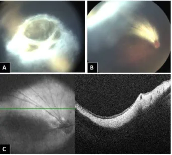
Figure 1: A) & B) Fundus photos showing a yellowish white shiny area superior to the disc extending till the superior ora and forming a membranous fold behind the lens in this area,
giving an appearance of retrolental fibroplasias. C) Intraoperative OCT showing thickened RNFL with compact retina, which confirms the diagnosis of myelinated nerve fibers.
On torch light, leukocoria with normal anterior segment and clear lens was noted. The white reflex was more prominent from the superior one third of the pupil, more evident in the dilated state. Keeping in mind the differen- tial diagnosis of retinoblastoma, retinopathy of prematur- ity and exudative retinopathy, an examination under an- esthesia was advised.
On examination under anesthesia, the fundus showed a yellowish white shiny area (Figure 1A) superior to the disc extending till the superior ora and forming a membranous fold behind the lens in this area, giving an appearance of retrolental fibroplasia (Figure 1B). Intraoperative OCT (Figure 1C) confirmed a thickened retinal nerve fiber layer (RNFL) with compact retina, which confirmed our diagno- sis of myelinated nerve fibers (MNF).
Discussion
Myelinated retinal nerve fiber is a congenital benign condition which is commonly found in conjunction with the optic nerve head and uncommonly noted in isolation over the fundus. It appears as a whitish or yellowish membrane over the retinal surface following the distribu- tion of ganglion cell axons, giving it a frayed appearance [1].
The normal myelination process begins at around eight months in embryonic period and ends before birth [1].
Various hypotheses have been postulated, out of which the most commonly accepted one is the inhibition of oli- godendrocyte migration due to cellular factors released from astrocytes just before birth [2], [3]. If this does not occur, the myelination continues beyond the lamina cribrosa around the optic nerve hypoplasia (ONH). In most cases, MNF is localized, but if the process continues, it can be as extensive as till the equator or beyond [1].
In our case, the MNF was extending from the ONH till the superior ora serrata and presumably might have formed a membranous fold at that site. This subsequently gave
1/2 GMS Ophthalmology Cases 2020, Vol. 10, ISSN 2193-1496
Case Report
OPEN ACCESS
an appearance of retrolental fibroplastic membrane and showed a very typical white reflex on torch light examina- tion. Taking the age and history into consideration, con- genital cataract, retinoblastoma, retinopathy of prematur- ity or exudative retinopathy would be the primary sus- pects. A dilated fundus examination with thorough peri- pheral fundus evaluation and photo documentation is imperative in such cases, preferably under anesthesia.
To add to our armamentarium of investigations, an intra- operative OCT was done and the thickened RNFL seen in the area of MNF was captured, which ascertained our diagnosis of MNF [4].
Since the retrolental membranous fold was fortunately neither involving the visual axis nor causing any concur- rent traction, the observation of the same was advised and the mother was henceforth reassured regarding the benign nature of the condition. Regular use of glasses with amblyopia therapy was advised. A six-monthly follow- up was recommended.
Conclusion
MNF usually is a benign localized condition, but can be as extensive as till the ora, giving a pseudoappearance of leukocoria and masquerade conditions like retino- blastoma or retinopathy of prematurity. A thorough dilated fundus examination is a must in all such cases, preferably under anesthesia, to prevent misdiagnosis and mistreat- ment. As long as the retrolental membrane is neither in the visual axis nor causing any coexisting traction, obser- vation is warranted in such cases.
Notes
Competing interests
The authors declare that they have no competing in- terests.
Informed consent
The patient has viewed the content and images of this case report and has consented to the submission of the case report for publication.
References
1. Shelton JB, Digre KB, Gilman J, Warner JE, Katz BJ.
Characteristics of myelinated retinal nerve fiber layer in ophthalmic imaging: findings on autofluorescence, fluorescein angiographic, infrared, optical coherence tomographic, and red- free images. JAMA Ophthalmol. 2013 Jan;131(1):107-9. DOI:
10.1001/jamaophthalmol.2013.560
2. FitzGibbon T, Nestorovski Z. Morphological consequences of myelination in the human retina. Exp Eye Res. 1997 Dec;65(6):809-19. DOI: 10.1006/exer.1997.0388 3. Hunter SF, Leavitt JA, Rodriguez M. Direct observation of
myelination in vivo in the mature human central nervous system.
A model for the behaviour of oligodendrocyte progenitors and their progeny. Brain. 1997 Nov;120(Pt 11):2071-82. DOI:
10.1093/brain/120.11.2071
4. Gharai S, Prakash G, Ashok Kumar D, Jacob S, Agarwal A, Arora V. Spectral domain optical coherence tomographic characteristics of unilateral peripapillary myelinated retinal nerve fibers involving the macula. J AAPOS. 2010 Oct;14(5):432-4. DOI:
10.1016/j.jaapos.2010.05.010
Corresponding author:
Dr. Parveen Sen
No. 41, Old 18, College Road, Nungambakkam, Chennai 600034, India, Phone: +91 044 42271500
drpka@snmail.org
Please cite as
Sen P, Shah D. Retrolental fibroplasias: What lies beneath? GMS Ophthalmol Cases. 2020;10:Doc14.
DOI: 10.3205/oc000141, URN: urn:nbn:de:0183-oc0001413
This article is freely available from
https://www.egms.de/en/journals/oc/2020-10/oc000141.shtml Published:2020-03-18
Copyright
©2020 Sen et al. This is an Open Access article distributed under the terms of the Creative Commons Attribution 4.0 License. See license information at http://creativecommons.org/licenses/by/4.0/.
2/2 GMS Ophthalmology Cases 2020, Vol. 10, ISSN 2193-1496
Sen et al.: Retrolental fibroplasias: What lies beneath?