U NTERSUCHUNGEN ZUR B IODIVERSITÄT ,
BIOGEOGRAPHISCHEN V ERBREITUNG UND
P HYLOGENIE VON C HOANOFLAGELLATEN - UNTER BESONDERER B ERÜCKSICHTIGUNG DER POLAREN
R EGIONEN .
Inaugural-Dissertation zur
Erlangung des Doktorgrades
der Mathematisch-Naturwisenschaftlichen Fakultät der Universität zu Köln
vorgelegt von Frank Nitsche
aus Salzburg
2007
Berichterstatter:
Prof. Dr. H. Arndt Prof. Dr. E. von Elert
Tag der letzten mündlichen Prüfung:22.06.2007
3
D ANKSAGUNG
• Prof. H. Arndt gilt mein besonderer Dank für die Möglichkeit zur Dissertation, die Betreuung der Arbeit, die vielen hilfreichen Diskussionen und Anregungen und die angenehme Zeit mit ihm als Mensch und Wissenschaftler.
• Meiner Frau möchte ich für die Unterstützung danken, die es mir erst ermöglicht hat, diese Arbeit zu erstellen.
• Claudia Wylezich und Frank Scheckenbach danke ich für Ihre Hilfestellung in molekular-biologischen Fragen.
• Der “Deutschen Forschungsgemeinschaft” (DFG) möchte ich für die im Rahmen dieser Arbeit benötigten Sachmittel und insbesondere für die Finanzierung meiner Stelle durch das geförderte Projekt (AR 288/8) danken.
• Herzlicher Dank geht an alle Verwandten, Freunde und Bekannten, die sich bereit erklärt haben, Wasserproben während Ihrer Urlaubs- und Geschäftsreisen zu nehmen und nach Köln zu transportieren. Besonders möchte ich meiner Schwester Sabine Nitsche und Michael Kern für Ihren unentwegten Einsatz in Sachen Probennahme danken.
• Barry Leadbeater danke ich für seine hilfreichen und motivierenden Kommentare.
• Mein ganz besondere Dank gilt meiner Mutter, ohne die diese Arbeit nicht zustande gekommen wäre
5
I NHALTSVERZEICHNIS
Einleitung 7
I. Global biogeography of acanthoecid choanoflagellates
(Eukaryota, Opisthokonta) 15
II. A new species of heterotrophic flagellates from Taiwanese brackish waters: Morphological and molecular biological studies of Diplotheca elongata nov. spec. and D. costata
(Choanoflagellida, Acanthoecidae) 27
III. Deep sea records of choanoflagellates with a description
of two new Species 41
IV. Bipolar comparisons of acanthoecid choanoflagellates 55 V. Studies on the phylogenetic relationships among
choanoflagellates 69
Abstract 83
Zusammenfassung 85
Anhang 89
7
E INLEITUNG
In der Protozoologie werden immer wieder drei grundlegende Fragen gestellt, die eng miteinander verbunden sind: Was ist eine Art, wie hoch ist die Artenzahl und wie verbreitet sind Protisten. Und schließlich stellt sich in Verbindung mit Choanoflagellaten noch die Frage nach dem Ursprung der mehrzelligen Lebensformen.
Das biologische Artkonzept nach Mayr (1942) ist bei Protisten, die sich asexuell oder klonal fortpflanzen nicht anwendbar. Mit der Einbindung der Molekularbiologie in die Protozoologie und unter Verwendung der entsprechenden Methoden zeigte sich, dass das Konzept der Morphoart alleine nicht ausreichend ist um die Mannigfaltigkeit der genetischen und ökologischen Variationen innerhalb einer Art wiederzugeben.
Dennoch bildet die Morphoart nach wie vor die Grundlage der Taxonomie (Finlay 2004). Vor 50 Jahren wurde der Vorläufer des modernen Artkonzepts durch die Einbezugnahme der asexuellen Fortpflanzung durch Sonneborn (1957) begründet.
Danach spricht man beim Vorhandensein einer auch noch so minimalen genetischen irreversiblen Distanz einer neuen Art. Molekularbiologische Untersuchungen der kleinen Untereinheit der ribosomalen DNA innerhalb von einer Morphoart haben eine sehr hohe Variabilität (über 10% p-Distanz) aufgezeigt (Scheckenbach et al. 2006, Massana et al. 2006). Taxonomie sollte daher aus der Bestimmung des Phänotyps, Genotyps und Ökotyps bestehen. Die Methode des genetischen Barcodings (z.B. Tautz et al. 2003) ist hierbei ein hilfreiches Instrument, ebenso wie das Elektronenmikroskop und ökologische Untersuchungen. Doch nur durch Kombination aller Methoden kann in Zukunft eine verlässliche Taxonomie erstellt werden (Will und Rubinoff 2004).
Untrennbar mit dem Artkonzept verbunden ist die Frage nach der Artenzahl.
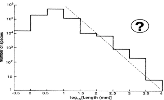
May (1988) hat eine Zusammenstellung der Anzahl beschriebener Arten unter Bezugnahme auf ihre Größe erstellt. Mit abnehmender Größe nimmt die Artenzahl in den logarithmischen Größenklassen wie erwartet zu. Allerdings nimmt die Artenzahl bei einem Schwellenwert von etwa 1mm, unter den die meisten Arten innerhalb der Protisten fallen, wieder ab. Würde man jedoch diese Berechnung extrapolieren, so würde sich bei einer Größe von 1mm eine Artenzahl von 106 ergeben und innerhalb der Größenklasse der Protisten eine Artenzahl von 108 (Abbildung I).
Derzeit sind 150.000 Protistenarten beschrieben, Schätzungen der Gesamtzahl liegen bei 5·106 Arten (Report on the Alfred P. Sloan Foundation Workshop on Protistan Barcoding, 2006). Ein Grund für die möglicherweise zu niedrige Artenzahl von Protisten könnte in dem Mangel an taxonomischen Arbeiten zu finden sein (May
Abbildung I: Schätzung der Artenzahl je Größenklasse (durchgezogene Linie) und des Verhältnisses S=L-2 (S=Artenzahl, L=Länge; gestrichelte Linie). Aus: Robert M. May (1988) „How many species are there on earth“ Science 241: 1441-1449.
1988) sowie in marinen Picoplankton ist eine morphologische Klassifizierung oberhalb des Klassenniveaus nicht möglich (Potter et al. 1997). Innerhalb der Eukaryoten stellen Protisten die individuenreichste Gruppe dar. Heterotrophe Flagellaten treten in Konzentrationen von etwa 102 - 105 Individuen • ml-1 auf (Berninger et al. 1991), Choanoflagellaten zeigen eine Abundanz von bis zu 104 Individuen • ml-1 auf (Marchant 2005).
Diese hohe Abundanz, wie sie bei Protisten sehr häufig auftritt zusammen mit der geringen Größe steigert die Wahrscheinlichkeit, dass diese ubiquitär auftritt. Nach Finlay und Fenchels Postulat „alles ist überall“ sollte Endemismus bei Protisten nur sehr vereinzelt vorkommen (Abbildung II). Bei entsprechendem Vorhandensein der ökologischen Bedingungen sollte eine Art in der Lage sein, diese Nische weltweit besiedeln zu können. Dies schließt marine wie limnische Gewässer mit ein.
Existierender Endemismus wurde bisweilen damit begründet, dass zu wenig Probenstellen untersucht wurden. Dieser Theorie stehen einige Forscher wie der α- Taxonomist Foissner (1999) kritisch gegenüber. Nach Foissner gibt es eine deutliche
9 noch andere Arten, die durchaus eine endemische Lebensweise aufweisen, von Fenchel und Finlay (2004) aber wegen möglichem `undersamplings´ als nichtendemisch bezeichnet werden.
Abbildung II: Schätzung des Artenanteils an ubiquitären und endemischen Arten im Verhältnis zur Größe. Aus Finlay, B.J. (2002) Global Dispersal of Free-Living Microbial Eukaryote Species. Science 296, 1061
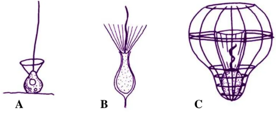
Als Gruppe von Organismen wurde für diese Arbeit die Klasse der Choanoflagellaten gewählt. Choanoflagellaten sind einzellige mikrobielle Eukaryoten mit einer globalen Verbreitung, die sowohl in marinen als auch in limnischen Systemen auftreten. Innerhalb der Ordnung der Choanoflagellaten gibt es drei Familien, die Acanthoecidae (Abbildung IIIC), deren Protoplast sich in einer mineralischen Hülle, der Lorica, befindet, und die nur marin oder im Brackwasser vorkommen. Weiters die Salpingoecidae (Abbildung IIIB), deren Protoplast von einer organischen Hülle, der Theka, umgeben wird, sowie die Codonosigidae (Abbildung IIIA), die „nackten“
Choanoflagellaten, die weder Theka noch Lorica besitzen und wie die Salpingoecidae sowohl marin bis limnisch verbreitet vorkommen. Hierbei eignen sich besonders die acanthoeciden Choanoflagellaten als Studienobjekt.
Wegen ihrer Lorica, die aus silikathältigen Stäben und Platten besteht, die art- charakteristisch angeordnet sind und damit ein eindeutiges morphologisches Merkmal
darstellen, zählen die Acanthoeciden zu der bestbeschriebenen Familie innerhalb der Choanoflagellaten. Derzeit sind 102 Arten in 30 Genera beschrieben.
Abbildung III: Schematische Darstellung von typischen Vertretern aus den drei Familien der Choanoflagellaten. A: Codonosigidae; B: Salpingoecidae; C: Acanthoecidae. Aus: Thomsen, H.A. (1992) Loricabærende choanoflagellater (Kraveflagellater). Plankton in Plankton i de indre danske farvande (ed. by HA Thomsen), Havforskning fra Miljøstyrelsen, 11,157-194
Zahlreiche Untersuchungen haben gezeigt, dass Protozoen eine bedeutende Position im Stoffumsatz aquatischer Systeme einnehmen (Azam et al., 1983; Güde, 1989; Weisse et al., 1990). Nach dem Konzept des „microbial loop“ (Azam et al., 1983) sind die HNF die wichtigsten Prädatoren der Bakterien im Pelagial (Abbildung IV).
A B C
11 In Studien zur Artzusammensetzung der Konsumenten von Bakterien findet man Zahlen von 10 bis 90% der Biomasse in Bezug auf Choanoflagellaten. Gerade im wichtigen polaren Nahrungsgewebe stellt diese Ordnung einen sehr hohen Prozentsatz an der Biomasse dar (Pace and Vaqué 1994; Sherr and Sherr 1994).
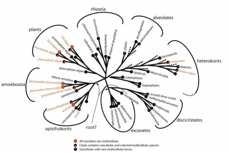
Abbildung V: Phylogenetische Position der Choanoflagellaten innerhalb der Eukaryoten. Aus:
Baldauf, S. L. (2003) The deep roots of eukaryotes. Science, 300: 1703-1706
Um einen aussagekräftigen phylogenetischen Stammbaum erstellen zu können, benötigt man ausreichend Sequenzen verschiedener Arten. Zu Beginn dieser Arbeit standen insgesamt neun verschiedene SSU (Small Sub Unit) rRNA Choanoflagellaten Sequenzen aus Gendatenbanken zur Verfügung. Mittlerweile ist dieser Datensatz auf dreizehn verschiedene Sequenzen angewachsen. Lediglich von zwei Choanoflagellatenarten liegt die LSU (Large Sub Unit) rRNA vor. Eines der großen Probleme bei der Isolation von Choanoflagellaten zur Erstellung monoxenischer Kulturen ist ihre Empfindlichkeit gegenüber starken Strömungen. Somit ist eine
Isolierung mittels Pipette bei den meisten Arten annähernd ausgeschlossen. Zudem sind viele Arten benthisch, wodurch die Vereinzelung ebenfalls erschwert wird. Um diese Probleme zu umgehen, wurde für diese Arbeit die Technik der Einzell-PCR (Polymerase Chain Reaction) weiterentwickelt. Mit Hilfe eines Mikromanipulators und einer im Spitzendurchmesser an die zu isolierende Art angepassten Kapillare wurden einzelne Organismen aus dem Medium entnommen und direkt der PCR zugeführt. Die so gewonnene rRNA ist rein klonal und frei von Kontaminationen (bei jeder PCR wurde eine Negativ-Kontrolle durchgeführt).
Die morphologische Eigenheit der acanthoeciden Choanoflagellaten, die Lorica, bietet die einmalige Möglichkeit, die Verbreitung einer Gruppe heterotropher Nanoflagellaten zu untersuchen. Weiters soll in dieser Arbeit am Beispiel von acanthoeciden Choanoflagellaten gezeigt werden, dass die Diversität von Protisten durch ein rein morphologisches Artkonzept möglicherweise unterschätzt wird. Anhand der Untersuchung von bipolaren Choanoflagellatenarten, Acanthocorbis nana und A unguiculata. sowie der kosmopolitischen Art Diaphanoeca grandis wird eine mögliche allopatrische Artbildung untersucht.
Bei der Untersuchung der oben beschriebenen Proben wurden drei neue Arten von Choanoflagellaten gefunden, die ebenfalls in dieser Arbeit beschrieben werden.
Diese neuen Arten sowie alle anderen Arten, die im Verlauf dieser Arbeit untersucht worden sind, werden sowohl zur Untersuchung der phylogenetischen Position der Choanoflagellaten innerhalb der Gruppe der Opisthokonta herangezogen als auch für eine Studie der Phylogenie und Taxonomie innerhalb der Choanoflagellaten.
Die Arbeit ist in fünf Kapitel aufgeteilt:
• Kapitel 1 analysiert die biogeographische Verbreitung von acanthoeciden Choanoflagellaten.
• Kapitel 2 beschreibt einen neuen Choanoflagellaten aus dem Genus Diplotheca.
• Kapitel 3 berichtet erstmalig über neue Choanoflagellatenarten aus der Familie der Salpingoecidae aus der Tiefsee.
• Kapitel 4 zieht einen morphologischen und molekularbiologischen Vergleich von Vertretern der bipolar verbreiteten Acanthoeciden (Acanthocorbis nana und A. unguiculata).
13
L
ITERATURAzam, F., Fenchel T., Field J. G., Gray J. S., Meyer-Reil L. A., Thingstad F. (1983) The ecological role of the water-column microbes in the sea. Mar. Ecol. Prog. Ser.
10: 257–263.
Baldauf, S. L. (2003) The deep roots of eukaryotes. Science, 300: 1703-1706
Berninger, U. G., Finlay, B. J., Kuuppo-Leinikki, P. (1991) Protozoan control of bacterial abundances in freshwater. Limnol Oceanogr 36: 139–147.
Fenchel, T. (1987) Ecology of Protozoa: The Biology of Free-living Phagotrophic Protists. Science Tech Publishers/Springer Verlag, Madison/Wisconsin, Berlin.
Fenchel, T., Finlay, B. J. (2004) Here and there or everywhere? Response from Fenchel and Finlay. Bioscience 54: 884–885.
Finlay, B. J. (2002) Global dispersal of free-living microbial eukaryote species.
Science, 296: 1061-1063.
Finlay, B. J. (2004) Protist taxonomy: an ecological perspective. Phi.l Trans. R.
Soc.Lond., Ser B 359: 599–610.
Foissner, W. (1999) Protist Diversity: Estimates of the near-imponderable. Protist 150: 363-368.
Güde, H. (1989) The role of grazing on bacteria in plankton succession, in U. Sommer (ed.) Plankton Ecology: Succession in Plankton Communities. Springer-Verlag, Berlin, Heidelberg, New York, pp.337-364.
Marchant, H. J. (2005) Antarctic marine protists. Choanoflagellates. (ed. by FJ Scott and H Marchant), pp 326-346. Australian Biological Resources Study, Canberra.
Massana, R., Terrado R., Vovejoy C., Pedros-Alio C. (2006) Distribution and abundance of uncultured heterotrophic flagellates in the world oceans. Environ.
Microbiol. 8: 1515-1522.
May, R. M. (1988) How many species are there on earth? Science, 247: 1441-49.
Mayr, E. (1942) Systematics and the Origin of Species. Columbia University Press, New York, USA.
Pace, M. L., Vaqué, D. (1994) The importance of Daphnia in determining mortality- rates of protozoans and rotifers in lakes. Limnol. Oceanogr. 39: 985–996.
Potter, D., Lajeunesse, T. C., Saunders, G. W., Anderson, R. A. (1997) Convergent evolution masks extensive biodiversity among marine coccoid picoplankton. Biodivers. Conserv. 6: 99–107.
Scheckenbach, F., Wylezich, C., Mylnikov, A. P., Weitere, M., Arndt, H. (2006) Molecular comparisons of freshwater and marine isolates of the same
morphospecies of heterotrophic flagellates. Appl. Environ. Microbiol. 72: 6638- 6643.
Sherr E. B., Sherr, B.F. (1994) Bacterivory and herbivory: key roles of phago- trophicprotists in pelagic food webs, Microbial Ecology. 28: 223-235.
Sonneborn, T. M. (1957) Breeding systems, reproductive methods, and species problems inProtozoa, in Mayr E. (ed.) The species problem. American Association for the Advancement of Science, pp. 155-324.
Tautz. D., Arctander, P., Minelli, A., Thomas, R. H., Vogler, A. P. (2003) A plea for DNA taxonomy. Trends Ecol. Evol. 18:70–74.
Thomsen, H. A. (1992) Loricabærende choanoflagellater (Kraveflagellater). Plankton in Plankton i de indre danske farvande (ed. by HA Thomsen), Havforskning fra Miljøstyrelsen, 11:157-194.
Weisse, T., Müller, H., Pinto-Coelho, R.M., Schweizer, A., Springmann, D. (1990) Response of the microbial loop to the phytoplankton spring bloom in a large prealpine lake, Limnol. Oceanogr. 35: 781-794.
Will, K. W., Rubinoff, D. (2004) Myth of the molecule: DNA barcodes for species cannot replace morphology for identification and classification. Cladistics 20:
47–55.
15
K APITEL I
Global biogeography of acanthoecid choanoflagellates
17 ABSTRACT
Aim The unicellular choanoflagellates form an important component of the plankton communities in surface waters of the world ocean. Acanthoecid choanoflagellates possess a lorica (size 5 - 100µm) composed of siliceous costae, a distinct feature which allows the morphological identification of most species. This offers the unique opportunity to study the biogeography of a group of heterotrophic nanoflagellates.
Location The study considered choanoflagellate distribution in the worldwide oceans.
Methods Data on the distribution of Acanthoecidae published in 80 papers within the last 42 years were used to create a taxonomically consistent data set. Analyses were based on the presence/absence of species in biogeographic regions. Clusters were distinguished using Bray-Curtis similarity values.
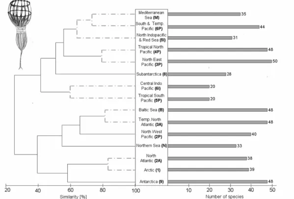
Results The analysis revealed distinct warm and cold water clusters. Out of 103 acanthoecid species fifteen were recorded until now only in the Arctic/North Atlantic waters and thirteen in the Subantarctic/Antarctic provinces. Nine species showed a bipolar distribution while 54 species were considered as cosmopolitan species. The species richness did not decrease with the latitude.
Main conclusion The data supported the idea that there are epibenthic/coastal species with a restricted geographic distribution. Various regions, especially the open sea, of the world ocean need further sampling and detailed electron microscopical investigations to verify this conclusion.
Key words Ocean biogeography, biodiversity, protists, heterotrophic flagellates, polar regions, species richness
INTRODUCTION
Acanthoecid choanoflagellates play a significant role as major consumers of bacteria in the open ocean, forming the basic transfer of carbon from DOC via bacteria to larger organisms such as ciliates and crustaceans. Heterotrophic flagellates are considered as part of the microbial loop in marine waters (Azam et al., 1983; Gasol &
Vaqué, 1993; Arndt, 2000) and have also been found to play a fundamental role in Antarctic waters (e.g. Anderson & Rivkin, 2001).
There is an intensive debate about the distribution patterns of unicellular organisms in literature (e.g. Finlay, 2002; Foissner, 2006). As the fundamental driver of random dispersal is high absolute abundance, and as organism size and abundance are inversely related, there may be some size range where ubiquitous dispersal becomes less likely and where species are more likely to be geographically restricted (Finlay, 2002). This would lead to the conclusion that acanthoecid choanoflagellates, mostly species smaller than 50 µm (protoplast generally smaller than 10 µm), should be ubiquitous or ‘common’ species. Such ‘common’ species are characterized by a combination of a broad distribution, an unspecialized habitat and large populations (May, 1988). Acanthoecids characterized by the presence of a species-specific lorica formed by siliceous costae (Leadbeater, 1991) and the limited morphospecies number of only 103 (29 genera) offer the unique opportunity to study the species richness and biogeographic distribution patterns of a family of heterotrophic nanoflagellates.
The only study on the global distribution of choanoflagellates has been carried out by Thomsen (1992). He found several taxa having a cosmopolitan distribution while others appeared to be confined to warm or cold waters. We used recent available data sets and the study of Thomsen (1992) and others for a more detailed analysis of the worldwide distribution of choanoflagellates.
METHODS
The basis of this research were 80 data collections of the last 42 years (see Appendix S2 in Supplementary Material) from all over the world plus our own data
19 microscopically examinations and the classification down to morphospecies. The dataset was corrected regarding synonyms considering the ‘check-list of the marine species’ by Brandt (2001) and the webpage of ‘micro*scope’
(http://starcentral.mbl.edu/microscope/portal.php) to filter out synonyms.
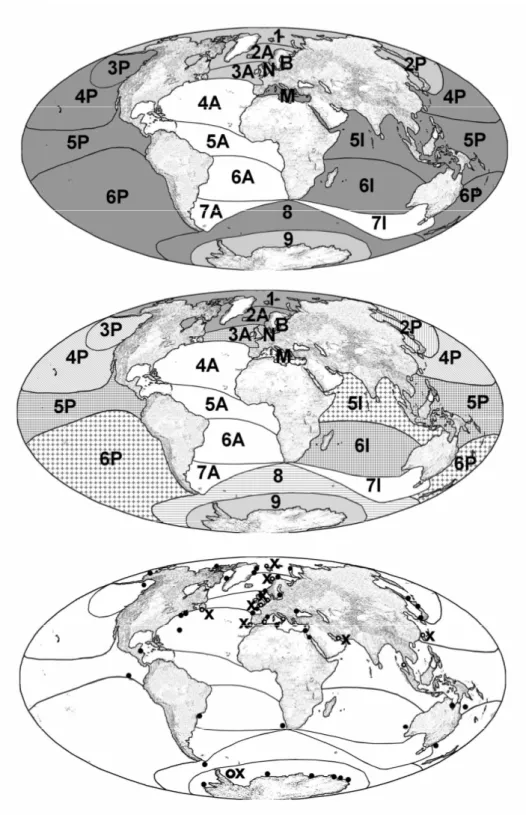
Presence/absence data were entered into Microsoft Excel 2003 datasheet (see Appendix S1 in Supplementary Material). To achieve the highest correlation between bioregions and sampling sites the bioregions (after Kelleher et al., 1995) were modified according to the prerequisites of choanoflagellates. The bioregions were defined as variables, the species as cases. Restricted distribution was defined as any species restricted to one bioregion or coherent bioregion temperature cluster (Fig. 2).
To be ‘common’ or ubiquitous the morphospecies had to occur in at least one warm or cold water province on both sides of the equator. Analysis of similarities among species assemblages was conducted using a cluster analysis based on the Bray-Curtis similarity coefficient. Coefficients were calculated with presence/absence-transformed data employing group average sorting (PRIMER version 6). Bioregions which were undersampled (e.g. less then 5 species) were excluded from the analysis and graphical display.
RESULTS
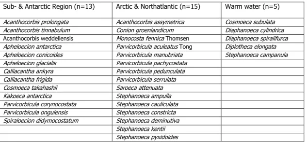
After filtering out all synonyms of the data collection, we established a list of 103 acanthoecid choanoflagellate morphospecies from 29 genera from marine and brackish waters. The survey of data collections showed that especially the Atlantic region is poorly investigated (Fig. 2C). Most sampling sites were close to coastal regions leaving the species composition of open sea mostly uninvestigated. Bearing this in mind and analysing the available data (Fig. 1) we found 13 out of 103 species which were only recorded from the Subantarctic and Antarctica and another 15 species which were present only in samples from the Arctic and Northern temperate provinces. All these 28 morphospecies did not show a bipolar distribution. Compared with these 28 non-bipolar cold-water species, 9 species were found in both polar regions. A total number of 54 out of 103 species were considered to be ubiquitous e.g.
they had a transtropical distribution according to their ecological needs (Table 1). Five morphospecies seemed to be restricted to warm-water provinces. For additional 7 acanthoecid species the restricted distribution is most probable, however, the sampling sites were at the boarder to the next bioregion and due to changes of surface currents a distinct assignment was impossible.
Table 1: List of endemic acanthoecid choanoflagellates in different marine provinces Sub- & Antarctic Region (n=13) Arctic & Northatlantic (n=15) Warm water (n=5) Acanthocorbis prolongata Acanthocorbis assymetrica Cosmoeca subulata Acanthocorbis tinnabulum Conion groenlandicum Diaphanoeca cylindrica Acanthocorbis weddellensis Monocosta fennica Thomsen Diaphanoeca spiralifurca Apheloecion antarctica Parvicorbicula aculeatus Tong Diplotheca elongata Apheloecion conicoides Parvicorbicula manubriata Stephanoeca campanula Apheloecion glacialis Parvicorbicula pachycostata
Calliacantha ankyra Parvicorbicula pedunculata Calliacantha frigida Parvicorbicula serrulata
Cosmoeca takahashii Saroeca attenuata
Kakoeca antarctica Stephanoeca ampulla
Parvicorbicula corynocostata Stephanoeca cauliculata Parvicorbicula ongulensis Stephanoeca constricta Spiraloecion didymocostatum Stephanoeca deminutiva
Stephanoeca kentii Stephanoeca pyxidoides
Cluster analysis revealed a clear separation into a warm and a cold water clusters (Fig. 2A). These clusters could be assigned to physical parameters such as temperature and ocean currents. The cold water cluster contained all cold and cold temperate water provinces except the Subantarctic. All tropical and warm temperate provinces formed the warm water cluster, in which also the Subantarctic was included.
Within these two main clusters, five distinct subclusters could be differentiated. The cold water cluster contained a specific Arctic/North Atlantic species community (84%
similarity of species community) and a Baltic/Temperate North Atlantic assemblage (83%). The warm water cluster was composed of the temperate Northeast Pacific/tropic North Pacific subcluster (82% similarity), the Northern Indopacific/South Pacific/Mediterranean Sea subcluster (65%), and the tropical North Atlantic/Central Indopacific subcluster (42%). Antarctica as well as the Subantarctic showed a highly specific species community and were separated from the other clusters. We established 21 marine bioregions modified after Kelleher et al. (1995) which match the distribution of acanthoecid choanoflagellates, considering the main ocean currents, temperature and sampling sites (Fig. 2B). There was no trend regarding the relationship of species richness of acanthoecid choanoflagellates and the latitude of the different regions (Fig.1).
21
Figure 1: Dendrogram of hierarchical cluster analysis of the worldwide distribution of acanthoecid choanoflagellate communities (left side, with % similarity given in the x- axis) compared with the corresponding number of different morphospecies for each bioregion (right side). The abbreviations of bioregions used for Fig. 2 are given in brackets.
DISCUSSION
Acanthoecid choanoflagellates are one of the few groups of heterotrophic nanoflagellates allowing the study of the geographic distribution. This is due to the unique morphology of their lorica. Though the sampling sites were largely restricted to coastal regions, pelagic species of the open sea were frequently collected. Care had to be taken considering the technical possibilities of researchers to determine morphospecies on the base of their lorica construction. Thus, several older publications had to be left unconsidered for the purpose of global comparisons. The authors are aware of the problematic use of the term “cosmopolitism” in the sense of Fenchel (2005). Despite a cautious interpretation of species records, we found one third of the species to show a restricted biogeo-
Figure 2: Global distribution patterns of acanthoecid choanoflagellate communities as a result of cluster analysis. Designations of bioregions refer to Fig. 1 A: Warm (dark grey) and coldwater (light grey) clusters; B: Distribution of subclusters; C: Sampling sites of data
23 graphic distribution compared two third of acanthoecid morphospecies meeting the premise of being cosmopolitans. The unexpected high number of acanthoecid species with a restricted distribution confirms studies on the distribution patterns of ciliates and diatoms (Hillebrand et al., 2001). The highest number of species with a presumably restricted distribution was found in polar regions. Abundances of acanthoecid choanoflagellates under the polar ice are several orders of magnitude higher than those from polar pelagic waters (e.g. Hewes et al. 1990, Esser 2006).
Even if dispersal via cysts in the deep ocean through the conveyor belt takes place, the number of cyst must be tremendously high to amortize the loss during the long transfer. Among choanoflagellates only salpingoecids and codonosigids had been reported from the deep sea (Nitsche et al. 2007) making it unlikely that deep sea flows are the major ways of distribution for acanthoecid choanoflagellates. The restricted exchange between choanoflagellate populations of polar regions may have contributed to the speciation process in the two cold water provinces.
For future studies, the analysis of the open sea regions, especially that of the Atlantic, would be desirable for a more complete understanding of acanthoecid distribution patterns. For instance, the clustering of the Subantarctic region in the warm water cluster had to be attributed to the fact that the only available data were recorded from the west wind drift meeting the Peru Current.
Our analysis of literature data included only morphospecies distributions leaving the potentially high number of cryptic species hidden in morphospecies complexes unconsidered. This might even underestimate the number of species with a restricted biogeographic distribution (Darling et al., 2002; Scheckenbach et al., 2005;
Nitsche et al., subm.).
ACKNOWLEDGEMENTS
We are greatly indebted to Barry S.C. Leadbeater for valuable discussions. The study was supported by a grant of the German Research Foundation (DFG) to H.A.
(AR 288/8 and AR 288/12)
REFERENCES
Anderson, M.R. & Rivkin, R.B. (2001) Seasonal patterns in grazing mortality of bacterioplankton in polar oceans: a bipolar comparison. Aquatic Microbial Ecology, 25, 195-206.
Arndt, H., Dietrich, D., Auer, B., Cleven, E., Gräfenhan, T., Weitere, M. & Mylnikov A.P.
(2000) Functional diversity of hetrotrophic flagellates in aquatic ecosystems. The Flagellates. (ed. by BSC Leadbeater and JC Green), pp240-268. Taylor & Francis Ltd, London.
Azaam, F., Fenchel, T., Field, J.G., Gray, J.S., Meyer-Reil, L.A. & Thingstad, F. (1983) The ecological role of water-column microbes in the sea. Marine Ecology Progress Series, 10, 257-263.
Brandt, S. (2001) Choanoflagellates. European register of marine species: a check-list of the marine species in Europe and a bibliography of guides to their
identification., (ed. by MJ Costello, C Emblow and R White), pp. 57-59.
Collection Patrimoines Naturels, 50.
Esser, M. (2005). Doctoral Thesis. University of Cologne.
Fenchel, T. (2005) Cosmopolitan microbes and their "cryptic" species. Aquatic Microbial Ecology, 41, 49-54.
Finlay, B.J. (2002) Global dispersal of free-living microbial eukaryote species. Science, 296, 1061-1063.
Foissner, W. (2006) Biogeography and dispersal of micro-organisms: A review emphasizing protists. ActaProtozoologica, 45, 111-136.
Gasol, J.M. & Vaqué, D. (1993) Lack of coupling between heterotrophic
nanoflagellates and bacteria: A general phenomenon across aquatic systems?
Limnology and Oceanography, 38, 657-665.
Hewes, C.D., Sakshaug, E., Reid, F.M.H. & Holm-Hansen, O. (1990) Microbial autotrophic and heterotrophic eucaryotes in Antarctic waters: relationships between biomass and chlorophyll, adenosine triphosphate and particulate organic carbon. Marine Ecology Progress Series, 63, 27-35.
Hillebrand, H., Watermann, F., Karez, R. & Berninger, U. (2001) Differences in species richness patterns between unicellular and multicellular organisms. Oecologia,
25 Leadbeater, B.S.C. (1991) Choanoflagellate organization with special reference to
loricate taxa.
Freeliving heterotrophic flagellates. (ed. by DJ Patterson and J Larsen ), pp 241–258.
Clarendon Press, Oxford.
Marchant, H.J. (2005) Antarctic marine protists. Choanoflagellates. (ed. by FJ Scott and H Marchant), pp 326-346. Australian Biological Resources Study, Canberra.
May, R. M. (1988) How many species are there on earth? Science, 247, 1441-49.
Nitsche, F. & Arndt, H (subm) Bipolar comparisons of acanthoecid choanoflagellates.
Nitsche, F., Weitere, M., Scheckenbach, F., Hausmann, K., Wylezich, C. & Arndt, H.
(2007) Deep sea records of choanoflagellates with a description of two new species. Acta Potozoologica, 46, in press.
Scheckenbach, F., Wylezich, C., Weitere, M., Hausmann, K. & Arndt H. (2005) Molecular identity of strains of heterotrophic flagellates isolated from surface waters and deep-sea sediments of the South Atlantic based on SSU rDNA.
Aquatic Microbial Ecology, 38: 239-247
Thomsen, H.A. (1992) Loricabærende choanoflagellater (Kraveflagellater). Plankton in Plankton i de indre danske farvande (ed. by HA Thomsen), Havforskning fra Miljøstyrelsen, 11,157-194
27
K APITEL II
A new species of heterotrophic flagellates from Taiwanese brackish waters:
Morphological and molecular biological studies of Diplotheca elongata nov. spec. and D. costata
(Choanoflagellida, Acanthoecidae)
29 Abstract
A new species of acanthoecid choanoflagellate isolated from brackish waters of the Danshui estuary in North Taiwan has a mineralised lorica which consists of two chambers with a total length of 19-36 µm. It shares with Diplotheca costata the features of a posterior lorica chamber formed from broad and flattened costal strips and an anterior chamber with spatula-shaped costal strips. The new species has therefore been placed in the same genus and named Diplotheca elongata. A phylogenetic analysis of partial SSU of rRNA sequences from Diplotheca costata and D. elongata supports this taxonomic affiliation. This is a large and distinctive choanoflagellate which has not been reported in any previous study, suggesting that it may be an endemic species of restricted distribution.
Key words. Choanoflagellates, biogeography, phylogeny, biodiversity
Introduction
Protists have been considered to have no restricted biographies as emphasized by a series of articles published by Finlay and Fenchel (e.g. 1999, 2004). Acanthoecid choanoflagellates seem to be a perfect model group within heterotrophic nanoflagellates to study the biogeographic distribution of nanoflagellates due to the species specific morphology of their lorica (Nitsche and Arndt subm.).
Choanoflagellates compose between 5 and 40 per cent of nanoflagellate biomass in marine, brackish and freshwater pelagic waters (e.g., Arndt et al. 2000). About 200 species of choanoflagellates are known, of which 102 species in 29 genera belong to acanthoecid choanoflagellates, which have mainly been described on the base of the specific shape of the lorica (Leadbeater 1991).
The genus Diplotheca belongs to the family Acanthoecidae containing up to now only one species, Diplotheca costata described by Valkanov (1970). D. costata has been found in the South Pacific Ocean, the North Sea, the Baltic, the Black Sea and the Mediterranean Sea (Valkanov 1970; Thomsen 1992, Tong, 1997). The posterior chamber of the lorica of Diplotheca typically consists of broad and flattened costal strips with rounded ends. We isolated a clone of Diplotheca elongata from the surface waters of the Chinese Sea in the Taiwan Strait near the city of Danshui, North Taiwan, which represents a new species of this genus. In addition to morphological data, we present also molecular biological studies of SSU rRNA from Diplotheca elongata and D.
costata.
Material and methods
Sampling sites. Material from Taiwan was collected in August 2005 and 2006 from the Taiwan Strait (courtesy of M. Kern). Surface water samples originate from the beach at the estuary of River Danshui near the city of Danshui, North Taiwan (25°10’N/121°26’E). One litre of water was taken in a sterile polyethylene bottle. For Diplotheca coststa additional water samples were taken from the coast of the English Channel at Calais, France, in September 2005, and from the coast of the Persian Gulf at Dubai, United Arabic Emirates, in May 2005 and August 2006 (courtesy of S.
Nitsche).
The salinity of the samples was about 12 PSU, 34 PSU and 41 PSU in samples from
31 populations as a food source for choanoflagellates. After one week, samples were examined by light microscopy (Zeiss Axiovert 100). Samples containing choanoflagellates were diluted with artificial seawater at the salinity of the sample site.
The samples were maintained at 18°C under a 12/12 hour day/night cycle.
Electron microscopy. The preparation of cells basically followed the protocol used by Stoeck et al. (2003). The samples were fixed at a ratio of one to one with Bouin’s fixative at 4°C for 45 minutes. The fixative contained three parts of saturated with picric acid, one part buffered formaldehyde (38%) and 2% glacial acetic acid, which was added immediately before fixation. To the final solution 0.1 to 0.2%
glutaraldehyde (38%) was added. Samples remained in the culture flask and a dehydration series of ethanol with 30%, 50%, 60%, 70%, 80%, 90%, 96% and pure ethanol (each step was done three times and lasted 10 minutes) was carried out. After this procedure, a 50:50 hexamethyldisilazane (HMDS)-ethanol solution was applied for 30 minutes followed by pure HMDS for 30 minutes. Afterwards, the samples were allowed to dry. The bottom of the flasks was cut to appropriate size and stuck to a sample holder. SEM samples were sputtered with a 120Å layer of gold before examination by SEM (Hitachi S-520).
Molecular biology. For single cell PCR, organisms were extracted using a micromanipulator. Cells were transferred to 27µl sterilized water and frozen at -20°C for three hours before PCR. The SSU rDNA fragment was amplified using 18SFor (AACCTGGTTGATCCTGCCAGT) and 28S-D5(rev) (CCGTAGGTGAACCTGCAGAAGGA) primer at a concentration of 1.6 nM followed by a reamplification with the primer pair 82F (GAAACTGCGAATGGCTC) and 18SRev-Ch (CCGTAGGTGAACCTGCAGAAGGA) for SSU. The PCR products were purified using the E.Z.N.A. Cycle-Pure-Kit (Peqlab, Erlangen, Germany). The sequencing of rDNA was done using Big Dye-Terminator v3.1 Cycle Sequencing Kit (Applied Biosystems, Weiterstadt, Germany) in accordance with the manufacturer’s instructions. Primers used for sequencing were 82F, 590F and 18SRev-Ch for SSU. Obtained sequence-parts were manually arranged.
Phylogenetic analyses. Alignments were carried out using ClustalX (Thompson et al. 1997) and corrections were made manually with BioEdit. The model (JC69) for maximum likelihood analysis was determined by MrAIC (Nylander 2004) and the ML analysis computed by PhyML (Guindon and Gascuel 2003), using 100 replicates for the bootstrap analysis. Neighbour joining (NJ) was calculated using MEGA 3.1 (Kumar et al. 2004) using the JC model and 100 replicates for bootstrap analysis.
Results
Description of Diplotheca elongata nov. spec.
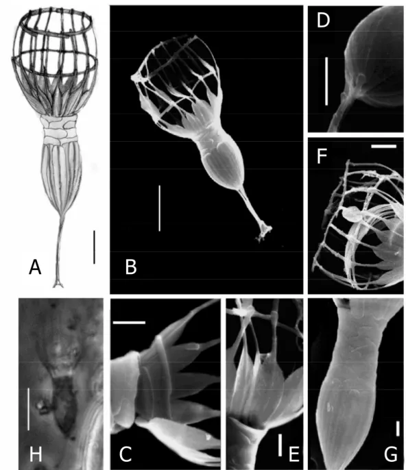
Diagnosis: Lorica-bearing protist with the characteristics of its genus (Fig. 1). The lorica of Diplotheca elongata is composed of two quite distinct chambers (total length 19-36 µm). The posterior chamber has a length of 5-13 µm and the anterior chamber is between 9 and 15 µm in length (Table 1). The anterior chamber contains 14 to 18 longitudinal costae, each being formed by two costal strips with spatulate ends (Fig.
1A, C). A transverse costa containing five flattened costal strips forms the anterior ring. A second transverse costa is situated in the middle of the anterior chamber (Fig.
1A, B) and is also composed of flattened costal strips. A third ring of costal strips is situated on the base of the spatulated ends of longitudinal strips concealed by the upper part of the posterior chamber (Fig. 1D). The upper part of the posterior lorica chamber is formed by a layer of scale-like flattened strips. The lower part consists of 12 to 16 broad and flattened costal strips with rounded ends. The stalk has a length of 4-8 µm and is built by 3-8 costal strips which are attached to the posterior end of the lorica (Fig. 1D).
Etymology: elongata (feminine) from Latin “elongated” in reference to the elongated posterior chamber of the lorica.
Type location: Estuary of the River Danshui near the city of Danshui, North Taiwan (25°10’N/121°26’E).
Holotype: The illustration of the specimen in Figure 1A.
SSU rRNA: Partial SSU rRNA fragments were used for a FASTA search. The result assigned Diplotheca elongata into the order of Choanoflagellida, but not into the family of Acanthoecidae (Fig. 3). NCBI accession numbers are: Diplotheca elongata EF483922 and D. costata EF483923.
Description: The description of Diplotheca elongata is based on the observation of cells using light and scanning electron microscopy. Cells are ovoid, with a size range of 8 to 10µm in length and 3.5 to 4 µm in width. The flagellum is 13 to 16 µm long. The collar measures 9 to 15 µm in length. The mineralised lorica of Diplotheca elongata consists of two chambers (total length 19-36 µm) (Fig. 1A). The anterior lorica chamber possesses 14 longitudinal costae each composed of two flattened costal strips. The basal coastal strips are spoon shaped (Fig. 1B). In addition to the anterior
33 form the upper part (Fig. 1D). Inside the lower anterior lorica chamber, a ring composed of crescent strips can be found in individuals, which are going to undergo cell division (Fig. 1E). These are the pre-produced costae for the lorica of the sister cell (Jackson and Leadbeater 1991). The stalk is attached to the posterior end of the lorica by the flattened ends of its costal strips. The number of costal strips forming the stalk ranges from 3-8µm, the length of the stalk is 4-8µm (Fig. 1C).
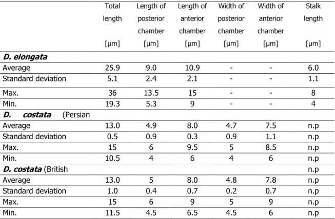
Table 1. Statistics of lorica measurements of Diplotheca elongata (based on 25 individuals) and D. costata from the Persian Gulf and the English Channel (based on 20 individuals each).Missing morphological feature are indicated by ‘n.p.’, not measured values by ‘-‘.
Total length
[µm]
Length of posterior chamber [µm]
Length of anterior chamber
[µm]
Width of posterior chamber [µm]
Width of anterior chamber
[µm]
Stalk length
[µm]
D. elongata
Average 25.9 9.0 10.9 - - 6.0
Standard deviation 5.1 2.4 2.1 - - 1.1
Max. 36 13.5 15 - - 8
Min. 19.3 5.3 9 - - 4
D. costata (Persian
Average 13.0 4.9 8.0 4.7 7.5 n.p
Standard deviation 0.5 0.9 0.3 0.9 1.1 n.p
Max. 15 6 9.5 5 8.5 n.p
Min. 10.5 4 6 4 6 n.p
D. costata (British n.p
Average 13.0 5 8.0 4.8 7.8 n.p
Standard deviation 1.0 0.4 0.7 0.2 0.7 n.p
Max. 15 6 9 5 9 n.p
Min. 11.5 4.5 6.5 4.5 6 n.p
In addition, we examined two populations of Diplotheca costata isolated from the English Channel at Calais (France) and from the coast of the Persian Gulf at Dubai.
The morphological appearance (Fig. 2) clearly resembled the original description of Valkanov (1970). Geographical differences could not be detected (Table 1).
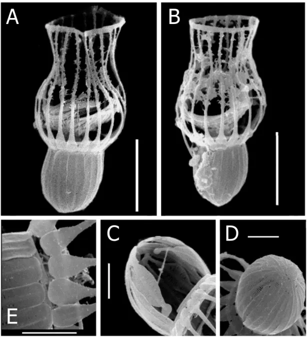
Figure 1. Drawings (A), scanning electron micrographs (B-G ) and light microscopic micrographs (H) of Diplotheca elongata from the estuary of River Danshui (Taiwan); A – drawing of a specimen; B - complete specimen (scale bars 5 µm); C - detail of costal strips forming the anterior lorica chamber with the typical spatula form; D - close-up of the posterior lorica chamber and the stalk; E – the third transverse costa at the base of the anterior lorica chamber; F – the bundle of crescentic strips inside the lorica; G – elongated upper section of the posterior lorica where the length growth takes place (scale bars 1 µm); H – light microscopical micrograph (scale bar 5 µm).
A B
H C
D
E G
F
35
Figure 2 A-F. Scanning electron micrographs of Diplotheca costata; A - complete specimen from the British Channel; B - complete specimen from the Persian Gulf; C - the posterior chamber built by flattened costal strips; D - detail of the costal strips from the posterior end; E - detail of the spatula like costal strips forming the anterior lorica (C-E specimens from the Persian Gulf; scale bars A-B 5µm, C-E 2µm)
A B
C D
E
Figure 3 Distance tree of available choanoflagellate sequences based on about 1200 bp long fragments of SSU rDNA using Dictyostelium rhizopodium as an outgroup. Bootstrap values represent maximum likelihood values (100 replicates) and neighbour joining (JC) values (100 replicates), values less than 75% are marked ‘-‘. Scale bar marks the genetic distance calculated for the NJ tree.
37 DISCUSSION
The new species from the Strait of Taiwan, Diplotheca elongata, obviously belongs to the same genus as Diplotheca costata. The lorica of Diplotheca elongata is comprised of on a posterior, an anterior chamber and a stalk. The broad and flattened costal strips which form the posterior chamber and the spatula-shaped costal strips of the anterior lorica chamber are typical for D. costata (see Valkanov 1970 and Tong 1997). There is no similarity to any other described acanthoecid choanoflagellate. Due to its size (about 20 to 40µm) this species can be distinguished easily by light microscopy. The biogeographic distribution is so far restricted to the brackish waters of the estuary of River Danshui. Samples from other Taiwanese river estuaries did not contain Diplotheca elongata, but it was found in the two following years in different samples from the Danshui estuary. Though this species has very prominent morphological characteristics, it has never been recorded before. It is assumed that this species has a very restricted biogeographic distribution. Considering the fact, that there are about 200 described choanoflagellate species, our recent observations of three new species (Nitsche et al. in press and present data) already add 1.5% new taxa to the dataset. Especially in polar regions, endemism seems to be far more common among acanthoecid choanoflagellates than estimated before. Our recent literature survey of more than 120 publications indicated that out of 102 acanthoecid morphospecies 26 were only found either in the Arctic or Antarctic. A total of 32 out of the 102 species were endemic to one biogeographic region (Nitsche and Arndt unpubl.). Considering this the everything-is-everywhere hypothesis seems to be only limited applicable for acanthoecid choanoflagellates.
The analysis of the partial SSU rRNA sequences did not display a clear taxonomic affiliation of both species from the genus Diplotheca to the family of Acanthoecidae (Fig. 3). For a more reliable phylogenetic analysis the number of sequenced species must be enlarged. Further studies with D. elongata and D. costata cultures were not possible as we could not establish continuous cultures. At present, Diplotheca elongata together with the sequence of D. costata from the Persian Gulf form a cluster out of the family of Acanthoecidae. This is supported by the specific morphology of the costae. There are two other species of acanthoecid-like choanoflagellates which own a similar type of lorica that is also composed of flattened costal strips - Syndetophyllum pulchellum, Parvicorbicula serrulata and Platypleura infundibuliformis (Leadbeater 1974; Manton and Leadbeater 1975; Thomsen and Boonruang 1983; Thomsen and Moestrup 1983). Future molecular studies of these species will be necessary to show whether these three taxa are related.
Acknowledgements. We thank Barry S.C. Leadbeater and Michael Sleigh for valuable discussions and constructive criticism. This study was financially supported by a grant of the German Research Foundation to H.A. We are very thankful to M. Kern and S. Nitsche for taking and transferring samples from Taiwan and Dubai. For technical support we are indebted to H. Bollhagen, C. Barth, and R. Bieg. Special thanks go to D. Tautz and his Department of Evolutionary Genetics (Institute for Genetics, University of Cologne) for the sequencer facilities.
REFERENCES
Arndt, H., Dietrich, D., Auer, B., Cleven, E., Gräfenhan, T., Weitere, M., Mylnikov A.P., 2000. Functional diversity of heterotrophic flagellates in aquatic ecosystems. In:
Leadbeater, B.S.C., Green, J.C. (Eds.), The Flagellates. Taylor & Francis Ltd, London, pp. 240-268.
Fenchel, T., Finlay, B.J., 2004. The ubiquity of small species: Patterns of local and global diversity. BioScience 54, 777-784.
Finlay, B.J., Fenchel, T., 1999. Divergent perspectives on protist species richness.
Protist 150, 229-233.
Guindon, S., Gascuel, O., 2003. A simple, fast and accurate algorithm to estimate large phylogenies by Maximum Likelihood. Systems Biology 52, 696-704.
Jackson, S. M., Leadbeater, B. S. C. (1991). Costal strip accumulation and lorica assembly in the marine choanoflagellate Diplothecacostata Valkanov. J. Protozool.
38, 97-104.
Kumar, S., Tamura, K., Nei, N., 2004. MEGA3: Integrated software for Molecular Evolutionary Genetics Analysis and sequence alignment. Briefings in Bioinformatics 5, 50-163.
Leadbeater, B.S.C., 1974. Ultrastructural observations on nanoplankton collected from the coast of Jugoslavia and the Bay of Algiers. J. Mar. Biol. Ass. U. K. 54, 179-196.
Leadbeater, B.S.C., 1991. Choanoflagellate organization with special reference to loricate taxa. In: Patterson, D.J., Larsen, J. (Eds.), Freeliving heterotrophic flagellates. Clarendon Press, Oxford, pp. 241–258.
Nitsche, F., Weitere, M., Scheckenbach, F., Hausmann, K., Wylezich, C., Arndt, H.,
39 Nylander, J.A.A., 2004. MrAIC.pl. Program distributed by the author. Evolutionary Biology Centre, Uppsala University, Uppsala.
Stoeck, T., Fowle, W.H., Epstein, S.S., 2003. Methodology of protistan discovery: from rRNA detection to quality scanning electron microscope images. Appl. Env.
Microbiol. 96, 6856-6863.
Thomsen, H.A., 1992. Loricabærende Choanoflagellater (Kraveflagellater). In:
Thomsen, H.A. (Ed.), Plankton i de indre danske farvande. Havforskning fra Miljøstyrelsen Vol. 11, Copenhagen, pp. 157-194.
Thomsen, H.A., Boonruang, P., 1983. A microscopical investigation of marine collared flagellates (Choanoflagellida) from the Andaman Sea, S.W. Thailand: species of Stephanacantha gen. nov. and Platypleura gen. nov. Protistologica 19, 193-214.
Thomsen, H.A., Moestrup, Ø., 1983. Electron microscopical investigations on two loricate choanoflagellates (Choanoflagellida), Calotheca alata gen. et. sp. nov. and Syndetophyllum pulchellum gen. et comb. nov., from Indo-Pacific localities. Proc.
R. Soc. Ser. B 219, 41-52.
Thompson, J.D., Gibson, T.J., Plewniak, F., Jeanmougin, F., Higgins, D.G., 1997. The ClustalX windows interface: flexible strategies for multiple sequence alignment aided by quality analysis tools. Nucleic Acids Research 24:4876-4882.
Tong, S.M. (1997) Choanoflagellates from Southampton Water, U.K., including the description of three new species. J. . Mar. Biol. Assoc.,UK, 77, 929-958.
Valkanov, A., 1970. Beitrag zur Kenntnis der Protozoen des Schwarzen Meeres. Zool.
Anz. 184, 241-250.
41
K APITEL III
Deep sea records of choanoflagellates with a description of
two new species
43
Abstract
Despite their high abundance and their high importance for the oceanic matter flux, heterotrophic nanoflagellates are only poorly studied in the deep-sea regions. Studies on the choanoflagellate distribution during two deep-sea expeditions, to the South Atlantic (5038 m) and Antarctica (Weddell Sea, 2551 m), revealed the deepest records of choanoflagellates so far. A new species, (Lagenoeca antarctica) with a conspicuous spike structure on the theca is described from deep Antarctic waters. Lagenoeca antarctica sp. n. is a solitary unstalked free living salpingoecid-like choanoflagellate.
The protoplast is surrounded by a typical theca with unique spikes only visible in SEM micrographs. The ovoid cell nearly fills the whole theca and ranges in size from 4 to 6µm. The collar measures 2-3 µm and the flagellum 3-5 µm. A second species, Salpingoeca abyssalis sp. n., was isolated from the abyssal plain of the South Atlantic (5038 m depth). Floating and attached forms were observed. The protoplast ranges from to 2 to 4 µm in length and 1 to 2 µm in width. The collar is about the same length as the protoplast and the flagellum has 2 to 2,5x the length of the protoplast.
Phylogenetic analyses based on a fragment of SSU rDNA revealed Salpingoeca abyssalis to cluster together with a marine isolate of Salpingoeca infusionum while Lagenoeca antarctica clusters separately from the other codonosigid and salpingoecid taxa. Salpingoeca abyssalis and an undetermined Monosiga species seems to be the first choanoflagellate species recorded from the abyssal plain.
Key words. Choanoflagellida, deep sea, Lagenoeca antarctica, sp. n., Salpingoeca abyssalis, phylogeny, SSU rDNA, abyssal plain, Antarctica.
INTRODUCTION
Heterotrophic nanoflagellates belong to the most important bacterial grazers in pelagic and benthic marine ecosystems (Azam et al. 1983, Fenchel 1987, Arndt et al.
2000). Though the deep-sea floor represents the largest part of the earth’s surface, very little is known about its most abundant eukaryotic inhabitants. Previous studies of the deep sea revealed an unexpected diversity of heterotrophic nanofauna (López- García et al. 2001, Hausmann et al. 2002, Arndt et al. 2003, Scheckenbach et al. 2005, Hausmann et al. 2006). Morphological and molecular biological studies showed that at least some widely distributed nanoflagellates of surface waters can also be found in the deep sea. A variety of protists exists in the deep-sea sediments up to a depth of 5400 m, but only a few species have been recorded up to now. Probably due to their small size (1-10 µm), choanoflagellates from abyssal regions have never been reported. Even molecular biological studies based on clone libraries revealed up to our knowledge no choanoflagellate sequences. This is surprising since choanoflagellates are common in surface marine, brackish and freshwaters forming between 5 to 40 per cent of total nanoflagellate biomass in these habitats (Arndt et al. 2000). Patterson et al. (1993) have recorded a variety of choanoflagellates from deep plankton and sediment samples indicating their potential occurrence in the deep sea.
About 200 morphospecies of choanoflagellates have been described of which about 50 belong to the family of Salpingoecidae. Members of this family greatly vary in size and shape. All are characterized by the presence of a theca, a layered substructure around the protoplast (Leadbeater 1990). Since recent molecular biological studies have indicated that morphospecies of heterotrophic nanoflagellates are often represented by very different genotypes with sometimes tremendous ecological differences (Scheckenbach et al. 2005, 2006; Hausmann et al. 2006, Massana et al. 2006), we tried to obtain living organisms which could be described on the base of their morphology and SSU rDNA sequence.
MATERIAL AND METHODS
Sampling sites. Material from Antarctica was collected by M. Esser on a cruise of the R/V Polarstern (ANT-XXII/2) in January 2005. Plankton samples were collected by sterilized polyethylene Niskin-bottles from the Weddell Sea from the surface to a depth
45 Abyssal Plain at a depth of 5038 m (28° 7’ S and 7° 21’ E), the Angola Abyssal Plain (9° 56’ S and 0° 54’ E), and the Guinea Abyssal Plain (0° 0’ S and 2° 25’ W). Also here, the open cores were washed for more than one hour in the deep sea to avoid contamination.
On board, samples were aliquoted and species were isolated at normal air pressure and transferred to cell culture flasks (50 ml, Sarstedt). A sterilized wheat grain was provided as nutrition for autochthonous bacteria populations as a food source for choanoflagellates. Each week, samples were examined by light microscopy (Zeiss Axiovert 200 or 40). Samples containing choanoflagellates were registered diluted with artificial seawater of the same salinity. Long-term storage of samples and cultures was carried at 4°C (Weddell Sea samples) or 10°C (abyssal plain samples) in the dark.
Electron microscopy. The preparation of cells basically followed the protocol used by Stoeck et al. (2003). Samples were fixed with 50:50 Bouin’s fixative mixture (buffered formaldehyde saturated with picric acid and 2% glacial acetic acid added immediately before fixation) with the addition of 0.1 to 0.2% glutaraldehyde, for 45 minutes at 4°C. Samples remained in the culture flask and a dehydration series of ethanol with 30%, 50%, 60%, 70%, 80%, 90%, 96% and pure ethanol (each step was done three times and lasted 10 minutes) was carried out. After this procedure, a 50:50 hexamethyldisilazane (HMDS)-Ethanol solution was applied for 30 minutes followed by pure HMDS for 30 minutes. Afterwards, the samples were allowed to dry.
The bottom of the flasks was cut to appropriate size and stuck to a sample holder.
SEM samples were sputtered with a 120Å layer of gold before examination by SEM (Hitachi S-520).
Molecular biology. For single cell PCR, organisms were extracted using a micromanipulator. Cells were transferred to 27µl sterilized water and frozen at -20°C for three hours before PCR. The SSU rDNA fragment was amplified using 18SFor (AACCTGGTTGATCCTGCCAGT) and 18SRev-Ch (CCGTAGGTGAACCTGCAGAAGGA) primer at a concentration of 1.6 nM followed by a reamplification with the primer pair 82F (GAAACTGCGAATGGCTC) and 18Srev-1 (CGTAACAAGGTTTCCGTAGGT). The PCR products were purified using the E.Z.N.A. Cycle-Pure-Kit (Peqlab, Erlangen, Germany).
The sequencing of rDNA was done using Big Dye-Terminator v3.1 Cycle Sequencing Kit (Applied Biosystems, Weiterstadt, Germany) in accordance with the manufacturer’s instructions. Primers used for sequencing were 82F and 590F. Obtained sequences were manually arranged.
Phylogenetic analyses. Alignments were carried out using ClustalX (Thompson et al. 1997) and corrections were made manually with BioEdit. The model (GTRG) for maximum likelihood analysis was determined by MrAIC (Nylander 2004) and the ML analysis computed by PhyML (Guindon and Gascuel 2003), using 100 replicates for the bootstrap analysis. Minimum evolution (ME) was calculated using MEGA 3.1 (Kumar et al. 2004) using the K2P model and 100 replicates for bootstrap analysis.
RESULTS
Four sampling sites, one pelagic (Weddell Sea) and three benthos samples (South Atlantic) were investigated for the presence of choanoflagellates using live- observation techniques. A salpingoecid choanoflagellate (Lagenoeca antarctica) could be isolated and cultivated from the Antarctic pelagial (2551 m depth) and grown at 4°C. From the South Atlantic stations, deep-sea cultures of choanoflagellates could be obtained from the Cape Basin (5034-5084 m depth) and from the Guinea Basin (5063- 5066 m depth). At both stations, the new species Salpingoeca abyssalis as well as an undefined Monosiga species were found. No choanoflagellates could be recorded from the Angola Basin (station at a depth of 5650 m). Unfortunately, Monosiga sp. did not grow in culture and we were not able to carry out detailed morphological and molecular biological studies with this species.
Rough abundance estimates using the liquid aliquot method (Butler and Rogerson 1995) revealed choanoflagellate densities in the deep sea lower than 1 ind./l in the pelagial of the Weddell Sea as well as in the bottom waters of the South Atlantic. Both choanoflagellate species of the deep sediments were not only found in the sediment surface layers (shells of foraminiferans) but also on small stones and shells of invertebrates. Remarkable was the slow beating rate of the flagella of the deep-sea choanoflagellates compared to choanoflagellates which we isolated from surface waters when cultivated under similar conditions.
Lagenoeca antarctica sp. n.
Diagnosis: Unstalked choanoflagellate with the characteristics of salpingoecids (Figs. 1A-E). The species specific characteristics of Lagenoeca antarctica are small thorn-like elevations on its theca visible by SEM. The size of the ovoid protoplast which fills the theca completely ranges from 4 to 6 µm in length and 2 to 3 µm in width. The
47 Type location: Bathypelagial of the Weddell Sea in Antarctica (63° 45,7’ S 50° 53,2’W) at a depth of 2551 m.
Holotype: The illustration of the specimen in Fig. 3.1A.
Taxonomic position: Although the morphological studies, especially the existence of a theca, support an affiliation of the new species to the Salpingoecidae, the phylogenetic examinations of the partial SSU rDNA (Fig. 3) indicate a significant phylogenetic distance of the species from other choanoflagellate taxa and do not allow yet a distinct assignment to the family of Salpingoecidae.
Description: The description of Lagenoeca antarctica sp. n. is based on the observation of cells using scanning electron microscopy and light microscopy. The protoplast is ovoid in shape and fills nearly the whole theca. The length of the protoplast ranges from 4-6 µm in length and from to 2-3 µm in width (Tab. 1). The theca is covered with spike or thorn like elevations only visible in EM (Figs 1B-E), which are irregularly spread over the whole surface. The relatively short flagellum (3-5 µm, Figs. 1B, C) emerges from a short collar (2-3 µm). The new choanoflagellate species appears only free swimming in cultures.
Salpingoeca abyssalis sp. n.
Diagnosis: Small choanoflagellate possessing a theca. Benthic forms are attached by a stalk to the substratum while free-swimming forms are unstalked (Figs 2A-G). Protoplasts are oval in shape and covered by a theca. The size of the protoplast ranges from 1-2 µm in width and 2-4 µm in length. The flagellum has a length of 2 to 2.5x the length of the protoplast, while the length of the collar is similar to the length of the protoplast.
Etymology: abyssalis in reference to the sampling site in the abyssal.
Type location: Cape Abyssal Plain (South Atlantic) at a depth of 5038 m (28° 6.7’
S and 7° 20.8’ E).
Holotype: The illustration of the specimen in Figs. 2 A, B.
Taxonomic position: According to the phylogenetic studies of the partial SSU rDNA (Fig. 3), Salpingoeca abyssalis clusters together with Salpingoeca infusionum, but not the other salpingoecids.
Description: The description of Salpingoeca abyssalis sp. n. is based on the observation of cells using scanning electron microscopy and light microscopy. The protoplast is ovoid shaped and surrounded by a theca which it fills completely (Figs. 2 B, C). The size of the cell ranges from 2 to 4 µm in length and 1 to 2 µm in width. The quite long flagellum measures 4-10 µm (Fig. 2D), the collar is 2 to 4 µm long (Tab. 1).
S. abyssalis was found free-swimming (theca without a stalk) but also frequently attached to the substratum (theca with a stalk). Both forms are illustrated in Figure 2.
Figure 1 A-F: Lagenoeca antarctica sp. n. A: Drawing. B–E: Scanning electron micrographs (B: complete specimen; C: close-up of collar and flagellum; D and E: close-up of the spiked structure on the theka; scale bars 1 µm). F: Light microscopical micrograph; A- F: c=collar, f=flagellum.
Table 1: Statistics of size measurements of Lagenoeca antarctica sp. n. and Salpingoeca.
abyssalis sp. n. (floating form) (n=25).
Protoplast Collar Flagellum
Length [µm] Width [µm] Length [µm] Length [µm]
L. antarctica
Average 5.05 2.43 2.47 4.03
Standard Deviation 0.76 0.41 0.39 0.81
Max. 6 3 3 5
Min. 4 2 2 3
S. abyssalis
Average 2.63 1.28 2.52 5.54
Standard Deviation 0.68 0.35 0.66 1.53
49
Figure 2 A-G: Salpingoeca abyssalis sp. n. A-B: Drawings (A: floating form, B: attached form).
C-F: Scanning electron micrographs (C and D: close-up of the collar; E: complete floating specimen with intact flagellum; F: complete attached specimen with a stalk;
scale bars 1 µm). G: Light microscopical micrograph of the floating form; A-G: c=collar, f=flagellum, s=stalk.
Figure 3: Distance tree of codonosigid and salpingoecid choanoflagellates based on about 1000 bp long fragments of SSU rDNA using an acanthoecid as an outgroup. Bootstrap values represent maximum likelihood values (100 replicates) and minimum evolution values (100 replicates).
The phylogenetic position of both species based on the SSU rDNA published in Genbank identified Salpingoeca abyssalis to cluster together with a marine isolate of Salpingoeca infusionum while the new species Lagenoeca antarctica clustered separately from the other codonosigid and salpingoecid taxa (Fig. 3). Acanthoecid taxa were chosen as an outgroup since this family forms a distinct cluster within the choanoflagellates.
DISCUSSION
Choanoflagellates should be considered as typical members of the deep-sea protistan fauna. Our records from the deep Atlantic abyssal plains are more than 2500m deeper than earlier findings of choanoflagellates (e.g. Patterson et al. 1993, Atkins et al. 2000). There is surprisingly little information about the community