J Gene Med2007;9: 335–344.
Published online in Wiley InterScience (www.interscience.wiley.com)DOI:10.1002/jgm.1032
A retroviral packaging cell line for pseudotype vectors based on glioma-infiltrating progenitor cells
Yvonne Heidemarie Fischer
1†Hrvoje Miletic
2†Tsanan Giroglou
3Sara Litwak
4Werner Stenzel
2Harald Neumann
4Dorothee von Laer
3*
1
University of Lund, Stem Cell Center, BMC B10, 22184 Lund, Sweden
2
University of Cologne, Department of Neuropathology,
Joseph-Stelzmann-Str. 9, 50931 Cologne, Germany
3
Georg-Speyer-Haus,
Paul-Ehrlich-Str. 42-44, 60596 Frankfurt am Main, Germany
4
University Bonn LIFE & BRAIN Center and Hertie Foundation, Institute of Reconstructive
Neurobiology, Neural Regeneration Unit, Sigmund-Freud-Str. 25, 53127 Bonn, Germany
*Correspondence to:
Dorothee von Laer, Georg-Speyer-Haus,
Paul-Ehrlich-Str. 42-44, 60596 Frankfurt, Germany.
E-mail: laer@em.uni-frankfurt.de
†These authors contributed equally to this publication.
Received: 6 October 2006 Revised: 5 February 2007 Accepted: 19 February 2007
Abstract
Background Early clinical trials for gene therapy of human gliomas with retroviral packaging cells (PC) have been hampered by low transduction efficacy and lack of dissemination of PC within the tumor. In the current approach, these issues have been addressed by creating a stable packaging cell line for retroviral vectors pseudotyped with glycoproteins of lymphocytic choriomeningitis virus (LCMV) based on tumor-infiltrating progenitor cells.
Methods Tumor-infiltrating progenitor cells, which had been isolated from adult rat bone marrow (BM-TIC), were modified to stably express Gag-Pol proteins of moloney murine leukemia virus (Mo-MLV) and glycoproteins of LCMV. Packaging of a retroviral vector was measured by titration experiments on human fibroblast cells as well as on mouse and human glioma cell lines.
Additionally, gene transfer was tested in a rat glioma model in vivo.
Results The BM-TIC-derived packaging cell line (BM-TIPC) produced retroviral vectors with titers between 2–8 × 10
3transducing units (TU)/ml.
Extended culturing of BM-TIPC over several weeks and freezing/thawing of cells did not affect vector titers. No replication-competent retrovirus was released from BM-TIPC. In a rat glioma model, BM-TIPC infiltrated the tumors extensively and with high specificity. Moreover, BM-TIPC mediated transduction of glioma cells in vivo.
Conclusion This proof-of-principle study shows that primary adult progenitor cells with tumor-infiltrating capacity can be genetically modified to stably produce retroviral LCMV pseudotype vectors. These BM-TIPC may be a useful tool to enhance specificity and efficacy of gene transfer to gliomas in patients. Copyright 2007 John Wiley & Sons, Ltd.
Introduction
Malignant gliomas are the most frequent primary brain tumors in adults and are associated with a high mortality rate [1]. These tumors are typically highly resistant to radiation and chemotherapy. Moreover, the infiltrative growth pattern of gliomas does not allow complete tumor resection [2].
Early gene therapy trials were initiated to find alternative treatment
strategies to relieve the dismal prognosis of glioma patients. In these
trials, retroviral vectors were produced in situ by packaging cells (PC)
grafted into the tumor or the resection cavity. The retroviral vectors
produced by these PC encoded the suicide gene herpes simplex virus thymi-
dine kinase (HSV-tk), which renders transduced tumor cells sensitive to
the prodrug ganciclovir. Despite very promising results in animal models [3–5], these trials failed in the clinical setting [6,7]. The major reason for this failure was that the mode of vector delivery did not allow efficient transduction of larger tumors or specific targeting of infiltrating glioma cells [8].
Neural stem cells (NSC) have been proposed for delivery of therapeutic molecules in vivo. NSC infiltrate gliomas in vivo after intratumoral injection and colocalize with invasive tumor cells [9–11]. Similar to NSC, stem cells of non-neural origin have shown to specifically infiltrate gliomas without migrating into normal brain tissue [12–14]. Accordingly, we recently found that a subpoplation of mesenchymal stem cells (MSC) infiltrate gliomas in vivo with high efficacy and specificity (Miletic, Fischer et al., submitted). This cell type was isolated from adult rat bone marrow according to the protocol for generating multipotent adult progenitor cells (MAPC) published by Jiang and collegues [15]. We subsequently referred to these cells as bone-marrow-derived tumor- infiltrating cells (BM-TIC). Unlike most types of adult progenitor cells, BM-TIC have a very high expansion capacity in vitro. Rapid and extensive expansion is required to create retroviral packaging cell lines.
Provided that both efficient and glioma-specific delivery of therapeutic vectors can be achieved, another critical step is the efficient entry of the vector into the glioma cells.
In previous studies on glioma gene therapy, retroviral vectors carried the amphotropic murine leukemia virus (MLV) envelope protein. However, this envelope allows transduction of human glioma cells with only moderate efficiency. In contrast, transduction of glioma cells can be enhanced by up to tenfold, if the amphotropic MLV envelope protein is exchanged by the glycoproteins of LCMV (LCMV-GP). Importantly, LCMV-GP is not cytotoxic, so that stable and constitutive expression of LCMV-GP in PC can be achieved [16,17]. Above all, LCMV-GP-mediated gene transfer to normal brain cells is extremely inefficient so that LCMV-GP pseudotypes transduce glioma cells in the brain with great specificity [17].
The present work describes the generation of stable PC for retroviral LCMV-GP-pseudotyped vectors based on BM-TIC. To establish this cell line, BM-TIC were genetically modified to express Mo-MLV Gag-Pol proteins and LCMV-GP. Viral vectors were produced in bulk cultures of BM-TIC packaging cells (BM-TIPC) with titers of 1–8 × 10
3transducing units (TU)/ml. BM-TIPC were passaged in vitro over several weeks without loss of vector titer. Replication-competent retrovirus (RCR) was not detected in BM-TIPC. Moreover, BM-TIPC infiltrated gliomas in vivo and mediated transduction of glioma cells. This report is the first to show that PC for LCMV- GP-pseudotyped retroviral vectors can be derived from tumor-infiltrating progenitor cells and that these BM-TIPC retain their specific migratory capacity and can be used for specific vector delivery to gliomas in vivo.
Materials and methods
Cell lines
The human embryonic kidney cell line 293T (ATCC number CRL-11 268), the human rhabdomyosarcoma cell line TE671 (ATCC number CCL-136) and the rat gliosarcoma cell line 9L (ATCC number CRL-2200) were obtained from the American Type Culture Collection (ATCC). 9L-DsRed cells were established by transduction of 9L cells with the retroviral DsRed-encoding vector pMP71DsRed as described previously [17]. The human glioblastoma cell line G62 was a gift from Prof. M.
Westphal, University Hospital Hamburg, Germany. All cell lines were maintained in Dulbecco’s modified Eagle’s medium (DMEM) supplemented with 10% fetal calf serum (FCS) and 1% glutamine and grown at 37
◦C in a humidified atmosphere of 5% CO
2. Rat MSC were kindly provided by Dr. C. Lange, UKE Hamburg, Germany, and were cultured in Ham’s-F12 medium (Promocell, Heidelberg, Germany) supplemented with 20% FCS.
BM-TIC isolation and characterisation
BM-TIC were prepared from bone marrow of rats following a protocol previously described [15], with minor modifications. Fisher 344 rats (4–6 weeks old, female) were killed and femurs and tibias were collected.
Bone marrow tissue was flushed out using an 18G syringe. A single-cell suspension was obtained and seeded into a T-75 flask covered with fibronectin (Sigma). Non-adherent cells were removed after 3 days of culture. Culture medium consisted of 60% DMEM/LG (Gibco), 40% MCDB-201 (Sigma) with 2% fetal bovine serum (FBS) (Pan Biotech), penicillin/streptomycin, 50 nM dexamenthasone (Sigma), 10
−4M ascorbic acid 2-phosphate (Sigma), linoleic acid-bovine serum albumin 0.5 mg/ml (Sigma), 1 × insulin transferrin selenium (Gibco), and was supplemented with 10 ng/ml platelet- derived growth factor-BB (Sigma), 10 ng/ml epidermal growth factor (Sigma) and 1000 U/ml leukemia inhibitory factor (Chemicon). Cells were grown at 37
◦C in 5% CO
2and maintained at low density by subculturing the cells every third day.
Antibody staining and flow cytometric analysis of BM-TIC and MSC
Rat-specific antibodies against CD44 (clone Ox-49), CD45 (clone Ox-1) and MHC class I (clone Ox-18) were purchased from BD Pharmingen (Heidelberg, Germany).
For flow cytometric analysis, 3 × 10
5cells were incubated
for 30 min with 1 µg of primary antibody at room
temperature. Cells were then washed three times with
phosphate-buffered saline (PBS) containing 3% FBS and
incubated for an additional 20 min with the approriate
APC-conjugated secondary antibody in a dilution of 1 : 50.
Cell-surface expression of the respective antigens was analyzed by flow cytometry on a FACSCalibur (Becton Dickinson, San Jose, CA, USA).
Construction of the lentiviral SIN vector encoding LCMV-GP
A synthetic LCMV-GP gene with optimized codons was obtained from Geneart GmbH, Regensburg, Germany. The LCMV-GPopt gene was cloned into EcoRI and NotI sites of pLCMV-GP-IRES-Puro, which was a gift from Dr. Winfried Beyer, University of Kiel, Germany [18]. The LCMV- GPopt/IRES/Puro cassette was inserted into pBluescript (Stratagene). From there, the LCMV-GPopt/IRES/Puro cassette was excised by BamHI and XhoI digestion and ligated into pHR’SIN-SEW to replace the eGFP gene [19]. The resulting lentiviral self-inactivating (SIN) vector pHR’SIN-GPopt contained an internal spleen focus forming virus (SFFV) promoter and allowed coexpression of LCMV-GPopt and the puromycin resistance gene (PuroR) via the internal ribosomal entry site (IRES).
Retroviral vector production by transient transfection of 293T cells
The 293T cell line was used for transient production of gammaretroviral or lentiviral vectors. For production of gammaretroviral vectors, the retroviral vector plasmid MP71-eGFP-wPRE [20] was cotransfected with the Gag- Pol expression plasmid pSV-Mo-MLVgagpol and the envelope expression plasmid pHCMV-LCMV-GP [16]
into 293T cells. Lentiviral vectors were generated by cotransfecting 293T cells with the lentiviral vector plasmid pHR’SIN-GPopt (5 µg), the HIV Gag-Pol expression plasmid pCMV-dR8.91 (15 µg) [21] and pHCMV-LCMV- GP (2 µg). Forty-eight hours after transfection, cell culture supernatants containing retroviral vectors were collected and filtered through 45 µM pore size filters. Vector supernatants were used directly to infect BM-TIC and other cell lines, or stored at − 80
◦C.
Titration of retroviral vectors
Titration of gammaretroviral and lentiviral LCMV-GP pseudotype vectors was performed on TE671 cells.
Titration of vectors produced in BM-TIPC was additionally performed on 9L and G62 cells. Serial dilutions of vector supernatants were prepared and 0.5 ml of each dilution was added to 5 × 10
4cells seeded in a well of a 24- well plate. Plates were centrifuged for 1 h at 1000 g.
For titration of vectors carrying the enhanced green fluorescent protein (eGFP) gene, cells were analyzed for eGFP expression 65 h after transduction by flow cytometry. For vectors carrying the LCMV-GP gene, cells were stained with a LCMV-GP-specific antibody as described below and analyzed by flow cytometry.
All flow cytometry measurements were performed on
a FACSCalibur (Becton Dickinson) using the CellQuest Pro software. Titers were calculated from dilutions which resulted in 0.5 to 20% of transduced cells. Within this range, a linear correlation between vector input and percentage of transduced cells can be expected.
Generation of a BM-TIC packaging cell line
For stable transfection of BM-TIC with the Mo-MLV Gag- Pol expression plasmid pGag-Pol-IRES-bsr [18], cells were seeded into 24-well plates at 1 × 10
3cells/well 2 days before transfection. Transfection was performed using the lipophilic reagent TransFectin (BioRad, Germany) according to the manufacturer’s instructions. For each well, 1 µg of plasmid DNA was used for transfection. Cells were incubated with DNA for 6 h, after which medium was changed. Forty-eight hours after transfection, BM-TIC were trypsinized, plated into 10 cm dishes and maintained in selection medium containing blasticidin (5 µg/ml).
After 10 days, colonies were harvested using sterile Q-tips and maintained as individual clones in selection medium.
For transduction of BM-TIC with the lentiviral vector pHR’SIN-GPopt or the gammaretroviral vector MP71- eGFP-wPRE, BM-TIC were plated into 24-well plates at 5 × 10
3cells/well on the day before transduction. The fol- lowing day, medium was replaced with vector-containing supernatants. Plates were centrifuged for 1 h at 37
◦C and 1000 g. Fresh medium was added 6 h after transduc- tion. For BM-TIC transduced with pHR’SIN-GPopt, cells were trypsinized 48 h after transduction and plated into 10 cm dishes in selection medium containing puromycin (2.5–5 µg/ml). For BM-TIC transduced with MP71-eGFP- wPRE, cells were analysed for eGFP expression 48 h after transduction by flow cytometry.
BM-TIC packaging cells (BM-TIPC) expressing both pGag-Pol-IRES-bsr and pHR’SIN-GPopt were perma- nently maintained under selection with puromycin and blasticidin (5 µg/ml each). To test vector production in BM-TIPC, cells were seeded into 6-well plates at 5 × 10
4cells/well in 2 ml of medium without selection antibiotics. The next day, cell culture medium of BM-TIPC was used to transduce TE671 cells as described above.
Flow cytometric analysis of Gag and LCMV-GP expression
Expression of Mo-MLV Gag and LCMV-GP in BM-TIC was
assessed by flow cytometric analysis. For Gag detection,
1 × 10
6cells were harvested, fixed and permeabilized
using the Fix&Perm Kit (Caltag). Staining for intracellular
Gag was performed with a polyclonal anti-p30 rabbit
serum, which was kindly provided by Dr. Carol Stocking,
University Clinic Hamburg-Eppendorf, Germany. The
secondary antibody was a phycoerythrin (PE)-conjugated
Fab-anti-rabbit (Caltag). For LCMV-GP detection, 5 × 10
5BM-TIC were harvested and fixed with 3% formaldehyde.
LCMV-GP on the cell surface was stained with the monoclonal antibody KL25 [22] and the PE-conjugated goat-anti-mouse serum (Caltag).
Test for replication-competent retrovirus
A sensitive polymerase chain reaction (PCR)-based assay was applied to test the presence of replication-competent retrovirus (RCR) in BM-TIPC supernatants. TE671 cells were plated into 24-well plates at 5 × 10
4cells/well and transduced with 0.5 ml supernatant of BM-TIPC.
As positive control, TE671 cells were transduced with supernatants from cells infected with amphotropic MLV (A-MLV). TE671 cells were then incubated for 9 days.
After this period, cell culture supernatants from TE671 cells were used to infect fresh TE671 cells. After 48 h, TE671 cells were harvested, genomic DNA was isolated using the DNeasy Kit (Qiagen) and used as templates for PCR. Primers pairs used to amplify actin, Mo-MLV Gag, amphotropic MLV envelope protein (A-MLVenv) and LCMV-GP sequences were: actin 5
$GCT CGT CGT CGA CAA CGG CTC 3
$and 5
$CAA ACA TGAT CTG GGT CAT CTT CTC 3
$; Mo-MLV Gag 5
$CTT CCT AGA GAG ACT TAA GG 3
$and 5
$GTT GGG ACC TCC TTC GTT CTC 3
$[23]; A-MLVenv 5
$TGT AAC CTG GAG AGT CAC CAA C 3
$and 5
$CTC CGA CCA GAT CAC ATA GAT C 3
$; LCMV-GP 5
$GCA GCC AGA CCA GCT ACC 3
$and 5
$GCT GTT CAC GGT GGT CTT G 3
$. The annealing temperature was 53
◦C. For each reaction, 40 amplification cycles were performed. PCR products were subjected to agarose gel electrophoresis in the presence of ethidium bromide and visualized under UV light.
Animal experiments
Adult female Fisher 344 rats (Harlan Winkelmann, Borchen, Germany) were anesthetized with intraperi- tonal (i.p.) injection of ketamine (50 mg/kg) and xylazine (2 mg/kg). Intracranial 9L-DsRed tumors were estab- lished by injection of 8 × 10
49LDsRed cells (in 2 µl PBS) into the right striatum using a Hamilton syringe in a stereotaxic apparatus (Stoelting, IL, USA). The coordi- nates used were 4 mm lateral to the bregma and 5 mm in depth to the dural surface. Five days after tumor implan- tation, rats were anesthetized, and 4 × 10
5BM-TIPC were injected using the same stereotactic coordinates. For control experiments, BM-TIC expressing only the gam- maretroviral eGFP vector or the eGFP vector and the lentiviral LCMV-GP vector were injected into rat tumors as described above. After 7 days, animals were eutha- nized and perfused with 4% paraformaldehyde. Brains were removed, suspended in 30% sucrose for 3 days, and then snap-frozen in isopentane chilled with liquid nitrogen. Coronal sections (12 µm) were prepared on a cryostat and examined under a fluorescence microscope (Zeiss, Jena, Germany). In addition, sections were ana- lyzed by confocal scanning laser microscopy (Leica, UK)
to detect in vivo transduction of tumor cells. Transduction efficiencies in infected tumor areas were determined by counting cells on confocal microscopic images from three different animals.
Results
Isolation and characterization of BM-TIC
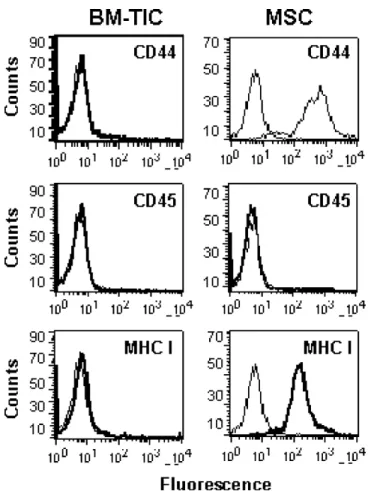
Tumor-infiltrating progenitor cells were isolated from adult rat bone marrow following a protocol established by Jiang et al. [15] to derive multipotent adult progenitor cells (MAPC). BM-TIC proliferated with a population dou- bling time of approximately 24 h and could be expanded for more than 60 passages without apparent signs of senescence (data not shown). To further characterize BM-TIC, expression of major histocompatibility complex class I (MHC I), CD44 and CD45 was assessed. As shown in Figure 1, BM-TIC are negative for MHCI, CD44 and CD45, thereby resembling MAPC [15]. In contrast, rat MSC express both MHCI and CD44, as previously reported [24].
Genetic modifications of BM-TIC to create a packaging cell line for
gammaretroviral LCMV-GP pseudotypes
To create a BM-TIC-based packaging cell line (BM-TIPC) for LCMV-GP pseudotypes, Mo-MLV Gag-Pol and LCMV- GP genes were introduced into BM-TIC. As a first step, BM- TIC were transfected with the Mo-MLV gag-pol expression plasmid pGag-Pol-IRES-bsr (Figure 2), which coexpresses a blasticidin resistance gene and Mo-MLV gag-pol via an IRES. Clones were grown in selection medium and exam- ined by flow cytometry for Mo-MLV Gag expression. Gag expression in clone BM-TIC-C1 is shown in Figure 3A.
To achieve stable LCMV-GP expression in BM-TIC, the lentiviral self-inactivating (SIN) vector pHR’SIN-GPopt with internal SFFV promoter and LCMV-GP/IRES/Puro cassette was created (Figure 2). BM-TIC-C1 were trans- duced with pHR’SIN-GPopt and subsequently transferred to selection medium. Transduced BM-TIC were expanded as bulk cultures without isolating single clones. LCMV- GP expression in BM-TIC-C1/GP was confirmed by flow cytometry at two different time points after transduc- tion. As shown in Figure 3B, LCMV-GPopt expression was detectable in BM-TIC-C1/GP and remained stable during 10 weeks of culture. The gammaretroviral vec- tor MP71-eGFP-wPRE (Figure 2) was then introduced by transduction into BM-TIC-C1/GP to create BM-TIPC.
Expression of the eGFP was determined by flow cytometry
and was present in >90% of cells (data not shown).
Figure 1. Staining of BM-TIC and MSC for cell-surface antigens. BM-TIC and MSC were stained with antibodies against CD44, CD45 or MHC I and with APC-conjugated secondary antibodies. Control samples were stained with secondary antibody only. Samples were analyzed by flow cytometry
Figure 2. Packaging constructs used to establish BM-TIPC. Promoters are shown as white arrows, open reading frames as grey boxes. The bicistronic expression plasmid pGag-Pol-IRESbsr (A) encodes Mo-MLV Gag-Pol and confers blasticidin resistance (bsr) to stable transfectants. The bicistronic lentiviral vector pHR’SIN-Gpopt (B) encodes a codon optimized LCMV-GP together with a puromycin resistance gene (Puro). The 3! HIV LTR has a deletion in the U3 region, which leads to self-inactivation of the promoter activity of the HIV LTR during reverse transcription. This prevents remobilization of the integrated lentiviral vector from the host cell. Transgene expression from pHR’SIN-Gpopt is driven by an internal SFFV promoter. The gammaretroviral reporter construct MP71-eGFP-wPRE (C) contains intact LTRs and the gammaretroviral packaging signal!and encodes the enhanced green fluorescent protein (eGFP). EF1a: elongation factor 1a promoter; IRES: internal ribosomal entry site from Encephalomyocarditis virus; LTR: long terminal repeats; MPSV: myeloproliferative sarcoma virus; SFFV: U3 promoter/enhancer region of the spleen focus forming virus; wPRE: woodchuck hepatitis virus posttranscriptional regulatory element
Vector production in BM-TIPC and transduction of glioma cell lines in vitro
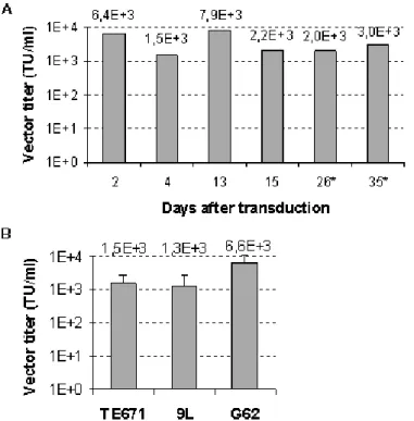
Efficiency and stability of vector production in BM-TIPC was tested by titration of supernatants on TE671 cells.
Vector titers ranged between 1.3 × 10
3and 7.9 × 10
3TU/ml and remained stable over 1 month in culture and after several cycles of freezing/thawing of BM-TIPC (Figure 4A). Vector titers on 9L rat glioma cells and G62 human glioma cells were subsequently measured. Vectors
produced in BM-TIPC mediated transduction of both glioma cell lines in vitro. The efficiency of transduction of 9L cells was comparable to TE671, whereas G62 human glioma cells were transduced with about 4-fold higher efficiency (Figure 4B).
The presence of replication-competent retrovirus (RCR)
in BM-TIPC supernatants was tested in a PCR-based
assay (refer to ‘Material and methods’ section), which
in previous studies reproducibly detected RCR in positive
controls (i.e. cell lines producing amphotropic replication-
competent MLV) and which is more sensitive than
Figure 3. Expression of MLV Gag and LCMV-GPopt in BM-TIC. (A) Gag expression in BM-TIC after stable transfection with pGag-Pol-IRES-bsr was determined by intracellular staining with an anti-p30 antibody and a PE-conjugated secondary antibody.
(B) Expression of LCMVGP in BM-TIC-C1 after transduction with pHR’SIN-GPopt was determined by staining of the cell surface with an a-LCMV-GP primary antibody (KL25) and a PE-conjugated secondary antibody. Staining against LCMV-GP was performed at two different time points (day 48 and day 72) after transduction. Samples were analyzed by flow cytometry
Figure 4. Production of retroviral vectors by BM-TIPC. (A) Cell culture supernatants from BM-TIPC were used to transduce TE671 cells at indicated time points after transduction with MP71-eGFP-wPRE. (∗) BM-TIPC had undergone three cycles of freezing/thawing.
(B) Vectors produced in BM-TIPC were titrated on TE671, 9L rat glioma and G62 human glioma cell lines in parallel. Data are shown as mean values and standard deviations from three independent experiments
the commonly used marker rescue assay (our own unpublished observations). BM-TIPC supernatants were free of any detectable RCR, as demonstrated by lack of MLV Gag, A-MLVenv and LCMV-GPopt sequences in genomic DNA of TE671 indicator cells (Supplementary Figure S1, see Supplementary Material).
Glioma-specific migration of BM-TIPC and transduction of glioma cells in vivo
For generation of the BM-TIPC, foreign (viral and non-
viral) genes were introduced and the cells were expanded
for numerous passages in vitro. The ability of these
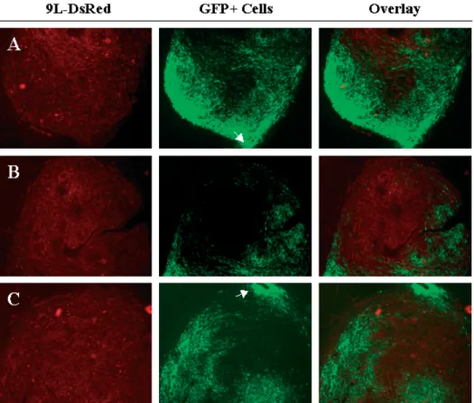
Figure 5. BM-TIPC infiltrate gliomasin vivowith high specificity and efficacy. BM-TIPC were injected intratumorally into established 9L-DsRed gliomas in Fischer rats. After 7 days, rats were killed, brain sections were prepared and analyzed by fluorescence microscopy. Panels A, B and C represent sections from three different tumors injected with BM-TIPC. Magnification 2.5×, white arrows indicate the site of injection of BM-TIPC
extensively manipulated BM-TIPC to specifically infiltrate gliomas in vivo was analyzed. BM-TIPC were injected into established 9L-DsRed gliomas in Fischer rats. After 7 days, examination of brain sections revealed an efficient infiltration of gliomas by BM-TIPC (Figure 5). BM-TIPC migrated far away from the injection site (indicated by arrows in Figures 5A and 5C) and infiltrated large proportions of the tumor. Moreover, the migratory behavior of BM-TIPC was highly specific for gliomas, as no BM-TIPC infiltration into normal brain was observed.
Transduction of glioma cells in vivo was examined by confocal microscopy on the same brain sections.
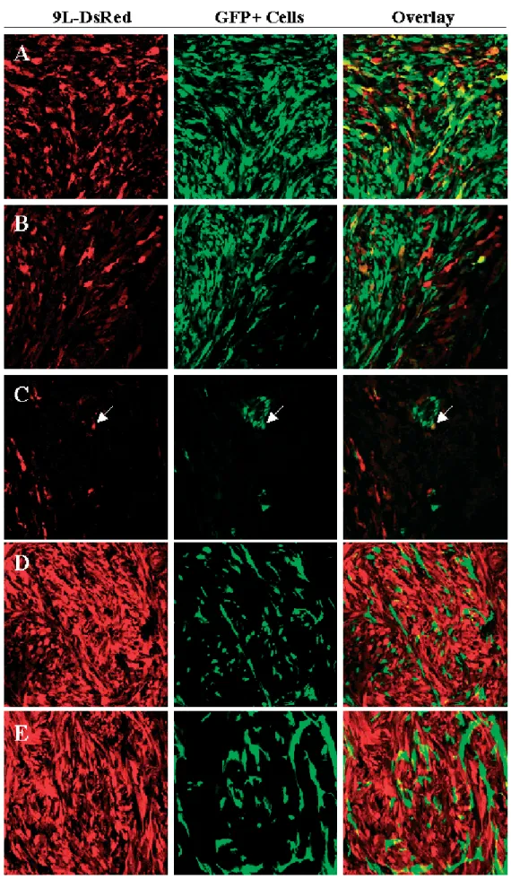
Colocalization of green and red fluorescence, indicative of transduced glioma cells, was detected in both solid (Figure 6A) and infiltrating (Figure 6B) tumor areas. In contrast, when BM-TIC expressing the gammaretroviral eGFP vector alone or together with LCMV-GP but lacking Mo-MLV Gag-Pol were injected into rat gliomas, no presence of red and green fluorescence in the same cell could be detected (Figures 6D and 6E). This shows that only BM-TIPC with complete packaging function can transfer the retroviral vector to glioma cells and that the colocalization of red and green fluorescence was not a result of cell fusion or uptake of fluorescent proteins from surrounding cells. The percentage of transduced glioma cells reached up to 33% in some tumor areas.
This shows that BM-TIPC continue to produce retroviral vectors in vivo and facilitate glioma cell transduction.
Importantly, BM-TIPC were also found adjacent to single infiltrating glioma cells and mediated their transduction (Figure 6C, arrow).
Discussion
The success of gene therapy for gliomas depends
primarily on the efficient and glioma-specific transfer
of the therapeutic gene. By injecting fibroblast-based
packaging cells (PC), efficient tumor cell transduction
can be achieved in animal models but not in clinical
settings, due to the non-migratory character of these PC
[6]. Stem cells of neural and non-neural origin can be
used to deliver therapeutic molecules to gliomas in vivo
[9,10,12]. However, the expression of toxic genes only
in the tumor-infiltrating cells but not the tumor itself
may not be sufficient to achieve a sufficient therapeutic
effect for the treatment of human gliomas. The aim of
the present work was therefore to create a PC based on
glioma-infiltrating progenitor cells. Most adult progenitor
cells, like neural and mesenchymal stem cells, however,
have only limited expansion capacity unless immortalized
with oncogenes. This limited expansion impedes the
establishment of clonal PC and the production of large
PC batches for clinical applications. In the present
study, we used tumor-infiltrating progenitor cells with
a high intrinsic expansion capacity to establish stable
Figure 6. BM-TIPC mediate in vivo transduction of rat glioma cells. BM-TIPC were injected intratumorally into established 9L-DsRed gliomas in Fischer rats. After 7 days, rats were killed, brain sections were prepared and analyzed by confocal microscopy.
Colocalization of green and red fluorescence demonstrates tumor cell transduction. Shown are images from solid (A) and infiltrating (B, C) tumor areas in which tumor tranduction could be readily detected. The white arrow in (C) indicates an infiltrating transduced glioma cell in close proximity to BM-TIPC. Injection of BM-TIC expressing only the gammaretroviral eGFP vector (D) or the gammaretroviral eGFP vector and LCMV-GP (E) did not result in colocalization of green and red fluorescence. Magnification 40×
PC. Progenitor cells were isolated from adult rat bone marrow according to a protocol established by Jiang and collegues [15] to isolate multipotent adult progenitor cells (MAPC). MAPC have the capability to differentiate into various cell types in vitro and in vivo. Moreover, MAPC have a very high expansion capacity and can be expanded to more than 100 population doublings without requiring exogenous immortalization [15]. Although the differentiation capacity of the BM-TIC was not analyzed in this study, marker expression and expansion capacity of BM-TIC strongly resemble that of MAPC. To establish BM-TIC-based PC, BM-TIC were genetically modified to express Mo-MLV Gag-Pol and LCMV-GPopt. The glycoprotein of LCMV was chosen because LCMV-GP mediates up to tenfold higher transduction rates in human glioma cell lines than the amphotropic retroviral envelope protein [16,25]. The expression of viral membrane proteins is often associated with cytotoxicity. Thus, the generation of PC for vectors pseudotyped with the glycoprotein of vesicular stomatitis virus (VSV-G) is hampered by the toxicity of VSV-G and requires inducible expression systems [26]. In contrast to VSV-G, LCMV-GP can be stably expressed in cell lines without causing cytotoxicity [16]. Moreover, LCMV-GP was shown to specifically mediate gene transfer to glioma cells but not to neuronal cells, while VSV-G was found to preferentially target neuronal cells [17]. A lentiviral self-inactivating (SIN) vector encoding LCMV-GP was created and used to transduce bulk cultures of BM-TIC. This led to stable and prolonged LCMV-GP expression in BM-TIC over more than 2 months in culture. Titers achieved on human glioma cells reached 1 × 10
4TU/ml and are therefore comparable to vector titers of PC used in previous clinical studies [27]. Importantly, as a consequence of tumor infiltration by BM-TIPC, local vector titers throughout the tumor are expected to be higher than titers achieved by injecting non-migratory PC.
When tested for glioma-specific migration in vivo, BM- TIPC behaved similarly to the parental BM-TIC cell line (Miletic, Fischer et al., submitted): after a single intratumoral injection, BM-TIPC were detected in most tumor areas, but never in normal brain parenchyma.
This shows that BM-TIPC retain their glioma-specific migratory capacity even after extensive culturing, genetic modification and subcloning. One important option for clinical application of BM-TIPC is their injection into gliomas during stereotactic biopsy. The very efficient tumor infiltration by BM-TIPC may then lead to widespread distribution of viral vectors within human gliomas and ensure the targeting of tumor cells distant from the injection site. This targeting of distant tumor cells can currently not be achieved by injection of replication- incompetent vectors due to only limited vector diffusion [6].
In a rat glioma model, tranduction was detected in both solid and infiltrating tumor regions, showing that BM-TIPC application is a suitable method to achieve gene transfer into glioma in vivo. Interestingly, BM-TIPC colocalized with single invasive glioma cells and mediated
transduction of these cells. Invasive glioma cells migrating into normal brain parenchyma are a major problem in glioma therapy, as they are not accessible to surgery and typically give rise to tumor spread and regrowth.
Transduction efficiencies achieved by BM-TIPC injection varied, ranging from undetectable to up to 33% of transduced glioma cells in different areas of the same glioma. In future experiments, the transduction efficiency in vivo may be enhanced by using lentiviral vectors instead of Mo-MLV-based gammaretroviral vectors. In contrast to gammaretroviral vectors, lentiviral vectors do not depend on cell proliferation for efficient transduction. Lentiviral gene transfer may thus enhance transduction of tumor areas with low proliferation rate. Specific gene transfer into glioma cells, but not into healthy brain, may thereby be achieved by pseudotyping lentiviral vectors with LCMV- GP [17].
In conclusion, the present study is the first to show that primary adult progenitor cells can be modified to stably produce retroviral LCMV pseudotype vectors while maintaining their high expansion capacity and their specific tropism for gliomas. In the presented model, specificity of gene transfer is conferred by the glioma- specific infiltration of the progenitor cells and by the selective tropism of LCMV-GP-pseudotyped vectors for glioma cells. In future studies, the efficiency of BM- TIPC-mediated gene transfer, shown here for the eGFP marker gene only, needs to be determined for therapeutic genes and extended to human glioma models. Provided that BM-TIPC show a good efficiency in these follow-up experiments, we are confident that these novel progenitor- cell-based packaging cells will be a valuable tool for gene therapy of human gliomas.
Acknowledgements
The authors would like to thank S. Bracharz, R. Seyd, M. Carstov and T. Merovci for expert technical assistance and F. Hermann for helpful discussions. The Neural Regeneration Group at the University Bonn LIFE & BRAIN Center is supported by the Hertie-Foundation and Walter-und-Ilse-Rose-Foundation. This work was additionally supported by the K¨oln Fortune Program (grant 108/2003 to H.M.). This publication was generated in the context of the CellPROM project, funded by the European Community as Contract No. NMP4-CT-2004-500039 under the 6th Framework Programme for Research andTechnological Development in the thematic area of ‘Nanotechnologies and nano-sciences, knowledge-based multifunctional materials and new production processes and devices’.’’ This publication reflects only the authors’ views;CellPROMis not liable for any use that may be made of the information contained therein.
Supplementary material
The supplementary electronic material for this paper is available in Wiley InterScience at: http://www.
interscience.wiley.com/jpages/1099-498X/suppmat/.
References
1. Kleihues P, et al. The WHO classification of tumors of the nervous system.J Neuropathol Exp Neurol2002;61: 215–225;
discussion 226–229.
2. Oertel J, et al. Prognosis of gliomas in the 1970s and today.
Neurosurg Focus2005;18: e12.
3. Culver KW, et al. In vivo gene transfer with retroviral vector- producer cells for treatment of experimental brain tumors.
Science1992;256: 1550–1552.
4. Ram Z, et al. Toxicity studies of retroviral-mediated gene transfer for the treatment of brain tumors.J Neurosurg1993;
79: 400–407.
5. Rainov NG, et al. Retrovirus-mediated gene therapy of experimental brain neoplasms using the herpes simplex virus- thymidine kinase/ganciclovir paradigm.Cancer Gene Ther1996;
3: 99–106.
6. Rainov NG. A phase III clinical evaluation of herpes simplex virus type 1 thymidine kinase and ganciclovir gene therapy as an adjuvant to surgical resection and radiation in adults with previously untreated glioblastoma multiforme.Hum Gene Ther 2000;11: 2389–2401.
7. Sandmair AM,et al. Thymidine kinase gene therapy for human malignant glioma, using replication-deficient retroviruses or adenoviruses.Hum Gene Ther2000;11: 2197–2205.
8. Rainov NG, Ren H. Clinical trials with retrovirus mediated gene therapy – what have we learned? J Neurooncol, 2003; 65:
227–236.
9. Aboody KS,et al. Neural stem cells display extensive tropism for pathology in adult brain: evidence from intracranial gliomas.
Proc Natl Acad Sci U S A2000;97: 12846–12851.
10. Ehtesham M,et al. The use of interleukin 12-secreting neural stem cells for the treatment of intracranial glioma.Cancer Res 2002;62: 5657–5663.
11. Lee J, et al. Cellular and genetic characterization of human adult bone marrow-derived neural stem-like cells: a potential antiglioma cellular vector.Cancer Res2003;63: 8877–8889.
12. Nakamura K, et al. Antitumor effect of genetically engineered mesenchymal stem cells in a rat glioma model.Gene Ther2004;
11: 1155–1164.
13. Moore XL, et al. Endothelial progenitor cells’ ‘‘homing’’
specificity to brain tumors.Gene Ther2004;11: 811–818.
14. Tabatabai G, et al. Lessons from the bone marrow: how malignant glioma cells attract adult haematopoietic progenitor cells.Brain2005;128: 2200–2211.
15. Jiang Y,et al. Pluripotency of mesenchymal stem cells derived from adult marrow.Nature2002;418: 41–49.
16. Beyer WR, et al. Oncoretrovirus and lentivirus vectors pseudotyped with lymphocytic choriomeningitis virus glycoprotein: generation, concentration, and broad host range.
J Virol2002;76: 1488–1495.
17. Miletic H, et al. Selective transduction of malignant glioma by lentiviral vectors pseudotyped with lymphocytic choriomeningitis virus glycoproteins.Hum Gene Ther2004;15:
1091–1100.
18. Morita S, Kojima T, Kitamura T. Plat-E: an efficient and stable system for transient packaging of retroviruses.Gene Ther2000;
7: 1063–1066.
19. Demaison C,et al. High-level transduction and gene expression in hematopoietic repopulating cells using a human immunodeficiency [correction of imunodeficiency] virus type 1-based lentiviral vector containing an internal spleen focus forming virus promoter.Hum Gene Ther2002;13: 803–813.
20. Schambach A, et al. Context dependence of different modules for posttranscriptional enhancement of gene expression from retroviral vectors.Mol Ther2000;2: 435–445.
21. Zufferey R,et al. Multiply attenuated lentiviral vector achieves efficient gene delivery in vivo. Nat Biotechnol 1997; 15:
871–875.
22. Bruns M,et al. Lymphocytic choriomeningitis virus. VI. Isolation of a glycoprotein mediating neutralization.Virology1983;130:
247–251.
23. Palu G, et al. Gene therapy of glioblastoma multiforme via combined expression of suicide and cytokine genes: a pilot study in humans.Gene Ther1999;6: 330–337.
24. Pittenger MF, et al. Multilineage potential of adult human mesenchymal stem cells.Science1999;284: 143–147.
25. Steffens S, et al. Transduction of human glial and neuronal tumor cells with different lentivirus vector pseudotypes. J Neurooncol2004;70: 281–288.
26. Yang Y, et al. Inducible, high-level production of infectious murine leukemia retroviral vector particles pseudotyped with vesicular stomatitis virus G envelope protein. Hum Gene Ther 1995;6: 1203–1213.
27. Ram Z,et al. Therapy of malignant brain tumors by intratumoral implantation of retroviral vector-producing cells.Nat Med1997;
3: 1354–1361.